premature ventricular contractions - osu center for ... ventricular contractions: acc/aha/esc 2006...
TRANSCRIPT

Orlando, Florida – October 7-9, 2011
Premature Ventricular ContractionsRalph Augostini, MD FACC FHRS

Premature Ventricular Contractions:
ACC/AHA/ESC 2006 Guidelines for Management of Patients With Ventricular Arrhythmias and the Prevention of Sudden Cardiac Death
J Am Coll Cardiol, 2006; 48:247-346.
BackgroundPVCs are ectopic impulses originating from an area distal to the His Purkinje systemMost common ventricular arrhythmiaSignificance of PVCs is interpreted in the context of the underlying cardiac conditionVentricular ectopy leading to ventricular tachycardia (VT), which, in turn, can degenerate into ventricular fibrillation, isone of the common mechanisms for sudden cardiac deathThe treatment paradigm in the 1970s and 1980s was to eliminate PVCs in patients after myocardial infarction (MI). CAST and other studies demonstrated that eliminating PVCs with available anti-arrhythmic drugs increases the risk of death to patients without providing any measurable benefit

PathophysiologyThree common mechanisms exist for PVCs, (1)
automaticity, (2) reentry, and (3) triggered activity:Automaticity: The development of a new site of depolarization in non-nodal ventricular tissue.Reentry circuit: Reentry typically occurs when slow-conducting tissue (eg, post-infarction myocardium) is present adjacent to normal tissue. Triggered activity: Afterdepolarization can occur either during (early) or after (late) completion of repolarization. Early afterdepolarizations commonly are responsible for bradycardia associated PVCs, but also with ischemia and electrolyte disturbance.

Triggered
Fogoros: Electrophysiologic Testing. 3rd ed. Blackwell Scientific 1999; 158.

EpidemiologyFrequencyThe Framingham heart study (with 1-h ambulatory ECG)
1 or more PVCs per hour was 33% in men without coronary artery disease (CAD) and 32% in women without CADAmong patients with CAD, the prevalence rate of 1 or more PVCs was 58% in men and 49% in women. 24-hour ambulatory monitoring showed a VPC prevalence rate of 41% in healthy teenage boys aged 14-16 years, 50-60% in healthy young adults, and 84% in healthy elderly persons aged 73-82 years.
PVCs also are common in patients with hypertension, ventricular hypertrophy, cardiomyopathy, and mitral valve prolapse. Data from the GISSI-2 study
64% of patients with prior MI had ventricular arrhythmia20% of patients had more than 10 PVCs per hour with 24h holter

Mortality/Morbidity
Prognosis depends on the frequency and characteristics of PVCs and on the type and severity of associated structural heart disease.PVCs are associated with increased risk of death, especially with CADThe relationship between VPC frequency and mortality is not robust and no benefit results in suppressing PVCs to improve survival in any populationIn asymptomatic patients, frequent ventricular ectopy was associated with 2.5-fold increased risk of cardiovascular deathMultiform PVCs confer a poorer prognosis than uniform PVCsPost-MI, frequent PVCs (>10/h) are associated with increased mortality in the pre-thrombolytic era, but the association in patients receiving thrombolytic is weak.

Mortality/MorbidityIn 2 studies, a frequent VPC during any given stage of stress testing, bigeminy, trigeminy, couplets, triplets, sustained or non-sustained ventricular tachycardia, ventricular flutter, torsade de pointes, or ventricular fibrillation was an independent predictor of death. Another study, frequent PVCs only during exercise did not independently predict an increased risk; Instead frequent PVCs during recovery was a stronger predictor of deathFrequent PVCs, especially when they occur in a bigeminal pattern, can precipitate tachycardia-induced cardiomyopathy that can be reversed by elimination of the PVCs through catheter ablationIn some circumstances, very frequent PVCs may decrease cardiac systolic function, and suppression by ablation may have a beneficial effect

Clinical Presentation
Palpitations: due to an augmented post-VPC beat and may be sensed as a pause rather than an extra beat
LightheadednessFatigueSustained tachycardia is not uncommonTrue syncope is infrequently seen
Physical ExaminationVariable or decreased intensity of heart sounds. The augmented beat following a dropped beat (pause) heard frequently. Bounding jugular pulse (cannon a wave) from a loss of AV synchrony may be present. The follow-up beat after a VPC is stronger due to the post-extra systolic compensatory pause, allowing greater left ventricular (LV) filling, causing greater intensity of that beat.Conversely, the VPC itself may be underperfused and consequently not perceived by radial pulse, resulting in a spurious documentation of bradycardia

Cardiac Causes
Acute myocardial infarction Valvular heart disease, especially mitral valve prolapse Cardiomyopathy (ischemic, dilated, hypertrophic, infiltrative) Myocardial stretch Cardiac contusion Bradycardia Tachycardia (high-catecholamine state)

Non-cardiac Causes
Electrolyte disturbances (hypokalemia, hypomagnesemia, or hypercalcemia) Medications (eg, digoxin, tricyclic antidepressants, aminophylline, amitriptyline, pseudoephedrine, fluoxetine) Other drugs (eg, cocaine, amphetamines, caffeine, alcohol) Anesthetics Surgery Infection Stress

Laboratory Studies
Look for correctable causes of PVCs:Medicationselectrolyte disturbancesinfectionmyocardial ischemia or MI
Check serum electrolyte and magnesium levels

Imaging Studies
Look for underlying structural heart abnormalities that can predispose to PVCsAssess the degree of LV dysfunction with echo radionuclide imagingEcho preferable because it also provides structural information about the heart

Other Tests
24-hour Holter monitorSeverity of LV dysfunction, along with the complexity and frequency of the PVCsSuppression of PVCs by beta-blocker or calcium blocker, together with a typical LBBB inferior axis, morphology helps tp establishing typical RVOT ectopy. Suppressing all PVCs themselves is not the focus of treatment
ECG performed to look for structural cardiac abnormalitiesWide beatsNo preceding premature P waves occurThe T wave usually is in the opposite direction from the R wave. Compensatory pause is common. PVCs originating from the left ventricle typically RBBB pattern.PVCs originating from right ventricle typically LBBB pattern.
Exercise stress testingcoronary ischemia, exercise-induced arrhythmia
Electrophysiology Study
Indicated for 2 types of patients with PVCs:
(1) those with a structurally normal heart with symptomatic PVCs, for whom pharmacological treatment or catheter ablation is indicated.
(2) those with PVCs and structural heart disease, for whom risk stratification for sudden cardiac death is indicated.
According to current ACC/AHA guidelines:class I indications for EPS are patients with CAD, low EF (< 0.36), and NSVT on ambulatory ECG. Class II indications for catheter ablation apply to patients with a highly symptomatic uniform PVCs, couplets, and NSVT

Classification
Classification according to frequency: Frequent - 10 or more PVCs per hour (by Holter monitoring) or 6 or more per minute Occasional - Fewer than 10 PVCs per hour or fewer than 6 per minute
Classification according to relationship to normal beats: Bigeminy - Paired complexes, VPC alternating with a normal beat Trigeminy - VPC occurring every third beat (2 sinus beats followed by VPC) Quadrigeminy - VPC occurring every fourth beat (VPC following 3 normal beats)
Couplet - 2 consecutive PVCs NSVT - 3 or more consecutive PVCs (< 30 s)
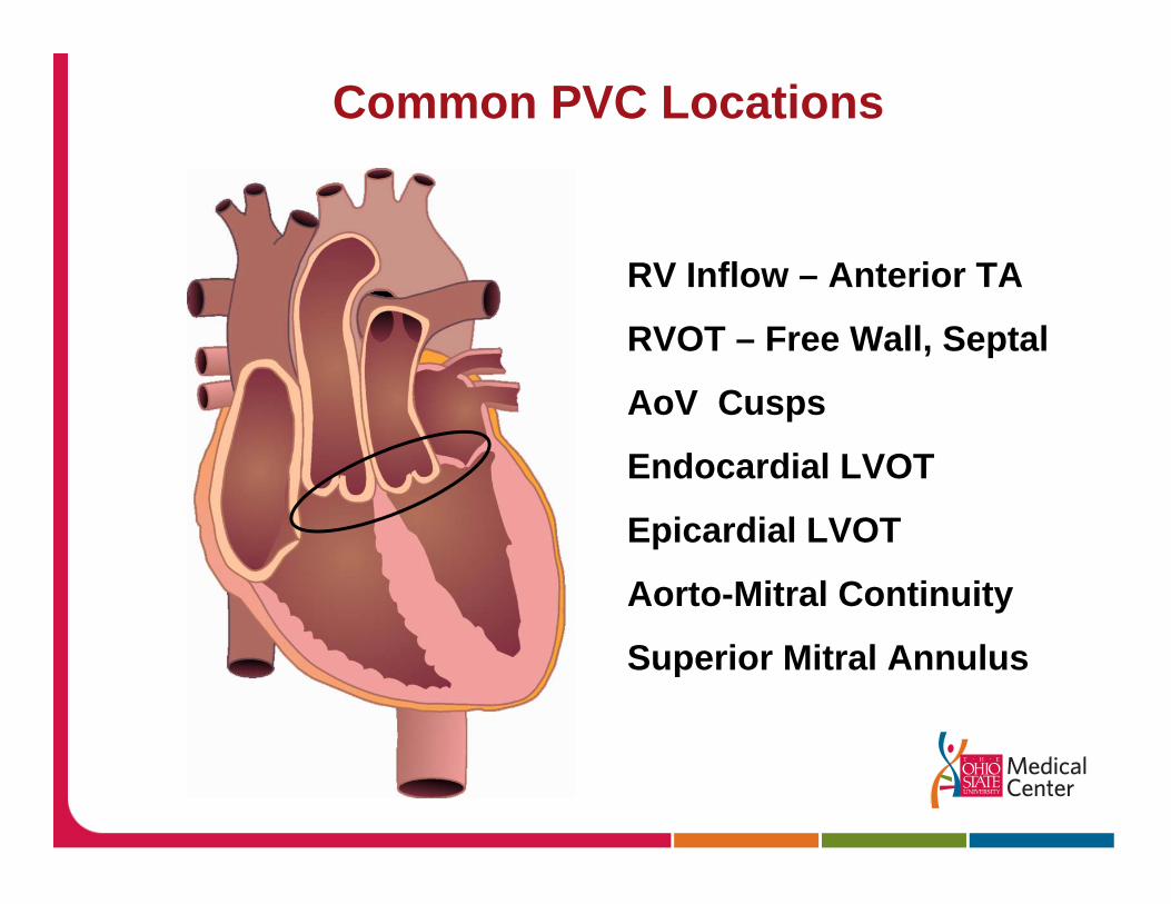
Common PVC Locations
RV Inflow – Anterior TA
RVOT – Free Wall, Septal
AoV Cusps
Endocardial LVOT
Epicardial LVOT
Aorto-Mitral Continuity
Superior Mitral Annulus

Outflow Tract Embryology – Neural Crest Cell Migration

Treatment
Absence of structural heart diseaseAsymptomatic = require no therapy. Symptomatic PVCs = patient education and reassurance, avoidance of aggravating factors, and anxiolytic drugs if neededBeta-blockers and non-dihydropyridine calcium channel blockers Anti-arrhythmic therapy is only used to prevent symptoms.
Presence of underlying heart diseaseThe presence of 2 or more of the following variables, (1) LV EF less than 0.40, (2) ventricular late potentials (on signal-averaged ECG), and (3) repetitive PVCs. Treatment of transient ischemia. Optimal treatment for congestive heart failure (CHF), CAD, or both should be instituted. Maintain electrolyte balance.Blood pressure control.
Treatment
The 2006 ACC/AHA/ESC guideline recommends that ablation therapy should be considered in the following:
Patients with frequent, symptomatic, and monomorphic PVCs refractory to medical therapy Patients who choose to avoid long-term medical therapy Patients with ventricular arrhythmia storm that is consistently provoked by PVCs of a similar morphology

Treatment
Patients deemed to be at high risk of sudden cardiac death may benefit from implantable cardioverter defibrillator (ICD) implantation

51 Year Old Female with Long History of PVC51 Year Old Female with Long History of PVC’’ssPresented with Palpitations/Near SyncopePresented with Palpitations/Near Syncope
Echo/Cardiolite/Cardiac MRI Echo/Cardiolite/Cardiac MRI –– NegativeNegative

51 Year Old Female with Palpitation/Near Syncope51 Year Old Female with Palpitation/Near SyncopeNSVT During Isuprel InfusionNSVT During Isuprel Infusion

51 Year Old Female 51 Year Old Female –– RVOT VTRVOT VT
Pacemap Vs. Spontaneous VT

51 Year Old Female 51 Year Old Female –– RVOT VT RVOT VT –– Carto MapCarto MapRF Sites

Benign PVCs? RFA of Frequent, idiopathic PVCs:
60 pts with PVCs referred for RFA. 22 with decreased EF
Mean PVC Burden: 21+ 17%
PVC location: RVOT 31 (52%), LVOT 9 (15%)
Other 20 (33%)
RFA Successful in 48 (80%)
LVEF normalized in 18 / 22 (82%) with baseline LV dysfunctionConclusions: LV dysfunction in the setting of frequent PVCs may be a reversible with catheter ablation.
Bogun et al, Heart Rhythm 2007

51 year old male with a Hx of “Benign” PVCsNIDCM Dx’d 2003 (EF = 20%). Meds = ACEI, Coreg, AldactoneHolter: Uniform PVCs = 20% of QRS complexes (18,000) Referred for ICD Implant

LAOPace Map of Distal CS / Proximal Aspect of
Anterior CS Branch

APPace Map of Distal CS / Proximal Aspect of
Anterior CS Branch

Arterial Pressure Recordings During PVC’s

Arterial Pressure Recordings Post RFA PVC’s

Follow-up 3 months following ablation: EF = 42%