preparation of a sialic acid-binding protein from streptococcus mitis
TRANSCRIPT

INFECTION AND IMMUNITY, Aug. 1986, p. 359-3650019-9567/86/080359-07$02.00/0Copyright © 1986, American Society for Microbiology
Preparation of a Sialic Acid-Binding Protein fromStreptococcus mitis KS32AR
PATRICIA A. MURRAY,'t MICHAEL J. LEVINE,'* MOLAKALA S. REDDY,' LAWRENCE A. TABAK,I-2AND E. JAMES BERGEY'
Departments of Oral Biology' and Endodontics,2 School ofDental Medicine, State University ofNew York at Buffalo,Buffalo, New York 14214
Received 26 September 1985/Accepted 15 April 1986
A recent report has identified a lectin on the surfaces of several strains of Streptococcus mitis andStreptococcus sanguis with specificity for an N-acetylneuraminic acid a2,3-galactose-,31,3-N-acetylga-lactosamine sequence (P. A. Murray, M. J. Levine, L. A. Tabak, and M. S. Reddy, Biochem. Biophys. Res.Commun. 106:390-396, 1982). In the present study, purification and characterization of this sialic acid-bindingprotein (SABP) was begun. A clinical isolate of S. mitis was grown to mid stationary phase in synthetic FMCmedium and then extracted with lithium 3,5-diiodosalicylate. Lyophilized extract was subjected to gel filtrationon a Sephadex G-200 column, giving four protein peaks (A to D). Peak B, shown by hemagglutination assayto contain SABP, was next subjected to affinity chromatography on a Sepharose-4B matrix coupled to fetuinglycopeptides. After an extensive washing, peak B materials bound to the affinity matrix were eluted withbuffered N-acetylneuraminic acid. Sodium dodecyl sulfate-polyacrylamide gel electrophoresis with 2-mercaptoethanol on 7.5% gels of affinity-purified materials revealed components of 96, 70, and 65 kilodaltons(kDa). Without reducing agent, only the 65-kDa band and materials which did not penetrate the gel werevisualized, suggesting that the 96- and 70-kDa components were disulfide linked. The chemical cross-linkingagent, disuccinimidyl suberate, was used to demonstrate specific interactions between the SABP preparationand [14C]fetuin glycopeptides. After cross-linking, sodium dodecyl sulfate-polyacrylamide gel electrophoresisand fluorography revealed the 96- and 70-kDa components, indicating that the SABP is at least bivalent. Thesefindings support our previous suggestion that human salivary glycoproteins facilitate clearance of selected oralstreptococci via specific interactions between sialic acid-containing oligosaccharides and a carbohydrate-binding protein on the bacterial cell surface.
The ability of a microorganism to adhere to oral surfaces isa prerequisite for successful colonization and subsequentplaque-mediated diseases (18). The failure of bacteria toadhere results in their being swept by mechanical means,such as swallowing and coughing, amid the fluids whichcontinuously bathe the oral tissues. Although it is recognizedthat surface components of oral bacteria participate in ad-herence and clearance phenomena (35), their propertiesremain largely obscure. Several oral bacteria have beenshown to contain surface adhesins or binding proteins, and alectinlike mechanism of adherence has been proposed on thebasis of the inhibition of attachment by specific sugars (2, 3,7, 10, 14, 16, 24, 36, 42). The purification and characteriza-tion of these binding proteins provides a more completeunderstanding of their role in host-parasite interactions.
Streptococcus sanguis and Streptococcus mitis are amongthe earliest colonizers of the tooth surface (6); consequently,these microorganisms appear to have a fundamental role inthe cascade of events leading to the formation of dentalplaque. Although available evidence indicates that the ad-herence of streptococci to saliva-coated hydroxyapatite sur-faces is complex and involves both specific and nonspecifictypes of interactions, the exact molecular mechanisms arenot fully understood. Electrostatic (4, 19) and hydrophobic(9, 15, 30, 34) forces are clearly involved in streptococcalsalivary pellicle binding, but these types of interactions do
* Corresponding author.t Present address: Department of Stomatology, School of Dentis-
try, University of California at San Francisco, San Francisco, CA94143.
not fully account for the tissue specificity observed inbacterial adherence. However, recent studies on the inter-actions of S. sanguis and salivary glycoproteins have begunto elucidate additional mechanisms of attachment involvinga bacterial recognition system which may function to alignsalivary glycoproteins with the bacterial surface (18, 24, 31).Earlier studies suggested that the carbohydrate moieties ofsalivary mucins play a role in both bacterial adherence andclearance (17) and that, in particular, sialic acids may bedeterminants necessary for certain types of saliva-bacteriainteractions (29). Later investigations (23, 45) with humansalivary mucin have shown that the agglutination of S.sanguis, but not that of S. mutans, is dependent upon thepresence of nonreducing terminal N-acetylneuraminic acid(NeuAc). More recently, we have presented evidence for alectin on the surface of several S. sanguis and S. mitis strainswith specificity for an N-acetylneuraminic acid a2,3-galactose-f31,3-N-acetylgalactosamine (NGG) sequence (31).The significance of this is suggested by the fact that themajor acidic oligosaccharide of the lower-molecular-weighthuman salivary mucin has this trisaccharide structure (37).Thus, interactions between a trisaccharide of human salivarymucin and a bacterial lectin may promote clearance ofstreptococci and may represent a nonimmune protectivemechanism in the oral cavity (46). In this study, we describethe purification and preliminary characterization of a sialicacid-binding protein (SABP) from S. mitis KS32AR.
(Preliminary results of this work have been presentedpreviously [P. A. Murray, M. J. Levine, L. A. Tabak, andM. S. Reddy, Proc. Annu. Meet. Am. Assoc. Dent. Re-
359
Vol. 53, No. 2

360 MURRAY ET AL.
search, Cincinnati, Ohio, 17 to 20 March 1983, in J. Dent.Res. 62:257, 1983.])
(This work constitutes a portion of the dissertation ofP. A. Murray, submitted in partial fulfillment of the require-ments for the Ph.D. degree from the State University ofNewYork at Buffalo, 1983.)
MATERIALS AND METHODS
Materials. Bovine fetuin (type II) was obtained fromCalbiochem-Behring, La Jolla, Calif., and from GIBCOLaboratories, Grand Island, N.Y. Pronase was obtainedfrom Calbiochem-Behring. HEPES (N-2-hydroxyethylpip-erazine-N'-2-ethanesulfonic acid), phenylmethanesulfonylfluoride, NeuAc (type VII), N-acetylneuramin-lactose, andN-(p-aminophenyl)-oxamic acid-agarose were purchasedfrom Sigma Chemical Co., St. Louis, Mo. ['4C]for-maldehyde (52.4 mCi/mmol) and En3Hance were obtainedfrom New England Nuclear Corp., Boston, Mass. Lithium3,5-diiodosalicylate (LIS) was purchased from Eastman Ko-dak Co., Rochester, N.Y. Sephadex resins, Sepharose-4B,and molecular weight standards were obtained fromPharmacia Fine Chemicals, Piscataway, N.J. Disuccinimidylsuberate was from Pierce Chemical Co., Rockford, Ill.
General chemical and analytical methods. General estima-tions of protein and neutral sugars were made by theprocedure of Lowry (27) and the anthrone reaction (39),respectively. Polyacrylamide gel electrophoresis (PAGE)was performed on 7.5% sodium dodecyl sulfate (SDS) diskor slab gels by the method of Weber and Osborn (49).Samples were prepared for electrophoresis by heating at100°C for 3 to 5 min in 0.01 M sodium phosphate (pH 7.0)containing 2% SDS with or without 5% (vol/vol) 2-mercaptoethanol. After electrophoresis, proteins were iden-tified by staining with Coomassie blue (12). Molecularweights were calculated from plots of the log molecularweights versus the relative mobility of standard referenceproteins. The following reference proteins (molecularweights) were used: unreduced immunoglobulin G (IgG),150,000; phosphorylase b, 94,000; bovine serum albumin,67,000; ovalbumin, 43,000; carbonic anhydrase, 30,000; andtrypsin inhibitor, 20,000.
Preparation of glycopeptides. Fetuin glycopeptides con-taining the NGG sequence were obtained by the methods ofSpiro and Bhoyroo (44). For this purpose, 1.5 g of fetuin (25mg/ml) was digested at 37°C with 1% (wt/vol) pronase in 0.15M Tris-acetate buffer (pH 7.8) containing 1.5 mM calciumacetate. After 72 h, this digest was fractionated on columnsof Sephadex G-50 (2.5 by 100 cm) in 0.1 M pyridine acetate(pH 5.1) to separate N-glycosidic units from the smaller0-glycosidic units. Elution was at 20 ml/h at 4°C with afraction size of 5.5 ml. Columns were monitored for hexoseby the anthrone method (39), for hexosamines on an aminoacid analyzer after hydrolysis in 2 N HCl for 6 h at 100°C(38), and for sialic acids (48) after acid hydrolysis (with 0.05N H2SO4 for 1 h at 80°C). N-Acetylgalactosamine-containingglycopeptides were further fractionated on columns of Seph-adex G-25 (1.5 by 120 cm) in 0.1 M pyridine acetate (pH 5.1)at 4°C. Fractions of 4.0 ml each were collected, and portionswere monitored as described above. NGG glycopeptideswere purified by chromatography on columns (1.25 by 80cm) of DE-52 cellulose equilibrated with the 0.002 Mpyridine acetate buffer (pH 5.1) and by using a linearpyridine acetate gradient to 0.15 M (44). The composition ofNGG glycopeptides was confirmed by chemical analyses bymethods previously described (38).
Labeling of NGG glycopeptides by reductive methylation.NGG glycopeptides (5 mg) were methylated with ['4C]for-maldehyde (1.3 ,umol at 0.94 x 10⁸ cpm/,umol) by the methodof Jentoft and Dearborn (20). Unreacted products wereremoved by gel filtration on columns (1.5 by 80 cm) ofBio-Gel P4 (200/400 mesh; Bio-Rad Laboratories, Rich-mond, Calif.) in 0.1 M pyridine acetate (pH 5.1). Columnswere monitored by scintillation spectroscopy, and the ap-propriate fractions were pooled and lyophilized. The specificactivity of the prepared 14C-labeled NGG glycopeptides was2.5 ,uCi/mg. Incorporation of 14C-labeled methyl groups ontolysine was verified by hydrolyzing labeled materials (with 6N HCl for 28 h at 105°C) and examining the hydrolysates formono- and dimethyllysine by paper chromatography with2-propanol-ammonia-water (8:1:1, vol/vol/vol) as the sol-vent system (22).
Bacteria and culture conditions. S. mitis KS32AR (ob-tained as a fresh clinical isolate from a 24-h human dentalplaque [32]) was used in these studies. This strain haspreviously been identified both as S. sanguis biotype 2 onthe basis of biochemical tests (5, 11) and as a dextran-producing S. mitis because of the presence of a cell-associated neuraminidase (32). For study, a 1% inoculum ofbacteria was grown for 16 to 18 h (mid to late stationaryphase) at 37°C in FMC chemically defined medium freshlybuffered with 0.02 M each of sodium carbonate and sodiumbicarbonate (47). Cells were harvested at 4°C by centrifuga-tion at 12,000 x g for 10 min and were washed three times incold 0.05 M Tris hydrochloride (pH 7.5). The yield wasapproximately 2.5 g of cells per liter (wet weight).LIS extraction of bacteria and gel filtration of extract on
Sephadex G-200. Bacteria were suspended in 0.3 M LIS in0.05 M Tris hydrochloride (pH 7.5) containing 100 ,uMphenylmethylsulfonyl fluoride at 2 x 10¹⁰ bacteria per ml andwere extracted as previously described (31). Examination ofthe bacterial pellet by phase-contrast microscopy verifiedthat LIS extraction did not rupture the cell walls. Lyophi-lized LIS extract (150 mg) was dissolved in 0.1 M Trishydrochloride (pH 7.5) (equilibration buffer) by shakingovernight at 4°C (20 mg/ml), centrifuged at 12,000 x g for 30min at 4°C to remove insoluble material, and then subjectedto gel filtration chromatography on columns of SephadexG-200 (1.5 by 110 cm). Elution in equilibration buffer wasachieved at 8 ml/h at 4°C with a fraction size of 4.5 ml.Portions of the collected fractions were monitored for pro-tein at A280. The appropriate tubes were then pooled, ex-haustively dialyzed against distilled water, and lyophilized.
Preparation of affinity matrices. NGG glycopeptides werecovalently coupled to Sepharose-4B after activation withcyanogen bromide (28) with 5 mg of NGG glycopeptides perml of resin in 0.1 M NaHCO3 (pH 8.3) containing 0.5 M NaCl(coupling buffer). After being mixed for 16 h at 4°C,unreacted sites were blocked by addition of an equal volumeof 1.0 M ethanolamine in coupling buffer and shaking over-night at 4°C. Excess adsorbed protein was removed fromresin by alternate washings (5 bed volumes) with 0.1 Msodium acetate (pH 5.0), containing 0.5 M NaCl, and cou-pling buffer. Combined washings were desalted by elutionwith water on columns (1.5 by 90 cm) of Sephadex G-10. TheNGG glycopeptide affinity matrix contained approximately375 ,ug of NeuAc per ml of gel and represented a couplingefficiency of 75%.
Affinity chromatography. Sephadex G-200 pool B wasdissolved (3 mg/ml) in 0.05 M Tris hydrochloride (pH 7.4)with 0.15 M NaCl, centrifuged at 12,000 x g for 30 min at 4°Cto remove any insoluble material, and incubated with NGG
INFECT. IMMUN.
SIALIC ACID-BINDING PROTEIN FROM S. MITIS KS32AR
I aB I C ID I
0
F.
_ 2 a
,
E
AB C D sG-200 Pools
I I I I I I I I I0 10 20 30 40 50 60 70 80
FRACTION # (3.1 ml)
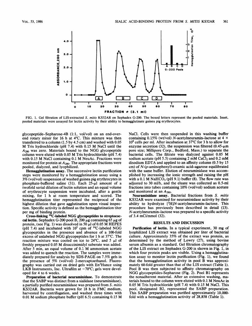
FIG. 1. Gel filtration of LIS-extracted S. mitis KS32AR on Sephadex G-200. The boxed letters represent the pooled materials. Inset,pooled materials were assayed for lectin activity by their ability to hemagglutinate guinea pig erythrocytes.
glycopeptide-Sepharose-4B (1:1, vol/vol) on an end-over-end rotary mixer for 16 h at 4°C. This mixture was thentransferred to a column (1.5 by 4.5 cm) and washed with 0.05M Tris hydrochloride (pH 7.4) with 0.15 M NaCl until theA280 was zero. Materials bound to the NGG glycopeptidecolumn were eluted with 0.05 M Tris hydrochloride (pH 7.4)with 0.15 M NaCl containing 0.1 M NeuAc. Fractions weremonitored for protein at A280. The appropriate fractions werepooled, dialyzed, and lyophilized.
Hemagglutination assay. The successive lectin purificationsteps were monitored by a hemagglutination assay using a3% (vol/vol) suspension of washed guinea pig erythrocytes inphosphate-buffered saline (31). Each 25-,I amount of atwofold serial dilution of lectin solution and an equal volumeof erythrocyte suspension were incubated, after a gentlemixing, for 1 h at room temperature and scored. Thehemagglutination titer represented the reciprocal of thehighest dilution that gave agglutination upon visual inspec-tion. Specific activity is defined as the hemagglutination titerper mg of binding protein.
Cross-linking 14C-labeled NGG glycopeptides to streptococ-cal lectin. Sephadex G-200 pool B, 200 jig containing 65 ,ug ofprotein, (see Fig. 1) was dissolved in 50 ,ul of 0.05 M HEPES(pH 7.4) and incubated with 105 cpm of "4C-labeled NGGglycopeptides in the presence and absence of a 100-foldexcess of unlabeled NGG glycopeptides for 1 h at 37°C. Thereaction mixture was cooled on ice to 24°C, and 5 RI offreshly prepared 0.05 M disuccinimidyl suberate was added.After 5 min, an equal volume of 0.1 M ammonium acetatewas added to quench the reaction. The samples were imme-diately prepared for analysis by SDS-PAGE on 7.5% gels inthe presence of 5% (vol/vol) 2-mercaptoethanol. Fluoro-graphy was carried out on dried gels with En3Hance usingLKB Instruments, Inc. Ultrafilm at -70°C; gels were devel-oped for 4 to 6 weeks.
Preparation of bacterial neuraminidase. To demonstratethat the SABP was distinct from a sialidase (neuraminidase),a partially purified neuraminidase was prepared from S. mitisKS32AR. Bacteria were grown for 18 h in FMC medium,harvested by centrifugation, and washed three times with0.01 M sodium phosphate buffer (pH 6.5) containing 0.15 M
NaCl. Cells were then suspended in this washing buffercontaining 0.15% (wt/vol) N-acetylneuramin-lactose at 4 x10⁹ cells per ml. After incubation at 37°C for 3 h to allow forenzyme secretion (32), the suspension was filtered (0.45-11mpore size; Millipore Corp., Bedford, Mass.) to separate thebacterial cells. The filtrate was dialyzed against 0.05 Msodium acetate (pH 5.5) containing 2 mM CaCl2 and 0.2 mMdisodium EDTA and applied to an affinity column (0.5 by 15cm) of N-(p-aminophenyl)-oxamic acid-agarose equilibratedwith the same buffer. Elution of neuraminidase was accom-plished by increasing the ionic strength and raising the pHwith a 0.1 M NaHCO3 (pH 9.1) buffer (8). The flow rate wasadjusted to 30 ml/h, and the eluate was collected as 0.5-mlfractions into tubes containing 10% (vol/vol) sodium acetateand monitored at an A280.Neuraminidase assay. Bacterial fractions from S. mitis
KS32AR were examined for neuraminidase activity by theirability to hydrolyze [3H]N-acetylneuramin-lactose. Thisprocedure has previously been described in detail (13).N-acetylneuramin-lactose was prepared to a specific activityof 3.4 mCi/mmol (32).
RESULTS AND DISCUSSIONPurification of lectin. In a typical experiment, 30 mg of
lyophilized LIS extract was obtained per liter of bacterialculture. Approximately 35% of the extract was protein, asdetermined by the method of Lowry (27), using bovineserum albumin as a standard. Gel filtration chromatographyof the LIS extract on Sephadex G-200 is shown in Fig. 1, inwhich four protein peaks are visible. Using a hemagglutina-tion assay to monitor lectin purification (Fig. 1), we foundthat the hemagglutination activity in pool B was approxi-mately 60-fold greater than that of the LIS extract (Table 1).Pool B was then subjected to affinity chromatography onNGG glycopeptides-Sepharose (Fig. 2). Pool Bi representsthe nonadherent material. After an extensive washing, ma-terials bound to the column were eluted with 0.1 M NeuAc in0.05 M Tris hydrochloride (pH 7.4) with 0.15 M NaCl. Thispool, designated B2, represented the SABP preparation.This SABP preparation was purified approximately 10,000-fold with a hemagglutination activity of 28,858 (Table 1).
4.0 -
Ec
00
z
4n000.4
3.0 -
2.0 -
1.0-
0
VOL. 53, 1986 361

362 MURRAY ET AL.
4.0 -
E
0a
0za'UUc0aSI'
3.0 -
2.0 -
1.0 -
0 -
0.1MNeuAc
0
IC
)0
es4-
a._
cob
E
Pools
I I I I I I I I I0 10 20 30 40 50 60 70 80
FRACTION # (3.1 ml)
FIG. 2. Affinity chromatography of Sephadex G-200 pool B on NGG glycopeptide-Sepharose-4B. The peaks were monitored at A280, andspecifically bound materials were eluted with 0.1 M NeuAc in 0.05 Tris hydrochloride (pH 7.4), containing 0.15 M NaCl. (Inset) Pooledmaterials were assayed for lectin activity by their ability to hemagglutinate guinea pig erythrocytes.
SDS-PAGE of lectin preparation. SDS-PAGE analyseswith 2-mercaptoethanol of pool B2 and the crude LIS extractare shown in Fig. 3. With Coomassie blue staining, acomplex protein pattern was obtained with the crude LISextract, whereas only three bands were seen with the SABPpreparation. The SABP did not stain with periodic acid-Schiff reagent. The estimated sizes of the three polypeptidecomponents in pool B2 were calculated to be 65, 70, and 96kilodaltons (kDa). When pool B2 was examined in theabsence of reducing agent, only the 65-kDa component, aswell as material that did not penetrate the 7.5% gel matrix,was seen. SDS-PAGE of SABP on 5% gels without 2-mercaptoethanol demonstrated a band with an Mr of -200kDa in addition to the 65-kDa component. These datasuggest that the 70- and 96-kDa components are disulfide-linked subunits associated noncovalently with the 65-kDapeptide.
Effects of repeated subculture. During a period of 2months, S. mitis KS32AR was subcultured 60 times in FMCsynthetic medium. No changes were observed in its reactiv-ity in various biochemical tests (32) after the 60 passages.The original isolate, as well as the 18-h broth cultures fromevery sixth subculture, was tested for SABP activity by thehemagglutination inhibition assay. SABP activity after 60transfers was comparable to that of the original isolate,indicating that repeated in vitro subcultures in syntheticFMC medium did not alter or affect the presence of lectinactivity. However, other studies of S. sanguis have demon-strated the effect of growth conditions on the bacterialsurface. For example, Liljemark and Bloomquist (26) foundthat an adherence-blocking component from S. sanguis was
TABLE 1. Purification of an SABP from S. mitis KS32ARTotal Total activity Sp act U/ Pufication
Prepn protein hemagglutination mg of (fold)(mg) Ua protein (od
LIS extract 150 4.5 x 10⁴ 2.7 1Pool B 23 4.3 x 10⁴ 166 61.5Pool B2 1.5 3.8 x 10⁴ 28,858 10,685
a A hemagglutination unit is defined as the reciprocal of the highest dilutionof lectin which produced visible agglutination of guinea pig erythrocytes.
often no longer detectable after several passages of freshisolates. Westergren and Olsson (50) also found that thehydrophobic properties of S. sanguis can diminish withrepeated subcultures in vitro; this most probably reflectschanges of different biochemical structures on the bacterial
7OK-0OSK-0
2 3
FIG. 3. SDS-PAGE analysis on 7.5% acrylamide gels in thepresence of 2-mercaptoethanol. Lanes: 1, 65 ptg of protein of crudeLIS extract from S. mitis KS32AR; 2, 73 ~Lg of protein of SABP(pool B2, Fig. 2); 3, 66 ~xg of protein of Sephadex G-200 pool B (Fig.1) incubated with 'IC-labeled NGG glycopeptides (2 x 101 cpm) andsubjected to the cross-linking agent disuccinimidyl suberate. Themolecular weights of the three polypeptide components in pool B2(in kilodaltons) are shown to the left of the gel.
INFECT. IMMUN.

SIALIC ACID-BINDING PROTEIN FROM S. MITIS KS32AR
TABLE 2. Streptococcal lectin versus enzyme: evidence for twodifferent molecules
Presence or reaction ofi:Parameter
Lectinb Neuraminidasec
Presence in organismS. sanguis G9B and 10556 +S. mitis KS32AR and 10557 + +
Extraction with LIS +
Elution on Sephadex G-200Excluded - +Included +
Heat labilityd - +
a +, Present or positive; -, absent or negative.b Reference 31.c Reference 32.d Neuraminidase activity of whole cells was inactivated at 80°C for 45 min
(31).
cell wall. For our studies, a chemically defined medium wasused. It may be that continuous passages in a more complexmedium or for a longer time produce surface changes thatresult in loss of lectinlike activity.
Characterization of SABP by covalent cross-linking with14C-labeled NGG glycopeptides. To identify which of thecomponents in the SABP preparation contained carbohy-drate-binding sites, Sephadex G-200 pool B (Fig. 1) wasincubated with 14C-labeled NGG glycopeptides (+ 100-foldmolar excess of unlabeled NGG glycopeptide). After 1 h at37°C, the covalent cross-linking agent, disuccinimidyl suber-ate, was added. If the glycopeptides were bound to theSABP, they would be cross-linked by the disuccinimidylsuberate and the 14C-labeled glycopeptide-adhesin com-plex(es) and then identified by fluorography after SDS-PAGE. The results of this experiment are depicted in Fig. 3,lane 3. Two radiolabeled bands which comigrated with the96- and 70-kDa components are shown. These results indi-cate that the two disulfide-linked constituents of the lectincontain carbohydrate-binding sites. The specificity of theseinteractions was verified by running this experiment in thepresence of a 100-fold excess of unlabeled glycopeptide.Under these conditions, no radiolabeled bands were visual-ized. The fact that the 65-kDa component was not radiola-beled indicates that this component does not contain acarbohydrate-binding site and confirms the earlier explana-tion that this band is either a noncovalently linked subunit ofthe lectin or, alternately, may represent a protein contami-nant.
Preparation of S. mitis neuraminidase. Because S. mitisstrains are known to possess a cell-associated, substrate-inducible neuraminidase (32), it was necessary to rule out thepossibility that the SABP was a neuraminidase. An activeenzyme preparation was prepared from the concentratedculture supernatant of strain KS32AR with N-(p-amino-phenyl)-oxamic acid-agarose as an affinity matrix (8). Thischromatography procedure resulted in a 160-fold purificationof the neuraminidase. When the neuraminidase preparationwas subjected to gel filtration on Sephadex G-200, enzymeactivity was eluted in the void volume fractions (in contrastto SABP, which was in the included fractions [Fig. 1, poolB]). In addition, neuraminidase was not extracted with LIS(31) but remained (albeit decreased) with the cell pellet.Previously, we have reported the presence of SABP in S.sanguis strains that do not produce neuraminidase (31).
Collectively, these data suggest that the neuraminidase andSABP from S. mitis KS32AR are two distinct molecules(Table 2).
In summary, this study was undertaken to purify and tobegin characterizing a component on the surface of S. mitisKS32AR that appears to specifically interact with the sialicacid-containing oligosaccharides of salivary glycoproteins.This SABP was proteinaceous in nature and contained atleast two disulfide-linked subunits of 96 and 70 kDa. Studiesusing the chemical cross-linking agent, disuccinimidyl suber-ate, confirmed the bivalent nature of SABP, since eachsubunit was shown to bind the NGG sequence. Studies fromseveral laboratories have demonstrated the presence ofadhesins on the surface of S. mitis or S. sanguis (1, 21, 33,40, 41, 43). The mechanism by which these adhesins interactwith saliva molecules is not completely understood. Indeed,the presence of at least two lectinlike adhesions on thesurface of S. mitis 10557 has been reported. These include agalactose-binding lectin (smaller than the SABP) (33) and alectin interactive with the NGG sequence (32). Both thegalactose-binding protein (33) and SABP (3) have beenshown to interact with the proline-rich glycoprotein ofhuman parotid saliva. The presence of terminal NeuAc andgalactose on the triantennary oligosaccharides of the proline-rich glycoprotein (25, 38) suggests that different ligands on asingle saliva molecule may interact with more than oneadhesin on the same bacterium. It is apparent that primarystructural determinants on saliva molecules (e.g., a definedoligosaccharide sequence) can be recognized by some bac-terial adhesins. In addition, conformational determinants onsaliva molecules may be important in saliva-bacteria inter-actions. It can be speculated that primary structural deter-minants function in clearance phenomena, whereas con-formational determinants play a role in the adherence ofbacteria to saliva-coated enamel or mucosal surfaces. Theseconformational determinants could form de novo as theresult of surface binding, or they might be buried within thesuprastructure of the molecule and become accessible onlyupon binding to an oral surface. Site-specific reagents suchas SABP could be used to examine the availability ofstructural or conformational determinants or both on salivamolecules before and after their binding to tissue surfaces.
ACKNOWLEDGMENTS
This study was supported in part by Public Health Service grantsDE04518, DE04971, DE07034, and DE06545 from the NationalInstitute of Dental Research.
LITERATURE CITED1. Appelbaum, B., and B. Rosan. 1984. Cell surface proteins of oral
streptococci. Infect. Immun. 46:245-250.2. Beachey, E. H. 1981. Bacterial adherence: adhesin-receptor
interactions mediating the attachment of bacteria to mucosalsurfaces. J. Infect. Dis. 143:325-345.
3. Bergey, E. J., M. J. Levine, M. S. Reddy, S. D. Bradway, and I.Al-Hashimi. 1986. Use of the photoaffinity crosslinking agent,N-hydroxysuccinimidyl-4-azidosalicylic acid, to characterizesalivary glycoprotein-bacterial interactions. Biochem. J. 234:43-48.
4. Bolton, R. W. 1980. Adherence of oral streptococci tohydroxyapatite in vitro via glycerol-teichoic acid. Arch. OralBiol. 25:111-114.
5. Carlsson, J. 1968. A numerical taxonomic study of human oralstreptococci. Odontol. Revy 16:348-358.
6. Carlsson, J., H. Grahnen, and G. Jonsson. 1975. Lactobacilliand streptococci in the mouth of children. Caries Res. 9:333-339.
VOL. 53, 1986 363

364 MURRAY ET AL.
7. Cisar, J. O., M. J. Brennan, and A. L. Sandberg. 1985. Lectin-specific interaction of Actinomyces fimbriae with oral strepto-cocci, p. 159-163. In S. E. Mergenhagen and B. Rosan (ed.),Molecular basis of oral microbial adhesion. American Societyfor Microbiology, Washington, D.C.
8. Cuatrecacas, P. 1972. Purification of neuraminidase (sialidases)by affinity chromatography. Methods Enzymol. 28:897-902.
9. Doyle, D. J., W. E. Nesbitt, and K. G. Taylor. 1982. On themechanism of adherence of Streptococcus sanguis to hydroxy-apatite. FEMS Microbiol. Lett. 15:1-5.
10. Fachon-Kalweit, S., B. L. Elder, and P. Fives-Taylor. 1985.Antibodies that bind to fimbriae block adhesion of Streptococ-cus sanguis to saliva-coated hydroxyapatite. Infect. Immun.48:617-624.
11. Facklam, R. R. 1977. Physiological differentiation of viridansstreptococci. J. Clin. Microbiol. 5:184-201.
12. Fairbanks, G., T. L. Steck, and D. F. H. Wallach. 1971.Electrophoretic analysis of the major polypeptides of the humanerythrocyte membrane. Biochemistry 10:2606-2617.
13. Frish, A., and E. F. Neufeld. 1979. A rapid and sensitive assay
for neuraminidase: application to cultured fibroblasts. Anal.Biochem. 95:222-227.
14. Gibbons, R. J., and I. Etherden. 1982. Enzymatic modificationof bacterial receptors on saliva-treated hydroxyapatite surfaces.Infect. Immun. 36:52-58.
15. Gibbons, R. J., I. Etherden, and E. C. Moreno. 1983. Associa-tion of neuraminidase sensitive receptors and putative hydro-phobic interactions with high-affinity binding sites for Strepto-coccus sanguis C5 in salivary pellicles. Infect. Immun. 42:1006-1012.
16. Gibbons, R. J., I. Etherden, and W. Peros. 1985. Aspects of theattachment of oral streptococci to experimental pellicles, p.77-84. In S. E. Mergenhagen and B. Rosan (ed.), Molecularbasis of oral mnicrobial adhesion. American Society for Micro-biology, Washington, D.C.
17. Gibbons, R. J., and J. V. Qureshi. 1978. Selective binding ofblood group-reactive salivary mucins by Streptococcus mutansand other oral organisms. Infect. Immun. 22:665-671.
18. Gibbons, R. J., and J. Van Houte. 1980. Bacterial adherence andthe formation of dental plaques, p. 61-104. In E. H. Beachey(ed), Bacterial adherence, ser. B, vol. 6. Chapman & Hall, Ltd.,London.
19. Hogg, S. D., and G. Embery. 1982. Blood-group-reactive glyco-protein from human saliva interacts with lipoteichoic acid on thesurface of Streptococcus sanguis cells. Arch. Oral Biol. 27:261-268.
20. Jentoft, N., and D. G. Dearborn. 1979. Labeling of protein byreductive methylation using sodium cyanobohydride. J. Biol.Chem. 254:4359-4365.
21. Knox, K. W., L. N. Hardy, L. J. Markevics, J. D. Evans, andA. J. Wicken. 1985. Comparative studies on the effect of growthconditions on adhesion, hydrophobicity, and extracellular pro-tein profile of Streptococcus sanguis G9B. Infect. Immun.50:545-554.
22. Kuehl, W. M., and R. S. Adelstein. 1969. Identification of theepsilon-N-monomethyllysine and epsilon-N-trimethyllysine inrabbit skeletal myosin. Biochem. Biophys. Res. Commun.37:59-65.
23. Levine, M. J., M. C. Herzberg, M. S. Levine, S. A. Ellison,M. W. Stinson, H. C. Li, and T. Van- Dyke. 1978. Specificity ofsalivary-bacterial interactions: role of terminal sialic acid resi-dues in the interaction of salivary glycoproteins with Strepto-coccus sanguis and Streptococcus mutans. Infect. Immun.19:107-115.
24. Levine, M. J., L. A. Tabak, M. Reddy, and I. D. Mandel. 1985.Nature of salivary pellicles in microbial adherence: role ofsalivary mucins, p. 125-130. In S. E. Mergenhagen and B.Rosan (ed.), Molecular basis of oral microbial adhesion. Amer-ican Society for Microbiology, Washington, D.C.
25. Levine, M. J., J. C. Weill, and S. A. Ellison. 1969. The isolationand analysis of a glycoprotein from parotid saliva. Biochim.Biophys. Acta 188:165-167.
26. Liljemark, W. F., and C. G. Bloomquist. 1981. Isolation of a
protein-containing cell surface component from Streptococcussanguis which affects its adherence to saliva-coated hydroxy-apatite. Infect. Immun. 34:428-434.
27. Lowry, 0. H., N. J. Rosebrough, A. L. Farr, and R. J. Randall.1951. Protein measurement with the Folin phenol reagent. J.Biol. Chem. 193:265-275.
28. March, S. C., I. Perikh, and P. Cuatrecasas. 1974. A simplifiedmethod for cyanogen bromide activation of agarose for affinitychromatography. Anal. Biochem. 60:149-152.
29. McBride, B. C., and M. T. Gisslow. 1977. Role of sialic acid insaliva-induced aggregation of Streptococcus sanguis. Infect.Immun. 18:35-40.
30. McBride, B. C., E. J. Morris, and N. Ganeshkumar. 1985.Relationship of streptococcal cell surface proteins to hydropho-bicity and adherence, p. 85-93. In S. E. Mergenhagen and B.Rosan (ed.), Molecular basis of oral microbial adhesion. Amer-ican Society for Microbiology, Washington, D.C.
31. Murray, P. A., M. J. Levine, L. A. Tabak, and M. S. Reddy.1982. Specificity of salivary-bacterial interactions. II. Evidencefor a lectin on Streptococcus sanguis with specificity for aNeuAc (a) 2,3Gal (P) 1,3GalNAc sequence. Biochem. Biophys.Res. Commun. 106:390-396.
32. Murray, P. A., M. J. Levine, L. A. Tabak, and M. S. Reddy.1984. Neuraminidase activity: a biochemical marker to distin-guish Streptococcus mitis from Streptococcus sanguis. J. Dent.Res. 63:111-113.
33. Nagata, K., M. Nakao, S. Shibata, S. Shizukuishi, R. Nakamura,and A. Tsunemitse. 1983. Purification and characterization of agalactosephilic component present on the cell surface of Strep-tococcus sanguis 10557. J. Periodontol. 54:163-172.
34. Nesbitt, W. E., R. J. Doyle, and K. G. Taylor. 1982. Hydropho-bic interactions and the adherence of Streptococcus sanguis tohydroxylapatite. Infect. Immun. 38:637-644.
35. Ofek, I., and E. H. Beachey. 1980. General concepts andprinciples of bacterial adherence in animals and man, p. 1-29. InE. H. Beachey (ed.), Bacterial adherence, ser. B, vol. 6,Chapman & Hall, Ltd., London.
36. Ofek, I., and A. Perry. 1985. Molecular basis of bacterialadherence to tissues, p. 7-13. In S. E. Mergenhagen and B.Rosan (ed.), Molecular basis of oral microbial adhesion. Amer-ican Society for Microbiology, Washington, D.C.
37. Reddy, M. S., M. J. Levine, and A. Prakobphol. 1985. Oligosac-charide structures of the low-molecular-weight salivary mucinfrom a normal individual and one with cystic fibrosis. J. Dent.Res. 64:33-36.
38. Reddy, M. S., M. J. Levine, and L. A. Tabak. 1982. Structure ofthe carbohydrate chains of the proline-rich glycoprotein fromhuman paroAld saliva. Biochem. Biophys. Res. Commun.104:882-888.
39. Roe, J. H. 1955. The determination of sugar in blood and spinalfluid with anthrone reagent. J. Biol. Chem. 212:335-343.
40. Rosan, B., and B. Appelbaum. 1982. Surface receptors ofselected oral streptococci and their role in adhesion to hy-droxyapatite, p. 342-345. In D. Schlessinger (ed.), Microbiol-ogy-1982. American Society for Microbiology, Washington,D.C.
41. Rosan, B., B. Appelbaum, L. K. Campbell, K. W. Knox, and A.J. Wicken. 1982. Chemostat studies of the effect of environ-mental control on Strepiococcus sanguis adherence to hydroxy-apatite. Infect. Immun. 35:64-70.
42. Rosan, B., R. Eifert, and E. Golub. 1985. Bacterial surfaces,salivary pellicles, and plaque formation, p. 69-76. In S. E.Mergenhagen and B. Rosan (ed.), Molecular basis of oralmicrobial adhesion. American Society for Microbiology, Wash-ington, D.C.
43. Scholler, M., J. P. Klein, P. Sommer, and R. Frank. 1983.Common antigens of streptococcal and nonstreptococcal oralbacteria: characterization of wall-associated protein and com-parison with extracellular protein antigen. Infect. Immun.40:1186-1191.
44. Spiro, R. G., and V. D. Bhoyroo. 1974. Structure of the0-glycosidically linked carbohydrate units of fetuin. J. Biol.Chem. 249:5704-5717.
INFECT. IMMUN.

SIALIC ACID-BINDING PROTEIN FROM S. MITIS KS32AR
45. Stinson, M. W., M. J. Levine, J. M. Cavese, A. Prakobphol,P. A. Murray, L. A. Tabak, and M. S. Reddy. 1982. Adherenceof Streptococcus sanguis to salivary mucin bound to glass. J.Dent. Res. 61:1390-1393.
46. Tabak, L. A., M. J. Levine, I. D. Mandel, and S. A. Ellison.1982. Role of salivary mucins in the protection of the oralcavity. J. Oral Pathol. 11:1-17.
47. Terleckyj, B., N. P. Willett, and G. D. Shockman. 1975. Growthof several cariogenic strains of oral streptococci in a chemically
defined medium. Infect. Immun. 11:649-655.48. Warren, L. 1959. The thiobarbituric acid assay of sialic acids. J.
Biol. Chem. 234:1971-1975.49. Weber, K., and M. Osborn. 1969. The reliability of molecular
weight determination by dodecyl sulphate-polyacrylamide gelelectrophoresis. J. Biol. Chem. 224:4406-4412.
50. Westergren, G., and J. Olsson. 1983. Hydrophobicity and ad-herence of oral streptococci after repeated subculture in vitro.Infect. Imm,un. 40:432-435.
VOL. 53, 1986 365