preparation of parenteral in situ gel …iapc-obp.com/assets/files/349066_14_bp_9.pdf · such as...
TRANSCRIPT

Chapter
9
PREPARATION OF PARENTERAL IN SITU GEL FORMULATIONS BASED ON SMART PLGA POLYMERS: CONCEPTS TO DECREASE INITIAL DRUG BURST AND EXTEND THE DRUG RELEASE
Tarek A Ahmed1,2* and Zahid Hussain3 1 Department of Pharmaceutics, Faculty of Pharmacy, King Abdulaziz University, Jeddah, KSA 2 Department of Pharmaceutics & Industrial Pharmacy, Faculty of Pharmacy, Al-Azhar University, Cairo, Egypt 3 Department of Pharmaceutics, Faculty of Pharmacy, Universiti Teknologi MARA, Puncak Alam Campus, Bandar Puncak Alam 42300, Selangor, Malaysia *Corresponding author: [email protected]; [email protected]

Chapter 9
316
Contents 9.1. INTRODUCTION ........................................................................................................................................ 317
9.2. SMART POLYMERS .................................................................................................................................. 318 9.2.1. Temperature-sensitive (thermosensitive) polymers ................................................ 318 9.2.2. Phase-sensitive polymers ....................................................................................................... 319 9.2.3. pH-sensitive polymers ............................................................................................................. 319 9.2.4. Photosensitive polymers ........................................................................................................ 320 9.2.5. Other stimuli-sensitive polymers ....................................................................................... 320
9.3. POLY (LACTIC-CO-GLYCOLIC ACID) (PLGA) ................................................................................ 321
9.4. MECHANISM AND PROFILE OF DRUG RELEASE FROM PLGA POLYMERIC MATRICES .................................................................................................................................................. 323
9.5. TECHNIQUES UTILIZED TO PREPARE PLGA-BASED ISG SYSTEMS WITH MINIMUM INITIAL BURST AND EXTENDED DRUG RELEASE ........................................... 324 9.5.1. Ratio of lactide to glycolide in PLGA .................................................................................. 324 9.5.2. Use of hydrophobic solvents ................................................................................................. 324 9.5.3. PLGA concentration and molecular weight (intrinsic viscosity) .......................... 326 9.5.4. Addition of plasticizers or surfactants .............................................................................. 327 9.5.5. Formation of in situ microparticles (ISM) and microglobules................................ 329 9.5.6. ISG systems based on drug nanocrystals and nanoparticles .................................. 331
9.6. APPLICATIONS OF PARENTERAL ISG SYSTEMS ........................................................................ 332 9.6.1. Application in veterinary products .................................................................................... 332 9.6.2. Application in vaccine and gene delivery ........................................................................ 332 9.6.3. Applications in protein and peptide delivery ................................................................ 332
9.7. CURRENT AND FUTURE DEVELOPMENT ..................................................................................... 333
9.8. CONCLUSION .............................................................................................................................................. 333
REFERENCES ...................................................................................................................................................... 334

317
9.1. INTRODUCTION In 1990, Dunn et al. developed a novel in situ implant system as an alternative to the existing solid implant or microparticle formulations [1]. In situ gel (ISG) systems have been introduced in recent decades for the purpose of drug delivery and injectable tissue engineering [2,3]. In tissue engineering, a biocompatible system is employed to repair or replace portions of tissue or whole tissues such as bone, cartilage, blood vessels, skin, muscle, etc., while their use in drug delivery is mainly directed at controlling and sustaining the release of the administered drug [4,5]. Pharmaceutically, ISG systems are liquid or syringeable semi-solid polymeric drug preparations that congeal or solidify upon administration into the body due to one of the following mechanisms: In situ crosslinking; where polymer crosslinks are formed due to temperature change (thermosets), absorption of photons (photo-irradiation, photo-crosslinked gels), ionic interaction between the anionic polymer and small cations (ion-mediated gelation) or the presence of enzymes [6]. Copolymers of D,L-lactide or L-lactide with ε-caprolactone are used to set up a thermosetting framework. Pluronics alone or in combination with other polymers such as Carbopol® [poly(acrylic acid)] have also been used as thermoset polymers. Alginates form a gel upon contact with divalent cations, e.g. calcium ions. Derivatives of polyacrylamide- and gelatin-based photopolymers (visible light-crosslinkable porcine gelatin) are used as photo-crosslinking polymers. In situ polymer precipitation; in which solvent exchange [7], temperature change [8] or pH change [9] is the triggering factor. Pluronic F127 exhibits a change in solubility with changing environmental temperature; some EUDRAGIT® [poly(methacrylic acid)] derivatives are used as pH-sensitive polymers. In situ solidifying organogels; these are water-insoluble amphiphilic lipids, which swell in water and form various types of lyotropic liquid crystals. The nature of the liquid crystalline phase that is formed depends on many factors such as the structural properties of the lipid, the nature of the drug incorporated, temperature and the amount of water in the system. The amphiphilic lipids examined to date for drug delivery are primarily glycerol esters of fatty acids, such as glycerol monopalmitostearate, glycerol monooleate and glycerol monolinoleate which are waxes at room temperature. These compounds form a cubic liquid crystal phase when they come into contact with an aqueous medium. This liquid crystalline structure is gel-like and highly viscous in nature [10]. Among the aforementioned mechanisms, in situ polymer precipitation based on the process of solvent removal or exchange is common and has been

Chapter 9
318
developed into commercially available products [5]. Leuprolide acetate, doxycycline hyclate, bupivacaine, risperidone and paclitaxel are common examples of drugs commercially available or in clinical trials as in situ forming implants [5]. In this review, the role of smart polymers, and more specifically poly(lactic-co-glycolic acid) (PLGA), in the preparation of ISG systems will be illustrated. An overview of the exact mechanism of drug release from these systems and evidence from previously published articles in the same field will be outlined. Also, the different techniques utilized to produce biocompatible ISG systems based on PLGA with low initial burst will be stressed. Finally, the applications of these systems in drug delivery will be addressed.
9.2. SMART POLYMERS These are macromolecules that exhibit a dramatic physicochemical change in response to small changes in the surrounding environment (external stimuli). The external stimuli may be temperature, light, pH, solvent, magnetic field, ions or pressure. These polymers have been widely utilized in the preparation of injectable formulations and have gained much attention over the past few years. The interest in smart polymers has been sparked by the advantages these delivery systems possess, which include ease of application, localized drug delivery for a site-specific action, extended delivery periods, relatively low drug dosage with concurrent reduction in the possible undesirable side effects common to most forms of systemic drug delivery, biodegradability and improved patient compliance and comfort [11].
9.2.1. Temperature-sensitive (thermosensitive) polymers These are polymers which exhibit a change in solubility due to a change in the surrounding environmental temperature. An aqueous polymer solution is prepared which undergoes reversible sol–gel transition in response to temperature changes [12]. Hydrogels based on temperature-sensitive polymers undergo a volume phase transition or a sol–gel phase transition at a critical temperature. This critical temperature may be a lower critical solution temperature (LCST) or upper critical solution temperature (UCST). LCST systems, which contract upon heating above the LCST, have received much more interest than UCST systems for drug delivery because mixing of UCST systems and drugs needs to be achieved at a relatively high temperature, which may be destructive to some unstable drugs or biomolecules. Also, the triblock poly(ethylene oxide)–poly(p-phenylene oxide)–poly(ethylene oxide) (PEO–PPO–PEO) copolymers (Pluronics® or Poloxamers®) have shown gelation at body temperature at concentrations greater than 15 % (w/w) [13]. Table 1 illustrates the typical LCST of commonly used thermosensitive polymers [14]. UCST hydrogels are those that contract upon cooling below the

Preparation of parenteral in situ gel formulations based on smart PLGA polymers…
319
UCST. Polymer networks of poly(acrylic acid) and polyacrylamide or poly(acrylamide-co-butyl methacrylate) have positive or upper temperature dependence of swelling.
Table 1. Typical LCST of commonly used thermosensitive polymers
Polymer LCST (°C) Poly(N-isopropylacrylamide) (PNIPAM) 32 Poly(N,N-diethylacrylamide) (PDEAM) 25
Poly(methyl vinyl ether) (PMVE) 34 Poly(2-ethoxyethyl vinyl ether) (PEOVE) 20
Poly(N-vinylcaprolactam) (PNVCa) 30–50 Poly(N-vinylisobutyramide) (PNVIBAM) 39 Poly(N-vinyl-n-butyramide) (PNVBAM) 32
9.2.2. Phase-sensitive polymers These are water-insoluble biodegradable polymers, such as poly(lactic acid) (PLA), PLGA, poly(ε-caprolactone) and the copolymers of poly(D,L-lactide-co-ε-caprolactone), dissolved in a biocompatible solvent to which a drug is added to form a solution or suspension. After injection of the formulation into the body, the water-miscible organic solvent disperses and water molecules penetrate into the organic phase which causes polymer phase separation and precipitation with subsequent formation of a depot drug delivery system at the site of injection [7].
9.2.3. pH-sensitive polymers These are smart polymers that dramatically change the conformation of the polymer network in response to minute changes in the pH of the aqueous environment. These pH-sensitive polymers contain an acidic or basic group that either accept or donate protons in response to environmental pH. In the case of polymers containing weakly acidic (anionic) groups, the process of hydrogel swelling increases as the external pH increases, but decreases if the polymer contains weakly basic (cationic) groups [15]. Most anionic pH-sensitive polymers are based on poly(acrylic acid) (Carbopol®) or its derivatives. In addition to Carbopol®, poly(methacrylic acid) (EUDRAGIT®), poly(ethylene imine), poly(L-lysine) and poly(N,N-dimethyl aminoethyl methacrylamide) have also been explored for use in drug delivery [16].
Chapter 9
320
A pH-sensitive polymeric system was introduced by Himmelstein and Baustian in 1997 as reported by Tahami and Singh [11]. The system exhibits sol–gel transition over physiologically compatible pH ranges. Common examples of pH-sensitive polymers include carboxylic acid-containing polymers such as monomers of acrylic acid and methacrylic acid.
9.2.4. Photosensitive polymers These are polymers that contain at least one water-soluble region, at least one region which is biodegradable and at least two free radical-polymerizable regions [11]. Photosensitive polymers are polymerized by free radical initiators in the presence of visible light excitation, ultraviolet light or thermal energy. The core water-soluble region may consist of poly(ethylene glycol) (PEG), poly(vinyl alcohol), PEO–PPO, polysaccharides such as hyaluronic acid or proteins such as albumin. The biodegradable region may be polymers made up of poly(lactic acid) (PLA), poly(glycolic acid), poly(amino acids) and polylactones. Preferred polymerizable regions include acrylates, diacrylates, methacrylates or other biologically accepted photopolymerizable groups. The monomer PEG–DL–lactic acid–diacrylate has been utilized as a photosensitive polymer for the release of lysozyme which exhibited a steady release over 8 days where the maximum release was observed within the first 48 h [11].
9.2.5. Other stimuli-sensitive polymers Some smart polymers are sensitive to other external stimuli. Ion-sensitive smart polymers such as carrageenan, gellan gum and alginic acid may undergo phase transition in the presence of various ions. Electric signal-sensitive hydrogels such as hydrated chitosan gels undergo shrinking or swelling in the presence of an externally applied electric field. Finally, intelligent stimuli-responsive delivery systems using hydrogels that can release insulin have been investigated and assigned the name glucose-sensitive hydrogels [17].

Preparation of parenteral in situ gel formulations based on smart PLGA polymers…
321
9.3. POLY (LACTIC-CO-GLYCOLIC ACID) (PLGA) The biodegradable copolymer of lactic and glycolic acid, PLGA, has been permitted for parenteral use by regulatory authorities all over the world [18]. The reason for the great interest and widespread use of this type of polymer is its biocompatibility, biodegradability and mechanical strength [19]. Jalil and Nixon mentioned that PLGA polymers degrade into the biocompatible lactic and glycolic acids [20]. Both acids are eliminated from the body as carbon dioxide and water after they have entered the tricarboxylic acid cycle. Glycolic acid may also be excreted unchanged in the kidney. Different PLGA formulations containing a variety of drug classes for drug delivery use have been approved by the Food and Drug Administration (FDA). Among these are microspheres, microcapsules, nanoparticles, pellets, implants and cylinders [21-26]. Although PLGA implants have been designed to deliver a variety of drug classes, they have not received much commercial success, primarily due to difficulty in the administration process since they require minor surgical incision or a special type of pellet injector (trocar) which is inconvenient to patients. Table 2 illustrates the commercial products containing PLGA in the form of microparticles, implants and ISG. ELIGARD® is a sterile polymeric matrix formulation of leuprolide acetate, a GnRH agonist, for subcutaneous injection. It consists of a biodegradable PLGA polymer dissolved in the biocompatible solvent N-methyl pyrrolidone (NMP). It is designed to deliver leuprolide acetate at a controlled rate over a therapeutic period of 1, 3, 4 or 6 months. ELIGARD® is prefilled and supplied in two separate sterile syringes. One syringe contains the polymeric delivery system that consists of the biodegradable PLGA polymer formulation dissolved in the biocompatible solvent NMP. The other syringe contains the active constituent leuprolide acetate. The contents of the two syringes are joined and mixed immediately prior to administration as a single homogenous dose product. ELIGARD® is administered subcutaneously, where it forms a solid drug delivery depot.
The Atridox® product is a subgingival controlled release product composed of a mixing system with two separate syringes. Syringe A contains 450 mg of the ATRIGEL® delivery system, which is a biocompatible, bioabsorbable, flowable polymeric formulation composed of 36.7 % PLGA dissolved in 63.3 % NMP. Syringe B contains 50 mg of the active constituent doxycycline hyclate which is equivalent to 42.5 mg doxycycline. After mixing, the constituted product is a pale yellow to yellow viscous liquid with a concentration of 10 % doxycycline hyclate. When it comes into contact with the crevicular fluid, the administered liquid product solidifies and allows controlled release of the drug doxycycline for a period of 7 days.

Chapter 9
322
Table 2. Common PLGA-based depot formulations available in the market Trade name Active constituent Use Pharmaceutical
form Lupron Depot® Leuprolide acetate
(gonadotropin-releasing hormone
(GnRH) agonist)
Anti-tumour
PLGA microparticles
Nutropin Depot® Recombinant growth hormone
(GH)
GH deficiency
Decapeptyl® Triptorelin acetate (luteinizing
hormone-releasing hormone analogue)
Prostate cancer Treatment of
endometriosis
TrelstarTM Depot Triptorelin pamoate
Prostate cancer Treatment of
endometriosis Sandostatin® LAR
Depot Octreotide
(a more potent inhibitor of GH,
glucagon and insulin than
somatostatin)
Reduce blood levels of GH
Carcinoid and vasoactive
intestinal peptide tumours
Somatuline® LA Lanreotide (long-acting analogue of
somatostatin)
Treatment of acromegaly
Treatment of neuroendocrine
tumours Profact® Depot Buserelin acetate Antineoplastic
agent
PLGA implants Zoladex® Goserelin acetate Endometriosis and breast cancer in
women Prostate cancer
ELIGARD® (injectable
suspension)
Leuprolide acetate Reduces levels of testosterone and is
used to treat prostate cancer
PLGA ISG Atridox® Doxycycline hyclate A locally applied antibiotic that is
placed gently into gum periodontal pockets where
bacteria thrive and cause infection
Preparation of parenteral in situ gel formulations based on smart PLGA polymers…
323
9.4. MECHANISM AND PROFILE OF DRUG RELEASE FROM PLGA POLYMERIC MATRICES
The term ‘release mechanism’ is used to describe the way or the process by which drug molecules are transported or released [27]. Fredenberg et al. identified the release mechanism from polymeric drug matrices as a description of the process or event that determines the release rate, and reported different processes and mechanisms in drug release such as dissolution of the drug (in combination with diffusion), diffusion through water-filled pores, diffusion through the polymer matrix, hydrolysis, erosion, osmotic pumping, water absorption/swelling, polymer–drug interactions, drug–drug interactions, polymer relaxation, pore closure, heterogeneous degradation, formation of cracks or deformation and collapse of the polymer structure [18]. For PLGA-based drug delivery systems, there are only three possible ways of drug release: (i) transport through water-filled pores, (ii) diffusion through the polymer and (iii) due to dissolution or degradation of the encapsulating polymer (which does not require drug transport) [18]. Diffusion and degradation/erosion are the two main pathways related to the process of drug release from PLGA drug delivery systems. D’Souza et al. reported that release from PLGA is initially diffusion-controlled followed by a degradation/erosion-controlled final stage [28]. Wang et al. also illustrated a two-phase release profile for metoclopramide and its salt (monohydrochloride) from PLGA/benzyl benzoate solutions following injection into buffer: an early diffusion, followed by erosion [29]. The release profile (shape of the release) for PLGA polymeric matrices is usually triphasic, sometimes biphasic and rarely monophasic [18]. For PLGA ISG systems, the most common profile is the triphasic one in which there is an initial burst release phase followed by a slow release phase and finally a rapid release phase. Ahmed et al. demonstrated a triphasic release pattern for haloperidol ISG prepared with PLGA [30]. Ibrahim et al. reported the same behaviour for meloxicam in situ implants prepared using PLGA dissolved in NMP [31]. Initial burst is the major disadvantage of polymeric solutions that solidify in the body [32]. It is a high release rate of the drug or administered material that is noticed at the beginning of the process. Drug toxicity and tissue irritation are the major drawbacks associated with the phenomenon [33]. The major causes of this behaviour include: the rapid release of drug adsorbed on the polymeric surface [34], unequal distribution of the drug within the polymeric network [35] and/or rapid diffusion of the drug to the surrounding medium prior to the solidification process [36]. Several attempts have been conducted to overcome this drawback; among the factors that should be considered are: the type of
Chapter 9
324
solvent used, the lactide-to-glycolide ratio (L:G) of the polymer, the concentration and molecular weight (viscosity) of the polymer, incorporation of plasticizers or surfactants and formation of in situ microparticles and microglobules.
9.5. TECHNIQUES UTILIZED TO PREPARE PLGA-BASED ISG SYSTEMS WITH MINIMUM INITIAL BURST AND EXTENDED DRUG RELEASE
9.5.1. Ratio of lactide to glycolide in PLGA It has been stated that the choice of PLGA may be considered as the key factor in the process of modifying drug release from PLGA-based ISG systems [30,36]. The ratio of lactide to glycolide in PLGA may be 50 : 50, 65 : 35, 75 : 25 or 85 : 15. As the ratio of lactic acid increases the hydrophobicity of the polymer is increased, as lactic acid is more hydrophobic than glycolic acid; consequently, the PLGA will absorb less water and degrade more slowly [19]. Madhu et al. studied the release of rosiglitazone from ISG formulations using different vehicles (NMP and triacetin), PLGA concentrations and L-G ratios (65 : 35, 75 : 25, 85 : 15); they illustrated that the ratio 85 : 15 showed more sustained release with comparatively less burst effect [37]. Patel et al. reported a similar finding for the release profile of fluorescein (model drug) from in situ forming implants consisting of PLGA dissolved in NMP. They demonstrated the effect of different formulations of components on drug release profile and reported that PLGA with a L : G ratio of 75 : 25 released drug at a slower rate compared to PLGA with a 50 : 50 L:G ratio formulation and mentioned that the ratio of the polymer subunit composition also affects drug release from these systems [38].
9.5.2. Use of hydrophobic solvents Solvents commonly used for dissolving PLGA can be classified into two main categories: water-miscible solvents and partially water-miscible ones. Common examples of the former include NMP, dimethyl sulfoxide (DMSO), propylene glycol, acetone, tetrahydrofuran (THF), 2-pyrrolidone, glycofurol and low molecular weight PEG [7,18,30,31,36], while the latter includes benzyl benzoate, ethyl benzoate, ethyl acetate, triacetin, triethyl citrate and benzyl alcohol [39-41]. Among these solvents, NMP is most frequently used due to its solvating power. Strickley reported the use of NMP, DMSO and PEG 400 in many commercial injectable products [42] while 2-pyrrolidone has been used in veterinary injectable products [43]. Brodbeck et al. explained the role of solvent properties in the dynamics of polymer precipitation and in vitro release of chicken egg white lysozyme

Preparation of parenteral in situ gel formulations based on smart PLGA polymers…
325
protein. The release of this protein from an NMP/PLGA-based system exhibited a distinct initial burst, while depots of PLGA in triacetin or ethyl benzoate (with low solvent /water affinity) showed a lower initial burst. They attributed the lower initial burst behaviour from triacetin and ethyl benzoate to the slower phase inversion process that produces an ISG system characterized by a less porous, more fluid, two-phase structure [41]. Wang et al. also reported the same interpretation for metoclopramide release with PLGA prepared in different solvents. The NMP/PLGA system showed the fastest release, followed by triacetin which migrated into the buffer phase more slowly and finally benzyl benzoate due to its limited water solubility [29]. Ahmed et al. studied the release of haloperidol from four different solvents: NMP, DMSO (used as water-miscible), triacetin and ethyl acetate (selected as partially water-miscible). They inferred that solvent type is among the formulation factors that have a marked effect on haloperidol initial burst and attributed this behaviour to the slow phase inversion rate, and so the aqueous solubility of the studied solvents affects the solvent exchange rate during conversion of the administered polymeric liquid into gel [30]. The same behaviour was reported for montelukast release from an ISG system prepared using PLGA in the same solvents [36]. The initial montelukast release was higher from DMSO, followed by NMP then ethyl acetate and finally triacetin. These solvents are arranged in descending order according to their miscibility in water: DMSO > NMP > ethyl acetate > triacetin. For more explanation to the role of solvent, following injection of the ISG system and during the solvent exchange process, some of the loaded drug dissipates into the medium/physiological fluid, and hence the faster the exchange rate, the higher the drug released (initial burst). By reducing the solvent/nonsolvent affinity for the prepared PLGA solutions, the rate of phase inversion is slowed and a more uniform release is obtained. Typical solvents for this behaviour include triacetin, benzyl benzoate, ethyl benzoate [41], triethyl citrate [44] and benzyl alcohol [40]. The process of polymer solidification can take from hours to days in these slow phase inverting systems. Morphological characterization of such systems has revealed that these depots possess a smaller pore size and are homogeneously dense. The main drawback of these solutions is their viscosity which makes them difficult to inject without previous warming to 37 °C [5].

Chapter 9
326
9.5.3. PLGA concentration and molecular weight (intrinsic viscosity)
Intrinsic viscosity [η] is a measure of a solute’s contribution to the viscosity of a solution. Intrinsic viscosity is calculated by
[η] = limφ→0
η− η0η0φ
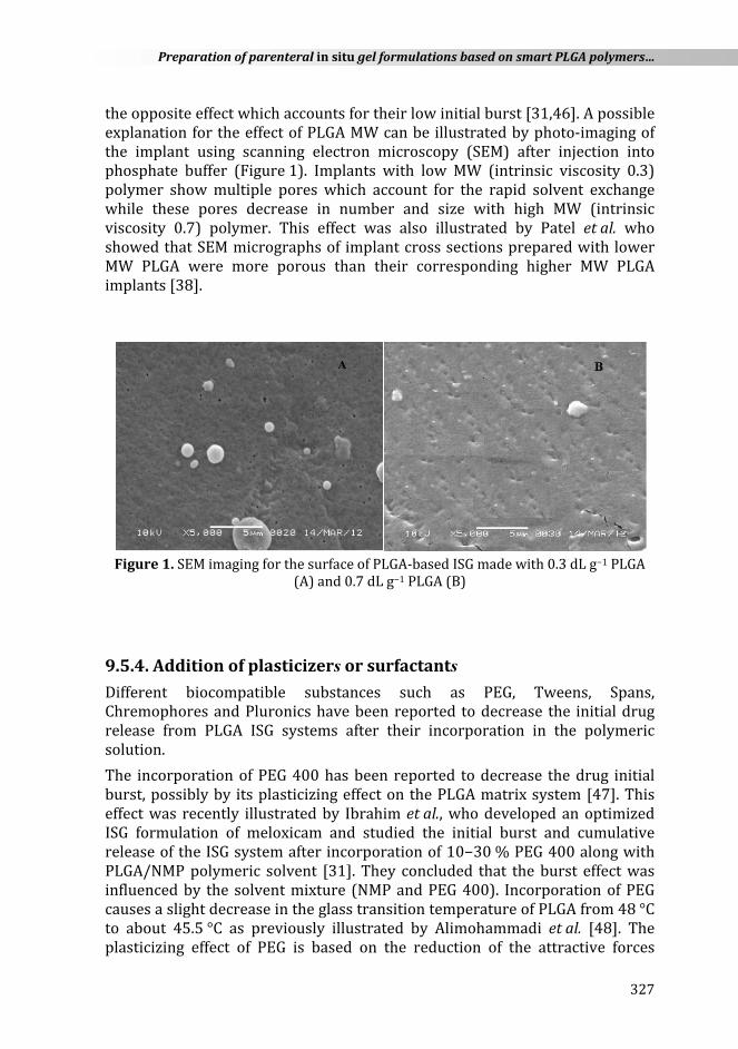
where η is the solution viscosity, η0 is the viscosity of the pure solvent and φ is the solute mass concentration (the volume fraction of the solute in the solution). The term [η−η0/η0] is the specific viscosity. Intrinsic viscosity [η] should not be confused with inherent viscosity, which is the ratio of the natural logarithm of the relative viscosity [η/η0] to the mass concentration of the polymer [ln ηrelative/φ]. The units of φ, sometimes referred to as c, are g dL−1 and the units of intrinsic viscosity [η] are dL g−1, otherwise known as inverse concentration. Intrinsic and inherent viscosity can be measured based on the flow time of a polymer solution [t] of known concentration through a narrow capillary relative to the flow time of the pure solvent [t0] through the capillary, where [η−η0/η0] = [(t−t0/t0]. The concentration of PLGA in ISG formulations normally ranges from 10−6 wt %. The viscosity of prepared polymeric solutions is greatly affected by the polymer concentration in the solution and its molecular weight [45]. High polymer concentrations up to 40−50 wt % result in decreased drug initial release but the viscosity should be considered since the injectability can be impaired [5,32]. Lambert and Peck studied the release of bovine serum albumin from PLGA solution; a relatively low initial burst effect was obtained when both high molecular weight PLGA (75−115,000 g mol−1) and high polymer loading in the solvent were used [39]. Ahmed et al. studied the release of haloperidol from a polymeric solution containing 20 %, 30 % and 40 % PLGA (50 : 50 L-:G ratio, molecular weight (MW) 60,000−70,000 g mol−1, intrinsic viscosity 0.5 dL g−1) in four different organic solvents, namely NMP, DMSO, triacetin and ethyl acetate. The initial release of the drug decreased as the polymer concentration was increased [30]. The same effect was also observed for montelukast, reported by the same research group [36]. The effect of different PLGA MW (intrinsic viscosity 0.3, 0.5 and 0.7 dL g−1) on the initial burst and cumulative release of meloxicam has been studied by Ibrahim et al. [31]. They mentioned that polymer MW and hence polymer viscosity has a great impact on meloxicam initial burst; as the intrinsic viscosity of the polymer increases the hydrophilicity decreases, and so PLGA grades that have low intrinsic viscosity (low MW) will have greater water solubility owing to the rapid uptake of water while high MW ones will possess
Preparation of parenteral in situ gel formulations based on smart PLGA polymers…
327
the opposite effect which accounts for their low initial burst [31,46]. A possible explanation for the effect of PLGA MW can be illustrated by photo-imaging of the implant using scanning electron microscopy (SEM) after injection into phosphate buffer (Figure 1). Implants with low MW (intrinsic viscosity 0.3) polymer show multiple pores which account for the rapid solvent exchange while these pores decrease in number and size with high MW (intrinsic viscosity 0.7) polymer. This effect was also illustrated by Patel et al. who showed that SEM micrographs of implant cross sections prepared with lower MW PLGA were more porous than their corresponding higher MW PLGA implants [38].
Figure 1. SEM imaging for the surface of PLGA-based ISG made with 0.3 dL g−1 PLGA
(A) and 0.7 dL g−1 PLGA (B)
9.5.4. Addition of plasticizers or surfactants Different biocompatible substances such as PEG, Tweens, Spans, Chremophores and Pluronics have been reported to decrease the initial drug release from PLGA ISG systems after their incorporation in the polymeric solution. The incorporation of PEG 400 has been reported to decrease the drug initial burst, possibly by its plasticizing effect on the PLGA matrix system [47]. This effect was recently illustrated by Ibrahim et al., who developed an optimized ISG formulation of meloxicam and studied the initial burst and cumulative release of the ISG system after incorporation of 10−30 % PEG 400 along with PLGA/NMP polymeric solvent [31]. They concluded that the burst effect was influenced by the solvent mixture (NMP and PEG 400). Incorporation of PEG causes a slight decrease in the glass transition temperature of PLGA from 48 °C to about 45.5 °C as previously illustrated by Alimohammadi et al. [48]. The plasticizing effect of PEG is based on the reduction of the attractive forces

Chapter 9
328
among the polymer chains. These effects could explain the decrease in the drug burst effect after incorporation of PEG. Another possible explanation for the effect of PEG is its solubilizing power, which allows the uniform distribution of drug particles inside the PLGA matrix and prevents adsorption of any drug particles at the surface. Morphological study of the ISG formulation without PEG shows a cracked surface (Figure 2A). Incorporation of PEG in the optimized formulation leads to the disappearance of these cracks as indicated by the smooth implant surface (Figure 2B). Tang and Singh also verified that the addition of PEG 400 to a PLGA ISG system significantly decreased the initial burst of aspirin from 36.9 ± 1.9 % to 30.9 ± 1.2 % [49].
Figure 2. SEM of the surface of an atorvastatin in situ gel system without PEG (A) and
the corresponding formulation with PEG (B) [50]. Reprinted from: Depot injectable atorvastatin biodegradable in situ gel: development, optimization, in vitro, and in vivo
evaluation. Drug Des. Devel. Ther. 10 (2016) 405–415. Copyright (2016), with permission from Dove Medical Press Ltd.
The incorporation of biocompatible surfactants such as Tweens, Spans, Chremophores or Pluronics can have a positive effect on the release profile and duration of activity. Elias-Al-Mamun et al. illustrated the effect of incorporating biocompatible excipients such as Tween 20, Tween 60, Span 20, Span 80, Chremophore EL and Chremophore RH 40 on the in vitro release of tamsulosin from biodegradable PLGA in situ implants. They stated that it was clearly observed that the studied surfactants lower the release rate of tamsulosin and prolong the drug activity [24]. Patel et al. also studied the effect of incorporating up to 5 % Pluronic P85 (P85, MW: 4600 Da) on the release profile of a sodium fluorescein (low MW mock drug molecule) in situ forming implant formulation prepared with PLGA dissolved in NMP. They verified that the P85 concentration showed a minimal effect on sodium fluorescein release during the first hour. However, after 1 h and up to 4 days (during the intermediate release stage), a P85 concentration in the range of 1−2.5 %
Preparation of parenteral in situ gel formulations based on smart PLGA polymers…
329
appears to lower the drug initial burst. Exactly at the end of the 4-day time point, in situ forming implants with 1 % and 2.5 % P85 released about 33.6 % and 28.2 %, respectively, of their drug when compared with the corresponding formulations without any P85 (38.2 %). They also stated that increasing the concentration of P85 beyond 5 % reversed any lowering of the drug burst release [38]. DesNoyer and McHugh studied the effect of variations in Pluronic concentration and MW on protein release from PLGA/NMP solutions. They used Pluronic L101 and L121 (hydrophobic), the only difference between them being the higher MW of the L121. They indicated that the Pluronic molecules direct themselves in such a way that the hydrophobic PPO parts are inserted into the polymer matrix while the hydrophilic PEO parts are extended in the surrounding aqueous phase, an effect that leads to segregation of the Pluronic molecules and formation of a phase boundary. This effect was more obvious with the higher MW Pluronics, with overall reduction in the burst effect [51].
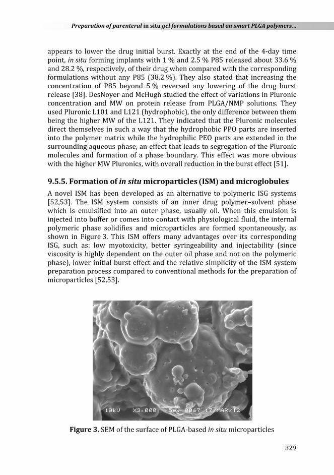
9.5.5. Formation of in situ microparticles (ISM) and microglobules A novel ISM has been developed as an alternative to polymeric ISG systems [52,53]. The ISM system consists of an inner drug polymer–solvent phase which is emulsified into an outer phase, usually oil. When this emulsion is injected into buffer or comes into contact with physiological fluid, the internal polymeric phase solidifies and microparticles are formed spontaneously, as shown in Figure 3. This ISM offers many advantages over its corresponding ISG, such as: low myotoxicity, better syringeability and injectability (since viscosity is highly dependent on the outer oil phase and not on the polymeric phase), lower initial burst effect and the relative simplicity of the ISM system preparation process compared to conventional methods for the preparation of microparticles [52,53].
Figure 3. SEM of the surface of PLGA-based in situ microparticles

Chapter 9
330
Kranz and Bodmeier studied the release of diltiazem hydrochloride and buserelin acetate from two different in situ forming systems, namely in situ implants (ISI) and ISM. Both PLA and PLGA in DMSO, NMP or 2-pyrrolidone were used to form polymeric drug solutions that were used as ISI. The ISM systems were prepared utilizing the previously described polymeric drug solutions which were emulsified into peanut oil at different polymeric solution to oil phase ratios. The release of both drugs from the ISI systems showed an initial high burst release compared to release from the ISM systems. They concluded that the ISM systems significantly reduced drug initial burst effect when compared to the ISI systems (polymer solutions) and attributed that effect to the presence of an outer oil phase which makes a partial barrier between the inner polymer solution and the outer aqueous medium. Another possible mechanism for the lower initial drug burst was the less porous surface of the ISM compared to the ISI system [52]. Another comparative study between both systems (ISI and ISM) was conducted on bupivacaine hydrochloride utilizing PLA as a biocompatible polymer which was dissolved in various organic solvents to prepare ISI, while the ISM was prepared using peanut oil as the external phase at different polymer phase to oil phase ratios as previously described. A reduced initial bupivacaine hydrochloride release was also exhibited from the ISM compared to the ISI and they also attributed this behaviour to the presence of the external oil phase and the less porous surface of the ISM [54]. Ahmed et al. also reported the same results for haloperidol and montelukast during the in vitro and in vivo release of both drugs from ISM and ISI systems [30,36]. Jain et al. developed a novel in situ method for the preparation of an injectable stable dispersion of PLGA microglobules (premicrospheres or embryonic microspheres). The preparation was made up of two oil phases. Oil phase I consists of a mixture of PLGA/triacetin/drug/PEG 400/Tween 80. This mixture is added dropwise to oil phase II which is composed of Miglyol 812 and Span 80 and homogenized to produce a rubbery injectable dispersion of PLGA microglobules. The produced embryonic or premicrospheres harden and shrink, and can entrap the drug and form true microspheres in situ within 17 min. One major advantage of this system is its ability to control the release of cytochrome c from a few days to weeks. The burst of the drug was less than 30 % (within the first 24 h) of the total drug load and they attributed the majority of that to unencapsulated drug [55].

Preparation of parenteral in situ gel formulations based on smart PLGA polymers…
331
9.5.6. ISG systems based on drug nanocrystals and nanoparticles Development of polymer-coated drug nanocrystals with subsequent loading into ISG systems is a technique recently reported to minimize the initial burst from these systems. Kurakula and Ahmed developed chitosan-coated atorvastatin nanocrystals loaded into an injectable ISG formulation composed of 45 % PLGA in a solvent mixture of NMP : benzyl benzoate (1 : 3) and observed lower initial burst, prolonged atorvastatin release and enhanced hypolipidemic effect from this formulation when compared to the corresponding pure drug-loaded ISG system [56]. In another study, Zein glimepiride nanoparticles were prepared utilizing a liquid–liquid phase separation technique and loaded into a PEGylated PLGA ISG system. The authors reported 3.3 % and 17.3 % drug release after 2 and 24 h, respectively. This ISG system exhibited pseudoplastic behaviour with a reduction of glimepiride release rate as the concentration of the polymer was increased [57]. Another factor that could play a role in the release of drugs from ISI systems is drug lipophilicity. Deadman et al. studied the effect of drug lipophilicity on the release profile from different controlled release vehicles such as PLGA microparticles and in situ forming depots. They reported that, although there was a minor effect of drug lipophilicity on the in vitro studies, the effect was obvious in vivo, which was attributed to the interaction between the formulation and biological tissue.

Chapter 9
332
9.6. APPLICATIONS OF PARENTERAL ISG SYSTEMS
9.6.1. Application in veterinary products Controlled release veterinary products characterized by a reduction of dosing frequency, long duration of action and reduced animal stress have emerged and continue to be developed to meet future market expectation. Around 15 % of the marketed veterinary products are controlled release dosage forms, of which parenteral sustained release products account for an estimated 40 % [58]. Solutions, suspensions, emulsions and implants are the most common preparations utilized for a parenteral sustained release effect. The main treatment areas are respiratory diseases, feed efficiency improvement/growth and milk enhancement. Revalor, Compudose and Synovex are hormonal solid implant marketed products used to improve growth in cattle. Milbemycin, a highly effective lipophilic anti-infective agent, has been formulated as an ISG parenteral long-acting formulation for dogs [59].
9.6.2. Application in vaccine and gene delivery Injectable sustained release ISG systems have been utilized for plasmid DNA release and gene expression. Roy et al. designed two ISG systems to modulate plasmid DNA release and gene expression for the delivery of DNA expression vectors and reported extended duration of expression after a single intramuscular administration [60]. In another study, the in vitro release of plasmid DNA and salmon sperm DNA from ISG formulations was investigated and the authors reported that ISG systems can be considered as a valuable injectable controlled delivery system for plasmid DNA in their role to provide protection from DNase degradation [61].
9.6.3. Applications in protein and peptide delivery Biodegradable injectable in situ forming drug delivery systems may offer an attractive method of protein delivery and could possibly extend the patent life of therapeutic drugs that are proteins in nature [4]. Joshi reported successful entrapment, protection and control of in vitro release of the model protein macromolecule alpha-amylase for up to 6 days and it can therefore be considered for in vivo investigation [62]. In another study, ISG systems composed of PLGA and PEG have been widely studied for delivery of proteins and peptides as these systems are biocompatible and biodegradable. The triblock copolymer of PLGA–PEG–PLGA has been utilized as a controlled release ISG system for porcine growth hormone; a single subcutaneous injection of polymer formulation provided almost steady state serum levels of the administered hormone for nearly 4 weeks [63].

Preparation of parenteral in situ gel formulations based on smart PLGA polymers…
333
9.7. CURRENT AND FUTURE DEVELOPMENT The demand for parenteral controlled release drug delivery systems is progressively increasing. This is apparent in the number of polymer-based drug delivery systems that are under clinical trials and those that have progressed to commercial production. ISG systems based on smart polymers are among the controlled release drug delivery systems that need further development to administer the prepared formulation as easily as possible and release the incorporated agent in a chemically and conformationally stable form for a longer duration with minimal side effects. New strategies are now under investigation such as:
• Preparation of novel polymer- and lipid-based nanoparticles with subsequent loading into ISG systems.
• Incorporation of new surfactants and plasticizers.
9.8. CONCLUSION Smart polymers have been investigated for their role in drug delivery systems, particularly in the formulation of ISG systems. Among those, the biocompatible and biodegradable polymer PLGA has found wide application in drug delivery, particularly via the parenteral route. PLGA-based ISG systems are efficient injectable biocompatible controlled release systems as they provide efficient tools for the delivery of small molecules and macromolecules. Several attempts have been investigated to lower the initial burst associated with these systems. Processing and formulation factors and a combination of two or more techniques can be optimized to produce the desired drug release profile.

Chapter 9
334
REFERENCES 1. R.L. Dunn, J.P. English, D.R. Cowsar, D.P. Vanderbilt, (Atrix Laboratories, Inc.),
US Patent No. 4938 763 (1990). 2. W. Xiaohong, S. Sui, Y. Yan, R. Zhang. J. Bioact. Compat. Polym. 25(3) (2010)
229−240. 3. D.Y. Ko, U.P. Shinde, B. Yeon, B. Jeong. Prog. Polym. Sci. 38(3-4) (2013)
672−701. 4. C.B. Packhaeuser, J. Schnieders, C.G. Oster, T. Kissel. Eur. J. Pharm. Biopharm.
58(2) (2004) 445−455. 5. S. Kempe, K. Mäder. J. Control. Release 161(2) (2012) 668−679. 6. A. Hatefi, B. Amsden. J. Control. Release 80(1) (2002) 9−28. 7. R.E. Eliaz, J. Kost. J. Biomed. Mater. Res. 50(3) (2000) 388−396. 8. B. Jeong, Y.H. Bae, S.W. Kim. J. Control. Release 63(1-2) (2000) 155−163. 9. R.A. Siegel, B.A. Firestone. Macromolecules 21(11) (1988) 3254−3259.
10. A.B. Kumbhar, A.K. Rakde, P. Chaudhari. Int. J. Pharm. Sci. Res. 4(2) (2013) 597−609.
11. K. Al-Tahami, J. Singh. Recent Pat. Drug Deliv. Formul. 1(1) (2007) 65−71. 12. G. Molinaro, J.-C. Leroux, J. Damas, A. Adam. Biomaterials 23(13) (2002)
2717−2722. 13. A. Bochot, E. Fattal, A. Gulik, G. Couarraze, P. Couvreur. Pharm. Res. 15(9)
(1998) 1364−1369. 14. C. He, S.W. Kim, D.S. Lee. J. Control. Release. 127(3) (2008) 189−207. 15. Y. Qiu, K. Park. Adv. Drug Deliv. Rev. 53(3) (2001) 321−339. 16. B. Jeong, A. Gutowska. Trends Biotechnol. 20(7) (2002) 305−311. 17. R. Masteiková, Z. Chalupová, Z. Sklubalová. Medicina (Kaunas) 39(2) (2003)
19−24. 18. S. Fredenberg, M. Wahlgren, M. Reslow, A. Axelsson. Int. J. Pharm. 415(1-2)
(2011) 34−52. 19. R.A. Jain. Biomaterials 21(23) (2000) 2475−2490. 20. R. Jalil, J.R. Nixon. J. Microencapsul. 7(3) (1990) 297−325. 21. Y.-Y. Huang, M. Qi, H.-Z. Liu, H. Zhao, D.-Z. Yang. J. Biomed. Mater. Res. A 80(4)
(2007) 909−915. 22. H. Murakami, M. Kobayashi, H. Takeuchi, Y. Kawashima. Int. J. Pharm. 187(2)
(1999) 143−152. 23. M. Husmann, S. Schenderlein, M. Lück, H. Lindner, P. Kleinebudde. Int. J. Pharm.
242(1-2) (2002) 277−280. 24. M. Elias-Al-Mamun, H.A. Khan, I. Dewan, R.-U. Jalil. Pak. J. Pharm. Sci. 22(4)
(2009) 360−367. 25. M. Shive, J. Anderson. Adv. Drug Deliv. Rev. 28(1) (1997) 5−24. 26. X. He, N. Kawazoe, G. Chen. Biomed Res. Int. (2014) 106082. 27. H. Kranz, N. Ubrich, P. Maincent, R. Bodmeier. J. Pharm. Sci. 89(12) (2000)
1558−1566. 28. S.S. D’Souza, J.A. Faraj, P.P. DeLuca. AAPS PharmSciTech 6(4) (2005) E553-564. 29. L. Wang, S. Venkatraman, L. Kleiner. J. Control. Release 99(2) (2004) 207−216. 30. T.A. Ahmed, H.M. Ibrahim, F. Ibrahim, A.M. Samy, A. Kaseem, M.T. H. Nutan,
M.D. Hussain. J. Pharm. Sci. 101(10) (2012) 3753−3762. 31. H.M. Ibrahim, T.A. Ahmed, M.D. Hussain, Z. Rahman, A.M. Samy, A. Kaseem,
M.T.H. Nutan. Drug Dev. Ind. Pharm. 9045 (2013) 1−8.

Preparation of parenteral in situ gel formulations based on smart PLGA polymers…
335
32. H. Kranz, G.A. Brazeau, J. Napaporn, R.L. Martin, W. Millard, R. Bodmeier. Int. J. Pharm. 212(1) (2001) 11−18.
33. X. Huang, C.S. Brazel. Chem. Eng. Commun. 190(4) (2003) 519−532. 34. K.J. Pekarek, J.S. Jacob, E. Mathiowitz. Nature 367(6460) (1994) 258−260. 35. S.K. Mallapragada, N.A. Peppas, P. Colombo. J. Biomed. Mater. Res. 36(1) (1997)
125−130. 36. T.A. Ahmed, H.M. Ibrahim, A.M. Samy, A. Kaseem, M.T.H. Nutan, M.D. Hussain.
AAPS PharmSciTech 15(3) (2014) 772−780. 37. M. Madhu, L. Shaila, B.J. Anwar. Int. J. Pharm. Pharm. Sci. 1(1) (2009) 103−107. 38. R.B. Patel, A.N. Carlson, L. Solorio, A.A. Exner. J. Biomed. Mater. Res. A 94(2)
(2010) 476−484. 39. W.J. Lambert, K.D. Peck. J. Control. Release 33(1) (1995) 189−195. 40. F. Kang, J. Singh. Int. J. Pharm. 304(1-2) (2005) 83−90. 41. K.J. Brodbeck, S. Pushpala, A.J. McHugh. Pharm. Res. 16(12) (1999)
1825−1829. 42. R.G. Strickley. Pharm. Res. 21(2) (2004) 201−230. 43. W.Y. Dong, M. Körber, V. López Esguerra, R. Bodmeier. J. Control. Release,
115(2) (2006) 158−167. 44. N.H. Shah, A.S. Railkar, F.C. Chen, R. Tarantino, S. Kumar, M. Murjani, D. Palmer,
M.H. Infeld, A.W. Malick. J. Control. Release 27(2) (1993) 139−147. 45. A.J. McHugh. J. Control. Release 109(1-3) (2005) 211−221. 46. D. Bodmer, T. Kissel, E. Traechslin. J. Control. Release 21(1-3) (1992) 129−137. 47. L.P. Tan, S.S. Venkatraman, P.F. Sung, X.T. Wang. Int. J. Pharm. 283(1-2) (2004)
89−96. 48. S. Alimohammadi, R. Salehi, N. Amini, S. Davaran. Bull. Korean Chem. Soc.
33(10) (2012) 3225−3232. 49. Y. Tang, J. Singh. Int. J. Pharm. 357(1-2) (2008) 119−125. 50. K.M. El-Say, Y. Alharby, A.-R. El-Helw, K.M. Hosny, T. Ahmed. Drug Des. Devel.
Ther. (2016) 405. 51. J.R. DesNoyer, A.J. McHugh. J. Control. Release 86(1) (2003) 15−24. 52. H. Kranz, R. Bodmeier. Int. J. Pharm. 332(1-2) (2007) 107−114. 53. H. Kranz, E. Yilmaz, G.A. Brazeau, R. Bodmeier. Pharm. Res. 25(6) (2008)
1347−1354. 54. H. Kranz, R. Bodmeier. Eur. J. Pharm. Sci. 34(2-3) (2008) 164−172. 55. R.A. Jain, C.T. Rhodes, A.M. Railkar, A.W. Malick, N.H. Shah. Eur. J. Pharm.
Biopharm. 50(2) (2000) 257−262. 56. M. Kurakula, T.A. Ahmed. Curr. Drug Deliv. 13 (2016) 1−11. 57. O.A.A. Ahmed, A.S. Zidan, M. Khayat. Int. J. Nanomedicine 11 (2016) 543−555. 58. N.J. Medlicott, N.A. Waldron, T.P. Foster. Adv. Drug Deliv. Rev. 56(10) (2004)
1345−1365. 59. C. Anne, F. Sautter. “Sustained release injectables formed in-situ for veterinary
use,” Faculty of Natural Sciences, University of Basel, 2006. 60. K. Roy. Mol. Ther. 7(3) (2003) 401−408. 61. F.A. Ismail, J. Napaporn, J.A. Hughes, G.A. Brazeau. Pharm Dev Technol. 5(3)
(2000) 391−397. 62. R. Joshi, D.H. Robinson, K.J. Himmelstein. Pharm. Dev. Technol. 4(4) (1999)
515−522. 63. S. Chen, J. Singh. Int. J. Pharm. 352(1-2) (2008) 58−65.

Chapter 9
336