presence of the myelin-associated glycoprotein correlates with
TRANSCRIPT

Presence of the Myelin-associated Glycoprotein Correlates with
Alterations in the Periodicity of Peripheral Myelin
BRUCE D. TRAPP and RICHARD H. QUARLESInfectious Diseases Branch and Developmental and Metabolic Neurology Branch, National Institute of Neurological andCommunicative Disorders and Stroke, National Institutes of Health, Bethesda, Maryland 20205
ABSTRACT
The myelin-associated glycoprotein (MAG) is an integral membrane protein (=100,000 molwt) which is a minor component of purified peripheral nervous system (PNS) myelin . In the presentstudy, MAG was localized immunocytochemically in 1-pm thick Epon sections of 7-d and adult ratperipheral nerves, and its localization was compared to that of the major structural protein (Po) ofPNS myelin . To determine more precisely the localization of MAG, immunostained areas in 1 [Lmsections were traced on electron micrographs of identical areas from adjacently cut thin sections .MAG was localized in periaxonal membranes, Schmidt-Lantermann incisures, paranodal membranes,and the outer mesaxon of PNS myelin sheaths. Compact regions of PNS myelin did not react withMAG antiserum . The results demonstrate MAG's presence in "semi-compact" Schwann cell or myelinmembranes that have a gap of 12-14 nm between extracellular leaflets and a spacing of 5 nm or morebetween cytoplasmic leaflets . In compact regions of the myelin sheath which do not contain MAG,the cytoplasmic leaflets are "fused" and form the major dense line, whereas the extracellular leafletsare separated by a 2 .0 nm gap appearing as paired minor dense lines. Thus, it is proposed that MAGplays a role in maintaining the periaxonal space, Schmidt-Lantermann incisures, paranodal myelinloops, and outer mesaxon by preventing "complete" compaction of Schwann cell and myelinmembranes . The presence of MAG in these locations also suggests that MAG may serve a function inregulating myelination in the PNS.
Myelin-associated glycoprotein (MAG) is an integral mem-brane glycoprotein (=100,000 mol wt) which is quantitativelya minor component of purified CNS and PNS myelin . Itspresence in purified CNS myelin was first demonstrated byradioactive labeling with sugar precursors (10) . Subfractiona-tion experiments suggested that MAG was not present incompact CNS myelin but was concentrated in closely associ-ated oligodendroglial membranes (11) . This hypothesis wasconfirmed by immunocytochemical studies (15) . When sectionsof developing rat brain were treated with MAG antiserum,oligodendrocytes, and myelin sheaths were selectively stained.When compact myelin sheaths grew in thickness, only periax-onal portions of the myelin sheaths were stained. Compactmyelin lamellae did not react with MAG antiserum . An un-expected finding in the immunocytochemical study (15) wasthe staining for MAG in PNS myelin sheaths, because previousbiochemical studies had not detected MAG in PNS myelin (3).However, reexamination with biochemical and immunochem-ical techniques has now also revealed the presence of MAG inthe PNS (4) . In the PNS, MAG antiserum stained periaxonalmembranes, Schmidt-Lantermann incisures, and paranodalportions of PNS myelin sheaths (14, 15) .
THE JOURNAL Of CELL BIOLOGY " VOLUME 92 MARCH 1982 877-882©The Rockefeller University Press " 0021-9525/82/03/0877/06 $1 .00
Integral membrane glycoproteins are believed to be involvedin cell-cell recognition and in interactions between cell mem-branes (5) . The periaxonal localization of MAG in CNS andPNS myelin sheaths suggests a possible role for this glycopro-tein in glia-axon interactions. The present study describes theimmunocytochemical localization ofMAG in 1-Itm thick Eponsections of developing and adult rat peripheral nerves. MAGlocalization is compared to that of the major structural protein(Po) of PNS myelin . To determine more precisely the locali-zation of MAG, immunostained areas in 1-tin thick Eponsections were tracted on electron micrographs of adjacent thinsections .
MATERIALS AND METHODS
Tissue
Seven-day-old and adult Sprague-Dawley rats were fixed by intracardiacperfusion with a solution containing 1 .5% glutaraldehyde and 0.5% paraformal-dehyde in 0.08 M Sorensen's buffer. Segments from the trigeminal and sciaticnerves were removed and placed in the fixative for an additional hour. To obtainbetter fixation of adult sciatic nerves, additional animals were perfused throughthe abdominal aorta. Thetissue waspostfixed in 2% osmium tetroxide, dehydratedin ethanol, and embedded in Epon. l pm-thick sections were cut with glass
877
on April 3, 2019jcb.rupress.org Downloaded from http://doi.org/10.1083/jcb.92.3.877Published Online: 1 March, 1982 | Supp Info:
knives, mounted on glass slides, and encircled with a diamond scribe . Six serialsections were cut from the 7-d-old and adult nerves . Even numbered sectionswere stained immunocytochemically with MAG antiserum ; the others werestained with Po antiserum. From the 7-d trigeminal nerve, additional 1-pm thicksections were cut adjacent to thin sections with silver interference colors . The I-lam thick sections were stained with MAG antiserum . The thin sections weremounted on formvar-coated slot grids, stained with uranyl acetate and leadcitrate, and examined in a Philips 400 electron microscope. Areas of MAGstaining in 1-pm thick sections were photographed, and their negative imageswere enlarged and compared to the fine structure in electron micrographs fromidentical areas of the adjacently cut thin sections .
Immunostaining Procedure
I-Am thick Epon sections mounted on glass slides were placed in a 60-80°Coven for 24-48 h. Epon was removed from the sections by sodium ethoxide aspreviously described (18). The slides were then placed in 0.2% hydrogen peroxidefor 5 min, rinsed in 0.5 M Tris buffer, and stained with a (1 :250) dilution ofMAGantiserum or a (1 :500) dilution of Po antiserum by the peroxidase-antiper-oxidase (PAP) method as previously described (18) . All sections were examinedmicroscopically with bright-field illumination.MAG and Po antisera were prepared in rabbits. The purity of the MAGand
Po proteins used in preparing the antisera and the immunological and immuno-cytochemical specificity of these antisera have been described (6, 13, 15, 17, l8) .
RESULTS
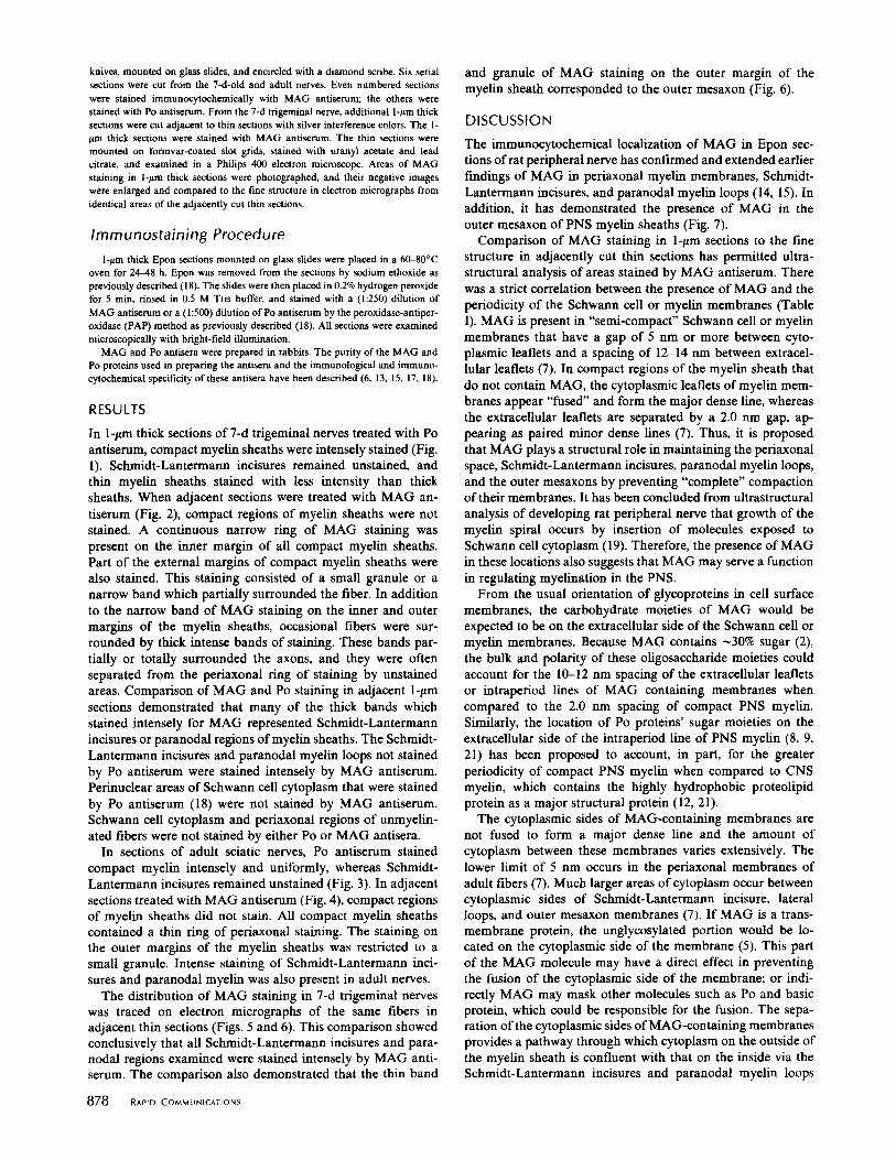
In 1-ltm thick sections of 7-d trigeminal nerves treated with Poantiserum, compact myelin sheaths were intensely stained (Fig .1) . Schmidt-Lantermann incisures remained unstained, andthin myelin sheaths stained with less intensity than thicksheaths. When adjacent sections were treated with MAG an-tiserum (Fig. 2), compact regions of myelin sheaths were notstained. A continuous narrow ring of MAG staining waspresent on the inner margin of all compact myelin sheaths.Part of the external margins of compact myelin sheaths werealso stained. This staining consisted of a small granule or anarrow band which partially surrounded the fiber. In additionto the narrow band of MAG staining on the inner and outermargins of the myelin sheaths, occasional fibers were sur-rounded by thick intense bands of staining. These bands par-tially or totally surrounded the axons, and they were oftenseparated from the periaxonal ring of staining by unstainedareas . Comparison of MAG and Po staining in adjacent 1-ltmsections demonstrated that many of the thick bands whichstained intensely for MAG represented Schmidt-Lantermannincisures or paranodal regions ofmyelin sheaths . The Schmidt-Lantermann incisures and paranodal myelin loops not stainedby Po antiserum were stained intensely by MAG antiserum.Perinuclear areas ofSchwann cell cytoplasm that were stainedby Po antiserum (18) were not stained by MAG antiserum.Schwann cell cytoplasm and periaxonal regions of unmyelin-ated fibers were not stained by either Po or MAG antisera .
In sections of adult sciatic nerves, Po antiserum stainedcompact myelin intensely and uniformly, whereas Schmidt-Lantermann incisures remained unstained (Fig. 3) . In adjacentsections treated with MAG antiserum (Fig . 4), compact regionsof myelin sheaths did not stain. All compact myelin sheathscontained a thin ring of periaxonal staining. The staining onthe outer margins of the myelin sheaths was restricted to asmall granule . Intense staining of Schmidt-Lantermann inci-sures and paranodal myelin was also present in adult nerves .The distribution of MAG staining in 7-d trigeminal nerves
was traced on electron micrographs of the same fibers inadjacent thin sections (Figs . 5 and 6) . This comparison showedconclusively that all Schmidt-Lantermann incisures and para-nodal regions examined were stained intensely by MAG anti-serum . The comparison also demonstrated that the thin band
878
RAPID COMMUNICATIONS
and granule of MAG staining on the outer margin of themyelin sheath corresponded to the outer mesaxon (Fig. 6) .
DISCUSSION
The immunocytochemical localization of MAG in Epon sec-tions of rat peripheral nerve has confirmed and extended earlierfindings of MAG in periaxonal myelin membranes, Schmidt-Lantermann incisures, and paranodal myelin loops (14, 15) . Inaddition, it has demonstrated the presence of MAG in theouter mesaxon of PNS myelin sheaths (Fig. 7) .Comparison of MAG staining in 1-/.Lm sections to the fine
structure in adjacently cut thin sections has permitted ultra-structural analysis of areas stained by MAG antiserum . Therewas a strict correlation between the presence of MAG and theperiodicity of the Schwann cell or myelin membranes (TableI) . MAG is present in "semi-compact" Schwann cell or myelinmembranes that have a gap of 5 nm or more between cyto-plasmic leaflets and a spacing of 12-14 nm between extracel-lular leaflets (7) . In compact regions of the myelin sheath thatdo not contain MAG, the cytoplasmic leaflets of myelin mem-branes appear "fused" and form the major dense line, whereasthe extracellular leaflets are separated by a 2.0 nm gap, ap-pearing as paired minor dense lines (7) . Thus, it is proposedthat MAG plays a structural role in maintaining the periaxonalspace, Schmidt-Lantermann incisures, paranodal myelin loops,and the outer mesaxons by preventing "complete" compactionoftheir membranes . It has been concluded from ultrastructuralanalysis of developing rat peripheral nerve that growth of themyelin spiral occurs by insertion of molecules exposed toSchwann cell cytoplasm (19) . Therefore, the presence of MAGin these locations also suggests that MAG may serve a functionin regulating myelination in the PNS.From the usual orientation of glycoproteins in cell surface
membranes, the carbohydrate moieties of MAG would beexpected to be on the extracellular side of the Schwann cell ormyelin membranes. Because MAG contains -30% sugar (2),the bulk and polarity of these oligosaccharide moieties couldaccount for the 10-12 nm spacing of the extracellular leafletsor intraperiod lines of MAG containing membranes whencompared to the 2.0 nm spacing of compact PNS myelin .Similarly, the location of Po proteins' sugar moieties on theextracellular side of the intraperiod line of PNS myelin (8, 9,21) has been proposed to account, in part, for the greaterperiodicity of compact PNS myelin when compared to CNSmyelin, which contains the highly hydrophobic proteolipidprotein as a major structural protein (12, 21) .The cytoplasmic sides of MAG-containing membranes are
not fused to form a major dense line and the amount ofcytoplasm between these membranes varies extensively . Thelower limit of 5 nm occurs in the periaxonal membranes ofadult fibers (7) . Much larger areas of cytoplasm occur betweencytoplasmic sides of Schmidt-Lantermann incisure, lateralloops, and outer mesaxon membranes (7) . If MAG is a trans-membrane protein, the unglycosylated portion would be lo-cated on the cytoplasmic side of the membrane (5) . This partof the MAG molecule may have a direct effect in preventingthe fusion of the cytoplasmic side of the membrane ; or indi-rectly MAG may mask other molecules such as Po and basicprotein, which could be responsible for the fusion. The sepa-ration ofthe cytoplasmic sides of MAG-containing membranesprovides a pathway through which cytoplasm on the outside ofthe myelin sheath is confluent with that on the inside via theSchmidt-Lantermann incisures and paranodal myelin loops
FIGURE 1
Transverse section of 7-d trigeminal nerve stained with Po antiserum (1 :500) . thick myelin sheaths are intensely stained ;thinner sheaths are surrounded by less intense staining . Areas of Schmidt-Lantermann incisures (arrows) are unstained . Brightfield . Bar, 10 l.m . X 950.
FIGURE 2
This section is adjacent to that shown in Fig . 1 ; it was stained with MAG antiserum (1 :250) . All myelinated axons aresurrounded by a thin ring of periaxonal staining . Portions of the external margins of compact myelin sheaths contain a granule orband of staining that partially surrounds the fiber. Areas of Schmidt-Lantermann incisures (arrows) and paranodal regions ofmyelin sheaths (arrowhead) are intensely stained . Bright field . Bar, 10 jm . X 950.
FIGURE 3
Transverse section, adult sciatic nerve, stained with Po antiserum (1 :500) . Myelin sheaths are intensely stained . Schmidt-Lantermann incisures are unstained (arrows) . Bright field . Bar, 10gm . X 1,500 .
FIGURE 4
This section is adjacent to that shown in Fig . 3 ; it was stained with MAG antiserum (1 :250) . All myelinated axons aresurrounded by a thin ring of periaxonal staining . Outer margins of myelin sheaths contain a single granule of staining (arrows) .Compact portions of myelin sheaths are unstained . Schmidt-Lantermann incisures are intensely stained . Bright field . Bar, 10 gm .X 1,500.
879

FIGURE 5
Electron micrograph of 7-d trigeminal nerve . The insert shows the same area in an adjacent 1-/im section that wasstained with MAG antiserum . Fiber A is surrounded by a Schmidt-Lantermann incisure which is intensely stained by MAGantiserum . Compact portions of the myelin sheath are unstained . Periaxonal ring of staining is also present. Fiber B is surroundedby paranodal myelin loops that are intensely stained by MAG antiserum . Outer mesaxons (arrows) are also stained by MAGantiserum . Bar, 1 ,um . EM ; x 12,250, insert ; x 3,200.
(Fig. 7) . These cytoplasmic channels may facilitate the trans-port of metabolites and degradation products throughout themyelin sheath.
880
RAPID COMMUNICATIONS
Recent experiments have indicated that Schwann cells re-quire a signal from the axon to produce myelin (1, 16). Becausemembrane glycoproteins are believed to be involved in recog-

FIGURE 6
Electron micrograph of a myelinated fiber from a 7-d trigeminal nerve . The insert shows the same fiber in an adjacent1-pm section that was stained by MAG antiserum . MAG staining is restricted to a periaxonal ring and to the outer mesaxon . Bar,1 gm . EM ; X 25,500, insert; X 5,000 .
nition and cell-cell interactions, the periaxonal location ofMAG is consistent with the hypothesis that MAG may beinvolved in glial-axonal interactions (12, 20). What role MAGmay play in the initiation of myelination in either the central
or peripheral nervous system remains to be clarified. The datapresented in this manuscript support a structural role for MAGin maintaining the periodicity of discrete regions of Schwanncell and PNS myelin membranes.
RAPID COMMUNICATIONS
881

FIGURE 7
Diagrammatic representation of MAG localization in faceand transverse views of the "unrolled" myelin sheath . In the faceview, the dark bands represent areas stained by MAG antiserum(OM-outer mesaxon, SL-Schmidt-Lantermann incisures, PL-para-nodal loops, PM-periaxonal membranes) . The stippled areas repre-sent compact regions of the myelin sheath . Transverse views A andB correspond to levels shown in the face view . Cytoplasmic side ofMAG containing membranes (thick lines) are separated by Schwanncell cytoplasm which is confluent with that of the cell soma .
882
RAPID COMMUNICATIONS
The authors wish to thank Mr . Raymond Rusten for excellent technicalassistance and Judy Hertler for typing the manuscript .Received for publication 25 September 1981, and in revised form 26October 1981.
REFERENCES
I . Aguayo, A. J., L. Charron, and G. M . Bray. 1976. Potential of Schwann cells fromuamyelinated nerves to produce myelin: a quantitative ultrastruclural and radiographicstudy . J. Neurocytol 5 :565-573 .
2 . Barbarash, G . R ., D. A. Figlewicz, and R . H . Quarles . 1981 . Myelin-associate d glycopro-tein: purification and partial characterization . Trans. Amer. Soc. Neurochem. 12 :165.
3 . Everly, J . L ., R. O . Brady, and R . H, Quarles . 1973 . Evidence that the major protein in ratsciatic nerve myelin is a glycoprotein. J. Neurochem. 21 :329-334 .
4 . Figlewicz, D. A., R. H . Quarles, D. Johnson, G . R. Barbarash, and N . H. Sternberger .1981 . Biochemical demonstration of the myelin-associated glycoprotein in the peripheralnervous system . J. Neurochem. 37 :749-758.
5 . Hughes, R . C. 1976 . Membrane glycoprotein : a review of structure and function . Butter-worth Publishers, Inc., Woburn, Mass .
6 . Johnson, D ., R. H. Quarles, and R . O . Brady . 1980. A radioimmunoassay for the myelinassociated glycoprotein, Fed. Proc. 39 :1831 .
7 . Peters, A ., S. L. Palay, and H . deF . Webster . 1976, The fine structure of the nervoussystem . The neurons and supporting cells . W . B . Saunders Company, Philadelphia, PA .
8 . Peterson, R. G ., and D. C . Pease . 1972 . Myelin embedded in polymerized glutaraldehyde-urea . J. Ultrasiruct. Res. 41 :115-132 .
9 . Peterson, R . G ., and R . W . Gruener. 1978 . Morphological localization of PNS myelinproteins . Brain Res. 152 :17-29 .
10 . Quarles, R . H ., 1. L . Everly, and R . O . Brady . 1973 . Evidence for the close association ofa glycoprotein with myelin in rat brain. J. Neurochem. 21 :1177-1191 .
11 . Quarles, R. H . 1979 . Glycoproteins in myelin and myelin-related membranes. In : ComplexCarbohydrates of the Nervous System. R. U. Margolis and R . K . Margolis, editors.Plenum Press, New York, NY 209-233.
12 . Quarles, R. H. 1980. Glycoproteins from the central and peripheral myelin . In: Myelin :Chemistry and Biology . G. A. Hashim, editor . Alan R . Liss, Inc ., New YorkNY 55-77 .
13. Quarles, R . H ., D . Johnson, R. O. Brady, and N . H . Stemberger. 1981 . Preparation andcharacterization of antisera to the myelin-associated glycoprotein. Neurochem . Res. 6 :1109-1121 .
14ShbRJItNHStbBDTEPRihdAKA. coer, ., . oyama, . . emerger, . . rapp, . . carson, . . sury,TABLE I R . H . Quarles, and H . deF . Webster. 1981 . Immunocytochemical study of Po glycoprotein,Membrane Periodicity and Immunocytochemical Localization P,, and P z basic proteins, and myelin-associated glycoprotein (MAG) in lesions of
of MAG in PNS Myelin
MAG No MAG
idiopathic polyneuritis. Neuropath. and Applied Neurobiol. 7 :437--051 .15 . Sternberger, N . H., R. H . Quarles, Y . Itoyama, and H . deF . Webster . 1979 . Myelin-
associated glycoprotein demonstrated immunocytochemically in myelin and myelin-form-ing cells of developing rats. Proc . Nail A cad. Sci. U S. A . 76:1510-1514.
16 . Spencer, P. S. 1979 . Neuronal regulation of myelinating cell function . Soc. Nmrosci.Membrane periodicity Syrup. 4:275-321 .
Separation of : 17 . Trapp, B . D., L . J . McIntyre, R. H . Quarles, N . H . Stemberger, and H . deF . Webster .1979 . Immunocytochemical localization of rat peripheral nervous system myelin proteins:
Extracellular leaflets 12-14 nm 1 .5-2 .5 nm P2 protein is not a component of all peripheral nervous system myelin sheaths . Proc. Nall.
Cytoplasmic leaflets >5 nm "Fused" Acad. Sci. U. S. A . 76:3552-3556 .18. Trapp, B. D ., Y. Itoyama, N . H, Sterberger, R. H . Quarles, and H . deF . Webster. 1981 .
Immunocytochemical localization of Po protein in Golgi membranes and myelin ofdeveloping rat Schwann ccells. J. Cell Biol. 90:1-6 .
Location Periaxonal Compact myelin 19. Webster, H . deF . 1971 . The geometry of peripheral myelin sheaths during their formationand growth in rat sciatic nerves . J. Cell Biol. 48 :348-367 .
Schmidt-Lanter- 20 . Webster, H. deF ., B. D. Trapp, N . H. Stemberger, and R . H . Quarles . 1981 . Myeli n
mann incisures forming glial cells : morphological and immunocytochemical observations . In : Symposiumon Development in the Nervous System . D . R . Garrod and 1 . D . Feldman, editors.
Paranodal loops Cambridge University Press, Cambridge, England . 265-288 .
Outer mesaxon 21 . Wood, 1 . G ., and B . 1 . McLaughlin . 1975. The visualization of concanavalin A. Bindingsites in the interperiod line of rat sciatic nerve myelin . J. Neurochem. 24:233-235 .