presented by gordon holt, ph.d. at the nonclinical studies subcommittee of the advisory committee...
TRANSCRIPT
Presented by
Gordon Holt, Ph.D.at the
Nonclinical Studies Subcommitteeof the
Advisory Committee for Pharmaceutical Science
March 9, 2000
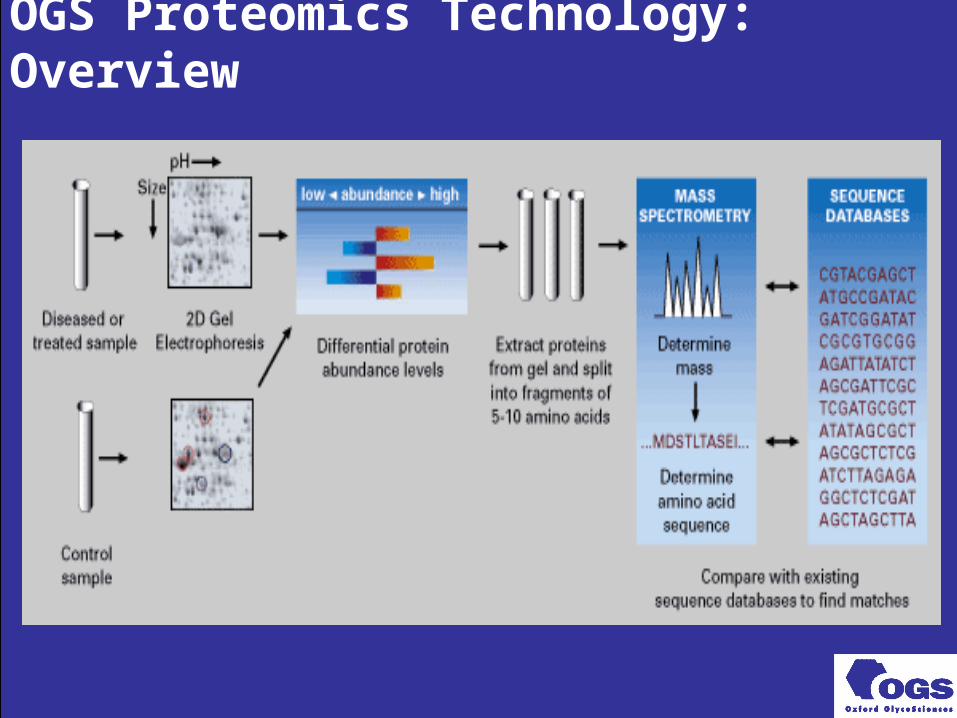
OGS Proteomics Technology: Overview
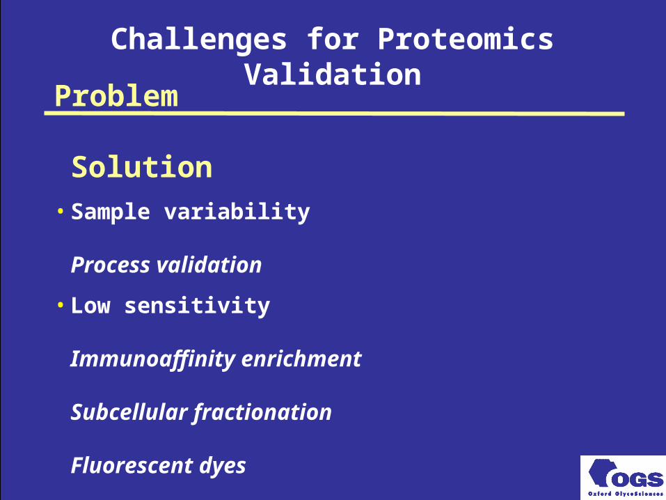
Challenges for Proteomics Validation
Problem
Solution• Sample variability
Process validation
• Low sensitivity
Immunoaffinity enrichment
Subcellular fractionation
Fluorescent dyes
Imaging
• Gel variability
Process validation
Image warping
• Low throughput
Robotics
• Data analysis overload
LIMS management
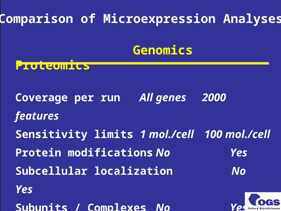
Genomics Proteomics
Coverage per run All genes 2000 features
Sensitivity limits 1 mol./cell 100 mol./cell
Protein modifications No Yes
Subcellular localization No Yes
Subunits / Complexes No Yes
Clinical samples No Yes
Comparison of Microexpression Analyses

Identification of Cardiotoxicity and Vasculitis Surrogate Markers
Work done in
collaboration with
Frank Sistare
CDER
FDA
Investigation of Doxorubicin-Induced Toxicity
Doxorubicin (Dxr) background
• commonly used anticancer agent
– effective against childhood leukemia
• causes dose-related cardiotoxicity
– precise toxicity mechanism unknown
– metal ions appear to be important
• metal chelation by ICRF-187 provides significant chemoprotection
>> Can proteomics identify clinically relevant early markers of cardiotoxicity?

Doxorubicin cardiotoxicity study design
• Three rats per group
• 1 mg/kg doxorubicin per week
• Treat for 7 weeks
• Sacrifice animals 24 hrs after last dose
• Proteomic analyses of plasma
– OGS plasma sample SOP
– serum enrichment applied
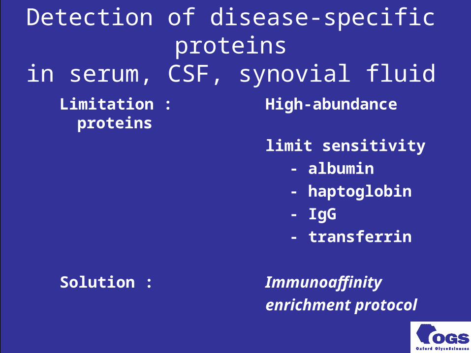
Detection of disease-specific proteinsin serum, CSF, synovial fluid
Limitation : High-abundance proteins
limit sensitivity
- albumin
- haptoglobin
- IgG
- transferrin
Solution : Immunoaffinity
enrichment protocol
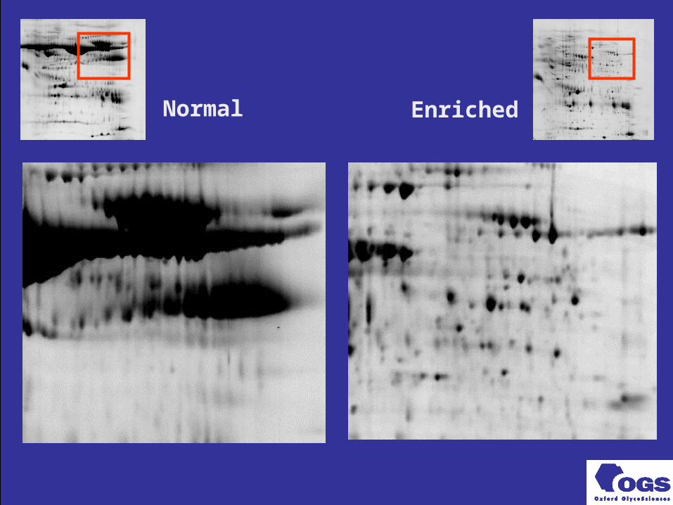
Normal Enriched
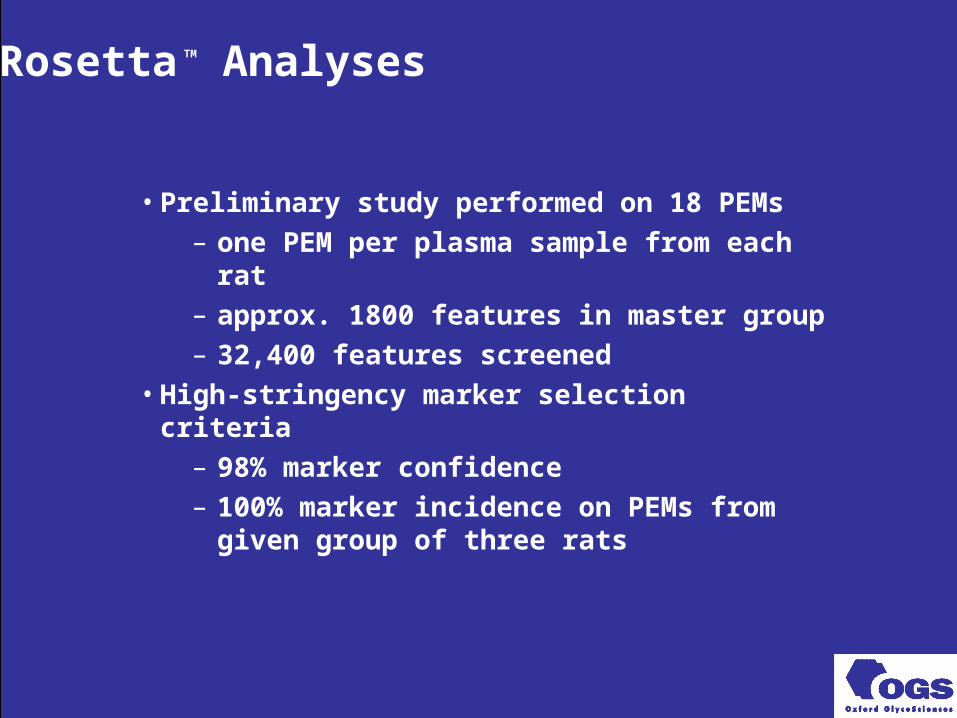
Rosetta
TM Analyses
• Preliminary study performed on 18 PEMs
– one PEM per plasma sample from each rat
– approx. 1800 features in master group
– 32,400 features screened
• High-stringency marker selection criteria
– 98% marker confidence
– 100% marker incidence on PEMs from given group of three rats
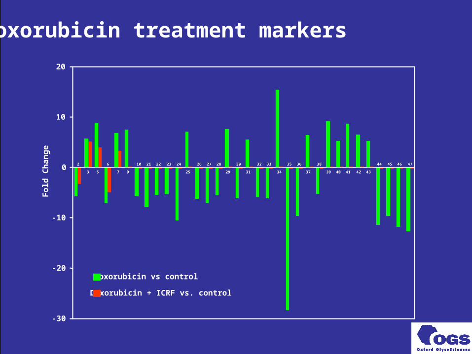
Doxorubicin + ICRF vs. control
Doxorubicin treatment markers
-30
-20
-10
0
10
20
Fold
Ch
an
ge
3 5 7 9 25 29 31 34 37 39 40 41 42 43
2 6 10 21 22 23 24 26 27 28 30 32 33 35 36 38 44 45 46 47
Doxorubicin vs control
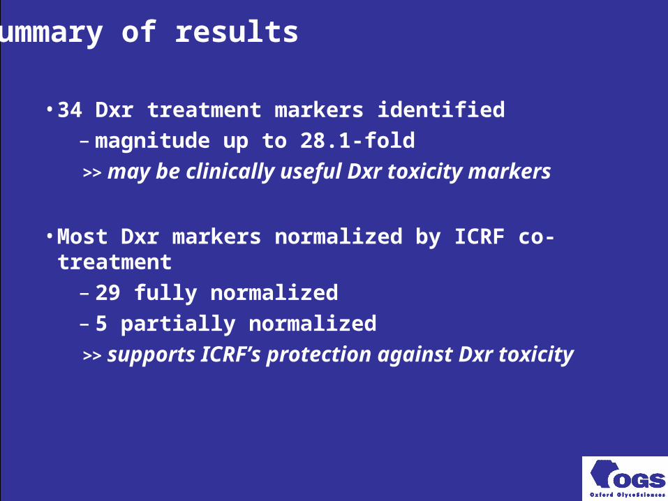
Summary of results
• 34 Dxr treatment markers identified– magnitude up to 28.1-fold
>> may be clinically useful Dxr toxicity markers
• Most Dxr markers normalized by ICRF co-treatment– 29 fully normalized– 5 partially normalized
>> supports ICRF’s protection against Dxr toxicity
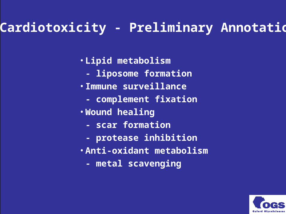
Dxr Cardiotoxicity - Preliminary Annotations
• Lipid metabolism
- liposome formation
• Immune surveillance
- complement fixation
• Wound healing
- scar formation
- protease inhibition
• Anti-oxidant metabolism
- metal scavenging
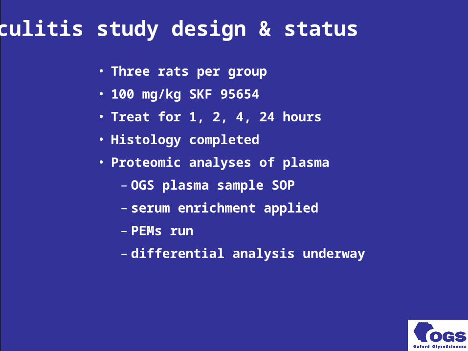
Vasculitis study design & status
• Three rats per group
• 100 mg/kg SKF 95654
• Treat for 1, 2, 4, 24 hours
• Histology completed
• Proteomic analyses of plasma
– OGS plasma sample SOP
– serum enrichment applied
– PEMs run
– differential analysis underway
Identification of Nephrotoxicity Surrogate Markers
Work done in
collaboration with
Quintiles UK

Gentamicin Background
Parenteral aminoglycoside active against gram-negative bacteria
Clinical important toxicity - potential for irreversible cumulative ototoxicity (manifest as hearing loss - initially of high frequencies) and vestibular damage
Reversible nephrotoxicity may occur and acute renal failure reported
Therapeutic index - individual monitoring of plasma concentrations generally required
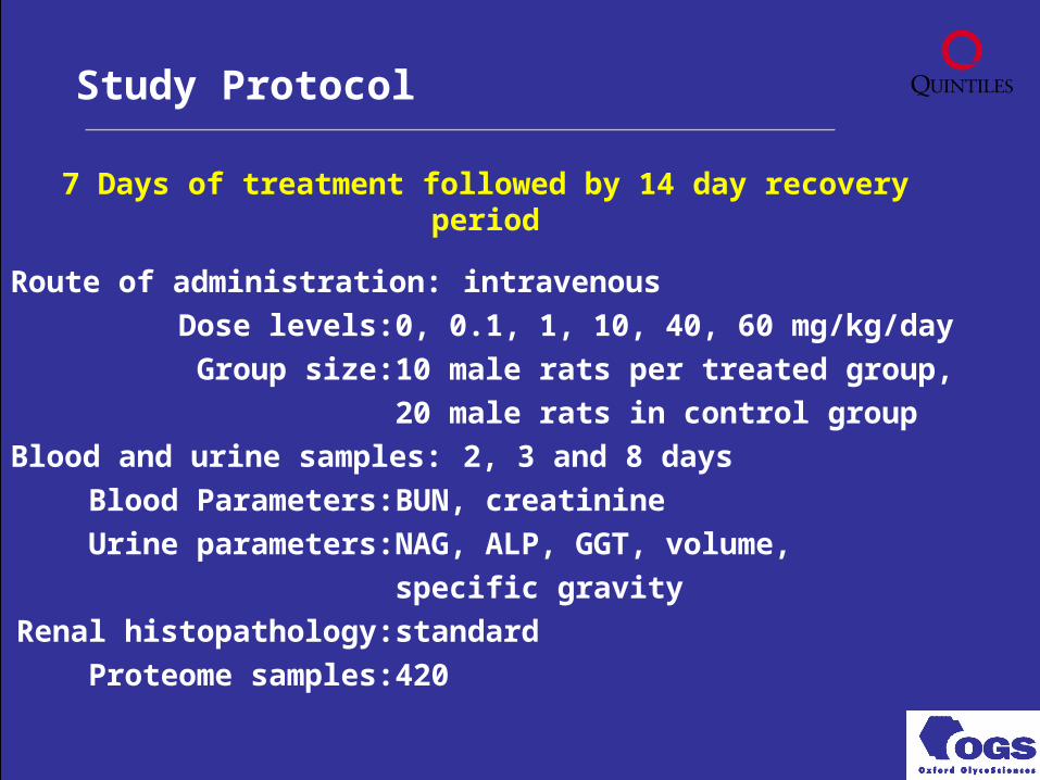
7 Days of treatment followed by 14 day recovery period
Route of administration: intravenous
Dose levels: 0, 0.1, 1, 10, 40, 60 mg/kg/day
Group size: 10 male rats per treated group,
20 male rats in control group
Blood and urine samples: 2, 3 and 8 days
Blood Parameters: BUN, creatinine
Urine parameters: NAG, ALP, GGT, volume,
specific gravity
Renal histopathology: standard
Proteome samples: 420
Study Protocol
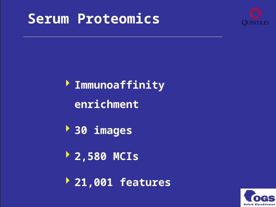
Immunoaffinity enrichment
30 images
2,580 MCIs
21,001 features
Serum Proteomics
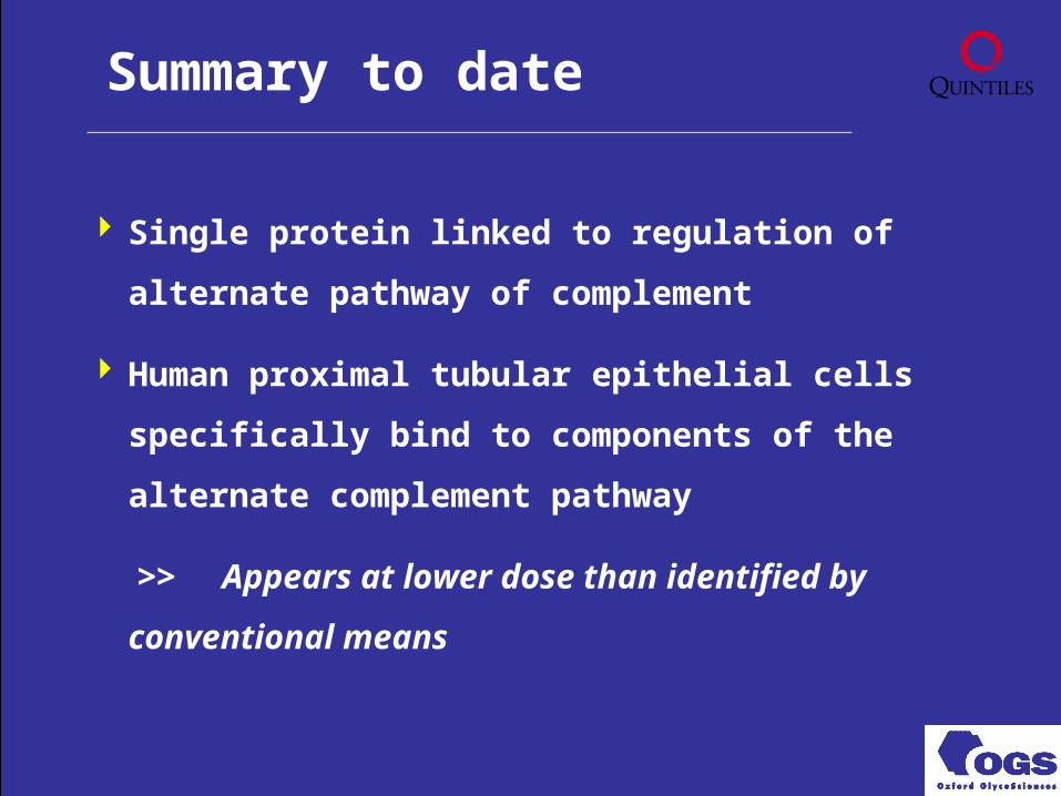
Single protein linked to regulation of alternate
pathway of complement
Human proximal tubular epithelial cells specifically
bind to components of the alternate complement
pathway
>> Appears at lower dose than identified by
conventional means
Summary to date
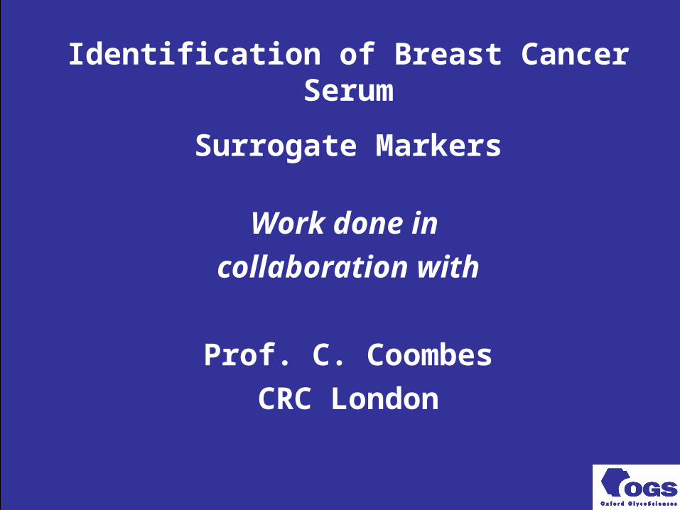
Identification of Breast Cancer Serum
Surrogate Markers
Work done in
collaboration with
Prof. C. Coombes
CRC London
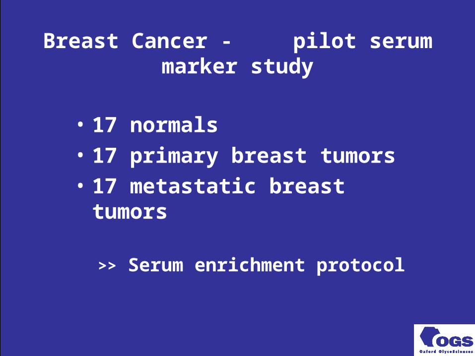
Breast Cancer - pilot serum marker study
• 17 normals
• 17 primary breast tumors
• 17 metastatic breast tumors
>> Serum enrichment protocol
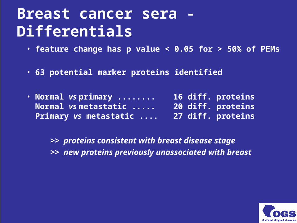
Breast cancer sera - Differentials
• feature change has p value < 0.05 for > 50% of PEMs
• 63 potential marker proteins identified
• Normal vs primary ........ 16 diff. proteinsNormal vs metastatic ..... 20 diff. proteinsPrimary vs metastatic .... 27 diff. proteins
>> proteins consistent with breast disease stage
>> new proteins previously unassociated with breast

Summary: OGS Proteomics
• Identify disease-specific proteins• Identify treatment-specific proteins
>> Quantitative and qualitative
>> Synergy with genomics data
>> Powerful tool for surrogate marker identification
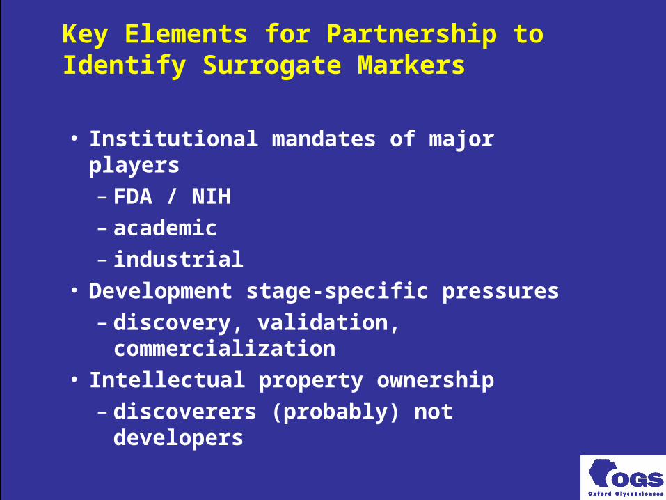
Key Elements for Partnership to Identify Surrogate Markers
• Institutional mandates of major players– FDA / NIH– academic– industrial
• Development stage-specific pressures– discovery, validation, commercialization
• Intellectual property ownership– discoverers (probably) not developers