prevention of biofilm associated infections and degradation of
TRANSCRIPT

22
Prevention of Biofilm Associated Infections and Degradation of Polymeric Materials used in
Biomedical Applications
Peter Kaali, Emma Strömberg and Sigbritt Karlsson Royal Institute of Technology,
Sweden
1. Introduction
Biomedical polymers have a wide variety of applications for external and internal use. Similar criteria must be fulfilled by biomedical polymeric materials used as internal or partly internal (invasive) devices, where the polymer gets in contact with the human environment. The material needs to be biocompatible, neutral to the human body and have to express excellent stability and resistance against tissues, cells, enzymes and different body fluids. The body response to the polymer can be acceptance or rejection and depending on the location of the material, these responses are influenced by different factors. Besides the body response, the microbiological effect and biofilm formation on the internal medical devices are of great importance. If biofilm adheres to the surface it can initiate a degradation process of the material, and due to the high concentration of microorganisms, infections and health related problems can be caused. The biocompatibility of polymers does not only depend on the chemical structure, the capability of microbes and the body environment to adhere or also initiate the degradation inside the human body is highly structure dependant. Once degradation occurs, along with the migration of additives and low molecular weight compounds, the polymer loses its biocompatibility and stability, which can lead to the failure of the device or could cause health related issues. Therefore the understanding of the different degradation processes that may occur inside the human body due to blood, tissue or biofilm interaction is very important. This chapter gives an overview on the mechanism of biofilm formation and adherence to surfaces, and means to characterize and determine its presence. Furthermore, the effect and the role of body-polymer interaction, the degradation mechanisms and the factors influencing the degradation of medical polymers are discussed. The factors that should be controlled are the biofilm formation and the prevention of infections caused by the microorganisms that usually generate intensive body reactions. Means to modify the polymeric materials by incorporating antimicrobial agents into the bulk of the polymer or right onto the surface as a coating is presented.
2. Biofilm
2.1 Characteristics and formation By definition, biofilms are aggregates of microorganisms, which are formed due to the attachment of cells to each other and/or to a host surface in an aqueous environment. (Lynch
22
www.intechopen.com
Biomedical Engineering, Trends in Materials Science
514
et al., 2003) In general, biofilms can host microorganisms such as bacteria, fungi, protozoa, algae and their mixtures, and usually the constituent cells require similar conditions to initiate and progress the cell growth. The factors that influence the biofilm formation are humidity, temperature, pH of the environment or medium, atmospheric conditions and nutrition sources. Besides microorganism cells, biofilms usually contain 80-90% of water and depending on the host surface their thickness may vary between 50-100om. Biofilm formation starts with the deposition of microorganisms on the surface of the material, followed by growth and spreading of the colonies. Microbial colony numbers are often very high and the emerging biofilms contain several layers of microorganisms, resulting in a highly complex structure (Flemming, 1998). The microbial cells are encased in an adhesive matrix produced by the microorganisms of the biofilm, called extracellular polymer substance or exopolysaccharide (EPS), which contains proteins, nucleic acids, lipids and polysaccharides. (Mayer et al., 1999, Beech, 2004). EPS influences the adhesion to the surface and plays an important role in the protection of the biofilm from outer environment. Therefore, the biofilms have an improved resistance against toxins, detergents and antimicrobial agents. In some cases the resistance of bacterial biofilms against antibiotics can be increased up to 1000 fold compared to isolated colonies.
2.2 Microbial adhesion Since biofilm plays a vital role in a wide variety of industrial, environmental and medical applications, the understanding of its formation mechanism and factors that influence the attachment to surfaces is essential. The environment which surrounds the surface may catalyze the biofilm formation; however, the process in most of the cases is similar. Fowler and Mckay were the first ones to investigate and describe the dynamic mechanism of bacterial adhesion. They took into account the initial physicochemical characteristics of the two surfaces that interact (Fowler and Mckay, 1980). The adhesion of the bacterial cell is a sequence of dynamic processes which involves characteristic forces, time scales and length scales (Denyer et al., 1993, Dickinson et al., 2000). In the first sequence, the cell is transported to the surface by gravitational force (sedimentation) and hydrodynamic forces (fluid flow, cell motility) where it reaches a diffusive boundary layer (Fig. 1). At this interface diffusion is the main driving force and, due to the small size of the cell, Brownian motion plays a vital role in the diffusive transport even closer to the surface. In the interval of the diffusive boundary layer there is a certain distance where direct interaction takes place between the cell surface and the substrate through attractive and repulsive forces (that includes Van der Waals and double layer interactions). At this distance the attachment of the cell to the surface is reversible since the interactions between both surfaces are weak (Oliveira, 1992). Initially both surfaces are negatively charged and therefore the attractive forces to ensure the adhesion must overcome an electrostatic repulse ion barrier. The interaction range between the cell and the surface is relatively small (<1 micron), however, the characteristic length of the stronger irreversible forces is around 5 to several hundreds of nanometers. The time scale of the transport process to the surface is flow dependent which is on the scale of 5x10⁻⁹ cm2/s for a cell having 1 micron in diameter. Once the cell has attached to the surface the strength of the attachment is governed by short range interactions (<5nm) which involves the resistance to detachment of the particle (irreversible) (Dickinson et al., 2000, Oliveira, 1992). These interactions include hydrogen bonding, shorter range Van der Waals forces, electrostatic, ionic and dipole interactions (Bos et al., 1999). The
514 Biomedical Engineering, Trends in Materials Science
www.intechopen.com

Prevention of Biofilm Associated Infections and Degradation of Polymeric Materials used in Biomedical Applications
515
Fig. 1. Cell attachment mechanism redrawn from (Dickinson et. al., 2000) long, short range forces and electrostatic interactions which play an important role in the bacterial attachment are described by the DLVO theory (Derjaguin and Landau, 1941, Verwey and Overbeek, 1948). The theory was developed originally to explain the coagulation behaviour of charged colloidal particles; however, it could also be applied to explain the interaction between a colloidal particle (as a bacterial cell) and a macroscopic surface (Fig.2).
Fig. 2. The DLVO theory
Diffusiveboundarylayer
Shorter
rangeforces
Longerrange
forcesAttachment Detachment
Convection (Motility)
Diffusion
Attraction
RepulsionElectrostatic force
van der Waals forcePrimary attraction
~5nm
Distance
515Prevention of Biofilm Associated Infections andDegradation of Polymeric Materials used in Biomedical Applications
www.intechopen.com

Biomedical Engineering, Trends in Materials Science
516
Type of interaction Interaction forces Approximate interaction energy (kJ/mol)
Reversible Long range, weak, low specificity
Van der Waals Electrostatic
20-50
Irreversible Short rage, high specificity
Dipole-dipole Dipole-induced dipole Ion-dipole Ionic Hydrogen bonds Hydrophobic
40-400
Table 1. Reversible and irreversible interaction during bacterial attachment(Oliveira, 1992)
2.3 Biodeterioration of polymeric materials Biofilm formation is common on most polymeric materials used in environments with high humidity. The nutrition sources necessary for successful colonisation may consist of the material itself or a variety of pollutants that end up on the surface of the material. The biofilm-polymer interactions depend on several factors which can be evaluated separately and in various combinations in authentic artificial environments that mimic the material’s end-use conditions(Wallström et al., 2002, Wallström et al., 2005). The characterization of the biofilm growth is also essential and is in general conducted by microscopic (i.e. optical, scanning electron microscope) methods, however, a few studies showed that the biofilm growth can be monitored by fluorescence lidar imaging as well (Bengtsson et al., 2005, Wallström and Karlsson, 2004). For polymers, biodegradation is usually a complex system, starting with consumption of accessible additives and propagating with the decomposition of the matrix (Figure 3)(Flemming, 1998).Although biofilm formation on some surfaces does not lead to polymer biodegradation it can result in the loss of functionality. Through biofouling, the spreading of the biofilm over the surface, the original properties of the material such as hydrophobicity may be altered. The deterioration of the medical function by clotting and disrupting the flow through for example a urinal catheter may cause pain for the patient or result in a serious infection. Medical implants are convenient surfaces for microbial growth, both the short-term devices (urinary catheters) and the long-term implants (artificial joints). The notorious biofilms consisting of various bacterial strains are protected from the attack by the immune system, antibiotics and other antimicrobial agents due to difficulties in penetrating into the biofilm. Plastic materials usually contain additives, low molecular weight compounds, residues of the polymer synthesis as well as shorter chains resulting from the degradation of the material, which migrate out of the material and interact with the biofilm. It is known that fillers such as polyesters, adiapates, epoxidised fatty acids, oleates, stearates and carbon-based plasticisers are perfect nutrition sources for microorganisms in the biofilm (Seal and Morton, 1986, Flemming, 1998). The most disputed material in biomedical applications is poly (vinyl chloride) (PVC), widely used for tubing purposes. During service life, toxic phthalate plasticisers tend to migrate out of the material, exposing the patient and providing nutrition to a growing biofilm. This leads to a harder and more brittle material, still insusceptible to biodegradation but instead sensitive to physical degradation.
516 Biomedical Engineering, Trends in Materials Science
www.intechopen.com

Prevention of Biofilm Associated Infections and Degradation of Polymeric Materials used in Biomedical Applications
517
Fig. 3. Effect of biofilm formation on polymer material surface (redrawn from (Flemming, 1998)
Factors influencing the rate of biodegradation are pH, environment, oxygen, salts, redox potential and temperature. Salts could be formed by anions which can be final products of microbial metabolism and react with cations (Wallström, 2005). An increase in salt concentration or significant change in pH highly assists the breakdown of the polymer (Sand, 1997). The transformed surface conditions, including the increasing humidity, induce the decomposition rate of the material. The excretion of enzymes from microorganisms may accelerate material degradation. Microorganisms are capable to cause enzymatic degradation of the polymer, this is the main biodegradation process for several medical polymers (e.g. polyurethane etc.) (Albertsson and Karlsson, 1994, Karlsson and Albertsson, 1998)). It has been reported that many fungi develop powerful enzyme systems to degrade highly stable polymers. These enzyme systems promote the reduction of peroxides to free radicals. Fungal hyphae can penetrate into the polymer, influencing mechanical stability and facilitate water diffusion into the material. Hyphal penetration provides mechanical degradation as a complement to chemical breakdown. (Flemming, 1998, Wallström et al., 2002, Gu, 2003, Gu et al., 1997) The microorganisms in a biofilm may cause a discolouration of the polymer surface, through diffusion of lipophilic pigments into the material. These substances do not alter the properties of the polymer, but are impossible to remove (Flemming, 1998). The discolouration can also be induced by other environmental factors such as oxidation of filler, additives or the polymer it self (Wallström, 2005). Another concern during biodegradation is the formation of low molecular weight compounds which may cause various odours. In biomedical context the main issue is the potential harmful effect such compounds may have to the patient.
2.4 Medical problems caused by biofilm Clinical trials on polyurethane tracheostomy tubes and silicone voice prosthesis showed that in most of the cases when a medical device is exposed to microorganisms, biofilm formation initiates and ultimately causes degradation of the material (Backman et al., 2009, Bjorling et al., 2007, Neu et al., 1993). Besides the negative effects of biofilm on polymeric medical devices, there is a high risk for emergence of infections. Recent research showed that
Process
Effect
Polymer
Fouling Degradation of
leachingcom
Change of surfaceproperties
Loss of stability
Loss of stability
ConductivitySwelling
Change in appearance and smell
Biotic degradation
Hydration Penetration
Colour Odour
Additives
Biofilm Enzymes Radicals
517Prevention of Biofilm Associated Infections andDegradation of Polymeric Materials used in Biomedical Applications
www.intechopen.com

Biomedical Engineering, Trends in Materials Science
518
biofilms are involved in 65% of microbial infections in the body (Potera, 1999), such as urinary tract infections, catheter associated and middle-ear infections, formation of dental plaque, gingivitis, coating contact lenses, and less common but more lethal processes i.e. endocarditis, infections in cystic fibrosis, and infections of permanent indwelling devices such as joint prostheses and heart valves (Costerton et al., 1999, Jarett et al., 2002, Potera, 1999). The body defence against infections is the production of antibodies (lymphocytes), however, the immune system is in capable of penetrating the biofilm and destroying the cells. Antibiotic therapy is effective only against free floating bacterial cells and the released antigen produced by the biofilm (Lynch and Robertson, 2008, Vergara-Irigaray et al., 2009). The reasons for biofilm resistance to antibiotic agents are: ‚ antibiotics are not able to penetrate the full depth of the biofilm and the diffusion of
antibiotics in EPS is relatively slow ‚ many antimicrobial agents are incapable of destroying slow growing or not growing
cells (stationary phase), in addition, some of the cells in the biofilm have a low nutrition intake or live in starved state, which render cell survival
‚ there are differences in cell wall protein between bacteria in biofilm and their free floating counterparts, some bacteria in biofilm can survive without dividing which makes them resistant to antibiotics that attack dividing cells or breakdown specific cell wall types.
3. Polymeric materials in medical applications
The biocompatibility of a medical device or implant, i.e. the ability of a material to perform without causing a host response, or having toxic or injurious effects, is highly important throughout the lifetime of the biomedical application. (Williams, 1999, Dorland, 1980).A non-biocompatible implant is rapidly encapsulated by collagen tissues, resulting in failure of the desired function of the product. The amount of the tissue growth around the material depends on the polarity; non-polar polymers are surrounded by less tissue, than polar ones (Akmal and Usmani, 2000). In case of rejection, the body tries to expel the polymer through chemical reactions by phagocytic or enzymatic activity (Gebelein, 1985, Akmal and Usmani, 2000), resulting in the emergence of inflammations. The body response is highly dependent on the form (foam, fibre, film), shape, and movement of the implant, as well as the location in the body. A smooth, rounded shape gives less interaction and reduces tissue adhesion around the material more than a rough-edged shape. Powdered polymers give high tissue interactions owing to the large surface area (Akmal and Usmani, 2000). Adsorption of various body chemicals (e.g. triglycerides and steroids) by the material can alter polymer properties and also lead to degradation (Gebelein, 1985). All polymeric materials degrade to some extent when in contact with the human body environment. Polymer implants, under normal circumstances, always undergo abrasion and stress (Hofmann et al., 2009). Poor long-term properties such as low resistance to wear and mechanical stress result in discomfort or pain for the patient, or costly replacement operations. The device or implant must be non-harmful during interactions with tissues and no toxic substances may be formed or leach out during the implementation of the application. Biomedical materials must be non-toxic, non carcinogenic, non-thrombogenic, non-inflammatory and non-immunogenic (King and Lyman, 1975, Venkatraman et al., 2008). Low molecular weight additives and degradation products produce significant tissue interactions due to their mobility and solubility in body chemicals. Therefore, polymers that
518 Biomedical Engineering, Trends in Materials Science
www.intechopen.com

Prevention of Biofilm Associated Infections and Degradation of Polymeric Materials used in Biomedical Applications
519
contain additives, residual monomers and polymerisation catalysts are not suitable for implant purposes. Many extracorporeal devices have to be biocompatible with blood due to constant blood exposure. The surface treatment of the biomedical devices by anticoagulants (e.g. heparin) is essential to reduce the probability of clotting the application. Only a few polymeric materials have good blood compatibility; hydrogels, polyether urethane ureas, and materials made by affixing biologically inactivated natural tissue to the polymer surface (Akmal and Usmani, 2000). The most commonly used materials for internal medical purposes are polyurethanes, polyolefins, silicones, fluoropolymers, vinyl- and acrylic polymers. Polyether type polyurethanes are used in a variety of applications (ligament replacements, heart valve prostheses, vascular graft prostheses, breast prosthesis, catheter, cannulae etc.) due to the materials good biocompatibility, high resistance against hydrolysis and body fluids, excellent mechanical properties (high tensile strength, highly elastomeric) and showing a low degree of degradation. The application of the polymer is versatile, from foam to film and as a bulk material. Silicone rubber, a medical elastomer (poly(dimethylsiloxane)), is as prevalent and versatile as polyurethanes. The material can be synthesized in very pure form, is highly inert and shows excellent chemical resistance (due to the high hydrophobicity). Besides poly(dimethylsiloxane), vinyl- and aromatic (phenyl) dimethylsiloxanes are also preferably used as a medical polymer due to the superior surface properties (e.g. super hydrophobicity). This surface characteristic and the aromatic groups in the structure make the silicone rubbers surface less attractive to microorganisms, thus avoiding biofilm formation. The silicone rubbers are used in artificial skin, joint replacement, vitreous replacement, artificial heart, breast implants, different types of catheters and cannulae. Polyolefins (polyethylene, polypropylene), fluoropolymers (teflon etc.) and acrylic polymers are mostly used as prosthetic devices. They express high degree of biocompatibility (almost totally neutral), excellent chemical resistance and superior mechanical properties. Compared to metallic implants the main advantage of polyolefins and fluoropolymers is the low friction coefficient and wear resistance due to their self-lubricating characteristic. The main medical applications are hip joints, knee implants etc. Acrlylic polymers show even better mechanical properties. They are mostly used as dental materials and bone cements to anchor artificial joints to the body. Due to the excellent optical properties methacrylates are also used in contact lenses. Polymers such as poly(vinyl chloride) (PVC), poly(lactic acid) (PLA), polycaprolactone (PCL) and poly(vinyl alcohol) (PVA)have a variety of different medical applications. PVC is commonly used for lung bypass sets, catheters and cannulae, tubing for dialysis, endotracheal feeding etc. PVC is preferably used since the material is easy to sterilise and simple to process into products that do not crack or leak. The main drawback of PVC is the necessity of plasticizers, phthalates, for achieving the required mechanical properties, softness and flexibility, since the material itself is stiff. The low molecular weight plasticizers can be a target for microbial attack and under certain circumstances they migrate out of material and cause toxic reactions in the human body. On the other hand, the loss of these additives deteriorates the mechanical properties of the material. PLA is used as a biodegradable polymer in controlled-drug release systems, resorbable sutures and resorbable bone plates.
519Prevention of Biofilm Associated Infections andDegradation of Polymeric Materials used in Biomedical Applications
www.intechopen.com

Biomedical Engineering, Trends in Materials Science
520
4. Degradation mechanisms of medical polymers
The human body contains a variety of enzymes and chemicals that may cause degradation of the polymer (Williams, 1992, Williams, 1991). Polymers containing ester or amide linkages (i.e. polyurethane) are more likely to hydrolyze or oxidize, while polyether type polymers are more stable, showing minimal degradation during long-term exposure to human body environment. Chain-scissions and/or crosslinking occur in addition to hydrolytic degradation (Kaali et al., 2010a). As previously discussed, the biofilm attachment to the surface of the polymeric materials plays a vital role in the initiation and propagation of the degradation process. The biofilm formation is more pronounced for invasive materials, however, implants, after the implantation, are rarely exposed to such aggressive biological milieus. Biofilm can also cause immunological response (i.e. infections) that changes the surrounding body environment resulting in a negative effect on the material properties (Gumargalieva et al., 1982). Based on the molecular, chemical and mechanical interactions with the human body environment, four types of degradation mechanisms of polymers used in medical applications can be distinguished: hydrolysis, oxidation, enzymatic- and physical degradation (Lyu and Untereker, 2009). The kinetics of the processes differ, and the key factors are the structure of the material and the surrounding environment (Göpferich, 1996). Body fluids represent the environment of the given location in the body or liquids (enzymes) produced by the body as an immunological response. In this case, the important factor in the material degradation is the change of pH of the surrounding environment, since some polymers (i.e. polyesters, polyamides) are highly pH sensitive (Göpferich, 1996, Williams, 1992). Another significant factor is the water uptake by the material (which is highly dependent on the hydrophobicity). The adsorbed water acts as a plasticizer, altering the physical properties of the material, swells the polymer, causing dimensional instability of the device or implant, and initiates the degradation of the polymer by hydrolysis. In general, the degradation of the polymer by hydrolysisoccurs through three stages, however, depending on the molecular weight and the type, some polymers undergo only one or two stages (Lyu and Untereker, 2009). In the first stage, the polymer adsorbs water and becomes saturated in a short period of time, thereafter reactions between the water molecules and the polymer chains initiate. At this stage no auto-acceleration occurs since the water content of the polymer is constant, the molecular weight is high and the chain-ends concentration is low. This is followed by the second stage, where the molecular weight of the polymer decreases, increasing the chain-end concentration to a certain level, where auto-acceleration initiates and catalyzes the degradation process. Due to the increase in the chain-end concentration, the water adsorption of the polymer increases extending the polymer-water interactions, resulting in further decrease in the molecular weight. There is a point where the molecular weight becomes so low that it becomes soluble in the media. This corresponds to stage three. The low molecular weight compounds that form due to the reactions dissolve in the media and the molecular weight of the polymer gradually decreases until the polymer is completely dissolved. Although the water uptake of medical polymers is low (i.e. polyesters 1%), hydrolysis and bond-cleavage in the polymer chain result in a material with a decreased molecular weight and increased number of hydrophilic chain-ends. The chain-ends may adsorb an increasing amount of water which can further catalyze the reaction and lead to the complete breakdown of the material. This is the typical degradation mechanism for polyesters, polyamides and polycarbonates, however, the hydrolytic degradation of the stable poly(dimethylsiloxane) may also occur
520 Biomedical Engineering, Trends in Materials Science
www.intechopen.com
Prevention of Biofilm Associated Infections and Degradation of Polymeric Materials used in Biomedical Applications
521
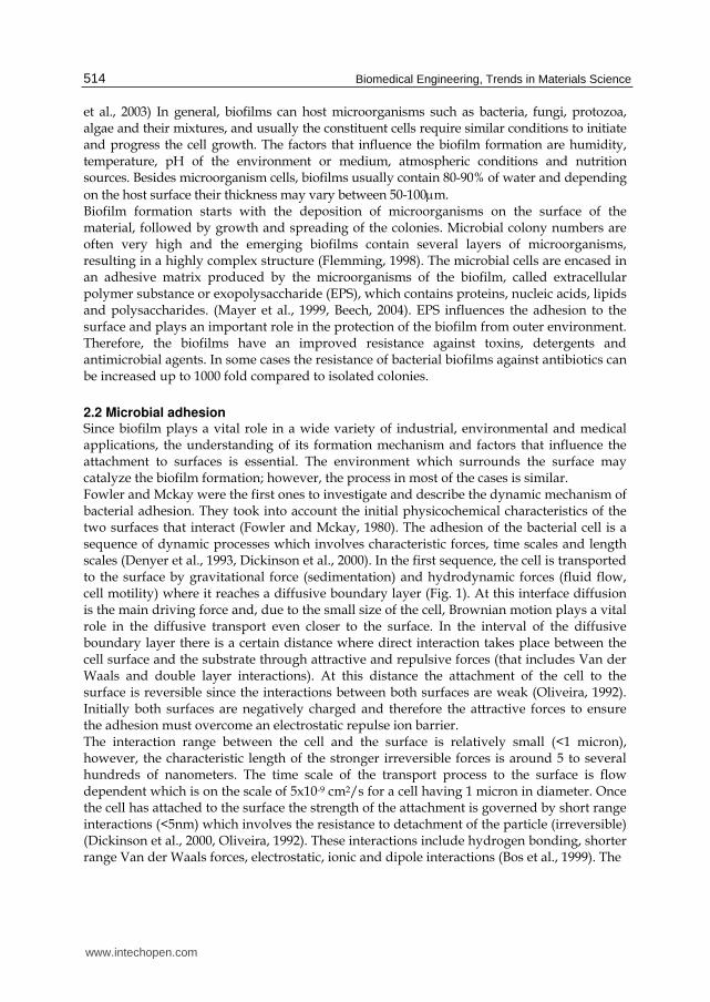
during in vivo use (Kaali et al., 2010a, Lukasiak et al., 2003). Recent long-term studies have confirmed that silicone tracheostomy tubes undergo hydrolytic degradation during use (Kaali et al., 2010a). Tubes, with an exposure time from one to six months, from several patients were collected and the analysis by Scanning Electron Microscopy (SEM), Fourier Transform Infrared Spectroscopy (FTIR) and Matrix Assisted Laser Desorption/Ionization Time of Flight Mass Spectrometry (MALDI TOF MS) showed degradation of silicone rubber after just 1 months exposure. The SEM micrographs clearly showed evidence of the surface alteration during the whole exposure period, which was also confirmed by the contact angle measurements, where the change in surface hydrophobicity was established.
Fig. 4. SEM micrographs of unexposed silicone rubber tracheostomy tubes (a) and exposed to human environment for (b) three and (c) six months. (Kaali et al., 2010a)
The contact angle slightly decreased as a function of time, however, it must be noted that the surface of the silicone remained hydrophobic. The evidence of hydrolytic degradation was established by MALDI and FTIR. The FTIR analysis showed the formation of –OH groups, which may correspond to materials water uptake, however unlikely due to the high hydrophobicity of the silicone material. Besides, traces of protein, resulting from the attachment of the biofilm during the service life of the material, were also identified from the FTIR spectra. The hydrolytic degradation of the material was confirmed by MALDI TOF MS. The formation of low molecular weight silicone compounds with hydroxyl groups was identified. These compounds were absent in the unexposed samples. The extended results of this study showed that the degradation of polymeric materials and the rate of degradation within the human body depend on the biotic degradation, on the surrounding body environment and also the applied drug treatment. In addition to MALDI TOF MS, Gas Chromatography (GC) and High-Performance Liquid Chromatography (HPLC) are widely used for determination of low molecular weight compounds, as reported in several studies (Haider and Karlsson, 2002, Hillborg et al., 2001, Khabbaz et al., 2000, Flassbeck et al., 2001, Flassbeck et al., 2003, Gruemping and Hirner, 1999). The oxidative degradation of medical polymers occurs inside the human body and can be monitored in simulated environments (Backman et al., 2009, Kaali et al., 2010b). The reaction is caused by the peroxides produced by the human body against “non-accepted” implant materials, the rejection mechanism (Lyu and Untereker, 2009, Santerre et al., 2005). Inflammation takes place at the implantation site, monocytes are migrating to the site and the production of macrophages initiates. If rejection is not possible, the body tries to encapsulate the material by foreign body giant cells. These cells and macrophages produce peroxides in order to try to break down the material to eliminate it from the body (Lyu and Untereker, 2009). The oxidation mechanism begins with the increasing number of free radicals due to the oxygen adsorbed from the surrounding tissues or blood. The oxygen
521Prevention of Biofilm Associated Infections andDegradation of Polymeric Materials used in Biomedical Applications
www.intechopen.com

Biomedical Engineering, Trends in Materials Science
522
molecules react with the existing free radicals (Lyu and Untereker, 2009), resulting in an accelerated process where each oxygen molecule produces two radicals. The formed free radicals are transported to different parts of the polymer chain causing chain scission and formation of new chain-ends, one carrying a free radical and the other containing a double bond. The double bonded end can react further and form acids, ketones, while the free radical end continues the before mentioned process. This reaction propagates until the chains become too short for further degradation. The most susceptible medical polymers for oxidation are polyolefins, vinyl polymers, polyethers and polyamides. Several authors have reported that polyether type polyurethanes are quite stable against hydrolysis without exposure to oxidation (Frautschi et al., 1993, Santerre et al., 2005, Wiggins et al., 2001). The oxidative degradation takes place primarily at the ether linkage of the polymer, where the peroxide radical attacks the ccarbon of the soft segment. This reaction leads to the formation of an ester linkage which is susceptible to hydrolysis. The oxidative degradation of polyether urethane is therefore followed by hydrolysis. The polymeric materials that are susceptible or less susceptible to certain degradation processes are summarized in Table 2. In the human body, materials undergo also enzymatic degradation(Christenson et al., 2006, Duguay et al., 1995, Santerre et al., 1995, Santerre et al., 1993). This is also a defensive response of the body against implant materials and can be linked to the activity of the tissues and cells. Although enzymes are produced for specific interaction, they are capable to recognize “unnatural” substrates such as polymers (Santerre et al., 2005). In order to interact with the polymer, the enzyme must diffuse into the material either by swelling or hydrolysis (Duguay et al., 1995). This is considered to be the primary contact between the enzyme and the polymers surface. At this stage the enzyme becomes inactive, forming an “enzyme-bond” complex by attaching to an enzymatically susceptible bond (i.e. urethane, ester etc.).If this complex is relatively stable, bond scission may occur between the interface bonds and the bound enzyme, which results in the formation and release of various compounds.
Susceptible Less susceptible
Hydrolysis
Polyanhydride Polyorthoester
Polyketal Polyester (aliphatic)
Polyolefin Polyether
Polysulfone PDMS
Polycarbonate Polyimide
Polyurethane Polyester (aromatic)
Polyamide
Oxidation
Polyolefins Vinyl polymers
Polyethers Polyamines
Fluoropolymers Polyesters
Methacrylates Silicone
Polysulfone Polyetheretherketone
Table 2. Polymeric materials susceptible to degradation
522 Biomedical Engineering, Trends in Materials Science
www.intechopen.com

Prevention of Biofilm Associated Infections and Degradation of Polymeric Materials used in Biomedical Applications
523
These compounds then undergo further degradation and cleavage. Two kinds of enzymatic degradation can be distinguished, oxidation or hydrolysis of the material, which are based on the type of the enzyme produced (Albertsson and Karlsson, 1994). The enzymatic systems are highly specific and are able to catalyze degradation of the particular polymer chains summarised in Table 3. Due to the complexity of the human body, the materials are exposed to most of the discussed degradation mechanisms simultaneously. The different degradation factors need to be evaluated separately and in various combinations in artificial environments that mimic the product’s end-use conditions, in order to predict and understand the property changes that will occur in the material during its lifetime. The negative body response to a foreign material is the production of peroxides, therefore the most commonly used solvent to simulate this oxidative environment is hydrogen peroxide (Christenson et al., 2006, Lyu et al., 2008). Different artificial body fluids are used to test the biocompatibility or degradability of the material, such as phosphate buffered saline (PBS) which is used to mimic the blood plasma, and artificial lysosomal fluid (ALF) and Gamlbe´s solutions for simulating more complex systems. ALF solution simulates the enzymes that may initiate the breakdown of the polymer while Gamble´s solution represents the environment of the deep lungs (Herting et al., 2007, Midander et al., 2007). In a recent study silicone rubber and polyester type polyurethane were exposed to both ALF and Gamble´s solution at 37oC (the body temperature) for 3 months (Kaali et al., 2010b). During the exposure the formation and increasing concentration of low molecular weight compounds in silicone rubber were observed. These substances were the same hydrolyzed compounds that were detected during the in vivo use of silicone rubber tracheostomy tubes (Kaali et al., 2010a). In addition, polyurethane showed chemical property changes due to the exposure to artificial body fluids and based on the results it was determined that oxidative degradation took place. These results confirmed that artificial body fluids and simulated environments give similar results to in vivo experiments and represent good tools for testing new materials that are going to be implanted into the patents. In addition the application of in vitro studies reduce the costs and experiment time significantly.
Polymer Enzyme
Polyurethanes Cholesterol esterase, xanthine oxidase,
cathepsin B, collagenase Polyglycolic acid Esterase, chymotrypsin, trypsin
Polyester Esterase Polyester urea Urease, pepsin, chymitrypsin
Polycaprolactone Lipase, carboxytic esterase Polyamide
Polymethylmethacrylate Esterase, papain, trypsin, chymotrypsin
Table 3. Polymers susceptible to enzymatic degradation (Santerre et al., 1995)
Besides chemical degradation, physical degradation of the polymers also occurs in the human body. This is most relevant for implants that are exposed to different mechanical forces during their use, and therefore excellent mechanical properties are key requirements. These materials are usually knee and hip joints or other kinds of orthopaedic implants. The most common failures of these materials are wearing, breaking or cracking and erosion
523Prevention of Biofilm Associated Infections andDegradation of Polymeric Materials used in Biomedical Applications
www.intechopen.com
Biomedical Engineering, Trends in Materials Science
524
(Göpferich, 1996). These failures may appear together or separately depending on the application, however, it is typical that the orthopaedic implants undergo mechanical friction which is associated with motion under pressure. Although UHMWPE is a superior material for joint purposes, some studies have reported high degree of mechanical degradation on hip and knee implants (Brach del Prever et al., 1996, Heisel et al., 2004, Kabo et al., 1993). It was also determined that the wear, friction and oxidative properties are better for cross-linked UMPWE than the conventional one (Heisel et al., 2004, Heisel et al., 2005, Markut-Kohl et al., 2009) and that during the mechanical wear, oxidative degradation of the polyethylene may occur.
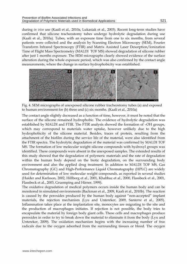
4.1 Effects of sterilization on polymer degradation During the manufacture biomedical materials are exposed to microorganisms and other substances even in a very pure production environment. Therefore they have to be sterilized and well sealed for storage in order to avoid any contamination or microbes that may cause infections or health problems right after the implantation. The sterilizationprocedures are presentedin Figure 5. Dry heat and autoclaving involve high temperature (~120-180oC) and pressure. The sterilization process by these methods could take from 3 minutes up to a couple of hours. During this exposure the materials may undergo thermal degradation caused by the temperature and hot steam that penetrates into the structure of the materials. Therefore commonly used sterilization methods for medical materials used in the human body are sterilization by either irradiation or gaseous chemicals (ethylene oxide).
Fig. 5. Sterilization methods of medical polymers
There are two types of irradiation sterilization processes; gamma ray and electron beam. During gamma sterilization gamma rays are produced from Co66 source and have a high penetration capability up to 50 cm into the material. Electron beam sterilization is performed by an electron beam generator (1MeV-12MeV), which generates high-energy electrons. The penetration depth is around 5 cm, however, compared to gamma rays at the same strength, the dosage rate for the electron source is many times greater. This is due to the characteristic of the electron beam, which is unidirectional and therefore more concentrated on a smaller area, while gamma rays are less focused and cover a bigger surface. Both electron and gamma rays have such a high energy that the microorganisms that remain in the material after the production are accurately destroyed. From the material point of view these high-
Sterilization methods
Dry heat Autoclaving Irradiation
Gamma ray Electron beam
Gaseous chemicals
524 Biomedical Engineering, Trends in Materials Science
www.intechopen.com
Prevention of Biofilm Associated Infections and Degradation of Polymeric Materials used in Biomedical Applications
525
energy impacts initiate changes in the structure of the material. These changes can be bonds scission, cross-linking, branching and degradation of additives. It has been reported that polyurethane catheters treated with electron beam sterilization undergo oxidative degradation, which leads to chain scissions in the hard and soft segments. This leads to the formation of smaller highly volatile soft segment fractions. In addition cross-linking occurs, that is thought to be influenced by the chain scissions of the hard segment, and forms on the urethane linkage sites. Most of the medical polymers contain additives that can be degraded by the electron beam and therefore can be easily released from the polymer. This causes a decrease in the stability of the polymer and could cause cytotoxicity (Guignot et al., 2001, Mrad et al., 2009a, Mrad et al., 2009b, Ravat et al., 2001a, Ravat et al., 2001b). Gamma irradiation has similar influence on the material. Due to the gamma sterilization, branching on polyurethanes (Haugen et al., 2007) and an extremely high rate of oxidation was observed on UHMWPE implants (Bracco et al., 2006, Goldman et al., 1998). During the irradiation, oxygen penetrates into the amorphous region of the polymer where misfit strain is developed. As a result the lamellae boundaries become tortuous which leads to further strain development and microcracking. Microcracking is a serious problem for materials designed for prosthetic purposes since it influences the mechanical properties negatively and the lifetime of the material decreases. Besides the types of irradiation, the dosage, flux and the outer environment have an effect on the degradation rate and degradation mechanisms that occur. For instance, the higher flux and the presence of oxygen increase the oxidation, since it generates anincreased formation of free radicals. Besides irradiation, the use of gaseous chemicals is also prevalent. For this purpose usually ethylene oxide is used which is a strong alkylating agent, toxic and carcinogenic gas. The effectiveness of this gas on sterilization depends on the sterilization method which includes several factors. These are regarded as the gas concentration, temperature, relative humidity, the permeability and absorbance of the polymer. It has been reported that sterilization with ethylene oxide has no or very minimal influence on the structure of the polymer (Abraham et al., 1997, Burgos and Jiménez, 2009, Gilding et al., 1980, Lucas et al., 2003). However, the residues of the gas that remain after the process could at a certain concentration (above 400 ppm)cause toxicity (Bolt, 2000, MacNeil and Glaser, 1997). In principle the amount of remaining residue depends on the applied sterilization method and the polymers absorbance. Therefore with a proper method development that suits for the polymer and allows the complete release of ethylene oxide is necessary. Among the currently used sterilization processes the treatment with ethylene oxide has a big potential compared to irradiation techniques due to the reduced risk for degradation and subsequent health related issues.
4.2 Effects of degradation products on the human body The degradation of the biomedical materials and formation of the degradation products have a serious influence on the human body (Lyu and Untereker, 2009). For instance, due to hydrolysis carboxylic acid and/or hydroxyl chain ends may form. Hydroxyl groups can be further oxidized and the reaction may produce different kinds of degradation products i.e. aldehydes, ketones or carboxylic acids. The degradation rate and its influence on the body depend on the size and location of the implant. However, if a biocompatible material starts to degrade it loses its stability and from the application point of view causes decreased service time. An example for body reaction is the formation of carboxylic acid, which
525Prevention of Biofilm Associated Infections andDegradation of Polymeric Materials used in Biomedical Applications
www.intechopen.com

Biomedical Engineering, Trends in Materials Science
526
changes the local pH, causing inflammatory response. As an example during the production of some polyurethanes, methylene diamine is used, which is a toxic compound. In case ofthese kind of toxic precursors biodegradation is not relevant since base monomer usually don´t form due to chemical degradation. However residues of these monomers may remain in the material after the production. This is a technological question and the ability of human body to handle these kind of compounds depends on several factors i.e. can the compound be diluted or transported to organs (i.e. kidney) where it can be further degraded or flushed away.
5. Inhibition of biofilm formation and prevention of related infections
5.1 Current applications The potential risk of biofilm formation is a big problem influencing the degradation of invasive materials and may cause health related issues. It has been reported (Kumon et al., 2001) that patients often face catheter associated infections (CAI). From the patient point of view, depending on the type of catheter (central venous, urinary etc.), the infection might be lethal. For the hospitals, these infections appear as a cost increase for patient therapy, which might be anticipated if the biofilm can be properly removed from the surface of the device or the material itself would have antimicrobial effect. Invasive materials that are permanently exposed to the human environment (especially tracheostomy tubes) require occasional checkups in order to determine the physical condition of the device and the biofilm growth on the surface. Usually biofilm formation is quite intensive right after the implantation, therefore a proper and regular cleaning is needed to remove the biofilm from the surface, reduce the biofilm related infections and the risk of the polymer degradation. If the biofilm can be completely removed, the surface will be less susceptible to microorganisms than in case where some residues or biofilm cells are remaining on the surface. In principle the cleaning procedure of medical devices is performed in two steps. The first step is the cleaning of the surface with oxidisers or detergents that weakens the physical stability of the biofilm and removes dead cells from the surface. In the second step the interaction between the surface and biofilm needs to be broken which can only be done by an intensive physical action i.e. brushing, scraping or flushing with high pressure of water (Wallström, 2005). Since some biofilm cells attach to the surface through ligands and chemical compounds, their removal is not possible without the physical surface damage of the material. Therefore in the field of biofilm inhibition, the development and improvement of cleaning techniques play only a secondary role. There are some promising methods i.e. mucus shaver and different sterilization treatments (argon plasma etc.) for the reduction or removal of biofilm after the attachment (Berra et al., 2006, Lee et al., 2009),however, the main priority is to inhibit the ability of the biofilm to attach to polymeric surfaces. Most of the currently used medical devices that express antimicrobial effect contain either biocides or silver. It is known that silver has antimicrobial effect, however, the mechanism is not fully understood. Silver ion has the capability to be exchanged by ions that exist in the environment (Ca+, Zn+ etc.) and attach to bacterial cells (Schierholz et al., 1998). It forms chelate-complexes with the DNA of the bacteria. These complex molecules block the main transport processes of the cell, which leads to its decay. The sensitivity of Gram negative and Gram positive strains to silver varies due to the structural differences and is influenced by the attachment of silver to murein in the cell wall by adsorption. If it reaches a certain concentration in the cell wall it forms the chelate complex with the DNA. By increasing the
526 Biomedical Engineering, Trends in Materials Science
www.intechopen.com

Prevention of Biofilm Associated Infections and Degradation of Polymeric Materials used in Biomedical Applications
527
silver ion concentration the antimicrobial activity can be increased, however, a certain concentration in the human environment (10mg/L ,(Schierholz et al., 1998)) is cytotoxic and may lead to argyria. The antimicrobial efficiency and the performance of antibiotics and silver treated catheters against CAI have been compared in several studies (Böswald et al., 1999, Lyu and Untereker, 2009, Paterson et al., 1999, Walder et al., 2002). The most commonly used antibiotics for treatment of the catheters are minocycline-rifampin, piperacillin, gentamicin, and ofloxacin. Silver indifferent forms (ion, compound, hydrogel, metallic) can be used inmedical devices. The most common ones for invasive materials are chlorohexidine-silver-sulfadiazine and impregnated silver. The studies showed that use ofmonociclyne-rifampin, ofloxacim and chlorohexidine-silver-sulfadiazine resulted in the least number of CAI compared to the other agents. Silver sulfadiazine expressed a high degree of biocompatibility and applicability in human environment (Böswald et al., 1999, Paterson et al., 1999), however, monocycline-rifampin showed better antiseptic properties. The antibacterial mechanism of silver-sulfadiazine is not completely understood. In a trial it was shown that in case of central venous catheters (CVC´s), probably the chlorine was responsible for the decreased CAI since it secedes from the surface of the polymer easier (Schierholz et al., 1998) Impregnated silver was less accurate because the mechanism is based on the migration and diffusion. Since silver-sulfadiazine is a salt it dilutes easily from the surface while impregnated silver ions must diffuse out in order to get in contact with the outer environment. Results of short and long term ion release studies of impregnated silver and silver-oxide coated catheters showed that during the first month the release of silver ions decreased significantly as a function of time while after four months it remained unaltered. During the exposure the impregnated catheter showed better antimicrobial results compared to a control sample where, the initial silver release was 2,5 ppm(Kubey et al., 1995). The use of hydrogels as antimicrobial agents in the medical industry is exceeding. Silver-hydrogels are preferably used as dressings for wound healing (Castellano et al., 2007, Ip et al., 2006), however, they show good antiseptic properties as polymer coatings on urinary catheters as well (Kwan and Fontecchio, 2002). Besides silver containing coatings, it was confirmed that nitrofurazone is also an excellent antibacterial agent that reduces the risk of urinary and blood-stream infections (Johnson et al., 2006). The different forms of application of silver as an antimicrobial and antiseptic agent for invasive materials is promising based on the clinical trials, however, the real efficiency is sometimes questionable (Walder et al., 2002). In order to use silver in medical applications, the standardization of this material is necessary. This standardization is difficult since the effect is influenced by several factors; the destruction of different bacterial strains requires different silver concentrations and the minimum inhibitory results may give broad intervals of concentration. As an example, the minimum inhibitory concentration (MIC) for Stapylococcusaureus and Pseudomonas aeruginosa can vary 8-80mg/L (Chopra, 2007). The broad interval depends on the variation of the silver concentrations in different products and the variation of the release as a function of time depending on the nature of the material (i.e. water absorption) and the physical, chemical form of the silver and the location in the material. Moreover, there are bacterial strains that are resistant to silver and this mechanism is yet not well understood. There are species that become resistant by the modification of gene mutation or identification of the silver as a toxic material when it gets into the cell resulting in a rejection mechanism. Besides silver, certain bacterial strain has resistance
527Prevention of Biofilm Associated Infections andDegradation of Polymeric Materials used in Biomedical Applications
www.intechopen.com

Biomedical Engineering, Trends in Materials Science
528
against specific antibiotics (e.g. MRSA: Methicillin-Resistant Staphylococcus aureus) or in extreme cases this resistance can expand to several groups of drugs. During the improvement and development of antimicrobial and antiseptic materials that inhibit the biofilm formation and infections, these factors must be well considered.
5.2 Research strategies and trends to decrease device related infections and biofilm formation There is a variety of developments and improvements in this field, however, the different forms and applications of silver or other antibacterial metals give the main direction of the research. Silver, copper and titanium nanoparticles are usually used and their in vitro antimicrobial efficiency is more or less confirmed, however silver is superior compared to other metals. The antimicrobial mechanism of the nanoparticles is not known but it can be assumed that it is similar to the ions. This is due to their small size and high specific surface area which makes them extremely reactive with the environment. If a nanoparticle interacts with a bacteria cell, ions may attach to the cell blocking the main transport phenomena and the replication of the DNA. The particles are incorporated into the material by mixing, bulk reduction (composite) or by a surface modification procedure (implantation, coating). During the production of composites aggregation of the particles takes place. This is a big problem, since the size of the particles increases due to the aggregation, which influences the diffusion and the antimicrobial properties (Jeong et al., 2005, Schmidt and Malwitz, 2003). Besides the size, the shape of the nanoparticles also influences the antimicrobial properties. It has been reported that truncated cone shaped particles express better efficiency against microbial growth than the spherical ones (Pal et al., 2007). In order to reach a homogenous dispersion of the particles by mixing, the use of a surfactant is necessary (Dirix et al., 1999). Therefore bulk reduction is a better procedure to prepare polymer matrix nanocomposites. In this process, theoretically, the polymer is responsible for the stabilization of the nanoparticles. In principle, the first step is the addition of silver in to the polymer by mixing a silver compound (i.e. AgNO3) with the polymer solution. As a second step, a reduction agent is added to the system and the formation of nanoparticles initiates. This change is usually followed by a colour change. The effectiveness of this procedure has been confirmed by several studies (Akamatsu et al., 2000, Babu et al., 2006, Mahapatra and Karak, 2008, Sambhy et al., 2006). Another perspective of silver-polymer composites is the incorporation of silver ions into zeolite which is then mixed into the bulk polymer. It is known that zeolite is a natural mineral and has a high ion-exchange capability. Research papers confirm high efficiency of Ag-zeolite polymer against microorganisms (Kam et al., 2008, Kawahara et al., 2000, Pehlivan et al., 2005, Kaali et al., 2010b). The efficiency is based on the silver content which is linked to the zeolite content. The higher the zeolite content the better the antimicrobial efficiency. However, zeolite is a hydrophilic material and therefore adsorbs moisture and water easily. An in vitro study showed that the increasing zeolite content in polymeric materials results in a higher rate of degradation due to the increased water uptake of the material (Kaali et al., 2010b). In this case, zeolite content above 1% showed significant degradation compared to an unfilled material. Therefore the only solution that can be applied for zeolite-polymer composites is the increase of the ion content. In case of composites, for microbial interaction diffusion is the main transport of the antimicrobial agent to the surface. It is usually governed by a concentration gradient, which forms between the material and the environment. In order to reach the equilibrium the agents are concentrated on the surface and if it is possible diffuse out. This process depends
528 Biomedical Engineering, Trends in Materials Science
www.intechopen.com

Prevention of Biofilm Associated Infections and Degradation of Polymeric Materials used in Biomedical Applications
529
on several factors such as the material, temperature, humidity etc. The problem with composites is that due to the diffusion, the release of the agent is slow and the antimicrobial effect might be limited. Therefore the bigger part of the research focuses on surface modification (i.e. plasma immersion)(Akamatsu et al., 2000, Huang et al., 2004, Zhang and Chu, 2008, Zhang, 2000) or coating of the materials. By the immersion techniques the antimicrobial agent can be incorporated onto the surface of the materials where they physically or chemically bond to the matrix. They remain at the interface region and can interact easier with the microorganisms than the antimicrobials in composite materials. In case of metallic coatings, the main challenge is the adhesion. Since polymeric and metallic surfaces are not compatible with each other, the development of coatings is based on the improvement of the adhesion between the polymer and metallic surface by different deposition techniques. The strength of adhesion between these two interfaces depends on the structure of the polymer, hydrophobicity and surface roughness. Some papers describe that the surface must be activated before the coating procedure which is usually done by plasma or neutral nuclear beam (Dowling et al., 2003, Dowling et al., 2001, Gray et al., 2005). The activation is followed by the deposition of the substrate. Dowling used magnetron to deposit silver coating and platinum. As a result, the thickness of the coating was estimated to 7-28nm and the coating passed the tape test. In case of physically bonded coatings, depending on application, the deposited layer may easily crack if the material is softer than the substrate. Most of the polymers are exposed to mechanical stress and during the use of a coated catheter, where bending of the device is common, the coating will sooner or later peel off the material. In contrast metallic coatings that are bonded chemically to functional groups of the polymer may show better elastic properties. Electroless deposition is a useful chemical process to coat polymer surfaces with metals without electric current. Studies have confirmed (Gray et al., 2005, Li et al., 2004)that the silver coating which is deposited by this technique bond chemically to the surface through carbonyl groups by forming Ag-O-C bond. The higher the amount of the carbonyl groups on the surface the better adhesion can be reached. Studying the antibacterial effect of these metallic antimicrobial coatings show promising results (MIC of Stapylococcusaureus 0,5-10mg/L). However, the investigation of cytotoxicity is a very important issue, since these surfaces may release a high amount of ions that may cause toxic reaction for the human body. The most common ways for detection of released metallic particles from polymers are atomic absorption spectroscopy (AAS) and ionic plasma coupled mass spectrometry (ICP-MS). These two techniques allow to monitor even very low concentrations of metallic ions. Since ICP has a lower detection limit it is used to monitor the ion release of both composites and coatings, AAS is applicable for metallic coatings and pure metals (Herting et al., 2007, Midander et al., 2007). Besides metallic coatings, research is focusingon variousnon-metallic coatings. One of the main interests in this field is based on multifunctional hydroxyapatite coatings that can be loaded with metallic particles or antibiotics (Brohede et al., 2009, Noda et al., 2009, Shimazaki et al., 2009). The application of this coating is mainly on hip joints and other metallic prostheses. Hydroxyapatite (HA) is a calcium apatite natural mineral that can be found in the body (teeth, bones) and therefore highly biocompatible. The role of hydroxyapatite as a coating is to ensure the tissue growth around the implant and decrease probability of rejection by the human body. Besides biocompatibility, if antibiotics or antimicrobial metallic particles are incorporated into the structure of HA the risk for infections and biofilm formation can be decreased. The trials showed that silver and antibiotics loaded HA coatings express superior antimicrobial properties on short term and
529Prevention of Biofilm Associated Infections andDegradation of Polymeric Materials used in Biomedical Applications
www.intechopen.com

Biomedical Engineering, Trends in Materials Science
530
due to the slow release of the agent this characteristic can also be obtained on long term. Polymeric hydrogel coatings have also a great perspective. The main advantage of these materials is similar to hydroxyapatite, they are biologically inert, have good physicochemical properties and can be incorporated with either different antibiotics, or metallic nanoparticles to ensure their antimicrobial efficiency. The antimicrobial mechanism of hydrogels is the release of the agent to the environment. This release is governed by diffusion and influenced by the swelling and surface properties of the hydrogels. In principle this mechanism is the same as for antimicrobial-composites, however, the diffusion rate is much higher. Moreover, due to the chemical structure of the hydrogels, the release of antimicrobial agents is more even and pulsatile than in polymer composites. There is a wide variety of hydrogels which have different structures and release properties and therefore, for certain applications, these materials can be optimized to the antimicrobial agent. Besides the surface and release properties, hydrogels have to express good mechanical properties to be suitable for coatings. Based on a study, where the different hydrogel structures were tested, hydroxyethyl methacrylate and isopropylacrylamide based copolymers seemed to have the best properties (swelling, pulsatile release) for medical applications or drug delivery (Jones et al., 2008). Besides invasive materials, hydrogels are currently used for wound healing and clinical trials have confirmed their efficiency against infections. The antimicrobial and antiseptic properties depend also on the type of agent. So far silver nanoparticles incorporated into hydrogel coatings show the best antiseptic efficiency (Thomas et al., 2007, Dabbagh et al., 2008, Wu et al., 2009, Varaprasad et al.). However, due to the risk for silver toxicity (that may happen in case of high release rate) new antimicrobial agents are currently under development which are non-cytotoxic for human cells but has good efficiency against a variety of microorganisms (i.e. chitosan and bioactive antibodies)(Rojas et al., 2000, Yang et al., 2007) Besides synthetic materials, biological coatings or surface modifications are also reported and this field is growing extensively to exchange the currently used synthetic materials with more “human friendly” ones. In these studies biosurfactants produces by bacteria strains and bacteriophages as a coating to inhibit the biofilm formation on the surfaces (Goldman et al., 2009, Rivardo et al., 2009, Rodrigues et al., 2006) give the main frame of the research. The efficiency of these materials against biofilm formation was confirmed; in some cases 90% of growth reduction was observed. The mechanism that is responsible for inhibition of the biofilm is based on the enzymes that are produced by the bacteriophages. These enzymes are capable to destroy the EPS of biofilms. The eradication efficiency of antibiotics against urinary infective strains was also increased by the biosurfactants. In contrast,biosurfactants are not capable of destroying planktonic cells and inhibit only the growth of certain strains. Moreover, bacteriophages are only efficient against specific strains and therefore the solution might be the combined use of different surfactants and/or bacteriophages together. The idea of biological biofilm inhibitors and antibacterial agents has a great perspective, but further development and improvement is necessary in order to be used in medical applications. Peptides can be placed into this group as well. They are known as agents of the immune system and can be found in a wide variety of living organisms (i.e. bacteria, insects, human body etc.). These peptides are usually obtained from different sources as macrophages, neutrophils, epithelial cells etc. and exhibit high activity against microorganisms. Peptides are either chemically bonded or incorporated into the surface. The main advantage of chemically bonded peptides is that they express antimicrobial activity locally and in contrast to the synthetic coatings, where antimicrobial agents are released via
530 Biomedical Engineering, Trends in Materials Science
www.intechopen.com
Prevention of Biofilm Associated Infections and Degradation of Polymeric Materials used in Biomedical Applications
531
mechanical or chemical processes, these peptides remain on the surface of the material. Therefore, no release takes place resulting in a higher stability, lower toxicity and longer activity against microorganisms. The other application of antimicrobial peptides is a single or multilayer coating, where each peptide layer is followed by a biodegradable polymer layer. Due to the exposure to human environment these polymer layers degrade one by one and slowly release the antimicrobial peptide. The main advantages of the application of antimicrobial peptides compared to metallic or synthetic agents are the high biocompatibility, very low toxicity and well controlled release.
6. Summary
It has been shown that biofilm formation on polymeric medical devices is an issue and its effects on both of the human body and the device are negative. The attachment and formation mechanism of biofilm on surfaces is a very complex phenomenon. In general it is based on colloid chemistry and surface-surface interactions, where the adhesion strength depends on short and long range interactions (Van der Waals forces, hydrogen bonding, electrostatic interactions etc.). After the cell attachment, the biofilm formation begins by colonization and production of extracellular polymeric substance that embeds the microorganisms, ensures the adhesion to the surface and protects the biofilm from the outer environment and impacts. The extracellular polymer substance provides stability for the biofilm and protection against antimicrobial agents, which in turn causes initiation of the material degradation and induction of serious infections. The requirements for an implant or invasive materials are biocompatibility, non-toxicity, non-carcinogenicity and extreme stability, however, degradation of the material may occur due to the body response. The acceptance or rejection response of the human body depends highly on the size, shape and location of the material in the body. The sensitivity of the material to rejection and degradation is highly dependant on the chemical structure of the polymer. There are four major mechanisms that are responsible for degradation of biomedical materials: hydrolysis, oxidative, enzymatic and mechanical degradation. Enzymatic degradation can also be caused by biofilms. The mechanical degradation is referring to the swelling and friction under motion and pressure of the polymer and not directly influenced by the human body. The sterilization procedure also is one of the factors promoting the degradation of the devises. Due to gamma and electron sterilization, oxidative degradation of the polymer can occur resulting in a decrease of the molecular weight and degradation of additives. Low molecular weight compounds can leach out of the material causing toxic reactions. The sterilization of medical polymers with ethylene oxide does not lead to any significant degradation of the polymer structure, but the residues of ethylene oxide can cause toxic reactions. In order to prevent the negative effects of biofilms on the polymer surfaces and in the human body, the inhibition and control of biofilmformation is necessary. For this purpose antibiotics, silver- nanoparticles, salts and compounds are incorporated into the surface or the bulk of the material. Silver nanoparticles are also used as metallic coatings on the surface in physically or chemically bonded form. Clinical trials confirm that certain antibiotics and silver products are efficient as antiseptic agents and decrease the number of medical device associated infections in healthcare. The efficiency of silver is based on the silver ions released from the material, however, this is material and application dependant and not well controlled yet. A high concentration of silver released into the body can cause toxicity.
531Prevention of Biofilm Associated Infections andDegradation of Polymeric Materials used in Biomedical Applications
www.intechopen.com

Biomedical Engineering, Trends in Materials Science
532
New coatings are under development for controlled and appropriately slow release of antibiotics or silver from the medical devices. Polymeric hydrogels can be one of the solutions for the controlled release due to their network structures, which allow a constant and sufficient release of the antimicrobial agents. Studies have shown that hydrogel dressings incorporated with antibiotics or nanoparticles assist the wound healing of the patients and decrease the risk for infections. Another recent development is extracellular polymeric substance that embeds the modification ofhydroxyapatite, a natural mineral that exists in the human body. Its pores can be filled with a variety of antimicrobial agents and provide a slow release mechanism. A new approach of research in inhibition of biofilm formation is the use of biological substances. Biological surfactants and bacteriophages are capable to inhibit the growth or destroy the biofilm. However, surfactants are not efficient against planktonic cells and not able to reduce the risk of infections caused by microorganisms. In addition bacteriophages can destroy only certain strains. The solution might be the combined use of different bacteriophages and surfactants to make these biological substances more universal against a variety of microorganisms. Their efficiency is confirmed, but since these solutions are newly introduced and developed, there is a big research potential in this field.
7. References
Electrostatic Charge and Bacterial Adhesion. Newcastle University, Department of Oral Biology
Abraham, G. A.; Frontini, P. & Cuadrado, T. (1997). Physical and mechanical behavior of sterilized biomedical segmented polyurethanes,Journal of Applied Polymer Science, Vol.65, 1193-1203
Akamatsu, K.; Takei, S.; Mizuhata, M.; Kajinami, A.; Deki, S.; Takeoka, S.; Fujii, M.; Hayashi, S. & Yamamoto, K. (2000). Preparation and characterization of polymer thin films containing silver and silver sulfide nanoparticles,Thin Solid Films, Vol.359, 55-60
Akmal, N. & Usmani, A. M. (2000).Medical polymers and diagnostic reagents, In:Handbook
of polymer degradation, Hamid, S. (Ed.)2 ed, Marcel Dekkel Inc.,New York Albertsson, A. C. & Karlsson, S. (1994). Chemistry and biochemistry of polymer
biodegradation, In:Chemistry and technology of biodegradable polymers, Griffin, G. J. I. (Ed.), Springer,0751400033, New York
Babu, R.; Zhang, J.; Beckman, E. J.; Virji, M.; Pasculle, W. A. & Wells, A. (2006). Antimicrobial activities of silver used as a polymerization catalyst for a wound-healing matrix Biomaterials, Vol.27, 4304-4314
Backman, S.; Björling, G.; Johansson, U. B.; Lysdahl, M.; Markström, A.; Schedin, U.; Aune, R. E.; Frostell, C. & Karlsson, S. (2009).Material wear of polymeric tracheostomy tubes: A six-month study,The Laryngoscope, Vol.119, 657-664
Beech, I. B. (2004). Corrosion of technical materials in the presence of biofilms--current understanding and state-of-the art methods of study,International Biodeterioration &
Biodegradation, Vol.53, 177-183 Bengtsson, M.; Grönlund, R.; Sjöholm, M.; Abrahamsson, C.; Dernfalk, A. D.; Wallström, S.;
Larsson, A.; Weibring, P.; Karlsson, S.; Gubanski, S. M.; Kröll, S. & Svanberg, S.
532 Biomedical Engineering, Trends in Materials Science
www.intechopen.com

Prevention of Biofilm Associated Infections and Degradation of Polymeric Materials used in Biomedical Applications
533
(2005). Fluorescence lidar imaging of fungal growth on high-voltage outdoor composite insulators,Optics and Lasers in Engineering, Vol.43, 624-632
Berra, L.; Curto, F.; Li Bassi, G.; Laquerriere, P.; Baccarelli, A. & Kolobow, T. (2006).Antibacterial-coated tracheal tubes cleaned with the Mucus Shaver,Intensive
Care Medicine, Vol.32, 888-893 Bjorling, G.; Axelsson, S.; Johansson, U.-B.; Lysdahl, M.; Markstrom, A.; Schedin, U.; Aune,
R. E.; Frostell, C. & Karlsson, S. (2007). Clinical Use and Material Wear of Polymeric Tracheostomy Tubes,Laryngoscope, Vol.9, 1552-1559
Bolt, H. M. (2000). Carcinogenicity and Genotoxicity of Ethylene Oxide: New Aspects and Recent Advances,Critical Reviews in Toxicology, Vol.30, 595-608
Bos, R.; van der Mei, H. C. & Busscher, H. J. (1999).Physico-chemistry of initial microbial adhesive interactions – its mechanisms and methods for study,FEMS Microbiology
Reviews, Vol.23, 179-230 Böswald, M.; Mende, K.; Bernschneider, W.; Bonakdar, S.; Ruder, H.; Kissler, H.; Sieber, E. &
Guggenbichler, J. P. (1999).Biocompatibility testing of a new silver-impregnated catheter in vivo,Infection, Vol.27, S38-S42
Bracco, P.; Brunella, V.; Luda, M. P.; Brach del Prever, E. M.; Zanetti, M. & Costa, L. (2006).Oxidation behaviour in prosthetic UHMWPE components sterilised with high-energy radiation in the presence of oxygen,Polymer Degradation and Stability, Vol.91, 3057-3064
Brach del Prever, E.; Crova, M.; Costa, L.; Dallera, A.; Camino, G. & Gallinaro, P. (1996). Unacceptable biodegradation of polyethylene in vivo,Biomaterials, Vol.17, 873-878
Brohede, U.; Forsgren, J.; Roos, S.; Mihranyan, A.; Engqvist, H. & Strømme, M. (2009).Multifunctional implant coatings providing possibilities for fast antibiotics loading with subsequent slow release,Journal of Materials Science: Materials in
Medicine, Vol.20, 1859-1867 Burgos, N. & Jiménez, A. (2009). Degradation of poly(vinyl chloride) plasticized with non-
phthalate plasticizers under sterilization conditions,Polymer Degradation and
Stability, Vol.94, 1473-1478 Castellano, J. J.; Shafii, S. M.; Ko, F.; Donate, G.; Wright, T. E.; Mannari, R. J.; Payne, W. G.;
Smith, D. J. & Robson, M. C. (2007). Comparative evaluation of silver-containing antimicrobial dressings and drugs,International Wound Journal, Vol.4, 114-122
Chopra, I. (2007). The increasing use of silver-based products as antimicrobial agents: a useful development or a cause for concern?,J. Antimicrob. Chemother., Vol.59, 587-590
Christenson, E. M.; Patel, S.; Anderson, J. M. & Hiltner, A. (2006). Enzymatic degradation of poly(ether urethane) and poly(carbonate urethane) by cholesterol esterase,Biomaterials, Vol.27, 3920-3926
Costerton, J. W.; Stewart, P. S. & Greenberg, E. P. (1999). Bacterial Biofilms: A Common Cause of Persistent Infections,Science, Vol.284, 1318-1322
Dabbagh, M. A.; Moghimipour, E.; Ameri, A. & Sayfoddin, N. (2008). Physicochemical Characterization and Antimicrobial Activity of Nanosilver Containing Hydrogels,Iranian Journal of Pharmaceutical Research, Vol.7, 21-28
533Prevention of Biofilm Associated Infections andDegradation of Polymeric Materials used in Biomedical Applications
www.intechopen.com

Biomedical Engineering, Trends in Materials Science
534
Denyer, S. P.; Hanlon, G. W. & Davies, M. C. (1993).Mechanisms of Microbial Adherence,
In:Microbial Biofilms: Formation and Control, Denyer, S. P., Gorman, S. P. & Sussman, M. (Eds.), Blackwell Scientific Publications,Cambridge
Derjaguin, B. & Landau, L. (1941). Theory of stability of strongly charged lyophobic sols and of the adhesion of strongly charged particles in solutions of electrolytes,Acta
Physicochimcal URSS, Vol.14, 733-762 Dickinson, R. B.; Ruta, A. G. & Treusdal, S. E. (2000). Physicochemical Basis of Bacterial
Adhesion to Biomaterial Surfaces, In:Antimicrobial/Anti-Infective Materials, Sawan, S. & Manivannan, G. (Eds.),1 ed, Technomic Publishing Company, Inc.,Lancaster
Dirix, Y.; Bastiaansen, C.; Caseri, W. & Smith, P. (1999).Preparation, structure and properties of uniaxially oriented polyethylene-silver nanocomposites,Journal of Materials
Science, Vol.34, 3859-3866 Dorland, W. A. N. (1980).Dorland's Medical Dictionary W.B. Saunders Company,
Philadelphia, Pennsylvania, USA, 0721631428, 9780721631424 Dowling, D. P.; Bettsa, A. J.; Popea, C.; McConnella, M. L.; Eloyb, R. & Arnaudb, M. N.
(2003). Anti-bacterial silver coatings exhibiting enhanced activity through the addition of platinum Surface and Coatings Technology, Vol.163-164, 637-640
Dowling, D. P.; Donnellya, K.; McConnella, M. L.; Eloyb, R. & Arnaudb, M. N. (2001).Deposition of anti-bacterial silver coatings on polymeric substrates,Thin
Solid Films, Vol., 602-606 Duguay, D. G.; Labow, R. S.; Santerre, J. P. & McLean, D. D. (1995). Development of a
mathematical model describing the enzymatic degradation of biomedical polyurethanes. 1. Background, rationale and model formulation,Polymer
Degradation and Stability, Vol.47, 229-249 Flassbeck, D.; Pfleiderer, B.; Grumping, R. & Hirner, A. V. (2001).Determination of Low
Molecular Weight Silicones in Plasma and Blood of Women after Exposure to Silicone Breast Implants by GC/MS,Anal. Chem., Vol.73, 606-611
Flassbeck, D.; Pfleiderer, B.; Klemens, P.; Heumann, K. G.; Eltze, E. & Hirner, A. V. (2003).Determination of siloxanes, silicon, and platinum in tissues of women with silicone gel-filled implants,Analytical and Bioanalytical Chemistry, Vol.375, 356-362
Flemming, H.-C. (1998). Relevance of biofilms for the biodeterioration of surfaces of polymeric materials,Polymer Degradation and Stability, Vol.59, 309-315
Fowler, H. W. & Mckay, A. J. (1980). The measurement of microbial adhesion, In:Microbial
Adhesion to Surfaces, Berkeley, R. C. W., Lynch, J. M., Melling, J., Rutter, P. R. & Vincet, B. (Eds.), Ellis Horwood,Chichester
Frautschi, J. R.; Chinn, J. A.; Phillips Jr, R. E.; Zhao, Q. H.; Anderson, J. M.; Joshi, R. & Levy, R. J. (1993). Degradation of polyurethanes in vitro and in vitro: comparison of different models,Colloids and Surfaces B: Biointerfaces, Vol.1, 305-313
Gebelein, C. G. (1985). Medical Applications of Polymers, In:Applied Polymer Science, Gilding, D. K.; Reed, A. M. & Baskett, S. A. (1980). Ethylene oxide sterilization: effect of
polymer structure and sterilization conditions on residue levels,Biomaterials, Vol.1, 145-148
534 Biomedical Engineering, Trends in Materials Science
www.intechopen.com
Prevention of Biofilm Associated Infections and Degradation of Polymeric Materials used in Biomedical Applications
535
Goldman, G.; Starosvetsky, J. & Armon, R. (2009). Inhibition of biofilm formation on UF membrane by use of specific bacteriophages,Journal of Membrane Science, Vol.342, 145-152
Goldman, M.; Gronsky, R.; Long, G. G. & Pruitt, L. (1998). The effects of hydrogen peroxide and sterilization on the structure of ultra high molecular weight polyethylene,Polymer Degradation and Stability, Vol.62, 97-104
Göpferich, A. (1996). Mechanisms of polymer degradation and erosion,Biomaterials, Vol.17, 103-114
Gray, J. E.; Norton, P. R. & Griffiths, K. (2005). Mechanism of adhesion of electroless-deposited silver on poly(ether urethane) Thin Solid Films, Vol.484, 196-207
Gruemping, R. & Hirner, A. V. (1999). HPLC/ICP-OES determination of water-soluble silicone (PDMS) degradation products in leachates,Fresenius' Journal of Analytical
Chemistry, Vol.363, 347-352 Gu, J.-D. (2003). Microbiological deterioration and degradation of synthetic polymeric
materials: recent research advances,International Biodeterioration & Biodegradation, Vol.52, 69-91
Gu, J. D.; Lu, C.; Mitchell, R.; Thorp, K. & Crasto, A. (1997). Fungal degradation of fiber-reinforced composite materials,Materials Performance, Vol.36, 37-42
Guignot, C.; Betz, N.; Legendre, B.; Le Moel, A. & Yagoubi, N. (2001).Degradation of segmented poly(etherurethane) Tecoflex® induced by electron beam irradiation: Characterization and evaluation,Nuclear Instruments and Methods in Physics Research
Section B: Beam Interactions with Materials and Atoms, Vol.185, 100-107 Gumargalieva, K. Z.; Moiseev, Y. V.; Daurova, T. T. & Voronkova, O. S. (1982).Effect of
infections on the degradation of polyethylene terephthalate implants,Biomaterials, Vol.3, 177-180
Haider, N. & Karlsson, S. (2002). Loss and transformation products of the aromatic antioxidants in MDPE film under long-term exposure to biotic and abiotic conditions,Journal of Applied Polymer Science, Vol.85, 974-988
Haugen, H. J.; Brunner, M.; Pellkofer, F.; Aigner, J.; Will, J. & Wintermantel, E. (2007). Effect of different ┛-irradiation doses on cytotoxicity and material properties of porous polyether-urethane polymer,Journal of Biomedical Materials Research Part B: Applied
Biomaterials, Vol.80B, 415-423 Heisel, C.; Silva, M.; dela Rosa, M. A. & Schmalzried, T. P. (2004).Short-Term in Vivo Wear
of Cross-Linked Polyethylene,J Bone Joint Surg Am, Vol.86, 748-751 Heisel, C.; Silva, M. & Schmalzried, T. (2005). In vivo wear of bilateral total hip
replacements: conventional versus crosslinked polyethylene,Archives of Orthopaedic
and Trauma Surgery, Vol.125, 555-557 Herting, G.; Odnevall Wallinder, I. & Leygraf, C. (2007).Metal release from various grades of
stainless steel exposed to synthetic body fluids,Corrosion Science, Vol.49, 103-111 Hillborg, H.; Karlsson, S. & Gedde, U. W. (2001).Characterisation of low molar mass
siloxanes extracted from crosslinked polydimethylsiloxanes exposed to corona discharges,Polymer, Vol.42, 8883-8889
535Prevention of Biofilm Associated Infections andDegradation of Polymeric Materials used in Biomedical Applications
www.intechopen.com

Biomedical Engineering, Trends in Materials Science
536
Hofmann, D.; Entrialgo-Castaño, M.; Kratz, K. & Lendlein, A. (2009). Knowledge-Based Approach towards Hydrolytic Degradation of Polymer-Based Biomaterials,Advanced Materials, Vol.21, 3237-3245
Huang, N.; Yang, P.; Leng, Y. X.; Wang, J.; Sun, H.; Chen, J. Y. & Wan, G. J. (2004). Surface modification of biomaterials by plasma immersion ion implantation Surface and
Coatings Technology, Vol.186, 218-226 Ip, M.; Lui, S. L.; Poon, V. K. M.; Lung, I. & Burd, A. (2006). Antimicrobial activities of silver
dressings: an in vitro comparison,J Med Microbiol, Vol.55, 59-63 Jarett, W. A.; Ribes, J. & Manaligod, J. M. (2002).Biofilm formation on tracheostomy
tubes,Ear , Nose & Throat Journal, Vol.81, 659-661 Jeong, S.; Yeo, S. & Yi, S. (2005). The effect of filler particle size on the antibacterial
properties of compounded polymer/silver fibers,Journal of Materials Science, Vol.40, 5407-5411
Johnson, J. R.; Kuskowski, M. A. & Wilt, T. J. (2006).Systematic Review: Antimicrobial Urinary Catheters To Prevent Catheter-Associated Urinary Tract Infection in Hospitalized Patients,Ann Intern Med, Vol.144, 116-126
Jones, D. S.; Lorimer, C. P.; McCoy, C. P. & Gorman, S. P. (2008). Characterization of the physicochemical, antimicrobial, and drug release properties of thermoresponsive hydrogel copolymers designed for medical device applications,Journal of Biomedical
Materials Research Part B: Applied Biomaterials, Vol.85B, 417-426 Kaali, P.; Momcilovic, D.; Markström, A.; Aune, R.; Czel, G. & Karlsson, S. (2010a).
Degradation of biomedical polydimethylsiloxanes during exposure to in vivo biofilm environment monitored by FE-SEM, ATR-FTIR, and MALDI-TOF MS,Journal of Applied Polymer Science, Vol.115, 802-810
Kaali, P.; Strömberg, E.; Aune, R. E.; Czél, G.; Momcilovic, D. & Karlsson, S. (2010b). Antimicrobial properties of Ag+ loaded zeolite polyester polyurethane and silicone rubber and long-term properties after exposure to in-vitro ageing,Polymer
Degradation and Stability, Vol.95, 1456-1465 Kabo, J. M.; Gebhard, J. S.; Loren, G. & Amstutz, H. C. (1993). In vivo wear of polyethylene
acetabular components,J Bone Joint Surg Br, Vol.75-B, 254-258 Kam, K.; Aksoy, E. A.; Akata, B.; Hasirci, N. & Baç, N. (2008).Preparation and
characterization of antibacterial zeolite-polyurethane composites,Journal of Applied
Polymer Science, Vol.110, 2854-2861 Karlsson, S. & Albertsson, A.-c. (1998). Biodegradable polymers and environmental
interaction,Polymer Engineering & Science, Vol.38, 1251-1253 Kawahara, K.; Tsuruda, K.; Morishita, M. & Uchida, M. (2000).Antibacterial effect of silver-
zeolite on oral bacteria under anaerobic conditions,Dental Materials, Vol.16, 452-455 Khabbaz, F.; Karlsson, S. & Albertsson, A. C. (2000). PY-GC/MS an effective technique to
characterizing of degradation mechanism of poly (L-lactide) in the different environment,Journal of Applied Polymer Science, Vol.78, 2369-2378
King, R. N. & Lyman, D. J. (1975). Polymers in Contact with the Body,Environmental Health
Perspectives, Vol.11, 71-74
536 Biomedical Engineering, Trends in Materials Science
www.intechopen.com

Prevention of Biofilm Associated Infections and Degradation of Polymeric Materials used in Biomedical Applications
537
Kubey, W.; Luneburg, P.; Ericson, S.; Brown, J. & Holmes, C. J. (1995). A longitudinal in vitro antimicrobial evaluation of two silver polymer surface treatments for peritoneal dialysis catheters,Advances in peritoneal dialysis. Conference on Peritoneal
Kumon, H.; Hashimoto, H.; Nishimura, M.; Monden, K. & Ono, N. (2001). Catheter-associated urinary tract infections: impact of catheter materials on their management,International Journal of Antimicrobial Agents, Vol.17, 311-316
Kwan, K. L. & Fontecchio, S. A. (2002). Use of silver-hydrogel urinary catheters on the incidence of catheter-associated urinary tract infections in hospitalized patients,American Journal of Infection Control Vol.30, 221-225
Lee, M. H.; Park, B. J.; Chang, S.; Kim, D.; Han, I.; Kim, J.; Hyun, S. O.; Chung, K.-H. & Park, J.-C. (2009).Removal and sterilization of biofilms and planktonic bacteria by microwave-induced argon plasma at atmospheric pressure,New Journal of Physics, Vol.11, 115-122
Li, Y.; Lu, Q.; Qian, X.; Zhu, Z. & Yin, J. (2004).Preparation of surface bound silver nanoparticles on polyimide by surface modification method and its application on electroless metal deposition,Applied Surface Science, Vol.233, 299-306
Lucas, A. D.; Merritt, K.; Hitchins, V. M.; Woods, T. O.; McNamee, S. G.; Lyle, D. B. & Brown, S. A. (2003). Residual ethylene oxide in medical devices and device material,Journal of Biomedical Materials Research Part B: Applied Biomaterials, Vol.66B, 548-552
Lukasiak, J.; Dorosz, A.; Prokopowicz, M.; Rosciewski, P. & Falkiewicz, B. (2003). Biodegradation of Silicones,Biopolymers, Vol.9, 539-568
Lynch, A. S. & Robertson, G. T. (2008). Bacterial and Fungal Biofilm Infections,Annual
Review of Medicine, Vol.59, 415-428 Lynch, J. F.; Lappin-Scott, H. M. & Costerton, J. W. (2003).Microbial biofilms, Cambridge
University Press, Cambridge,
Lyu, S.; Schley, J.; Loy, B.; Luo, L.; Hobot, C.; Sparer, R.; Untereker, D. & Krzeszak, J. (2008). In vitro biostability evaluation of polyurethane composites in acidic, basic, oxidative, and neutral solutions,Journal of Biomedical Materials Research Part B:
Applied Biomaterials, Vol.85B, 509-518 Lyu, S. & Untereker, D. (2009). Degradability of Polymers for Implantable Biomedical
Devices,International Journal of Molecular Sciences, Vol.10, 4033-4065 MacNeil, J. R. & Glaser, Z. R. (1997).Comparison of health care-based sterilization
technologies: safety, efficacy and economics,J Healthcare Safety Compliance Infect
Control Vol., 91-106 Mahapatra, S. S. & Karak, N. (2008).Silver nanoparticle in hyperbranched polyamine:
Synthesis, characterization and antibacterial activity,Materials Chemistry and
Physics, Vol.112, 1114-1119 Markut-Kohl, R.; Archodoulaki, V. M.; Seidler, S. & Skrbensky, G. (2009).PE-UHMW in Hip
Implants: Properties of Conventional and Crosslinked Prosthetic Components,Advanced Engineering Materials, Vol.11, B148-B154
Mayer, C.; Moritz, R.; Kirschner, C.; Borchard, W.; Maibaum, R.; Wingender, J. & Flemming, H.-C. (1999). The role of intermolecular interactions: studies on model systems for bacterial biofilms,International Journal of Biological Macromolecules, Vol.26, 3-16
537Prevention of Biofilm Associated Infections andDegradation of Polymeric Materials used in Biomedical Applications
www.intechopen.com

Biomedical Engineering, Trends in Materials Science
538
Midander, K.; Wallinder, I. O. & Leygraf, C. (2007).In vitro studies of copper release from powder particles in synthetic biological media,Environmental Pollution, Vol.145, 51-59
Mrad, O.; Saunier, J.; Aymes Chodur, C.; Rosilio, V.; Agnely, F.; Aubert, P.; Vigneron, J.; Etcheberry, A. & Yagoubi, N. (2009a). A comparison of plasma and electron beam-sterilization of PU catheters,Radiation Physics and Chemistry, Vol.79, 93-103
Mrad, O.; Saunier, J.; Chodur, C. A.; Agnely, F. & Yagoubi, N. (2009b). Influence of electron beam sterilization on polymers when incubated in different media,Journal of
Applied Polymer Science, Vol.111, 3113-3120 Neu, T. R.; Van der Mei, H. C.; Busscher, H. J.; Dijk, F. & Verkerke, G. J.
(1993).Biodeterioration of medical-grade silicone rubber used for voice prostheses: a SEM study,Biomaterials, Vol.14, 459-464
Noda, I.; Miyaji, F.; Ando, Y.; Miyamoto, H.; Shimazaki, T.; Yonekura, Y.; Miyazaki, M.; Mawatari, M. & Hotokebuchi, T. (2009). Development of novel thermal sprayed antibacterial coating and evaluation of release properties of silver ions,Journal of
Biomedical Materials Research Part B: Applied Biomaterials, Vol.89B, 456-465 Oliveira, D. R. (1992). Physico-chemical aspects of adhesion, In:Biofilms: Science and
Technology, Melo, L. F., Bott, T. R., Fletcher, M. & Capdevile, B. (Eds.), Kluwer Academic Publishers,Dordrecht
Pal, S.; Tak, Y. K. & Song, J. M. (2007).Does the Antibacterial Activity of Silver Nanoparticles Depend on the Shape of the Nanoparticle? A Study of the Gram-Negative Bacterium Escherichia coli,Appl. Environ. Microbiol., Vol.73, 1712-1720
Paterson, D. L.; Bach, A.; Maury, E.; Offenstadt, G.; Yasukawa, T.; Fujita, Y.; Sari, A.; Darouiche, R. O. & Raad, I. I. (1999). Antimicrobial-Impregnated Central Venous Catheters,N Engl J Med, Vol.340, 1761-1762
Pehlivan, H.; Balköse, D.; Ülkü, S. & Tihminlioglu, F. (2005).Characterization of pure and silver exchanged natural zeolite filled polypropylene composite films,Composites
Science Technology, Vol.65, 2049-2058 Potera, C. (1999). MICROBIOLOGY:Forging a Link Between Biofilms and Disease,Science,
Vol.283, 1837-1839 Ravat, B.; Grivet, M. & Chambaudet, A. (2001a). Evolution of the degradation and oxidation
of polyurethane versus the electron irradiation parameters: Fluence, flux and temperature,Nuclear Instruments and Methods in Physics Research Section B: Beam
Interactions with Materials and Atoms, Vol.179, 243-248 Ravat, B.; Grivet, M.; Grohens, Y. & Chambaudet, A. (2001b).Electron irradiation of
polyesterurethane: study of chemical and structural modifications using FTIR, UV spectroscopy and GPC,Radiation Measurements, Vol.34, 31-36
Rivardo, F.; Turner, R.; Allegrone, G.; Ceri, H. & Martinotti, M. (2009). Anti-adhesion activity of two biosurfactants produced by <i>Bacillus</i> spp. prevents biofilm formation of human bacterial pathogens,Applied Microbiology and
Biotechnology, Vol.83, 541-553 Rodrigues, L.; Van Der Mei, H.; Banat, I. M.; Teixeira, J. & Oliveira, R. (2006).Inhibition of
microbial adhesion to silicone rubber treated with biosurfactant from Streptococcus thermophilus A,FEMS Immunology & Medical Microbiology, Vol.46, 107-112
538 Biomedical Engineering, Trends in Materials Science
www.intechopen.com

Prevention of Biofilm Associated Infections and Degradation of Polymeric Materials used in Biomedical Applications
539
Rojas, I. A.; Slunt, J. B. & Grainger, D. W. (2000).Polyurethane coatings release bioactive antibodies to reduce bacterial adhesion,Journal of Controlled Release, Vol.63, 175-189
Sambhy, V.; MacBride, M. M.; Peterson, B. R. & Sen, A. (2006). Silver Bromide Nanoparticle/Polymer Composites: Dual Action Tunable Antimicrobial Materials,Journal of the American Chemical Society, Vol.128, 9798-9808
Sand, W. (1997). Microbial Mechanisms of Deterioration of Inorganic Substrates- A General Mechanistic Overview,International Biodeterioration & Biodegradation, Vol.40, 183-190
Santerre, J. P.; Duguay Daniel, G.; Labow Rosalind, S. & Brash John, L. (1995). Interactions of Hydrolytic Enzymes at an Aqueous?Polyurethane Interface, In:Proteins at Interfaces
II, American Chemical Society, Santerre, J. P.; Labow, R. S. & Adams, G. A. (1993). Enzyme-biomaterial interactions: Effect
of biosystems on degradation of polyurethanes,Journal of Biomedical Materials
Research, Vol.27, 97-109 Santerre, J. P.; Woodhouse, K.; Laroche, G. & Labow, R. S. (2005). Understanding the
biodegradation of polyurethanes: From classical implants to tissue engineering materials,Biomaterials, Vol.26, 7457-7470
Schierholz, J. M.; Lucas, L. J.; Rumpc, A. & Pulverer, G. (1998). Efficacy of silver-coated medical devices,Journal of Hospital Infection, Vol.40, 257-262
Schmidt, G. & Malwitz, M. M. (2003).Properties of polymer-nanoparticle composites,Current
Opinion in Colloid & Interface Science, Vol.8, 103-108 Seal, K. J. & Morton, L. G. H. (1986). Chemical Materials, In:Microbial Degradations,
Schönborn, W. (Ed.), VCH,Dusseldork Shimazaki, T.; Miyamoto, H.; Ando, Y.; Noda, I.; Yonekura, Y.; Kawano, S.; Miyazaki, M.;
Mawatari, M. & Hotokebuchi, T. (2009). In vivo antibacterial and silver-releasing properties of novel thermal sprayed silver-containing hydroxyapatite coating,Journal of Biomedical Materials Research Part B: Applied Biomaterials, Vol.92B, 386-389
Thomas, V.; Yallapu, M. M.; Sreedhar, B. & Bajpai, S. K. (2007). A versatile strategy to fabricate hydrogel-silver nanocomposites and investigation of their antimicrobial activity,Journal of Colloid and Interface Science, Vol.315, 389-395
Varaprasad, K.; Mohan, Y. M.; Ravindra, S.; Reddy, N. N.; Vimala, K.; Monika, K.; Sreedhar, B. & Raju, K. M. Hydrogel–silver nanoparticle composites: A new generation of antimicrobials,Journal of Applied Polymer Science, Vol.115, 1199-1207
Venkatraman, S.; Boey, F. & Lao, L. L. (2008).Implanted cardiovascular polymers: Natural, synthetic and bio-inspired,Progress in Polymer Science, Vol.33, 853-874
Vergara-Irigaray, M.; Valle, J.; Merino, N.; Latasa, C.; García, B.; Mozos, I. R. d. l.; Solano, C.; Toledo-Arana, A.; Penades, J. R. & Lasa, I. (2009).Relevant Role of Fibronectin-Binding Proteins in Staphylococcus aureus Biofilm-Associated Foreign-Body Infections,Infection and Immunity, Vol.77, 3978-3991
Verwey, E. J. W. & Overbeek, J. T. G. (1948).Theory of the Stability of Lyophobic Colloids,
Elsevier, New York, 0-486-40929-5 Walder, B.; Pittet, D. & Tramer, M. (2002).Prevention of bloodstream infections with central
venous catheters treated with anti-infective agents depends on catheter type and
539Prevention of Biofilm Associated Infections andDegradation of Polymeric Materials used in Biomedical Applications
www.intechopen.com

Biomedical Engineering, Trends in Materials Science
540
insertion time: evidence from a meta-analysis.,Infection Control and Hospital
Epidemiology, Vol.23, 748-756 Wallström, S. (2005). Biofilms on silicone rubber materials for outdoor high voltage
insulation,Department of Fibre and Polymertechnology. Royal Institute of Technology, Stockholm
Wallström, S.; Dowling, K. & Karlsson, S. (2002). Development and comparison of test methods for evaluating formation of biofilms on silicones,Polymer Degradation and
Stability, Vol.78, 257-262 Wallström, S. & Karlsson, S. (2004). Biofilms on silicone rubber insulators; microbial
composition and diagnostics of removal by use of ESEM/EDS: Composition of biofilms infecting silicone rubber insulators,Polymer Degradation and Stability, Vol.85, 841-846
Wallström, S.; Strömberg, E. & Karlsson, S. (2005).Microbiological growth testing of polymeric materials: an evaluation of new methods,Polymer Testing, Vol.24, 557-563
Wiggins, M. J.; Wilkoff, B.; Anderson, J. M. & Hiltner, A. (2001). Biodegradation of polyether polyurethane inner insulation in bipolar pacemaker leads,Journal of Biomedical
Materials Research, Vol.58, 302-307 Williams, D. F. (1991). Interfacial reactions in the degradation of polymers by cells and
bacteria,Biofouling: The Journal of Bioadhesion and Biofilm Research, Vol.4, 225 - 230 Williams, D. F. (1992). Mechanisms of biodegradation of implantable polymers,Clinical
Materials, Vol.10, 9-12 Williams, D. F. (1999).The Williams Dictionary of Biomaterials, Liverpool University Press,
Liverpool, 0853237344 Wu, J.; Hou, S.; Ren, D. & Mather, P. T. (2009). Antimicrobial Properties of Nanostructured
Hydrogel Webs Containing Silver,Biomacromolecules, Vol.10, 2686-2693 Yang, S. H.; Lee, Y. S. J.; Lin, F. H.; Yang, J. M. & Chen, K. s. (2007). Chitosan/poly(vinyl
alcohol) blending hydrogel coating improves the surface characteristics of segmented polyurethane urethral catheters,Journal of Biomedical Materials Research
Part B: Applied Biomaterials, Vol.83B, 304-313 Zhang, W. & Chu, P. K. (2008). Enhancement of antibacterial properties and
biocompatibility of polyethylene by silver and copper plasma immersion ion implantation,Surface and Coatings Technology, Vol.203, 909-912
Zhang, X. (2000). Anti-Infective Coatings Reduce Device-Related Infection,
In:Antimicrobial/Anti-Infective Materials, Sawan, S. & Manivannan, G. (Eds.),1 ed, Technomic Publishing Company, Inc.,Lancaster
540 Biomedical Engineering, Trends in Materials Science
www.intechopen.com

Biomedical Engineering, Trends in Materials ScienceEdited by Mr Anthony Laskovski
ISBN 978-953-307-513-6Hard cover, 564 pagesPublisher InTechPublished online 08, January, 2011Published in print edition January, 2011
InTech EuropeUniversity Campus STeP Ri Slavka Krautzeka 83/A 51000 Rijeka, Croatia Phone: +385 (51) 770 447 Fax: +385 (51) 686 166www.intechopen.com
InTech ChinaUnit 405, Office Block, Hotel Equatorial Shanghai No.65, Yan An Road (West), Shanghai, 200040, China
Phone: +86-21-62489820 Fax: +86-21-62489821
Rapid technological developments in the last century have brought the field of biomedical engineering into atotally new realm. Breakthroughs in materials science, imaging, electronics and, more recently, the informationage have improved our understanding of the human body. As a result, the field of biomedical engineering isthriving, with innovations that aim to improve the quality and reduce the cost of medical care. This book is thesecond in a series of three that will present recent trends in biomedical engineering, with a particular focus onmaterials science in biomedical engineering, including developments in alloys, nanomaterials and polymertechnologies.
How to referenceIn order to correctly reference this scholarly work, feel free to copy and paste the following:
Peter Kaali, Emma Strömberg and Sigbritt Karlsson (2011). Prevention of Biofilm Associated Infections andDegradation of Polymeric Materials Used in Biomedical Applications, Biomedical Engineering, Trends inMaterials Science, Mr Anthony Laskovski (Ed.), ISBN: 978-953-307-513-6, InTech, Available from:http://www.intechopen.com/books/biomedical-engineering-trends-in-materials-science/prevention-of-biofilm-associated-infections-and-degradation-of-polymeric-materials-used-in-biomedica

© 2011 The Author(s). Licensee IntechOpen. This chapter is distributedunder the terms of the Creative Commons Attribution-NonCommercial-ShareAlike-3.0 License, which permits use, distribution and reproduction fornon-commercial purposes, provided the original is properly cited andderivative works building on this content are distributed under the samelicense.