primary bone tumors: spine surgery live -video techniques ... ames... · 1 primary bone tumors:...
TRANSCRIPT

1
Primary Bone Tumors:
Spine Surgery Live -Video Techniques
Mobile Spine
Christopher Ames MD
Professor of Neurosurgery and Orthopedic Surgery
Director of Spine Tumor And Deformity Surgery
UCSF Department of Neurosurgery
Disclosures
DePuy
Medtronic
Stryker
Docters Research Group
Fish & Richardson
StrykerBiomet Spine
Consultant
Consultant
Consultant
Stock Shareholder (excluding mutual funds)
P.C.
Consultant

2
Outline
Background of Grading and Staging
Examples with Treatment Implications (Mobile Spine)
Modern Spine Tumor Surgery
Metastatic Spine Tumors Primary Spine Tumors

3
Primary tumors rely more on surgical treatment at initial surgery
Diagnosis
Histology
Staging System
Oncological
Location
Surgical
Treatment Planning
Adjuvant yes/no/pre/post
Surgery wide/marginal/intralesional (part/whole)
Surgical Technique
Primary Tumors
Histology
• Chondrogenic
• Osteogenic
• Fibrogenic
• Fibrohistiocytic
• Osteoclastic GC-rich
• Vascular
• Neuro/Ectodermal
• Notochordal
• Undefined/Pseudotumoral

4
Surgical staging system
Stage Definition
1 G0 T0 M0 Benign latent
2 G0 T0 M0 Benign active
3 G0-1 T0-1 M0 Benign aggressive
I A G1 T1 M0 Malignant, low grade, intracompartmental
I B G1 T2 M0 Malignant, low grade, extracompartmental
II A G2 T1 M0 Malignant, high grade, intracompartmental
II B G2 T2 M0 Malignant, high grade, extracompartmental
III G any T any M1 Malignant, any grade, any extent, distant
metastasis
Enneking et al,1983
1 G0 T0 M0 Latent
2 G0 T0 M0 Active
3 G0 T1/2 M0/1 Aggressive
1.Capsule, 2.Pseudocapsule
C
B
A
D E
Benign Tumor

5
Grade 1
Hemangioma
Grade 2
Grade 3
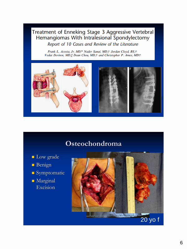
6
Osteochondroma
Low grade
Benign
Symptomatic
Marginal
Excision
20 yo f

7
Benign Tumor-Osteoid Osteoma
Treatment
NSAIDS
RFA
Intralesional
curettage
Associated
scoliosis
13 yo female
ABC
Modern Tx
Serial Embolization
Intralesional embolization
Intralesional resection
Not able to embolize
Mechanical Instabiliy
Denosumab?

8
Posterior resection and reconstruction of C1
lateral mass for ABC
9
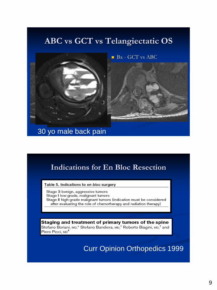
ABC vs GCT vs Telangiectatic OS
Bx - GCT vs ABC
Rebx- GCT vs OS
3 months denosumab
30 yo male back pain
Indications for En Bloc Resection
Curr Opinion Orthopedics 1999

10
GCT
Chordoma
Semin Spine Surg 21:76-85
Terminology En bloc resections without tumor-free margins
Intralesional:
The tumor periphery is violated and the tumor is not
covered by healthy tissue.
Marginal:
The pathologist describes
histologically a thin layer of healthy muscle,
bone, or an endothelial membrane continuously covering
the tumor mass
Wide:
A fascial barrier represents
a wide margin… 1 cm of muscle or cancellous
bone sometimes is not enough and a 2 cm barrier
may be required to consider the margin wide. Boriani Seminars in Oncology

11
WBB Staging
Osteoblastoma
Benign
Type 3
20% recurrence
intralesional
En Bloc Excision
Associated ABC

12
Operative Plan

13

14

15
Giant cell tumor
• Chondrogenic
• Osteogenic
• Fibrogenic
• Fibrohistiocytic
• Osteoclastic GC-rich
• Vascular
• Neuro/Ectodermal
• Notochordal
• Undefined/Pseudotumoral

16
Recurrent GCT L1-2
Options?

17

18
Giant cell Tumor (location)
Denosumab
• The efficacy assessment included 49
patients who had the opportunity to be
on denosumab treatment for at least 6
months.
• After 6 months, 47 patients (96%) were
free of disease progression based on
subjective assessment of disease status
0
20
40
60
80
100
No DiseaseProgression
Disease Progression
Pro
po
rtio
n o
f Pa
tie
nts
(%
)

19
IA Intra-comp; thin capsule, tumor in pseudocapsule
IB Extra-comp (Zone A,D,E) chordoma, chondrosarcoma
IIA Intra-comp; tumor in PC, Osteosarcoma, ES
IIB Extra-comp (Zone A,D,E)
1.Capsule, 2.Pseudocapsule, 3.Tumor invation to pseudocapsule, 4.Skip metastases
C
B A
D E
High grade
Low grade
Malignant Tumor

20
How to access contralateral
nerve root ?
Lateral retraction of specimen

21

22

23
Chordoma
• Chondrogenic
• Osteogenic
• Fibrogenic
• Fibrohistiocytic
• Osteoclastic GC-rich
• Vascular
• Neuro/Ectodermal
• Notochordal
• Undefined/Pseudotumoral
En Bloc Resection Technique Technique
1. Single stage, posterior
High to mid thoracic, sacrectomy
2. 2 stage, posterior/anterior
Cervical, Lower thoracic, lumbar, ? Vessel or
large extracompartmental disease

24
Tomita Threadwire Saw System
Tomita Threadwire Saw System

25
First Step-pass wire saw

26
Anterior Finger Dissection

27
Single stage, posterior
High to mid thoracic

28

29

30
Standard 2 stage P/A Chordoma

31

32

33

34
Chordoma 5 level
35
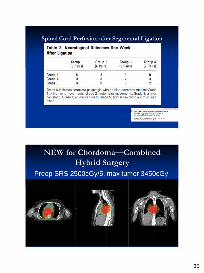
Spinal Cord Perfusion after Segmental Ligation
NEW for Chordoma—Combined
Hybrid Surgery
Preop SRS 2500cGy/5, max tumor 3450cGy
36
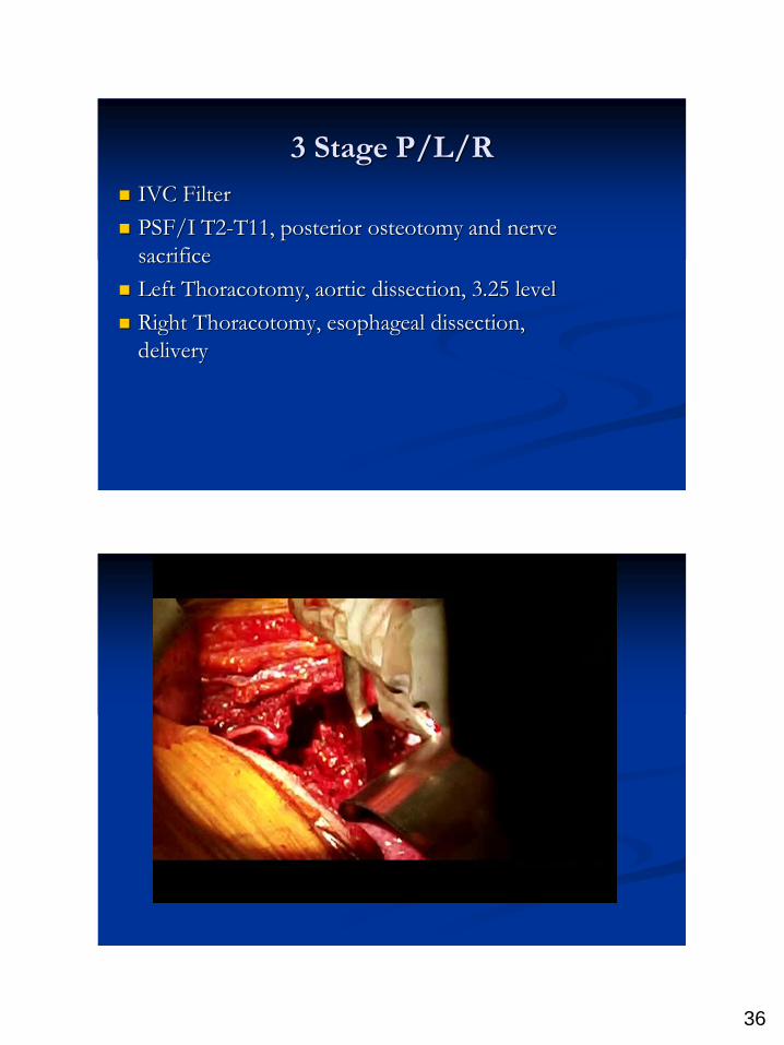
3 Stage P/L/R
IVC Filter
PSF/I T2-T11, posterior osteotomy and nerve
sacrifice
Left Thoracotomy, aortic dissection, 3.25 level
Right Thoracotomy, esophageal dissection,
delivery

37

38
Chordoma (Location)

39
Case 4 Spondylectomy Technique
En Bloc marginal -contaminated at vertebral a.
SAC
Preserve
Bilateral Transverse Cervical Exposure

40
4 rod reconstruction

41
3
4
5
6

42
Chondrosarcoma
• Chondrogenic
• Osteogenic
• Fibrogenic
• Fibrohistiocytic
• Osteoclastic GC-rich
• Vascular
• Neuro/Ectodermal
• Notochordal
• Undefined/Pseudotumoral
43
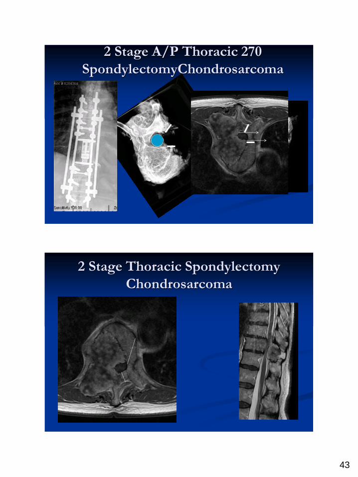
2 Stage A/P Thoracic 270
SpondylectomyChondrosarcoma
2 Stage Thoracic Spondylectomy
Chondrosarcoma

44
270 degree resection
L2-3 chondrosarcoma
A/P
32 yo male
Needle biospy with chondrosarcoma

45

46
Osteosarcoma Preoperative adjuvant
therapy
Response to therapy
predictive of outcome
Refinement of surgical
margins • Chondrogenic
• Osteogenic
• Fibrogenic
• Fibrohistiocytic
• Osteoclastic GC-rich
• Vascular
• Neuro/Ectodermal
• Notochordal
• Undefined/Pseudotumo
ral
Ewings Sarcoma
Preoperative Adjuvant Therapy
• Chondrogenic
• Osteogenic
• Fibrogenic
• Fibrohistiocytic
• Osteoclastic GC-rich
• Vascular
• Neuro/Ectodermal
• Notochordal
• Undefined/Pseudotumoral

47
P32 Brachytherapy
Ex: Leiomyosarcoma
Adjuvant therapy
P32 to margin
Dural margin
Shape radiation targets
for SRS
48
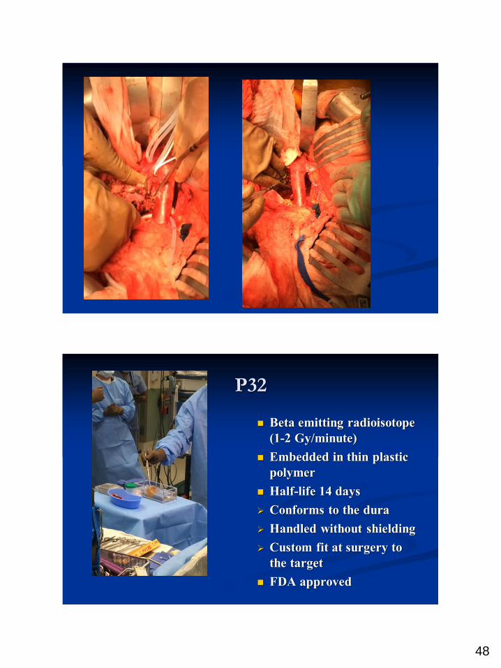
P32
Beta emitting radioisotope
(1-2 Gy/minute)
Embedded in thin plastic
polymer
Half-life 14 days
Conforms to the dura
Handled without shielding
Custom fit at surgery to
the target
FDA approved

49
Soft Tissue Sarcoma
Primary vs Metastatic
Bone grafting demand
Recurrent CS 5 years later…

50
Thanks to the Primary Tumor Team!
Vedat Deviren Orthopedics
Igor Barani Rad Onc
Pierre Theodore Thoracic
Ivan El Sayed ENT
Mika Varma Colorectal
Bill Hoffman Plastics
Charles Eichler Vascular
Rebecca Mustille PT