primary care ent
TRANSCRIPT

Primary Care ENT
Dr Layth Delaimy

EAR NOSE THROATExaminations

Inspecting the external ear
Swab any discharge, and remove any wax. Look for obvious signs of abnormality:
Size and shape of pinna Extra cartilage tags/pre-auricular sinuses or pits Signs of trauma to pinna Suspicious skin lesions on the pinna including
neoplasia Skin conditions of the pinna and external canal Infection/inflammation of external ear canal with
discharge Signs/scars of previous surgery

The normal ear
Normal drum the following structures can be identified: Handle/lateral process of the malleus Light reflex/cone of light Pars tensa and pars flaccida (attic) Occasionally, in a healthy, thin drum, it is possible to see
the following: Long process of incus Choridatympani Eustachian opening Promontory of the cochlea
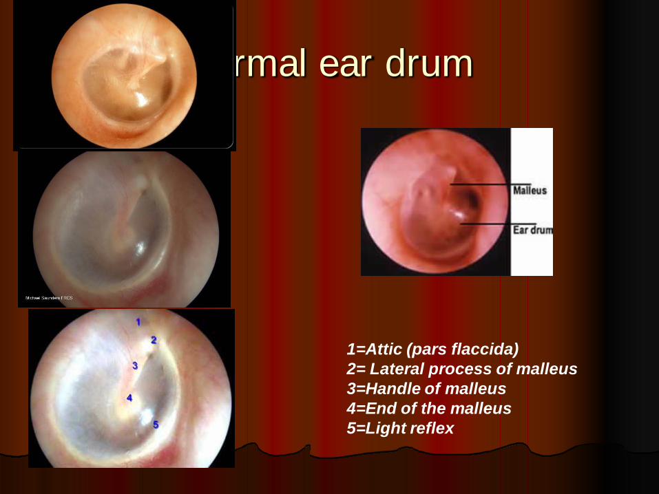
Normal ear drum
1=Attic (pars flaccida) 2= Lateral process of malleus 3=Handle of malleus 4=End of the malleus 5=Light reflex
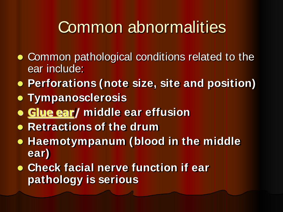
Common abnormalities
Common pathological conditions related to the ear include:
Perforations (note size, site and position) Tympanosclerosis Glue ear/ middle ear effusion Retractions of the drum Haemotympanum (blood in the middle
ear) Check facial nerve function if ear
pathology is serious

Perforation
Perforation
GrommetUS: TubeTraumatic


Tympanosclerosis
Common
Multiple

Otitis Media
Early (mild)
Late (severe)
Haemotympanum (not infection)

Retraction of Eardrum Pocket & Erosion Cholesteatoma

Inspection of the nose
First look at the external nose. Ask patient to remove glasses. Look at nose from front and side for any signs of the following:
Size and shape Obvious bend or deformity: a deviated
nose is often best looked at from above Swelling Scars or abnormal creases Redness (evidence of skin disease) Discharge or crusting Offensive smell

Inspection of the throat Ask patient to remove dentures and examine mouth systemically (use a
bright torch): tongue, hard and soft palate, tonsillar fossa, gingivolabial/gingivobuccal sulci, floor of mouth/undersurface of tongue as follows:
Examine mouth and note condition of tongue Examine back of tongue and tonsils (press down on tongue with a
tongue depressor) Palate the base of tongue (look for tumours that may not be easily
visible) Inspect uvula and soft palate Inspect hard palate (ask patient to tip their head backwards, until
the whole hard palate is visible) Examine buccal area and the gingivolabial (gingivobuccal) sulcus,
(space between cheek and gums) Examine the floor of mouth, check for submandibular duct stones
or masses (ask patient to stick their tongue out) Examine the nasopharynx and larynx with a mirror or flexible
fibre-optic nasendoscope
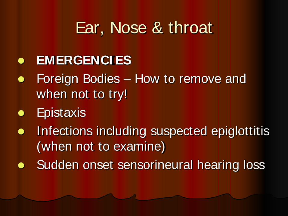
Ear, Nose & throat
EMERGENCIES Foreign Bodies – How to remove and
when not to try! Epistaxis Infections including suspected epiglottitis
(when not to examine) Sudden onset sensorineural hearing loss

COMMON GP PRESENTATIONS
1. Sore ear – Adult including Atypical e.g. TMJ problems &Child
2. Sore throat – Who to refer for tonsillectomy, When to use antibiotics.
3. Discharging Ears – Otitis externa, CSOM4. Hearing Loss including wax management5. Vertigo6. Tinnitus7. Nasal obstruction, polyps, allergy8. Sinus problems9. Facial pain

SPECIFIC CASES TO HIGHLIGHT
Dysphagia Foreign Bodies, Fishbone Neck lumps Hoarseness Head and Neck Cancers

Appreciation of Roles of Others:
Audiologist

Specific Skills:
Use of diagnostic set Epley’s manouevre
http://www.youtube.com/watch?v=eOuzUi5ckrk&feature=relatedhttp://www.youtube.com/watch?v=ZqokxZRbJfw&eurl=http://search.live.com/video/resu
lts.aspx?q=epley's+maneuver&docid=2486314867552&FORM=VIVR3
Micro-suction of auditory canal Audiogram interpretation

What is this?

Epley’s Manuevre
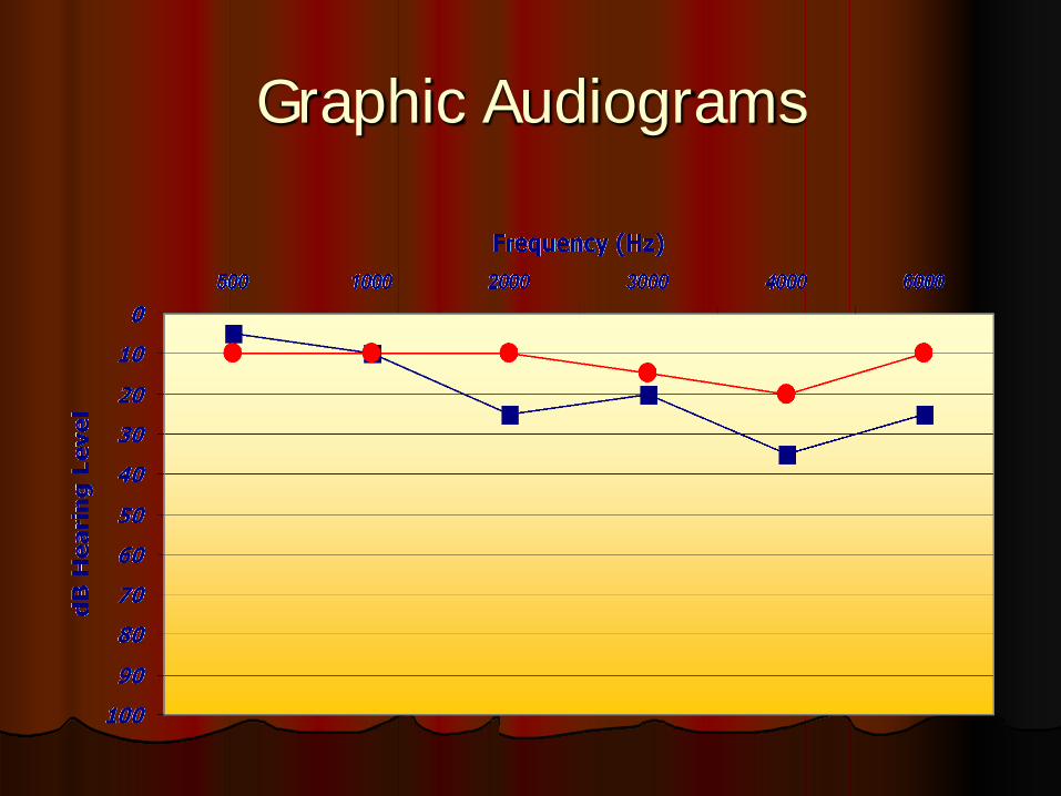
Graphic Audiograms

Self-recording Audiogram
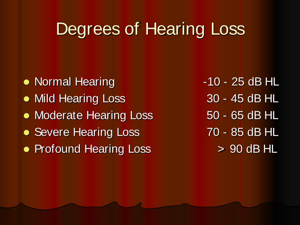
Degrees of Hearing Loss
Normal Hearing -10 - 25 dB HL Mild Hearing Loss 30 - 45 dB HL Moderate Hearing Loss 50 - 65 dB HL Severe Hearing Loss 70 - 85 dB HL Profound Hearing Loss > 90 dB HL