primary research open access antiproliferative and ... · primary research open access...
TRANSCRIPT
Dai et al. Cancer Cell International 2013, 13:27http://www.cancerci.com/content/13/1/27
PRIMARY RESEARCH Open Access
Antiproliferative and apoptotic effects ofβ-elemene on human hepatoma HepG2 cellsZhi-Jun Dai1*, Wei Tang2†, Wang-Feng Lu1†, Jie Gao3†, Hua-Feng Kang1, Xiao-Bin Ma1, Wei-Li Min1,Xi-Jing Wang1* and Wen-Ying Wu4*
Abstract
Background: β-elemene, a natural sesquiterpene extracted from the essential oils of Curcuma aromatica Salisb, hasbeen shown to be effective against a wide range of tumors. In this study, the antitumor effect of β-elemene on ahuman hepatoma cell line, HepG2, and the mechanism involved have been investigated.
Methods: MTT assay was used to determine the growth inhibition of hepatoma HepG2 cells in vitro. Apoptosis ofHepG2 cells were demonstrated by fluorescence microscope with Hoechst 33258 staining and flow cytometry withAnnexin V-FITC/PI double staining. Flow cytometry was performed to analyze the cell cycle distribution of HepG2cells. The mRNA and protein expression of Fas and FasL were measured by RT-PCR and Western blot analysis.
Results: MTT results showed that β-elemene could inhibit the proliferation of HepG2 cells in a time- and dose-dependent manner. Our results showed β-elemene had positive effect on apoptosis through fluorescencemicroscope and flow cytometry assay. Furthermore, β-elemene could induce the cell cycle arrest of the HepG2 cellsin the G2/M phase. Fas and FasL expression were obviously increased after β-elemene treatment in both mRNA andprotein level.
Conclusion: The present study indicates that β-elemene can effectively inhibit proliferation and induce apoptosis inhepatoma HepG2 cells, and the apoptosis induction is related with up-regulating of Fas/FasL expression.
Keywords: Hepatoma, β-elemene, Apoptosis, Fas, FasL
BackgroundHepatocellular carcinoma (HCC) is the fifth most commoncancer and the second leading cause of cancer-relateddeaths worldwide [1]. The causes of HCC includes HBV orHCV infection, alcohol intake, smoking and aflatoxin. InChina, HBV is the most important risk factor for the devel-opment of HCC [2].The treatment of patients with HCC is particularly
challenging because of the array of patient-specific(medical comorbidities), tumor-specific (size, number,location, and vascular involvement), and liver-specific(parenchymal reserve) variables that impact our abilityto treat patients safely and effectively [3]. Liver resection
* Correspondence: [email protected]; [email protected]; [email protected]†Equal contributors1Department of Oncology, the Second Affiliated Hospital of Xi’an JiaotongUniversity, Xi’an 710004, China4Department of Pharmacology, the Second Affiliated Hospital of Xi’anJiaotong University, Xi’an 710004, ChinaFull list of author information is available at the end of the article
© 2013 Dai et al.; licensee BioMed Central LtdCommons Attribution License (http://creativecreproduction in any medium, provided the or
remains the gold standard for patients with resectableHCC that develops in the setting of normal liver sub-stance. However, most patients with HCC have diseasedliver parenchyma and resection in this population ismore fraught, with the potential for complications.Eventually, most patients will receive some forms of
chemotherapy in hope of prolonging life. However, theeffect of chemotherapy is not satisfied, and the side ef-fects are often difficult to tolerate. Sorafenib is the firstmolecular inhibitor to be approved by the FDA for thetreatment of advanced HCC. While being considered anadvance over conventional chemotherapy, sorafenibonly shows approximate 3 months survival advantageover the nontreated group [4,5]. Prior to the arrival ofsorafenib, doxorubicin was routinely used as a singledrug for advanced HCC, but has shown inefficacy, witha response rate of about 15-20%. Other chemotherapyagents, such as epirubicin, cisplatin, 5-fluorouracil andtheir combinations, demonstrate even lower efficacy [6].
. This is an Open Access article distributed under the terms of the Creativeommons.org/licenses/by/2.0), which permits unrestricted use, distribution, andiginal work is properly cited.
Dai et al. Cancer Cell International 2013, 13:27 Page 2 of 10http://www.cancerci.com/content/13/1/27
With that in mind, people have being paying more atten-tion to searching for new antitumor agents from naturalproducts [7]. Many Chinese herbs have been discovered tobe potential sources of antitumor drugs [8,9].It was reported that many Chinese herbs had anticancer
properties and induce apoptosis [10]. Three apoptotic path-ways have been addressed, including the mitochondrialpathway, death receptor pathway, and endoplasmicreticulum stress-mediated apoptosis pathway [11]. Fas is amember of the death receptor family. Stimulation of Fasleads to induction of apoptotic signals, such as caspase 8activation, as well as “non-apoptotic” cellular responses,notably NF-κB activation [12]. Fas ligand (FasL) is a type IItransmembrane protein and signals through trimerizationof FasR, which spans the membrane of the “target”cell. This trimerization usually leads to apoptosis, orcell death [13].Curcuma aromatica Salisb. is a popular type of trad-
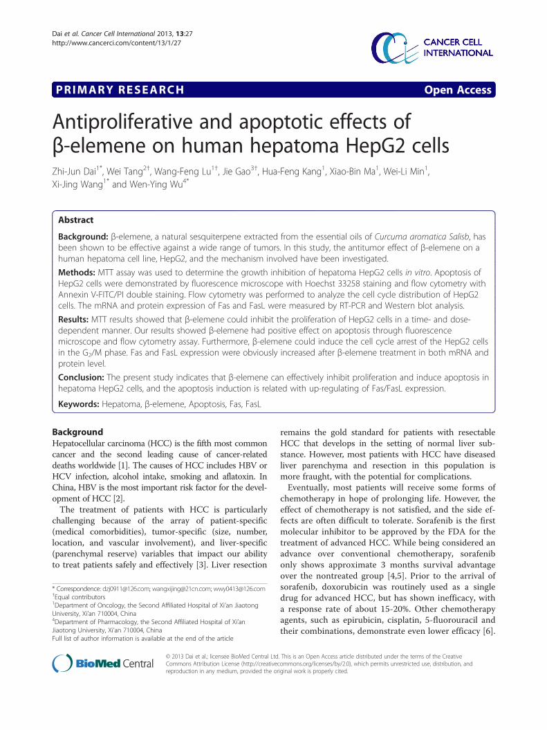
itional Chinese medicine plant whose essential oils arewidely used in cancer treatment in China [14]. β-elemene(1-methyl-1-vinyl-2,4-diisopropenyl-cyclohexane), a sesqui-terpene extracted from the essential oils of Curcumaaromatica Salis., accounts for 60-72% of elemene inCurcuma aromatica Salisb [15]. Among the active ingredi-ents, β-elemene is the most widely studied, whereas δ-elemene, furanodiene, furanodienone, curcumol, andgermacrone have just recently attracted the attention of re-searchers [14]. The molecular formula of β-elemene isC15H24 and its molecular weight 204.34. The chemicalstructure of β-elemene was showed in Figure 1. Meta-analysis results suggest that β-elemene can improve theeffect of chemotherapy in lung cancer as an adjunctivetreatment [16]. The combined treatment can improve qual-ity of life and prolong survival. In vitro, β-elemene hasshown promising anti-cancer effects against a broadspectrum of tumors, such as lung, breast, prostate, cervical,gastric, ovarian cancer and osteosarcoma [17-22]. However,it was few reported in hepatoma cells. The aim of the
Figure 1 Chemical structure of β-elemene. The molecular formulaof β-elemene is C15H24 and its molecular weight 204.34.
present study is to investigate the antitumor effect of β-elemene on human hepatoma HepG2 cells and the molecu-lar mechanism involved.
MethodsReagentsFetal bovine serum (Gibco, USA); RPMI1640 medi-um (Gibco, USA); 3-(4,5)-dimethylthiahiazo (−z-y1)-3,5-diphenyte-trazoliumromide (MTT) and propidium iodide(PI) were purchased from Sigma Chemical (St. Louis, MO).Annexin V-FITC/PI apoptosis detection kit (Becon DickinsonFacsalibur, USA); RT-PCR kit (ampliqon, Denmark); Trizol(Invitrogen, USA); β-elemene was obtained from DalianHolley Kingkong Pharmaceutical Co., Ltd (Dalian, China).Anti-Fas and Anti-FasL were purchased from Santa CruzBiotechnology.
Cell line and cell cultureThe protocol in this study was approved by the Committeeon the Ethics of Animal Experiments of Medicine College,Xi’an Jiaotong University (Certificate No. 22-9601018).Human hepatoma HepG2 cell line was obtained from
Cancer Institute of the Fourth Military Medical University(Xi’an, China). Cells were cultured in RPMI 1640 mediumcontaining 10% inactived fetal bovine serum in a humidifiedatmosphere with 5% CO2 incubator at 37°C.
MTT assay for the proliferation of HepG2 cellsViability of HepG2 cells was assessed using MTT dyereduction assay (Sigma, USA), which was conducted asdescribed previously [23]. Cells were seeded in a 96-wellplate at a density of 1 ×10⁴ cells/well, cultured for 12 h,and then treated with different concentration (0, 40, 80and 120 μg/mL) β-elemene for 0-96 h. At the end of thetreatment, MTT, 50 μg/10 μL, was added and the cellswere incubated for another 4 hours. Dimethylsufloxide(DMSO; 200 μL) was added to each well after removalof the supernatant. After shaking the plate for 10 min,cell viability was assessed by measuring the absorbanceat 490 nm using an Enzyme-labeling instrument (EX-800 type); all measurements were performed three times.The results represented as the average of three inde-pendent experiments done over multiple days.The growth inhibition was calculated according to the
following formula: The Growth Inhibition Ratio (IR%) =[(the absorbance of blank control group − the absorbanceof experimental group)/the absorbance of blank control -group] × 100%.
Detection of morphological apoptosis with Hoechst33258 stainingHepG2 cells were harvested by centrifugation, washedwith PBS and fixed with 1% glutaraldehyde for 1 h atroom temperature. Fixed cells were washed with PBS,

24 h 48 h 72 h 96 h
** **
*
**
#
##
#
*
**
*
Cel
l via
bilit
y (%
)
0
20
40
60
80
100 10 μg/mL β-elemene
20 μg/mL β-elemene
40 μg/mL β-elemene
***
Figure 2 Growth inhibiting effects of β-elemene on HepG2 cells. HepG2 cells were treated with different concentrations drug for 0-96 h. Cellviability was determined by MTT method. This assay was performed in triplicate. Dose- and time-dependent inhibition of cell growth could beobserved after 96 h (P<0.05, ANOVA analysis). *P<0.05, **P<0.01 versus 24 h; #P<0.05 versus 10 μg/mL β-elemene group.
Dai et al. Cancer Cell International 2013, 13:27 Page 3 of 10http://www.cancerci.com/content/13/1/27
stained with 200 μM Hoechst 33258 for 10 min, and thechanges in the nuclei of cells after stained with Hoechst33258 were observed using a fluorescence microscope(Olympus, BX-60, Japan).
Apoptosis detection by flow cytometry (FCM)Apoptotic cells were differentiated from viable or nec-rotic ones by combined application of annexin V-FITCand propidium iodide (PI) (BD Biosciences Clontech,USA) [13]. The samples were washed twice and adjustedto a concentration of 1×10⁶ cells/mL with 4°C PBS. TheFalcon tubes (12 mm×75 mm, polystyrene round-bottom) were used in this experiment, 100 μL of suspen-sions was added to each labeled tube, 10 μL of annexinV-FITC and 10 μL PI (20 μg/mL) were added into thelabeled tube, incubated for at least 20 min at roomtemperature in the dark, then 400 μL of PBS bindingbuffer was added to each tube without washing and ana-lyzed using FCM analysis (BD Biosciences Clontech,USA) as soon as possible (within 30 min). This assaywas done triplicate.
Cell cycle analysisHepG2 cells were incubated at 5 × 10⁵ cells/well in 6-well plates with serum-free RPMI1640 medium for 12 h,then treated with a homologous drug for 48 h. The de-tached and attached cells were harvested and fixed in70% ice-cold ethanol at −20°C overnight. After fixation,cells were washed with PBS, resuspended in 1 mL PBScontaining 1 mg/mL RNase (Sigma) and 50 μg/mL PI(Sigma), and incubated at 37°C for 30 min in the dark.Samples were then analyzed for DNA content by flowcytometry (Beckman, USA), and cell cycle phase distri-butions were analyzed with the Cell Quest acquisitionsoftware (BD Biosciences).
Reverse transcription-polymerase chain reaction (RT-PCR)HepG2 cells were seeded in 6 cm culture capsules andtreated with various concentration of β-elemene (10, 20and 40 μg/mL) separately for 48 h. Each group contained 3culture capsules. As previously described [24], cells col-lected at specified time to extract total RNA using theTrizol reagent following the manufacturer’s instructions.1 μg RNA synthetized cDNA through reverse transcriptaseundergo listed below condition: 70°C 5 min, 42°C extendedfor 60 min, 95°C enzyme inactivated for 3 min and 4°Cterminated reaction. Synthetical cDNA as template to carryout polymerase chain reaction. Fas primer sequence: 50
TTCTGCCATAAGCCCTGTC −30 (forward) and 50 TTGGTGTTGCTGGTGAGT −30 (reverse), amplificationfragment was 320 bp, renaturation temperature was 55°C(cycling 25 times). FasL primer sequence: 50 TTCAGCTCTTCCACCTACAG −30 (forward) and 50 ACATTCTCGGTGCCT GTAAC-30 (reverse), amplificationfragment was 599 bp. β-actin primer sequence was 50
GGTGCTGAGTATGTCGTGGAG −30 (forward), 50 ATGCA GGGATGATGTTCTAGG −30 (reverse), amplificationfragment was 390 bp. Renaturation temperature was 55°C(cycling 20–25 times). Amplification condition was below:pre-denaturized for 3 min at 95°C, denaturized for 30 s at95°C, renaturated for 30 s at 55°C and extended for 30 s at72°C. PCR product was detected on agarose gel electro-phoresis and ethidium bromide imaging system was usedto make density index analysis. The expression intensity ofdestination gene mRNA was denoted with the ratio of thephotodensity of the RT-PCR products of destination geneand β-actin.
Western blot analysisHepG2 cells were treated with various concentration of β-elemene (10, 20 and 40 μg/mL) for 48 h. As previously
A B
C D
Apo
ptos
is(%
)
**
**
*
A B C D0
10
20
30
40
50
60
70
80
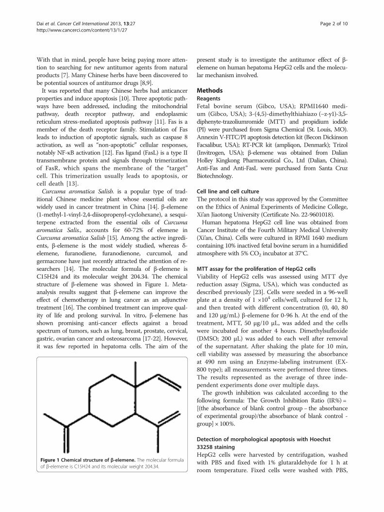
Figure 3 Cell apoptosis observed by Hoechst 33258 staining using a fluorescence microscope (200×). After cells were treated withdifferent dose of β-elemene for 72 h, Hoechst 33258 staining was used to observe the apoptotic cells under a BX-60 fluorescence microscope.Values represent mean ± SEM from three independent experiments. A: blank control group; B: 10 μg/mL β-elemene group; C: 20 μg/mLβ-elemene group; D: 40 μg/mL β-elemene group. *P<0.05, **P<0.01 versus blank control group. The arrow shows apoptotic cells.
Dai et al. Cancer Cell International 2013, 13:27 Page 4 of 10http://www.cancerci.com/content/13/1/27
described [23], cells were lysed in a sample buffer followedby denaturation. Protein concentrations were determinedusing the PIERCE BCA protein assay kit and equalamounts of protein (50 μg) were subjected to SDS-PAGEon 10% gel. The proteins were then electrophoreticallytransferred to nitrocellulose membranes. The membraneswere blocked with 5% skimmed milk, respectively
incubated with anti-Fas, anti-FasL (1:500; Santa CruzBiotechnology) at 4°C overnight. And then followed by in-cubation in goat antimouse secondary antibody conjugatedwith horseradish peroxidase (1:1000; Santa Cruz Biotech-nology). Equal loading of each lane was evaluated byimmunoblotting the same membranes with β-actin anti-bodies after detachment of previous primary antibodies.
Apo
ptos
is(%
)
**
**
*
0
10
20
30
40
50
60
70
A B C D
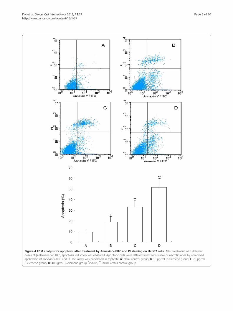
Figure 4 FCM analysis for apoptosis after treatment by Annexin V-FITC and PI staining on HepG2 cells. After treatment with differentdoses of β-elemene for 48 h, apoptosis induction was observed. Apoptotic cells were differentiated from viable or necrotic ones by combinedapplication of annexin V-FITC and PI. This assay was performed in triplicate. A: blank control group; B: 10 μg/mL β-elemene group; C: 20 μg/mLβ-elemene group; D: 40 μg/mL β-elemene group. *P<0.05, **P<0.01 versus control group.
Dai et al. Cancer Cell International 2013, 13:27 Page 5 of 10http://www.cancerci.com/content/13/1/27
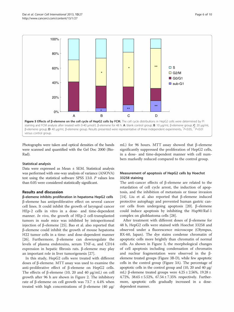
A B C D0%
20%
40%
60%
80%
100%
S
G2/M
G0/G1
sub-G1
**
**
*****
Figure 5 Effects of β-elemene on the cell cycle of HepG2 cells by FCM. The cell cycle distributions in HepG2 cells were determined by PIstaining and FCM analysis after treated with 0-40 μmol/L β-elemene for 48 h. A: blank control group; B: 10 μg/mL β-elemene group; C: 20 μg/mLβ-elemene group; D: 40 μg/mL β-elemene group. Results presented were representative of three independent experiments. *P<0.05, **P<0.01versus control group.
Dai et al. Cancer Cell International 2013, 13:27 Page 6 of 10http://www.cancerci.com/content/13/1/27
Photographs were taken and optical densities of the bandswere scanned and quantified with the Gel Doc 2000 (Bio-Rad).
Statistical analysisData were expressed as Mean ± SEM. Statistical analysiswas performed with one-way analysis of variance (ANOVA)test using the statistical software SPSS 13.0. P values lessthan 0.05 were considered statistically significant.
Results and discussionβ-elemene inhibits proliferation in hepatoma HepG2 cellsβ-elemene has antiproliferative effect on several cancercell lines. It could inhibit the growth of laryngeal cancerHEp-2 cells in vitro in a dose- and time-dependentmanner. In vivo, the growth of HEp-2 cell-transplantedtumors in nude mice was inhibited by intraperitonealinjection of β-elemene [25]. Bao et al. also reported thatβ-elemene could inhibit the growth of mouse hepatomaH22 tumor cells in a time- and dose-dependent manner[26]. Furthermore, β-elemene can downregulate thelevels of plasma endotoxins, serum TNF-α, and CD14expression in hepatic fibrosis rats. β-elemene may playan important role in liver tumorigenesis [27].In this study, HepG2 cells were treated with different
doses of β-elemene. MTT assay was used to examine theanti-proliferative effect of β-elemene on HepG2 cells.The effects of β-elemene (10, 20 and 40 μg/mL) on cellgrowth after 96 h are shown in Figure 2. The inhibitoryrate of β-elemene on cell growth was 73.7 ± 4.4% whentreated with high concentrations of β-elemene (40 μg/
mL) for 96 hours. MTT assay showed that β-elemenesignificantly suppressed the proliferation of HepG2 cells,in a dose- and time-dependent manner with cell num-bers markedly reduced compared to the control group.
Measurement of apoptosis of HepG2 cells by Hoechst33258 stainingThe anti-cancer effects of β-elemene are related to theretardation of cell cycle arrest, the induction of apop-tosis, and the inhibition of metastasis or tissue invasion[14]. Liu et al. also reported that β-elemene inducedprotective autophagy and prevented human gastric can-cer cells from undergoing apoptosis [20]. β-elemenecould induce apoptosis by inhibiting the Hsp90/Raf-1complex on glioblastoma cells [28].After treatment with different doses of β-elemene for
48 h, HepG2 cells were stained with Hoechst 33258 andobserved under a fluorescence microscope (Olympus,BX-60, Japan). The dye stains condense chromatin ofapoptotic cells more brightly than chromatin of normalcells. As shown in Figure 3, the morphological changesof cell apoptosis including condensation of chromatinand nuclear fragmentation were observed in the β-elemene treated groups (Figure 3B-D), while few apoptoticcells in the control group (Figure 3A). The percentage ofapoptotic cells in the control group and (10, 20 and 40 μg/mL) β-elemene treated groups were 4.25 ± 2.56%, 19.28 ±4.72%, 38.65 ± 5.52%, 67.54 ± 7.35% respectively. Further-more, apoptotic cells gradually increased in a dose-dependent manner.
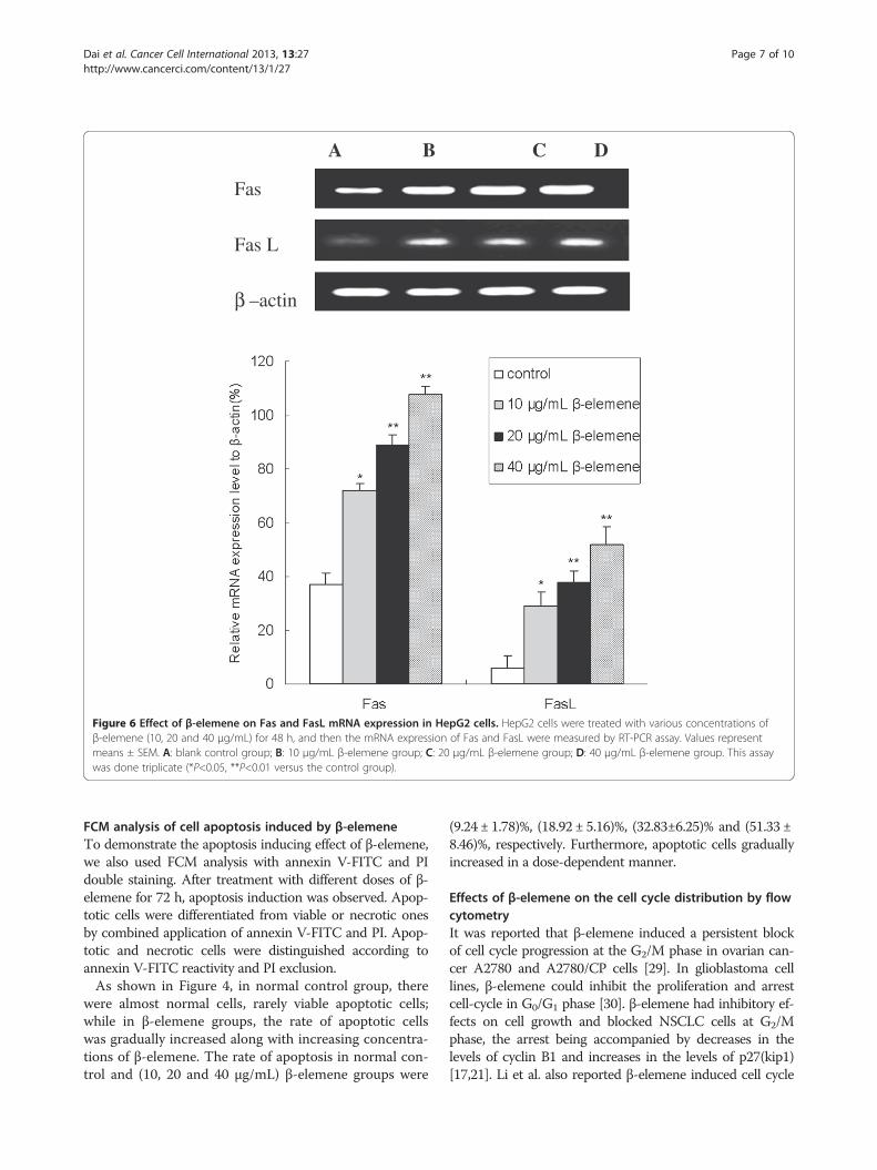
A B C D
Fas
Fas L
β –actin
Figure 6 Effect of β-elemene on Fas and FasL mRNA expression in HepG2 cells. HepG2 cells were treated with various concentrations ofβ-elemene (10, 20 and 40 μg/mL) for 48 h, and then the mRNA expression of Fas and FasL were measured by RT-PCR assay. Values representmeans ± SEM. A: blank control group; B: 10 μg/mL β-elemene group; C: 20 μg/mL β-elemene group; D: 40 μg/mL β-elemene group. This assaywas done triplicate (*P<0.05, **P<0.01 versus the control group).
Dai et al. Cancer Cell International 2013, 13:27 Page 7 of 10http://www.cancerci.com/content/13/1/27
FCM analysis of cell apoptosis induced by β-elemeneTo demonstrate the apoptosis inducing effect of β-elemene,we also used FCM analysis with annexin V-FITC and PIdouble staining. After treatment with different doses of β-elemene for 72 h, apoptosis induction was observed. Apop-totic cells were differentiated from viable or necrotic onesby combined application of annexin V-FITC and PI. Apop-totic and necrotic cells were distinguished according toannexin V-FITC reactivity and PI exclusion.As shown in Figure 4, in normal control group, there
were almost normal cells, rarely viable apoptotic cells;while in β-elemene groups, the rate of apoptotic cellswas gradually increased along with increasing concentra-tions of β-elemene. The rate of apoptosis in normal con-trol and (10, 20 and 40 μg/mL) β-elemene groups were
(9.24 ± 1.78)%, (18.92 ± 5.16)%, (32.83±6.25)% and (51.33 ±8.46)%, respectively. Furthermore, apoptotic cells graduallyincreased in a dose-dependent manner.
Effects of β-elemene on the cell cycle distribution by flowcytometryIt was reported that β-elemene induced a persistent blockof cell cycle progression at the G2/M phase in ovarian can-cer A2780 and A2780/CP cells [29]. In glioblastoma celllines, β-elemene could inhibit the proliferation and arrestcell-cycle in G0/G1 phase [30]. β-elemene had inhibitory ef-fects on cell growth and blocked NSCLC cells at G2/Mphase, the arrest being accompanied by decreases in thelevels of cyclin B1 and increases in the levels of p27(kip1)[17,21]. Li et al. also reported β-elemene induced cell cycle
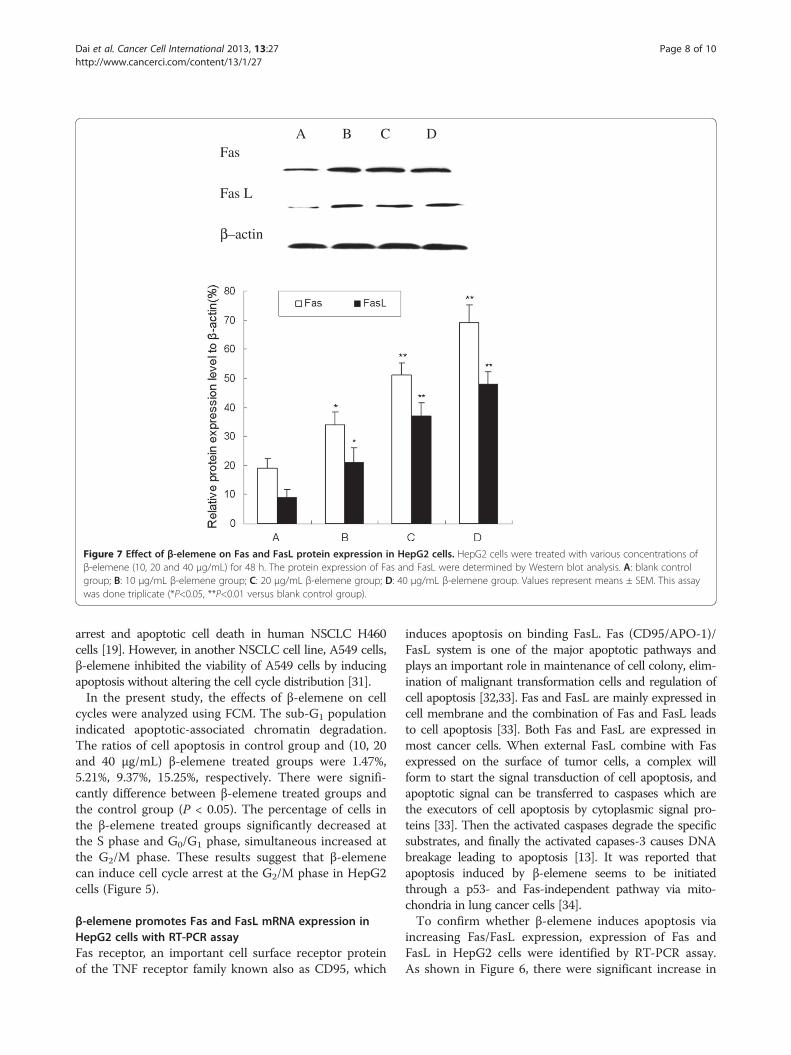
A B C D Fas
Fas L
β–actin
Figure 7 Effect of β-elemene on Fas and FasL protein expression in HepG2 cells. HepG2 cells were treated with various concentrations ofβ-elemene (10, 20 and 40 μg/mL) for 48 h. The protein expression of Fas and FasL were determined by Western blot analysis. A: blank controlgroup; B: 10 μg/mL β-elemene group; C: 20 μg/mL β-elemene group; D: 40 μg/mL β-elemene group. Values represent means ± SEM. This assaywas done triplicate (*P<0.05, **P<0.01 versus blank control group).
Dai et al. Cancer Cell International 2013, 13:27 Page 8 of 10http://www.cancerci.com/content/13/1/27
arrest and apoptotic cell death in human NSCLC H460cells [19]. However, in another NSCLC cell line, A549 cells,β-elemene inhibited the viability of A549 cells by inducingapoptosis without altering the cell cycle distribution [31].In the present study, the effects of β-elemene on cell
cycles were analyzed using FCM. The sub-G1 populationindicated apoptotic-associated chromatin degradation.The ratios of cell apoptosis in control group and (10, 20and 40 μg/mL) β-elemene treated groups were 1.47%,5.21%, 9.37%, 15.25%, respectively. There were signifi-cantly difference between β-elemene treated groups andthe control group (P < 0.05). The percentage of cells inthe β-elemene treated groups significantly decreased atthe S phase and G0/G1 phase, simultaneous increased atthe G2/M phase. These results suggest that β-elemenecan induce cell cycle arrest at the G2/M phase in HepG2cells (Figure 5).
β-elemene promotes Fas and FasL mRNA expression inHepG2 cells with RT-PCR assayFas receptor, an important cell surface receptor proteinof the TNF receptor family known also as CD95, which
induces apoptosis on binding FasL. Fas (CD95/APO-1)/FasL system is one of the major apoptotic pathways andplays an important role in maintenance of cell colony, elim-ination of malignant transformation cells and regulation ofcell apoptosis [32,33]. Fas and FasL are mainly expressed incell membrane and the combination of Fas and FasL leadsto cell apoptosis [33]. Both Fas and FasL are expressed inmost cancer cells. When external FasL combine with Fasexpressed on the surface of tumor cells, a complex willform to start the signal transduction of cell apoptosis, andapoptotic signal can be transferred to caspases which arethe executors of cell apoptosis by cytoplasmic signal pro-teins [33]. Then the activated caspases degrade the specificsubstrates, and finally the activated capases-3 causes DNAbreakage leading to apoptosis [13]. It was reported thatapoptosis induced by β-elemene seems to be initiatedthrough a p53- and Fas-independent pathway via mito-chondria in lung cancer cells [34].To confirm whether β-elemene induces apoptosis via
increasing Fas/FasL expression, expression of Fas andFasL in HepG2 cells were identified by RT-PCR assay.As shown in Figure 6, there were significant increase in
Dai et al. Cancer Cell International 2013, 13:27 Page 9 of 10http://www.cancerci.com/content/13/1/27
both Fas and FasL mRNA expression when treated with10, 20 and 40 μg/mL β-elemene in comparison with thecontrol group (P<0.05). Furthermore, the mRNA expres-sion of Fas and FasL in 40 μg/mL β-elemene group wereenhanced up to 192% and 550% respectively, thisenhancement effect was in a dose-dependent manner(P<0.05).
β-elemene increases Fas and FasL protein expression withWestern blot analysisAs mentioned above, the Fas and FasL mRNA expres-sion were increased after β-elemene treatment. Toexamine whether β-elemene increased the expression ofFas and FasL in protein level, we tested the expressionwith Western blot analysis. As shown in Figure 7, West-ern blot results showed that both Fas and FasL proteinexpression significantly increased in β-elemene treatedgroup comparing with the control group (P<0.05). Fur-thermore, the protein expression of Fas and FasL in40 μg/mL β-elemene group were enhanced 263% and433%, this enhancement effect was in a dose-dependentmanner (P<0.05).
ConclusionIn conclusion, MTT assay showed that β-elemene couldinhibit the proliferation of HepG2 cells in vitro in atime- and dose- dependent manner. Furthermore, β-elemene could induce apoptosis and cell cycle arrest atthe G2/M phase in HepG2 cells. The present study sug-gests that β-elemene can effectively inhibit proliferationand induce apoptosis in hepatoma cells, and the apop-tosis induction is related with up-regulating of Fas/FasLexpression. However, further studies are necessary toclarify the detailed mechanism involved in the antitumoreffects of β-elemene.
Competing interestsThe authors declare that they have no competing interests.
Authors’ contributionsDZJ, WXJ and WWY designed the research. DZJ, TW, LWF, GJ, and MXBperformed the experiments throughout this research. MWL, KHF and WXJcontributed to the reagents, and participated in its design and coordination.TW and LWF analyzed the data; DZJ and GJ contributed to the writing ofthe manuscript. Co-first authors: DZJ, TW, LWF and GJ. All authors have readand approved the final manuscript.
AcknowledgmentsThis study was supported by National Natural Science Foundation of China,No. 81102711; the Fundamental Research Funds for the Central Universities,China, No. xjj2011039; Sci-tech Program of Administration of TraditionalChinese Medicine of Shaanxi Province, China, No. 2009jc86.
Author details1Department of Oncology, the Second Affiliated Hospital of Xi’an JiaotongUniversity, Xi’an 710004, China. 2Department of life science, Shaanxi NormalUniversity, Xi’an 710061, China. 3Department of Nephrology, the SecondAffiliated Hospital of Xi’an Jiaotong University, Xi’an 710004, China.4Department of Pharmacology, the Second Affiliated Hospital of Xi’anJiaotong University, Xi’an 710004, China.
Received: 17 January 2013 Accepted: 8 March 2013Published: 14 March 2013
References1. Jemal A, Bray F, Center MM, Ferlay J, Ward E, Forman D: Global cancer
statistics. CA Cancer J Clin 2011, 61:69–90.2. Tanaka M, Katayama F, Kato H, Tanaka H, Wang J, Qiao YL, Inoue M:
Hepatitis B and C virus infection and hepatocellular carcinoma in China:a review of epidemiology and control measures. J Epidemiol 2011,21:401–416.
3. Maluccio M, Covey A: Recent progress in understanding, diagnosing, andtreating hepatocellular carcinoma. CA Cancer J Clin 2012, 62:394–399.
4. Llovet JM, Ricci S, Mazzaferro V, Hilgard P, Gane E, Blanc JF, de Oliveira AC,Santoro A, Raoul JL, Forner A, Schwartz M, Porta C, Zeuzem S, Bolondi L,Greten TF, Galle PR, Seitz JF, Borbath I, Häussinger D, Giannaris T, Shan M,Moscovici M, Voliotis D, Bruix J, SHARP Investigators Study Group: Sorafenibin advanced hepatocellular carcinoma. N Engl J Med 2008, 359:378–390.
5. Cheng AL, Kang YK, Chen Z, Tsao CJ, Qin S, Kim JS, Luo R, Feng J, Ye S,Yang TS, Xu J, Sun Y, Liang H, Liu J, Wang J, Tak WY, Pan H, Burock K, Zou J,Voliotis D, Guan Z: Efficacy and safety of sorafenib in patients in the Asia-Pacific region with advanced hepatocellular carcinoma: a phase IIIrandomised, double-blind, placebo-controlled trial. Lancet Oncol 2009,10:25–34.
6. Cao H, Phan H, Yang LX: Improved chemotherapy for hepatocellularcarcinoma. Anticancer Res 2012, 32:1379–1386.
7. Yu JQ, Liu HB, Lei JC, Tan WJ, Hu XM, Zou GL: Antitumor activity ofchloroform fraction of Scutellaria barbata and its active constituents.Phytother Res 2007, 21:817–822.
8. Vickers A: Botanical medicines for the treatment of cancer: rationale,overview of current data, and methodological considerations for phase Iand II trials. Cancer Invest 2002, 20:1069–1079.
9. Dai ZJ, Gao J, Li ZF, Ji ZZ, Kang HF, Guan HT, Diao Y, Wang BF, Wang XJ: InVitro and In Vivo antitumor activity of Scutellaria Barbate extract onmurine liver cancer. Molecules 2011, 16:4389–4400.
10. Yu ZH, Wei PK, Xu L, Qin ZF, Shi J: Anticancer effect of jinlongshe granuleson in situ-transplanted human MKN-45 gastric cancer in nude mice andxenografted sarcoma 180 in Kunming mice and its mechanism. World JGastroenterol 2006, 12:2890–2894.
11. Gupta S: Molecular signaling in death receptor and mitochondrialpathways of apoptosis. Int J Oncol 2003, 22:15–20.
12. Liu F, Bardhan K, Yang D, Thangaraju M, Ganapathy V, Waller JL, LilesGB, Lee JR, Liu K: NF-κB directly regulates Fas transcription tomodulate Fas-mediated apoptosis and tumor suppression. J BiolChem 2012, 287:25530–25540.
13. Dai ZJ, Gao J, Ji ZZ, Wang XJ, Ren HT, Liu XX, Wu WY, Kang HF, Guan HT:Matrine induces apoptosis in gastric carcinoma cells via alteration ofFas/FasL and activation of caspase-3. J Ethnopharmacol 2009, 123:91–96.
14. Lu JJ, Dang YY, Huang M, Xu WS, Chen XP, Wang YT: Anti-cancer properties ofterpenoids isolated from Rhizoma Curcumae. J Ethnopharmacol 2012,143:406–411.
15. Zhang R, Tian A, Zhang H, Zhou Z, Yu H, Chen L: Amelioration ofexperimental autoimmune encephalomyelitis by beta-elemenetreatment is associated with Th17 and Treg cell balance. J MolecularNeuroscience 2011, 44:31–40.
16. Wang B, Peng XX, Sun R, Li J, Zhan XR, Wu LJ, Wang SL, Xie T: Systematicreview of β-Elemene injection as adjunctive treatment for lung cancer.Chin J Integr Med 2012, 18:813–823.
17. Wang G, Li X, Huang F, Zhao J, Ding H, Cunningham C, Coad JE, Flynn DC,Reed E, Li QQ: Antitumor effect of beta-elemene in non-small-cell lungcancer cells is mediated via induction of cell cycle arrest and apoptoticcell death. Cell Mol Life Sci 2005, 62:881–893.
18. Zhang B, Zhang X, Tang B, Zheng P, Zhang Y: Investigation of elemene-induced reversal of tamoxifen resistance in MCF-7 cells throughoestrogen receptor α (ERα) re-expression. Breast Cancer Res Treat 2012,136:399–406.
19. Li QQ, Wang G, Huang F, Banda M, Reed E: Antineoplastic effect of beta-elemene on prostate cancer cells and other types of solid tumour cells.J Pharm Pharmacol 2010, 62:1018–1027.
20. Liu J, Zhang Y, Qu J, Xu L, Hou K, Zhang J, Qu X, Liu Y: β-Elemene-inducedautophagy protects human gastric cancer cells from undergoingapoptosis. BMC Cancer 2011, 11:183.

Dai et al. Cancer Cell International 2013, 13:27 Page 10 of 10http://www.cancerci.com/content/13/1/27
21. Li X, Wang G, Zhao J, Ding H, Cunningham C, Chen F, Flynn DC, Reed E, LiQQ: Antiproliferative effect of beta-elemene in chemoresistant ovariancarcinoma cells is mediated through arrest of the cell cycle at the G2-Mphase. Cell Mol Life Sci 2005, 62:894–904.
22. Liang D, Yang M, Guo B, Yang L, Cao J, Zhang X: HIF-1α induced by β-elemene protects human osteosarcoma cells from undergoingapoptosis. J Cancer Res Clin Oncol 2012, 138:1865–1887.
23. Dai ZJ, Ma XB, Kang HF, Gao J, Min WL, Guan HT, Diao Y, Lu WF, Wang XJ:Antitumor activity of the selective cyclooxygenase-2 inhibitor, celecoxib,on breast cancer in vitro and in vivo. Cancer Cell Int 2012, 12:53.
24. Guan HT, Xue XH, Dai ZJ, Wang XJ, Li A, Qin ZY: Downregulation ofsurvivin expression by small interfering RNA induces pancreatic cancercell apoptosis and enhances its radiosensitivity. World J Gastroenterol2006, 12:2901–2907.
25. Tao L, Zhou L, Zheng L, Yao M: Elemene displays anti-cancer ability onlaryngeal cancer cells in vitro and in vivo. Cancer Chemother Pharmacol2006, 58:24–34.
26. Bao F, Qiu J, Zhang H: Potential role of β-elemene on histone H1 in theH22 ascites hepatoma cell line. Mol Med Report 2012, 6:185–190.
27. Liu J, Zhang Z, Gao J, Xie J, Yang L, Hu S: Downregulation effects ofbeta-elemene on the levels of plasma endotoxin, serum TNF-alpha,and hepatic CD14 expression in rats with liver fibrosis. Front Med2011, 5:101–105.
28. Zhao YS, Zhu TZ, Chen YW, Yao YQ, Wu CM, Wei ZQ, Wang W, XuYH: Β-elemene inhibits Hsp90/Raf-1 molecular complex inducingapoptosis of glioblastoma cells. J Neurooncol 2012, 107:307–314.
29. Lee RX, Li QQ, Reed E: β-elemene effectively suppresses the growth andsurvival of both platinum-sensitive and -resistant ovarian tumor cells.Anticancer Res 2012, 32:3103–3113.
30. Yao YQ, Ding X, Jia YC, Huang CX, Wang YZ, Xu YH: Anti-tumor effect ofbeta-elemene in glioblastoma cells depends on p38 MAPK activation.Cancer Lett 2008, 264:127–134.
31. Liu J, Hu XJ, Jin B, Qu XJ, Hou KZ, Liu YP: β-Elemene induces apoptosis aswell as protective autophagy in human non-small-cell lung cancer A549cells. J Pharm Pharmacol 2012, 64:146–153.
32. Pinti M, Troiano L, Nasi M, Moretti L, Monterastelli E, Mazzacani A, Mussi C,Ventura P, Olivieri F, Franceschi C, Salvioli G, Cossarizza A: Geneticpolymorphisms of Fas (CD95) and FasL (CD178) in human longevity:studies on centenarians. Cell Death Differ 2002, 9:431–438.
33. Fumarola C, Guidotti GG: Stress-induced apoptosis: toward a symmetrywith receptor mediated cell death. Apoptosis 2004, 9:77–82.
34. Zhao J, Li QQ, Zou B, Wang G, Li X, Kim JE, Cuff CF, Huang L, Reed E,Gardner K: In vitro combination characterization of the new anticancerplant drug beta-elemene with taxanes against human lung carcinoma.Int J Oncol 2007, 31:241–252.
doi:10.1186/1475-2867-13-27Cite this article as: Dai et al.: Antiproliferative and apoptotic effects ofβ-elemene on human hepatoma HepG2 cells. Cancer Cell International2013 13:27.
Submit your next manuscript to BioMed Centraland take full advantage of:
• Convenient online submission
• Thorough peer review
• No space constraints or color figure charges
• Immediate publication on acceptance
• Inclusion in PubMed, CAS, Scopus and Google Scholar
• Research which is freely available for redistribution
Submit your manuscript at www.biomedcentral.com/submit