primary squamous cell carcinoma of kidney: a rare case … · diagnosis while evaluating cases of...
TRANSCRIPT

International Journal of Medical Science Research and PracticeUNIT OF AXIS JOURNALS
International Peer Reviewed Medical Journal Committed for ExcellencePrint ISSN: 2349-3178 Online ISSN: 2349-3186
49 International Journal of Medical Science Research and Practice • Vol 2 • Issue 1 • 2015
same region since 6 months. There was no history of any renal calculus disease or calculus surgery in the past.
Her general examination was normal. Per abdominal examination preoperatively had revealed, a lump of 10.0 cm × 8.0 cm was palpable in the right lumbar and hypochondriac regions. On examination, it was fi rm, moving with respiration and non-ballotable. The surface was bosselated, with well-defi ned medial, lower, and lateral borders. The upper border could not be palpated.
Hence, with a clinical diagnosis of a renal lump, most probably renal cell carcinoma (RCC), the patient was investigated. All routine hematological investigations were normal. Ultrasonography (USG) of the abdomen showed a solid mass at the upper pole of the right kidney abutting the liver segments VI and VII of liver show a hyperechoic lesion, likely to be a metastasis. Contrast-enhanced computed tomography (CECT) of abdomen revealed an ill-defi ned solid mass with necrotic areas showing heterogeneously moderate enhancement, noted arising from renal pelvis and superior pole of right kidney measuring 8.8 cm × 8.8 cm × 6.4 cm. The mass showed well-defi ned rounded calcifi cation and has breached the perinephric fat to involve segment VI and VII of liver. Both renal veins and inferior vena cava were normal. USG guided tru-cut biopsy was inconclusive,
INTRODUCTIONCancers of the kidney amounts to 2% of all the total human cancer.1 Primary tumors of the renal pelvis are quite rare accounting for only about 4-5% of all urothelial tumors and include transitional cell carcinomas, squamous cell carcinomas (SCC), and adenocarcinomas.2 Renal SCC (RSCC) of pelvis is a rare malignancy of the upper urinary tract characteristically presenting with advanced stage. Of these 90% are of transitional cell origin while SCCs constitute about 6-15%.3 Among malignant renal tumors, SCC are decidedly rare neoplasms and form only about 0.5-8%. These are rare and very few cases have been reported in the literature. We report two cases of primary RSCC of the renal pelvis.
CASE REPORTS
Case 1A 44-year-old lady presented to our Outdoor Patients Department (OPD) with a history of complaints of pain in the right fl ank since around 6-7 months. The pain was of dull aching type, continuous in nature. There was no history of weight loss, loss of appetite, hematuria or any other urinary complaints. The patient also had complaints of a lump in the
Primary squamous cell carcinoma of kidney: A rare case report
Ashok Kumar Diwan, Rohit Santosh Kabre
Department of Radiation Therapy & Oncology, Government Medical College & Hospital, Nagpur, Maharashtra, India
Corresponding Author:Dr. Ashok Kumar Diwan, Assistant Professor, Department of Radiation Therapy, Government Medical College & Hospital, Nagpur - 440 003, Maharashtra, India. E-mail: [email protected]
© 2015 International Journal of Medical Science Research and Practice available on www.ijmsrp.com
Case Report
ABSTRACT
Neoplasms of the kidney are most commonly adenocarcinomas. Carcinomas of the renal pelvis and ureter are rare, accounting for only 4% of all urothelial cancers. Squamous cell carcinoma (SCC) of renal pelvis and ureter are very rare and account for 6-15% of all renal tumors of which transitional carcinomas constitute the majority followed by adenocarcinoma and SCC. Renal SCC (RSCC) is a rare malignancy of the upper urinary tract characteristically presenting with advanced stage, the reported incidence being 1.4% of all renal malignancies. Very few cases have been reported in the literature. Most patients have a history of chronic urolithiasis, renal infection or abuse of analgesics. This tumor is aggressive in nature and usually has a poor prognosis. We report two cases of RSCC one with characteristic history of chronic pyelonephritis and one without characteristic etiologic factors. These cases have been reported due to their extreme rarity and also highlight the silent presentation of these tumors and the need to keep it as a differential diagnosis while evaluating cases of nephrolithiasis and chronic infl ammations.
Keywords: Chronic pyelonephritis, renal pelvis, squamous cell carcinoma
Diwan and Kabre: Primary squamous cell carcinoma of kidney
International Journal of Medical Science Research and Practice • Vol 2 • Issue 1 • 2015 50
showing only dysplastic cells. Rest of the metastatic workup was normal.
Hence, with a diagnosis of RCC, a right nephroureterectomy with wedge resection of liver was done (Figures 1 and 2). Histopathology was suggestive of well-differentiated SCC of the renal pelvis extending into the upper pole of kidney and liver. Patient was planned for platinum-based chemotherapy after wound healing indication being hepatic invasion. However, the patient was lost for follow-up and on enquiry patient passed away 3 months after surgical management.
Case 2A 60-year-old female presented to our OPD with complaints of on and off fever since 14 months and right-sided fl ank pain and passage of clots in urine since 2 months, pain was dull and intermittent in nature. There was no history of weight loss, loss of appetite or any other urinary complaints. There was no history of any renal calculus disease or calculus surgery in the past. Her general examination was normal. Per abdominal examination preoperatively had revealed, a bosselated lump of 8.0 cm × 8.0 cm was palpable in the right lumbar and hypochondriac regions. On examination, it was fi rm, moving with respiration and non-ballotable.
With clinical diagnosis of renal mass patient was evaluated. All routine hematological investigations were normal. Urine culture and sensitivity was positive for Escherichia coli. Urine cytology was negative for malignant cells. USG abdomen
and pelvis showed mass noted in the upper pole of the right kidney measuring 5.1 cm × 4.7 cm × 3.3 cm with mild vascularity and heterogeneous echotexture. CECT abdomen and pelvis revealed well-defi ned moderately enhancing mass lesion of size 5.9 cm × 5.4 cm × 6.3 cm in transverse, AP and CC axis noted arising from upper pole of right kidney with reniform shape of kidney well maintained with few non-enhancing areas noted s/o necrosis (Figures 3-5). There is thickening of Gerota’s fascia with perinephric fat stranding. Right renal vein and right suprarenal gland appear normal. Chest X-ray and electrocardiogram were normal.
With a diagnosis of RCC, right radical nephrectomy was done (Figure 6). Histopathology report though confi rmed diagnosis of SCC of kidney with chronic pyelonephritis with section from tumor with pelvis showing squamous metaplasia of pelvis with full thickness dysplasia (Figures 7-9).
Post-operative CECT Abdomen was within normal limits with confi rming postoperative status of the right kidney (Figure 10). In accordance with that patient was confi rmed to be staged as p(T3N0M0) and is kept on keen follow-up. As per, limited experience in the past chemoradiotherapy is indicated in metastatic setup.
Figure 1: C1 - Intraoperative view of liver extension
Figure 2: C1 - Gross specimen
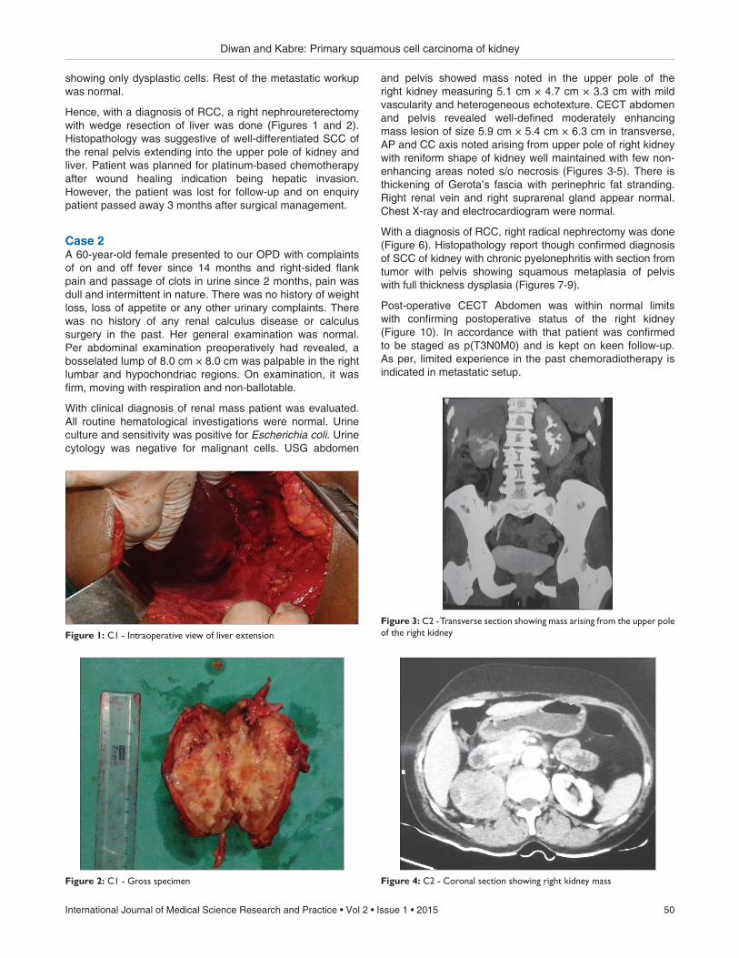
Figure 3: C2 - Transverse section showing mass arising from the upper pole of the right kidney
Figure 4: C2 - Coronal section showing right kidney mass

Diwan and Kabre: Primary squamous cell carcinoma of kidney
51 International Journal of Medical Science Research and Practice • Vol 2 • Issue 1 • 2015
DISCUSSIONSCC of the renal collecting system is a rare malignancy with poor prognosis accounting for about 10% of renal pelvic
tumors and 0.5% of all renal tumors.1,2 Primary RSCC of the renal pelvis is a rare malignancy with a variable incidence of about 7-8% of all urothelial cancers.3 Pure SCC is very rare, though it is the second most common tumor in the renal pelvis.4 Women are affected more frequently than men, predominant age group being 50-70 years.5
Figure 6: C2 - Gross specimen of radical nephrectomy
Figure 7: C2 - A: Thyroidization of glomeruli, B: Keratotic cellular debris
Figure 8: C2 - A: Normal glomeruli, B: Malignant squamous cells
Figure 9: Hyperchromic nuclei of squamous cells
Figure 5: C2 - Coronal section showing right kidney renal fossa with no signifi cant LNpathy
Figure 10: C2 - Post-operative scan showing empty right mass

Diwan and Kabre: Primary squamous cell carcinoma of kidney
International Journal of Medical Science Research and Practice • Vol 2 • Issue 1 • 2015 52
Chronic irritation of urothelium is presumed to be a cause of squamous metaplasia with subsequent malignant progression to SCC. Common causes of chronic irritation are renal calculus disease for long duration, previous history of renal calculi surgery, chronic analgesic abuse or radiotherapy. Li et al. reported incidence of co-existing renal stone in 100% of cases.6 Smoking or tobacco chewing was also observed in 60% of the patients as a known predisposing factor. Hypercalcemia, leukocytosis, and thrombocytosis have been reported as a part of paraneoplastic syndromes in RSCC cases.7,8
Overall median survival for renal pelvis SCC was 7.25 months.9 Using the current 7th edition AJCC staging, the 5-year renal pelvis and ureter cancer survival is as follows: Stage 0a - 72.3%, Stage 0 - 70%, Stage I - 63.9%, Stage II - 56.7%, Stage III - 38.5%, Stage IV - 10.2%. Their generally poor prognosis can be attributed to the typically advanced stage at diagnosis with high local recurrence rates, but the stage for stage, prognosis is similar for squamous, and usual urothelial carcinoma.10
As per literature, there are two entities when it comes to SCC of kidney, one being intraparenchymal SCC which is much rarer and pathognomonic sign of, which is normal histopathological features of renal pelvis which was not the case with our patients. Second, being primary renal SCC of pelvis, which may or may not be associated with squamous metaplasia/dysplasia. Hence, both of the reported cases are in primary renal SCC of the renal pelvis.
The diagnosis of SCC of the renal pelvis is restricted to tumors showing extensive squamous differentiation. The histologic hallmarks of pearl formation, intercellular bridges, and keratotic cellular debris are those of squamous carcinoma at any site. However, the conspicuous presence of keratinizing squamous metaplasia of the adjacent fl attened urothelium, especially if associated with dysplasia, supports a diagnosis of primary SCC of the renal pelvis which is rare11,12 which were characteristically seen in our case report. Thyroidization of renal glomeruli, which is the hallmark of chronic pyelonephritis, was also one of the fi ndings of our case report.
Hence, a tumor with squamous cell morphology in the kidney in a middle aged to the elderly patient should be meticulously sampled to differentiate among the primary urothelial carcinoma with squamous differentiation, primary SCC of the renal pelvis, metastatic SCC to kidney and primary intraparenchymal SCC of the kidney. Both of our cases were negative for metastatic work up, hence excluding above differential diagnosis.
Lee et al. found that the specifi c feature in CT of RSCC was presence of enhancing extra luminal exophytic mass or in some cases, an intraluminal component.13 They further suggested that intravenous urography (IVU) should be carried out periodically, especially, in patients with long-standing stones. Because the fi lling defects, delay in appearance of pyelogram, or renal parenchymal thickening in IVU may indicate a renal tumor despite the absence of mass effect and preservation of renal contour, warranting further studies.14 One of the cases showed mass arising
from upper pole of right kidney with reniform shape of kidney well-maintained.
Extensive review of the available medical literature on this rare malignant entity revealed a poor prognosis.9 Nativ et al. in their study divided renal SCC in three groups, reported 1- and 2-year survival rates of locally invasive renal SCC 33% and 22%, respectively.9 Review of literature suggested current primary treatment of RSCC being nephrectomy.9,10,12 Adjuvant chemotherapy or radiotherapy indicated in metastatic disease.15 First case reported had pathological staging as T4N1M0 (Stage IV) in which radical nephrectomy with hepatic wedge resection was done as special mention. Second case had pathological staging as T3N1#M0 (Stage IV) in which radical nephrectomy was done. Nephrectomy with ureterectomy is the treatment of choice in these patients, even in the face of metastatic disease. There is a lack of evidence regarding survival benefi t with chemo-radiation following surgery.
Adjuvant treatment in the form of radiotherapy has been used in management of renal pelvis cancer, clinical target volume should include the renal fossa, the course of ureter to bladder, the entire bladder and the para-caval, para-aortic lymph nodes to the dose of 45-50 Gy at 1.8 to 2 Gy per day for subclinical and microscopic disease, a boost of 5-10 Gy for more extensive disease, multiple positive nodes, microscopic positive margins, macroscopic residual margin.
In the setting of metastatic disease, adjuvant chemotherapy has been used which was platinum-based but defi nite chemotherapy protocol do not exist due to being rare clinical entity with poor prognosis. The possibility of anti-epidermal growth-factor receptor therapy in renal SCCs expressing the receptor needs further evaluation in a research setting.
CONCLUSIONPrimary RSCC of pelvis has silent presentation, often detected at an advanced stage hence cases of nephrolithiasis should be kept on follow-up with IVU.
ACKNOWLEDGMENTSNil
PEER REVIEWDouble blind externally peer reviewed.
CONFLICTS OF INTERESTNil
FUNDINGNil

Diwan and Kabre: Primary squamous cell carcinoma of kidney
53 International Journal of Medical Science Research and Practice • Vol 2 • Issue 1 • 2015
REFERENCES 1. Blacher EJ, Johnson DE, Abdul-Karim FW, Ayala AG.
Squamous cell carcinoma of renal pelvis. Urology 1985;25:124-6.2. Busby JE, Brown GA, Tamboli P, Kamat AM, Dinney CP,
Grossman HB, et al. Upper urinary tract tumors with nontransitional histology: A single-center experience. Urology 2006;67:518-23.
3. Verma N, Yadav G, Dhawan N, Kumar A. Squamous cell carcinoma of kidney co-existing with renal calculi: A rare tumour. BMJ Case Rep 2011;2011.
4. Karabulut A, Emir L, Gonultas M, Incel N, Germiyanoglu C, Erol E. Squamous cell carcinoma located in the renal calyceal system. A case report and review of the literature. Turk J Cancer 2002;32:20-4.
5. Talwar N, Dargan P, Arora MP, Sharma A, Sen AK. Primary squamous cell carcinoma of the renal pelvis masquerading as pyonephrosis: A case report. Indian J Pathol Microbiol 2006;49:418-20.
6. Li MK, Cheung WL. Squamous cell carcinoma of the renal pelvis. J Urol 1987;138:269-71.
7. Cadeddu JA, Jarrett TW. Hypercalcemia associated with squamous cell carcinoma of the renal pelvis. J Urol 1998;160:1798.
8. Er O, Coskun HS, Altinbas M, Akgün H, Cetin M, Eser B, et al. Rapidly relapsing squamous cell carcinoma of the renal pelvis associated with paraneoplastic syndromes of leukocytosis, thrombocytosis and hypercalcemia. Urol Int 2001;67:175-7.
9. Nativ O, Reiman HM, Lieber MM, Zincke H. Treatment of primary squamous cell carcinoma of the upper urinary tract.
Cancer 1991;68:2575-8.10. Kose F, Bal N, Ozyilkan O. Squamous cell carcinoma of the
renal pelvis. Med Oncol 2009;26:103-4.11. Lopez-Beltran A, Montironi R, Vidal-Jimenez A, Cheng L.
Pathology of tumors of the urinary bladder. In: Mikuz G, editor. Clinical Pathology of Urologic Tumors. Boca Raton, FL, USA: Informa Healthcare; 2007. p. 57-89.
12. Maclennan GT, Cheng L. Renal pelvis and ureter. In: Maclennan GT, Cheng L, editors. Atlas of Genitourinary Pathology. New York, USA: Springer; 2011. p. 123-40.
13. Lee TY, Ko SF, Wan YL, Cheng YF, Yang BY, Huang DL, et al. Renal squamous cell carcinoma: CT fi ndings and clinical signifi cance. Abdom Imaging 1998;23:203-8.
14. Jain A, Mittal D, Jindal A, Solanki R, Khatri S, Parikh A, et al. Incidentally detected squamous cell carcinoma of renal pelvis in patients with staghorn calculi: Case series with review of the literature. ISRN Oncol 2011;2011:620574.
15. Reuter VE. The urothelial tract: Renal pelvis, ureter, urinary bladder and urethra. In: Mills SE, Carter D, Greenson JK, Oberman HA, Reuter V, Stoler MH, editors. Sternberg’s Diagnostic Surgical Pathology. 4th ed. Philadelphia: Lippincott Williams and Wilkins; 2004. p. 2058-9.
How to cite this article: Diwan AK, Kabre RS. Primary squamous cell carcinoma of kidney: A rare case report. Inter J Medical Sci Res Prac 2015; 2(1):49-53.
Received: 20 Feb 2015; Accepted: 10 Mar 2015; Published: 31 Mar 2015