primary tumours the optic nerve and of the …jnnp.bmj.com/content/jnnp/s1-5/19/209.full.pdf ·...
TRANSCRIPT
PRIMARY TUMOURS OF THE OPTIC NERVE
PRIMARY TUMOURS OF THE OPTIC NERVEAND OF THE CHIASMA; WITH A REPORTOF A CASE.*
BY W. G. WYLLIE, LONDON.
INTRODUCTION.
THE subject of tumours of the optic nerve is one of peculiar interest bothfrom a clinical and from a histological point of view. In those instancesin which swelling of the intraorbital portion of the nerve occurs suchsymptoms as protrusion of the eyeball, impairment of vision, and of theocular movements naturally invite the attention of the ophthalmologist.On the other hand, cases in which the tumour affects chiefly the intra-cranial portion of the nerve and the chiasma constitute a difficult problemfor the neurologist. This is on account of the difficulties arising in thedifferential diagnosis of such tumours from other growths, which mayoriginate in the same locality but in entirely different structures. Thesymptoms are regional, and are exemplified by those produced by atumour of the hyophysis. The question, however, of a correct diagnosiscannot only be one of personal satisfaction, as in the interests of cranialsurgery it is desirable to be able to recognize what structures are primarilyaffected. Operative interference may be beneficial in the one case,whereas it may not be justified in the other, as will be pointed out later.
Under the heading of primary tumours it is intended to include onlythose of subdural origin. It is true that tumours of this class are ofuncommon occurrence, but we are fortunate in possessing such carefulcompilations of the recorded cases as those of Byers and of Hudson.Byers,' in 1901, collected from the literature 102 cases of primary tumourof the optic nerve. In 1912, Hudson 2 reached a total of 182. His workis of special importance, because of the fact that he made a classificationof the tumours according to the histological details recorded. The firstcases to be described were those of Wishart,3 in 1833, Middlemore,4 in1838, and Heymann,5 in 1842.
It has long been recognized that tumours of the optic nerve arepossessed of an extremely low degree of malignancy. In the first place,they grow very slowly. What is more striking, however, as has been
* From the Pathological Department, the Hospital for Sick Children, GreaOrmond Street, London.
209
Protected by copyright.
on 7 July 2018 by guest.http://jnnp.bm
j.com/
J Neurol P
sychopathol: first published as 10.1136/jnnp.s1-5.19.209 on 1 Novem
ber 1924. Dow
nloaded from

ORIGINAL PAPERS
shown in the case of intraorbital tumours, is that after their removalrecurrence of the growth is almost unkinowin. Even after an obviouslyincomplete removal the duration of life may vary from months to msanyyears.
From the large number of recorded cases which have been success-fully operated on by the ophthalmic surgeon the imprcssion is gainedthat the nmajority of the tumours are confined to the intraorbital part ofthe nerve. This, however, is far fronm being correct. Numerous instancesare on record wherc, after rcnoval of the tunm'our in the orbit, followedby death from accidental causes, such as meningitis, autopsy hasrevealcd the growth to extend as far as, or to include, the chiasma. Forcxample, in a case of Fischer's,6 a left intraorbital tunmour was remioved,probably a glionma. At autopsy the intracranial part of the left nerve,the chiasma and the right optic tract were found also to be involved.Axenfeld and Busch I report a case of a walnut-shaped tunmour whichfillcd the right optic cavity. Renmoval of the whole of the swollen portionof the nerve was not possible, as, although the nerve anteriorly was ofnormal thickness for a distance of 6 nim. behind the globe, posteriorlyit was twice its normal thickness and immovable in the foramcn opticum.
Up to a fairly recent date great diversity of opinlioni existed as to thenature of these tumours. In nmost of the early reported cases they werethought to bc of mesodernmal origin. From their appearanice they wrerevariously described as nmyxomnas, fibromas, fibromyxomas, fibrosarcomasand nmyxosarcomas. Hudson, however, having paid special attenitionto their histological features, canme to the conclusion that the majorityare glial in origin, and therefore ectodernmal. 'More recentlv Fleischer andScheerer,8 anmong others, have supported this view, and it is now generallyacknowledged tbat the type of growth w\hich affects the optic nerve mnostcommonly is a glioma.
Alnmost the only other typc of tuimiour with which ve are concernedcis of nmesoblastic origin, 'Viz., eidothelionma. In his classification of 182tumours, Hudson considered that 118 werc of glial structure, and thatonily 29 werc cndothelionmas. A snmall group he placed under the headiingof fibromatosis. The remnainder could not bc classified with ccrtainty.Sarcomas and other tumours of dural and extradural origini with seconl-(lary involv\Tement of the optic nerve do niot eniter into the scope of thepreseint article.
Sonme facts in regard to the age of oiiset are w\orth nmentioninig.Glionlas of the optic nerve usually occur in youilg people, a large niumilberof the cases beinig in childrein below the age of five. Enidothelieomas havea nmore cvelnly distributed age-incidence, but o01 the whole are miorecomnmon after than before the age of thirty.
Certaini differenices betweeni the glionmas anid the endotheliciias intheir nmode of growth may be indicated. Glionmas originating in the
210
Protected by copyright.
on 7 July 2018 by guest.http://jnnp.bm
j.com/
J Neurol P
sychopathol: first published as 10.1136/jnnp.s1-5.19.209 on 1 Novem
ber 1924. Dow
nloaded from

PRIMARY TUMOURS OF THE OPTIC NERVE
intraorbital portion of the nerve appear to have a greater tendency tospread in a central than in a peripheral direction along the nerve. It isa curious fact that in a large number of the recorded cases of intraorbitalglioma a small portion of the nerve immediately behind the eyeballhas been found to be unaffected except by secondary changes. Endo-thelial tumours, on the other hand, show no such predilection, and notinfrequently are found in close apposition to the back of the eye. Cen-trally, however, tumours of this type do not extend beyond the nerve.I can find no case recorded in which the chiasma was affected by exten-sion of the growth. Multiple endothelial tumours of the optic nerveshave been found. Apparently glioma of both nerves does not occurwithout the chiasma being involved in the growth. Willemer 9 recordsan unusual case of glioma in which, in addition to involvement of thewhole of the left optic nerve and chiasma, there were two small swellingsof the right optic nerve within the orbit.
Primary tumours of the optic nerve very rarely extend into thehead of the nerve in the eye. Gliomas and endotheliomas, however,originating in the nerve head tend to spread backwards along the nerve.Endotheliomas, according to Sidler-Huguenin,10 develop more often inthe nerve than at its head. Gliomas of the retina and papilla, on theother hand, are probably more common than those of the nerve itself.Secondary invasion of the nerve occurs either by way of the laminacribosa, or along the path of the posterior ciliary vessels, the growthinfiltrating the fibres of the selera and spreading into the dura and sub-dural spaces around the nerve (Snydacher 11, Neame 12). In the case ofprimary gliomas of the optic nerve infiltration of the dura does not occur.
Not infrequently growth may be present in other parts of the brainor its meninges, of a type similar to the tumour of the optic nerveaccording as it is glioma or endothelioma. The cerebral gliomas in suchcases may develop independently of the nerve growth or as extensionsof a chiasmal tumour into the neighbouring substance of the brain. Asexamples of the latter, in a case of v. Graefe's 13 there was a large massof tumour in front of the left corpus striatum which was in immediateconnection with a glioma of the left optic nerve and chiasma. Schott-Mauthner 14 reported a case in which the parts involved by the growthincluded the whole of the right optic nerve, the chiasma and the undersurface of both frontal lobes.
HISTOLOGY.
Glioma of the optic nerve usually involves the pial and arachnoidsheaths as well as the nerve itself, in which it originates. Cases such asPollack's,15 in which the growth does not extend through the pia, areunusual. Invasion of the dura by the tumour substance does not occur,and I can find no case recorded in which this membrane has been pene-VOL. V.-NO. 19. 2
211
Protected by copyright.
on 7 July 2018 by guest.http://jnnp.bm
j.com/
J Neurol P
sychopathol: first published as 10.1136/jnnp.s1-5.19.209 on 1 Novem
ber 1924. Dow
nloaded from

ORIGINAL PAPERS
trated. On entering the swelling the nerve fibres become widelyseparated, and in sections at the thickest part of the tumour it is oftenimpossible to detect the presence of axis cylinders at all. The interseptalspaces of the nerve are widened and irregular in shape, and the fibroustissue composing the septa is often swollen owing to the presence oftumour cells between its component fibres. A similar swelling andirregularity of arrangement of the pial and arachnoid membranes are alsocommonly observed. Very frequently interruptions in the continuity ofthe pia or arachnoid are present, and tumour tissue may distend the sub-dural space. Here and there the arachnoid miay have a thick laminatedappearance as if stimulated to hypertrophy by the growth within.
The essential substance of the tumour consists of glial cells alndfibres. Cell bodies are difficult to distinguish under ordinary magni-fications owing to the scanty protoplasm nmost of these cells possess.Various types of nuclei are met with, some oval or nearly round, stainingnmoderatcly well, and others more elongated in shape, which stain moredarkly. In the former type dark chromatin granules can be seen atthe periphery, apparently just under the nuclear membrane. No directcontinuity can be made out between the nuclei and the glial fibres amongwhich they lie. On the other hand, long fibres sonmetimes appear toproceed from the ends of the elongated nuclei. Much larger irregularly-shaped cells with eccentric pale-staining nuclei are occasionally seen.
The consistency of these tumours varies greatly. The majority arehard and deinse, being composed chiefly of closely set glial fibres, thecellular element being relatively small. Areas of degeneration having amyxomatous appearance are sometimes present. For the most part theglial fibres run in the long axis of the nerve except in the meningealspaces, where their course may bc most irregular. The vascular supplyof the tumours is generally meagre.
Endothelial tumours originate in the sheath of the nerve, anid,having surrounded it, bring about a gradual atrophy of the visual fibresby compressioni. Some hypertrophy of the nieuroglial tissue may takeplace. Penietration of the pia with invasion of the nerve itself seldonmoccurs. The masses of endothelial cells are found chiefly in the subduralspace, in the dura, and occasionally extend inito the structures outsidethis menmbrane.
The comlposition of this type of tunmour is in everv way sinmilar tothe endotheliomas of the membranes of the brain. There is the samegrouping of the cells in whorl-formationi. It is nowadays generallyaccepted that the majority of endotlelionmas develop from the cellswhich cover the arachnoid nmemnbranie. Histologicallv the cellular tissueof the tumour bears a strong resemblanice to those clusters of endothelialcells which are often present on the arachnioid in the niormal state,especially in relation to the Pacchioinian bodies. These cell clusters in
212
Protected by copyright.
on 7 July 2018 by guest.http://jnnp.bm
j.com/
J Neurol P
sychopathol: first published as 10.1136/jnnp.s1-5.19.209 on 1 Novem
ber 1924. Dow
nloaded from
PRIMARY TUMOURS OF THE OPTIC NERVE
growing 'may become attached to and enter the dura mater, sometimeslosing aIll connection with their point of origin. In this manner it canbe undoerstood how endothelial tumours may come to involve the duraalone. This method of development has been specially referred to byCushirlg 16 in discussing the origin of the cerebral nieningiomas (endo-thelior' a).
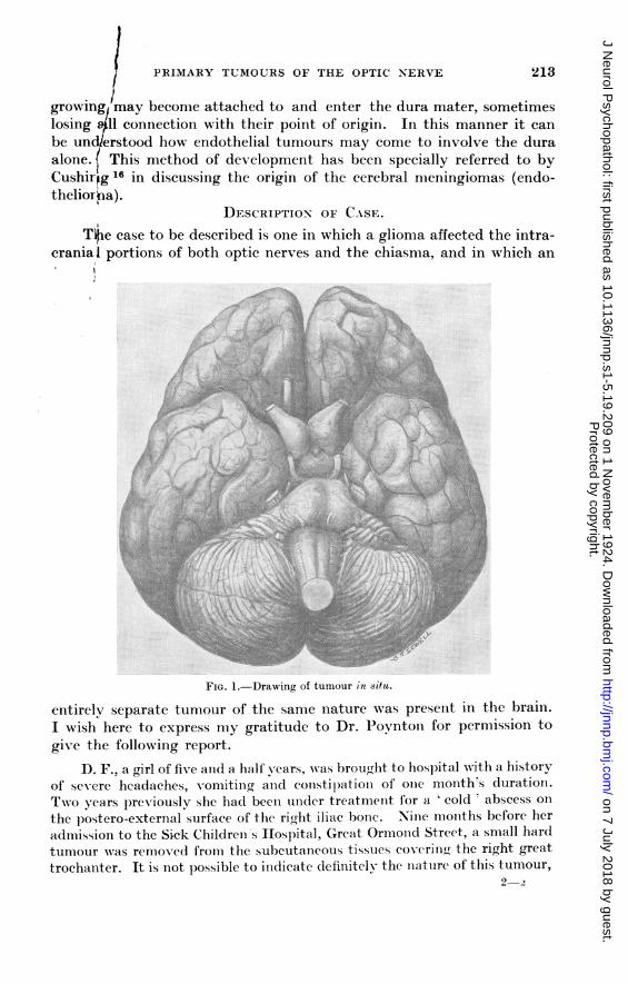
DESCRIPTION OF CASE.
T~e case to be described is one in which a glioma affected the intra-
crania,l portions of both optic nerves and the chiasma, and in which an
FIG. 1.-Drawing of tumour in situ.
entirely separate tumour of the same nature was present in the brain.I wish here to express my gratitude to Dr. Povntoni for permission togive the following report.
D. F., a girl of five an{d a half years, was brought to hospital -with a historyof se-ere headaches, vomitinlg and constipationi of ooe month's duratioin.Two years previously she had beeni uinder treatmenit for a ' eold ' abscess oInthe postero-external surface of the right iliac bone. Ninie montlhs before heradmission to the Sick Children's Hospital, Great Ormi-ond Street, a small hardtumour was removed from the subcutaneouis tissues coverin(r the right greattrochanter. It is not possible to inidicate definitelv the niature of this tumour,
2-2
2S13
Protected by copyright.
on 7 July 2018 by guest.http://jnnp.bm
j.com/
J Neurol P
sychopathol: first published as 10.1136/jnnp.s1-5.19.209 on 1 Novem
ber 1924. Dow
nloaded from
214 ORIGINAL PAPERS
but froml examiniationi of a snmall part it appeared to be chicfly co-T posed offibrous tissue. There was lno historv of trauma iM this ease, anid ti e familyhistory wvas unliml)ortant.
Onl admissioni to hospital the child was founid to be rather be. ow theav-eragye heieght for her age, but was decicledlv obese. Her wveight waw 51 lb.,the niormlal weeight for her a(re beiing, roughly, 41 lb. I3er hanids wer( ratherbroad an(l thiel, wvith short stumllpy fingers. She was not troubled by abnormalthirst, or by l)olyuiria. A larre I)atch of browii pigrnmenitationi of the skin wasl)resenit oni the left side of the abdomiieni. Bevonid this no other pigtmentedareas or other peculiarities of the skinl anid subeutanieouis tissues wvere ob served.
The patient w\Nas usually -ery drowsy and diffilcult to examine, akid anyattempts to test the visual fields were (lefeated. Visual acuity appe.ared tobe good, as she coul(l see anld recognis;e leople moving(r about the wardl. On
ophthalmoscopic examiniationi. theeddges of the discs were seenl to be alittle blurred, and about + 2 D. ofswelling vas presenlt in each eye.The discs appearecl to be under-going secoindary atrophy. Bothpupils were large anid inactive tolight, and there was paresis of theleft externial rectus muscle. A lefthemiparesis, inieludingr the face, waslpresent, aind progressed rapidly inseverity. Spasticity of the left
... extremitics was abscnt; they wvere,*AFM. S N-p,on the conitrary, rather hypotoniic.
Sensation was unaffected. Bothknee jerks were very sluggish, and
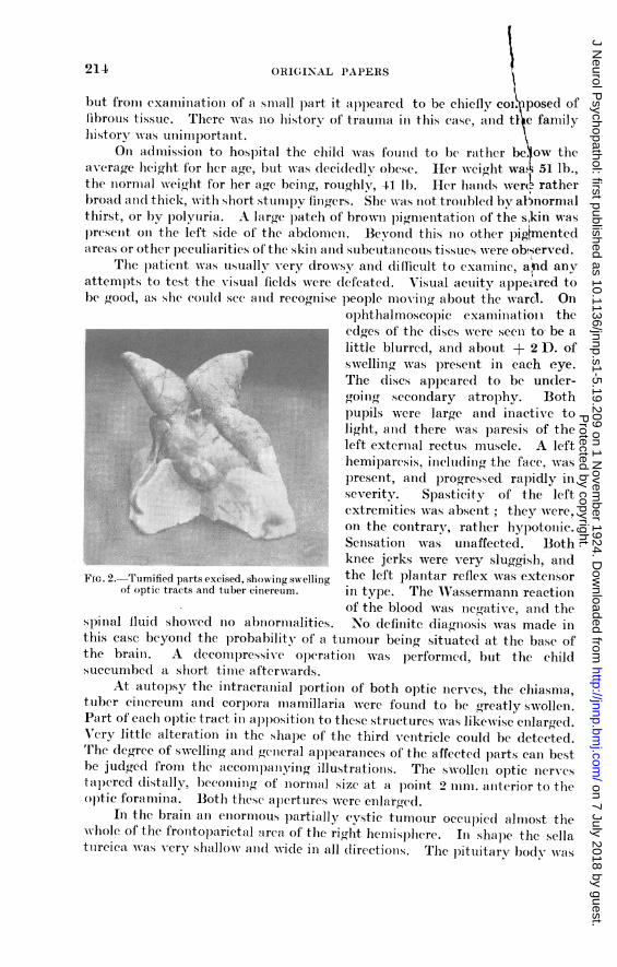
FIG. 2.-Tumified parts excised, showing swelling the left plantar reflex was extcisorof optic tracts and tuber cinereum. in type. The AVassermann reaction
of the blood was incgative, and thesp)inal flui(d showved Ino abniormlialities. No defiinitc diamiiosis was made illthis case beyond the probability of a tumiiour beiing situated at the base ofthe braini. A (lecomipressi-c ol)erationi was performed, but the childsuccumbed a short time afterwrards.
At autopsy the intracraniial lportion of both optic ner-es, the chiasma,tuber ciniereum aind corpora mamillaria were found to be gYreatly swollen.Part of each optic tract ini apposition to these structures was likewvisc cnlargred.Very little alterationi in the shape of the third venitricle could be detected.The degree of swvellinig anid genieral al)learaniecs of the affeetedlparts cani bestbe jud(rg(e from the accompaniyinig illustrationis. The swollen optic nervestapered distally, becomingo of niormal size at a poinit 2 immli. aniterior to th-eoptic foraminia. Both these ap)crtures werc enllarged.
In the brain an enormous p-artially cystic tumour occupied almost thewhole of the frontoparictal nrca of the rigrht hemisphere. Ini shape the sellatiircica, was verv shallow and( wide in all directionis. The pittlitary body was
Protected by copyright.
on 7 July 2018 by guest.http://jnnp.bm
j.com/
J Neurol P
sychopathol: first published as 10.1136/jnnp.s1-5.19.209 on 1 Novem
ber 1924. Dow
nloaded from

/ PRIMIARY TUMOURS OF THE OPTIC NERVE 215
flattened, 4nd the pars anterior aind infuinidibular stalk w\Nere of a semitranis-parent and gelatinous consistency.
HISTOLOGY OF THE TUMOURS.
Stailned by Bielschowskvys method, sectionis of the optic nerve andl chiasnmashow a goxeat reduction in the number of ner-e fibres. They are widely separatedand are more numerous in the optic nerves than in the chiasma, where thev arepractically absent in the central area. Wl'ith Weigert-Pal's staini the myelitisheaths a~re seen to be very scanty, anid in several patches in the chiasma theyare entilWely absent. Neuroglial tissue, stained with V'ictoria blue, is seen tobe enormously increased in quantity. This overgrowth consists chiefly ofneurogFial fibres. These form a denise meshwork. of which the fibres for the
FIG. 3.-Dense overgrowth of neuroglial fibres in centralarea of chiasma.
most part run transversely in the central areas of the chiasma, and longi-tudinally in the optic nerves. In the nerves, howe-er, the glial fibres at manypoiints take a course perpendicular to the fibrous tissue septa. Relatively tothe fibres the cells are few in number. The nuclei consist chiefly of two
kinds, small round or oval nuclei, and a more elongated form. An occasional
larger neuroglial cell of irregular shape writh an eccentric pale-staining nucleuscan be distin(ruished.
Sectionis stained by vani Gieson's method show great distortion of theconnective tissue septa, but Ino evidence that they have been infiltrated bythe new grrowth. The sheaths do not appear to be a-ffected. The vascularsupply of the affected parts is poor.
Histologically the appearanices of the tumiiour in the right hemisphere are
those of a soft. verv cellular glioma.
Protected by copyright.
on 7 July 2018 by guest.http://jnnp.bm
j.com/
J Neurol P
sychopathol: first published as 10.1136/jnnp.s1-5.19.209 on 1 Novem
ber 1924. Dow
nloaded from

216 ORIGINAL PAPERS
SYMPTOMATOLOGY.The symiptoms produced bv glioma and by endotheliomN of the
intraorbital portioni of the optic nerve are essen-tiallv similar, butconmmonly show slight nmodifications accordinig to the tvpe of (tunmourpreseint. Exophthalmos is the nmost obvious feature of these casles, and,secoIndly, inmpaired visionl or blindniess upoIn the side affected. Wherethe tumnour is a glionma vision is apt to be interfered with be-fore theoccurrence of exophthalmlos. which is onily natural considerilng the originof the growth is in the nervoous structures. Conversely, end othelialgrowths conmmonly give rise to exoplhtlhalnios prior to any im-pctirmentof vision, the destructioni of the visual fiLres beinig mlore slovly brought
J~~~~~~~~~
.4
abouit by the swellinig of the nerve sheaths. The occurrenice of temiporallhemyianopia progressing to blindniess, with chianges at the (lisc in the eyeopposite to the one primiarily affected, inidicates in the case of a gliomathat the growth has extenided back to and in-\ olh\ ed the chiasmia. Stra-bismius may occur in either case, but is apparentlvx not a conuniion eveI-nt.Restrictioni of the ocular m-iovements is miore frequentlv caused by enido-thelial tumours, which can be explained by the fact that they more ofteninvolve the portioni of the nerve imimediately behiind the eyeball than dothe tumours of glial origin. Pain is not usually complained of. Thechanges at the disc in both cases consist either of slight swellinig withevidence of secondary atrophy, or of atrophyv alonie.
Protected by copyright.
on 7 July 2018 by guest.http://jnnp.bm
j.com/
J Neurol P
sychopathol: first published as 10.1136/jnnp.s1-5.19.209 on 1 Novem
ber 1924. Dow
nloaded from

PRIMARY TUMOURS OF THE OPTIC NERVE
Gliomas affecting the chiasma and the intracranial portions of theoptic nerves may attain a considerable size. Owing to the pressureeffected upon surrounding structures the symptoms they produce arealmost identical with those caused by tumour of the hypophysis. Besidesthe disturbance of v-ision, the symptonms consist of drowsiness, obesity,defective growth, and frequieintly some degree of polvuria and polvdipsia.It is, therefore, unusual even to suspect clinically that the chiasma anidoptic nerves are the seat of growth. MIartin and Cushing,17 in a period ofeight years, nmet with seveen cases of glioma of the chiasnma and opticnierves, in two of whichi the growth did not extenid through the opticforamina. There are certain points with special reference to which thevconsider it nmight be possible to distinguiish tumouirs of the chiasma alndoptic nerves from those of the hvpophvsis. In three of the seven casesw-here perimiietrv Nas possible, although one eve was blinid, the helcnia-IIopic defect in the visual field of the othcer was not so conmpletc and clearcut as it is so ofteni found to be in cases of pituitary tumouir. The lossof \-ision in both eves due to a chiasmal tumour tenids to be mlore rapidlyprogressive, wlhereas with a pituitary tumour partial visioni in onle ceyat least is usually retained over a coinsiderable period. Anl imiportanitfeature obser-ed with the aid of stereoscopic plates of the skull takenilaterally was that the pituitary fossa appeared to be lengthenied owinhgto the distenision of the optic foramiina. Minor distinctions such asthese are of importanice fronm the surgical point of view. XVhile advo-cating the beiiefits of operatioin in the case of a pituitary neoplasnm. oniewould not hesitate to condeniii the renmov-al of a chiasmal growth as lonigas any sight renmained. When blindniess is conmplete operationi is use-less, as by then the tumnour will probably have invaded neighbourinigstructures.
RELATIONSHIP OF GLIOMIA NERVI OPTICI TO NEUROFIBROMATOSIS.
Whether any relationship exists between glioma of the optic nerv-eanid von Recklinghausen's disease, neurofibromatosis, is a point of greatiniterest. The actual number of cases in. which tumours of the optic aswell as of the other nerves have been present is extremely small. Whencompared by their histological features, it does not appear that the twotypes of tumour are in any -ay allied. Differences in developmentbetween the optic and other nerves must, however, be taken into account.The optic nerves form part of the central nervous system, and have acomposition unlike that of the peripheral nerves. It is conceivable thatthe tumours in either case are the result of a similar disturbance ofembryonal development. Both conditions are probably of congenitalorigin. The earlv onset in the majority of cases of glioma of the opticnerve supports this view. With regard to neurofibromatosis, Thomson 18considered that it is a developmental disease of the peripheral nervous
217
Protected by copyright.
on 7 July 2018 by guest.http://jnnp.bm
j.com/
J Neurol P
sychopathol: first published as 10.1136/jnnp.s1-5.19.209 on 1 Novem
ber 1924. Dow
nloaded from
ORIGINAL PAPERS
systenm conineciing in initrauterine life. Aniother feature which tenidsto form a linlk between the two conditions is the frequency in either casewith which a glionma of the central nervous system is presenit. AVithregard to the occurrence of gliomas of the braini or spinial cord in casesof neurofibronmatosis VAerocav 19 considers that thev formi no accidenitalconmbinationi, but are inl close histogenietic relationiship witlh the niervetumlours.
The v-iew that glionmas of the optic nerve anid nmultiple tunmours ofthe peripheral nerves are in the nature of honmologous fornmations ofcoingenital origin was first advanced by Goldnmani.20 In connection withneurofibroniatosis, BruInS 21 accepts the possible occurrence of blindnessdue to ' neuromas ' of the optic nerve.
Of the cases of optic nerv-e tunmour in association with neurofibro-matosis the nmost inmportant is Cushing's (Case No. VlII.). A tunmourinrVolving the chiasma and both optic nerves up to the optic foraminawas seen at operation, aind from histological exanminatioin of a small pieceremnoved subsequenitly proved to be a glionma. In PruddeIi'S,22 Serres',23and WestphalenI'S 24 cases, one or other optic nierve preseited a slightenlargemeint just before piercing the sclerotic ill two of the cases, andbefore entering the optic foramen in the third. The histological natureof the swelliings in these cases is unfortunately ilot stated. It is possiblethat in Westphalen's case the growth was of endothelial origin, as a snlalltumour of the dura showing concentric arrangenlent of cells was presentover the riglit frontal lobe. Michel 25 recorded the case of a sixteen-year-old girl, who developed elephantiasis ileuronlatosa of one leg at the ageof six nlonths. At autopsy a glioma was found in-olving the chiasmaand right optic nierve. In a case reported by Enmanuel,26 that of a child ofthree with a tunmour, probably a glioma, of the optic iierves, the child'sfather presenited the typical features of fibronma nmolluscunm, aild thegrandfather was said to have been similarly affected. It is suggestedthat heredity, which is a common featurc in neurofibronmatosis, nmayhave been a factor in the production of the child's condition.
In coilelusioni, while agreeing that a definite relationiship betweenthe two conditions is hard to prove, I believe there is nevertheless somejustification ill assuming that it does exist.
REFERENCES.BYERS, Studies Roy. Vict. Hosp., Montreal, 1901, i, 3.
2 HUDSON-, Roy. Lond. Ophth. Hosp. Reports, 1912, xviii, 317.3 WVISHART, -Edin. Med. Su1rg. Jour., 1833, xl. 274.4MIIDDLEMORE, Lond. Med. Gazette, 1837-8, xxii, 897.5HEYMANN.* Inaugural Dissert.. Berlin, 1842.6FISCHER, Arch. f. Augenheilk., 1908. lix, 181.7AXENFEL-BUJ.S_CH, Arch. of Ophth., 1901, xxx, 265.8 FLEISCHER-SCHEERER, Arch. f. Ophth.. 1920, ciii, 16.
* Not studied in the originial.
218
Protected by copyright.
on 7 July 2018 by guest.http://jnnp.bm
j.com/
J Neurol P
sychopathol: first published as 10.1136/jnnp.s1-5.19.209 on 1 Novem
ber 1924. Dow
nloaded from

PRIMARY TUMOURS OF THE OPTIC NERVE 219
9X\ ILLEMIER. Arch. f. Ophthl.. 1879. xxv, 195.10 SI)LER-HUvGUEN.IN. Arch. f. Ophth.. 1919-20, ci, 1 1i3.11 SNYDACHER. Arch. of Ophth., 1901. xxx. 130.12 NEA-ME, Brit. Jotur. Op1hthal., 1923, vii, 209.13 V. GRAEFE. Arch. f. Ophth., 1866. xii, 2, 100.14 8CHOTT-M1AUTHNER, Arch. f. Augenheilk., 1878. vii. 81.'1 POLLACK. Berl. klin. 1I-och., 1921, lviii, 210.16 CUSHINGx. Brain. 1922. xlv. 282.17 MARTIN-CUSHING, Arch. of Ophth.. 192'3 1ii. 909.18 TiiOMSON, On Neifroma and Neuirofibromato0is, 1900. 105).19 VEROCAY, Beit. z. path. Anat. u. z. ally. Path., 1910. xlvtiii, 1.20 GOLDMANN. Beit. z. 1kin. Chir., 1893. x, 13.21 BRUINS. Die GeschlicPlste des Nervzensystenis 190S, 44-53.22 PRUDDEN, Amer. .Joutr. Mlled. Sci.. 1880. lxxx. 134.23 SERRES, C'. R. Acad. Sci.. 1845. xxi. 17.24 WESTPHALEN, l irch. Arch., 1887. cx, 99.
MICHEL, Arch. f. Ophth.. 1873. xix. 3, 14.26 EANIUNIEL. Arch. f. Ophth. 1902, liii. 199.
Protected by copyright.
on 7 July 2018 by guest.http://jnnp.bm
j.com/
J Neurol P
sychopathol: first published as 10.1136/jnnp.s1-5.19.209 on 1 Novem
ber 1924. Dow
nloaded from