primate mandibular reconstruction with prefabricated ... · primate mandibular reconstruction with...
TRANSCRIPT

TitlePrimate mandibular reconstruction with prefabricated,vascularized tissue-engineered bone flaps and recombinanthuman bone morphogenetic protein-2 implanted in situ
Author(s) Zhou, M; Peng, X; Mao, C; Xu, F; Hu, M; Yu Guangyan, GY
Citation Biomaterials, 2010, v. 31 n. 18, p. 4935-4943
Issued Date 2010
URL http://hdl.handle.net/10722/123819
Rights Creative Commons: Attribution 3.0 Hong Kong License
Q1
Q3
Q2
lable at ScienceDirect
ARTICLE IN PRESS
Biomaterials xxx (2010) 1 9
JBMT11950_proof ■ 11 March 2010 ■ 1/9
Contents lists avai
Biomaterials
journal homepage: www.elsevier .com/locate/biomater ia ls
Primate mandibular reconstruction with prefabricated, vascularizedtissue-engineered bone flaps and recombinant human bone morphogeneticprotein-2 implanted in situ
Miao Zhou a,b, Xin Peng a, Chi Mao a, Fang Xu c, Min Hu d,**, Guang-yan Yu a,*
aDepartment of Oral and Maxillofacial Surgery, Peking University School and Hospital of Stomatology, Beijing 10081, ChinabDepartment of Oral and Maxillofacial Surgery, The Second Affiliated Hospital of Sun Yat-sen University, Guangzhou, ChinacCollege of Pharmaceutical Science, Zhejiang University of Technology, Hangzhou, ChinadDepartment of Oral and Maxillofacial Surgery, Chinese PLA General Hospital, Beijing, China
a r t i c l e i n f o
Article history:Received 16 February 2010Accepted 28 February 2010Available online xxx
Keywords:Mandibular reconstructionPrefabricated vascularized bone flapsrhBMP-2Demineralized freeze-dried bone allograftCoralline hydroxyapatite
* Corresponding author. Tel.: þ86 10 62191099; fax** Corresponding author.
E-mail address: [email protected] (G.-y. Yu).
0142-9612/$ see front matter � 2010 Elsevier Ltd.doi:10.1016/j.biomaterials.2010.02.072
Please cite this article in press as: Zhou M, etBiomaterials (2010), doi:10.1016/j.biomateri
a b s t r a c t
Several studies have validated successful mandibular reconstruction with prefabricated tissue engineered bone flaps and recombinant human bone morphogenetic protein 2 (rhBMP 2) implanted in situ.Whether rhBMP 2 applied with the prefabrication technique enables faster ossification of mandibulardefects than rhBMP 2 applied in situ is unknown. We aimed to compare mandibular reconstruction withprefabricated, vascularized tissue engineered bone flaps with rhBMP 2 and rhBMP 2 applied in situ inprimates (Rhesus monkey). We also compared the use of the carriers demineralized freeze dried boneallograft (DFDBA) and coralline hydroxyapatite (CHA) for applying rhBMP 2. After computed tomographyof the monkey head, custom meshes were made, loaded with rhBMP 2 incorporated DFDBA or CHA, andimplanted in the latissimus dorsi muscle. Meanwhile, contralateral segmental mandibular defects werecreated, and custom meshes loaded with DFDBA, CHA, or rhBMP 2 incooperated DFDBA and CHA wereimplanted in situ. Thirteen weeks later, the bone flaps with rhBMP 2 incorporated DFDBA or CHA weretransferred to repair segmental mandibular defects. The meshes loaded with DFDBA or CHA aloneshowed no bone regeneration 13 weeks after implantation in latissimus dorsi muscle. Radiography,angiography and histological analysis were used to evaluate the repair and vascularization of the implant.Segmental mandibular defects were successfully restored with prefabricated bone flaps and rhBMP 2incorporated CHA in situ, but other segmental mandibular defects remained with rhBMP 2 incorporatedDFDBA, DFDBA and CHA in situ. Moreover, mandibles reconstructed with rhBMP 2 incorporated CHAbone flaps revealed more regenerated and homogeneous bone formation than did other reconstructions.The study suggested that the prefabrication technique induced better mandibular reconstruction andbone regeneration in quantity and quality.
� 2010 Elsevier Ltd. All rights reserved.
1. Introduction
The restoration of mandibular defects caused by ablativesurgery for oral and maxillofacial tumor, trauma, infection andcongenital deformity remains a challenge for surgeons [1]. Autologous grafting and distraction osteogenesis are the most commontechniques used to restore mandibular defects [2]. Autologousgrafts have been considered the “gold standard” because of theadvantages of osteogenesis, osteoinduction and osteoconduction.
: þ86 10 62173402.
All rights reserved.
al., Primate mandibular recoals.2010.02.072
However, the major drawbacks of these methods are severemorbidity, as well as donor site availability, which calls for newmethods for mandibular reconstruction [1]. At present, bone tissueengineering has gone from the bench to the bedside and showneffectiveness in mandibular reconstruction [3e5].
Recombinant human bone morphogenetic protein 2 (rhBMP 2)is the growth factor most widely used for bone regeneration inclinical practice [6,7]. Two major techniques for use of rhBMP 2 inmandibular reconstruction are (1) applying rhBMP 2 in situ [8,9]and (2) applying rhBMP 2 to muscles adjoining major vessels forvascularized tissue engineered bone flap prefabricated in vivo,which is transferred to the jaw defect [3,4,10]. Both of these techniques achieved initial clinical success. The prefabrication technique provided a stable blood supply, mature bone and more soft
nstructionwith prefabricated, vascularized tissue engineered bone...,

M. Zhou et al. / Biomaterials xxx (2010) 1 92
ARTICLE IN PRESS JBMT11950_proof ■ 11 March 2010 ■ 2/9
tissues for restoring large compound mandibular defects withpreceding radiotherapy and scars in situ. Animal studies also validated the effectiveness of the 2 methods in mandibular reconstruction [11e13]. Whether the prefabrication technique enablesfaster ossification than with recombinant human bone morphogenetic proteins (rhBMPs) implanted in situ is still unknown,especially for large compound mandibular defects.
Carriers are used to increase the retention of rhBMPs at orthopaedic treatment sites for a sufficient period of time to allowregenerative tissue to form cells that migrate to the area of injuryand to proliferate and differentiate [14]. The four major categoriesof carrier materials for rhBMPs include natural polymers, inorganicmaterials, synthetic polymers and composites of these materials[15]. Demineralized freeze dried bone allograft (DFDBA) andcoralline hydroxyapatite (CHA) have been studied as carriers forrhBMP 2. Clokie et al. reconstructed 10 mandibular defects witha construct containing osteogenetic protein 1 (OP 1) in a demineralized bone matrix suspended in a reverse phase medium to effectsustained rhBMPs delivery [9]. Heliotis et al. reconstructed a humanmandibular defect with a prefabricated tissue engineered bone flapby implanting OP 1 coated CHA in the pectoralis major muscle [10].However, none of the research compared the ossification withectopic or orthotopic rhBMP 2 incorporated DFDBA or CHAcarriers.
We aimed to compare the effectiveness of rhBMP 2 applied insitu and applied in muscle of a prefabricated, vascularized tissueengineered bone flap for mandibular reconstruction. DFDBA andCHA were also evaluated as constructs in mandibularreconstruction.
2. Materials and methods
2.1. Manufacture and specification of DFDBA and CHA
DFDBA was made from the control group of a pharmacokinetics study ofrecombinant human stem cell growth factors in Rhesus monkeys (Beijing JoinnLaboratories, China). The donor Rhesus monkeys were healthy and 6 8 years old.Briefly, the spongy bone was obtained from the extremities and reserved at �70 �Cfor 2 months to extinguish immunogenicity. The spongy bone was separated fromthe head of extremities and cut into 2 sizes: scaffold “A” with upper plane 10 mm �3 mm, lower plane 10 mm � 6 mm and height 5 mm; and scaffold “B” with size 10mm � 6mm � 5 mm. The bone blocks were then soaked in acetone for 48 hr toremove the fatty composition. Distilled water was used to wash out bone marrowand rudimentary blood, then the bone block sample was demineralized with 0.6 M
HCL and washed with distilled water, then freeze-dried. The CHA scaffold wasprovided by Yihuajian Commercial Co. Ltd. (China). It was manufactured as for theDFDBA scaffold. Then, the samples were prepared by hydrothermal chemical ex-change with phosphate as per the manufacturer's protocol (Yihuajian CommercialCo. Ltd.). The porous morphology of the carriers was analyzed by scanning electronmicroscopy (SEM, JOEL, Model JSM-5600LV). The sample was freeze-dried and pre-pared for rhBMP-2 incorporation.
2.2. rhBMP-2 delivery
rhBMP-2 was produced by recombinant expression in Escherichia coli at theGenetic Institute of Huadong Medicine (China) and purified to more than 98%.Hangzhou Future Biotech Co. Ltd. (China) supplied the rhBMP-2 used in the exper-iment. rhBMP-2 (72 mg) was dissolved in gelatin solution in acetic acid (30 mg/ml).One block of each carrier was soaked in 1 ml gelatin solution with rhBMP-2 (1.5mg/ml) (DFDBA-BMP and CHA-BMP, respectively). rhBMP-2-incorporated scaffoldswere freeze-dried in frozen tubes and kept at 2 7 �C until use.DFDBA and CHA scaffolds coated with gelatin but without rhBMP-2 were alsoprepared. Particles of CHA and DFDBA (size: 0.4 1.0 mm) were prepared.
2.3. Experimental design
Nine adult male healthy Rhesus monkeys (6 9 years old, 6 12 kg) were providedby the Laboratory Animal Center of the Chinese PLA General Hospital (China).Surgical techniques and animal care conformed to the principles of the LaboratoryAnimal Center (NIH publication No. 85-23, revised 1985). For prefabricating tissue-engineered bone flaps, custom meshes loaded with DFDBA-BMP (n 3), CHA-BMP (n3), DFDBA (n 3) and CHA (n 3) were implanted in animals'
Please cite this article in press as: Zhou M, et al., Primate mandibular recoBiomaterials (2010), doi:10.1016/j.biomaterials.2010.02.072
ambilateral latissimus dorsi muscle. Thirteen weeks later, ossification was validatedin the groups of DFDBA-BMP and CHA-BMP, while no ossification was found in theother groups. Then, the prefabricated tissue-engineered bone flaps with ossificationwere transferred to repair homolateral segmental mandibular defects. At the time ofsurgical implantation in the latissimus dorsi muscle, contralateral segmentalmandibular defects described below were implanted with DFDBA-BMP, CHA-BMP,DFDBA and CHA respectively.
The bilateral mandibular defects in 9 monkeys were randomized into 6 groupsfor treatment: P-DFDBA-BMP and P-CHA-BMP: mandibular defect repaired by pre-fabricated tissue-engineered bone flap with DFDBA-BMP and CHA-BMP, respectively(n 3 each); S-DFDBA-BMP and S-CHA-BMP: mandibular defect repaired by in situDFDBA-BMP and CHA-BMP, respectively (n 3 each); and S-DFDBA and S-CHA:mandibular defect repaired by in situ DFDBA and CHA, respectively (n 3 each). Thestudy was approved by the Animal Care and Experiment Committee of PekingUniversity Health Science Center.
2.4. Manufacture of custom titanium mesh
3-D computed tomography (CT) images were taken of the heads of monkeys.The scanning data were uploaded to a computer-aided design program, 3-D medi-cine surface rendering (3DMSR, JIMAFEI Science and Technology Development Co.Ltd., China) software system. A 20-mm-long mandibulectomy was virtually createdin the mandibular body (Fig. 1a and b). Then, the data were transferred to a millingmachine (GSVM6540, Gold SunMould & CNCmachinery Co. Ltd., China). The customtitanium meshes for internal fixation of the stumps of the mandible were made bythe machine. The meshes were used to shape the tissue-engineered bone in thelatissimus dorsi muscle and internal fixation of mandibular stumps. The buccal andlingual height of the mesh was 12 and 10 mm, respectively (Fig. 1c and d).
2.5. Surgical procedures
General anesthesia was induced with ketamine hydrochloride (20 mg/kgintramuscularly or subcutaneously) and maintained with pentobarbital sodium(1 2%, intravenously). The monkeys were prepared for extraction of mandibularteeth from the bicuspid tooth to the back molar. Three months after extraction, thewounds of the socket healed for the next segmental mandibulectomy.
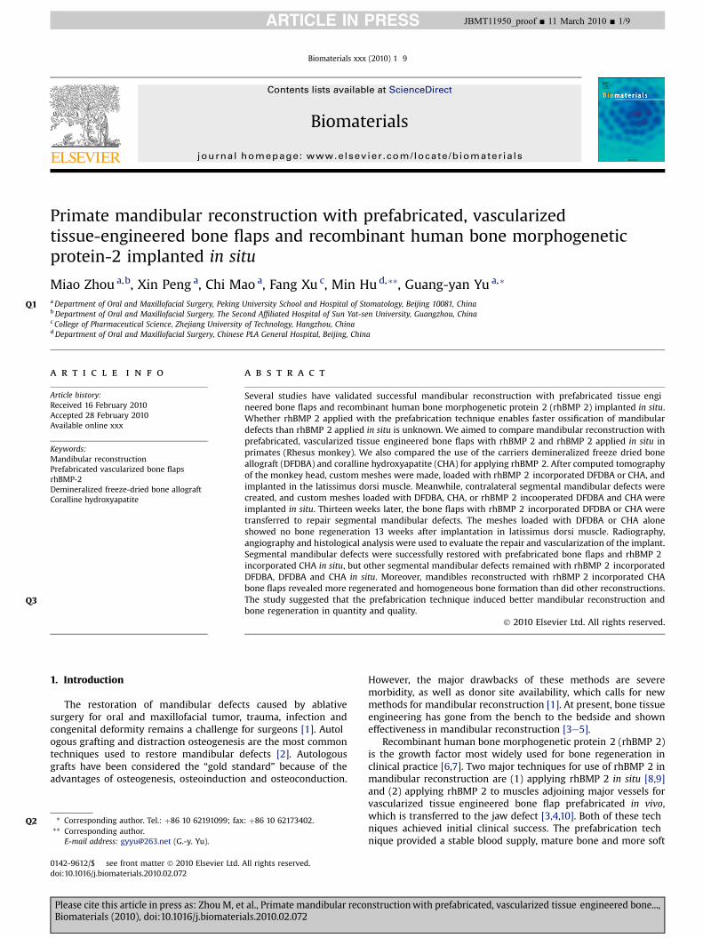
With monkeys under general anesthesia, the latissimus dorsi muscle wasvisualized through a lateral approach, and a pouchwasmade inside themuscle closeto the thoracodorsal artery. The custom mesh was loaded with 4 blocks of scaffold(containing 2 A and B, respectively). The particles of DFDBA or CHA scaffolds weremixed with the blood from the surgery. Then the mixture was used to fill the gapbetween the block and the custom mesh. The pre-loaded titanium mesh wasimplanted into the pouch of the latissimus dorsi muscle of anesthetized monkeys(Fig. 2a c). Thirteenweeks after implantation, the flap was harvested along with theadjoining part of the latissimus dorsi muscle containing the thoracodorsal artery andvein, which supplied blood to the bone flap. The bone flapwas biopsied (Fig. 2d). Thesegmental mandibular defect was carried out as follows. After a sub-mandibularincision, the mandible was exposed extra-orally through a full-thickness flap thatincluded the periosteum. A 20-mm-wide section of the mandible from the bicuspidtooth to the backmolar teeth was resected with a fissure bur (Fig. 2e). The volume ofthe defect was approximately 3 cm3 (20 mm� 10 mm� 15 mm). The wound of thedefect was irrigated with normal saline. The prefabricated tissue-engineered boneflap was then transferred to the mandibular defect via a tunnel created beneath themajor pectoral muscle (Fig. 2f). For study the bone regeneration with rhBMPs orscaffold alone implanted in situ, the titanium mesh loaded with DFDBA-BMP, CHA-BMP, DFDBA or CHA was fixed onto the stumps of the contralateral mandibulardefects with titanium micro-osteosynthesis screws.
2.6. Clinical view, angiography and radiography examinations
Postoperative activities, emotional response, food intake and would healing ofsurgical sites were observed in all animals. The status of bone regeneration of thecustom meshes loaded with DFDBA-BMP and CHA-BMP were examined duringtransplantation. At 26 weeks after implantation, animals were sacrificed by intra-venous overdose of sodium pentobarbitone, and mandibles and loaded custommeshes were excised. In 2 Rhesus monkeys, the lingual side of the mesh was inte-grated with the regenerated bone and was saved.
Mandibular radiogram was obtained (X-mind AC, ACTEON, France). The area ofthe bone regenerated in the mandibular defect was analyzed by the Leica imageanalysis system.
Angiography was performed before sacrifice in 4 animals to validate the bloodsupply of the prefabricated tissue-engineered bone flap. The latissimus dorsimuscles containing the titanium meshes were dissected. Two veins and 1 arterywere in the thoracodorsal vascular bundle. One thoracodorsal artery was cannu-lated. Subsequently, the graft was perfused at 37 �C with 130 mmHg pressure and100 ml of 49 ml normal saline, 50 ml Ultra-vist370 (Bayer Schering Pharma.,Germany) and 10000 U heparin. In 2 Rhesus monkeys, angiography of the distal endof the thoracodorsal artery was performed to validate the relation of the thor-acodorsal artery to the transferred tissue-engineered bone flap.
nstructionwith prefabricated, vascularized tissue engineered bone...,

Fig. 1. Virtual segmental osteotomy of Rhesus monkey mandible and designed custom titanium meshes. (a) Resecting the mandible with length 20 mm; (b) Mandibular defect aftervirtual osteotomy; (c) Lateral view of the mesh on the outer surface of the mandible; (d) Occlusional view of the mesh.
M. Zhou et al. / Biomaterials xxx (2010) 1 9 3
ARTICLE IN PRESS JBMT11950_proof ■ 11 March 2010 ■ 3/9
2.7. Histological analysis
For simultaneous sequential intravital staining of the regenerated bone, intra-peritoneal injection of fluorochromes began at 2 weeks after implantation with thecustommeshes. Alizarin-complexion (3% in 2% NaHCO3solution, 0.8ml/kg bodyweight)was injected 2 and 4 weeks after implantation. Tetracycline (1% in normal saline, 1ml/kg body weight) was injected 8 and 10 weeks after implantation. Xylenol orange(6% in 2% NaHCO3 solution, 1.5 ml/kg body weight) was injected 14 and 18 weeksafter implantation. Calcein (1% in 2% NaHCO3 solution, 5 ml/kg body weight)was injected 22 and 24 weeks after implantation (all fluorochromes from Sigma).
Mandibular biopsy specimens taken during surgery were fixed in 10% neutralbuffered formalin. After dehydration, specimens were immersed in Technovit7200VLC (Heraeus-Kulzer, Germany). After solidification for 24 hr, specimens werecut from the longitude of mandibles. Tissues with P-CHA and P-DFDBA inside thetitanium meshes were separated for histological examination. Histological prepara-tion was previously described [16]. The embedded samples were cut into thin sec-tions approximately 40 80 mm, and mounted on glass slides. Three ground sectionsfrom each specimen block, 300 mm apart, were stained with haematoxylin and eos-in (H&E). Histomorphometry involved the Leica image analysis system.
2.8. Statistical analysis
Data from radiography and histology are expressed as mean � s.e.m and wereanalyzed by one-way ANOVA. The differences between 2 groups among the 6 groupswere assessed with the Student Newman Kewls test. P < 0.05 was considered stat-istically significant.
3. Results
3.1. Properties of DFDBA and CHA
To study the retention of ectopic and orthotopically administered rhBMP 2, we chosed the two classical carriers forrhBMPs, DFDBA and CHA. The DFDBA prepared from theextremities of rhesus monkeys was considered an allograft. Theporous structure of DFDBA enabled rhBMP 2 to infiltrate deeplyinto the carrier and release slowly after implantation. SEM ofDFDBA revealed no osteoblast or crystal structure on the scaf-fold surface. The mean size of the pores in the trabecular zone
Please cite this article in press as: Zhou M, et al., Primate mandibular recoBiomaterials (2010), doi:10.1016/j.biomaterials.2010.02.072
was larger than that in cortical bone (400e600 mm [Fig. 3a]and 100e200 mm, respec tively). SEM of CHA revealed a porousstructure. The pore size of CHA was 270e550 mm (Fig. 3b), whichwas similar to the structure of human trabecular bone. Manyhydroxyapatite crystals adhered to the surface. The porousstructure enables blood capillary sprout, nutrient diffusion andbone ingrowth to the scaffold.
3.2. Gross view, angiography and radiography examinations
All wounds healed. No sign of flap necrosis was visible duringthe postoperative period. Postoperative swelling was noted in allanimals. The swelling of the latissimus dorsi muscle with DFDBABMP and CHA BMP implants lasted longer than that with DFDBA orCHA implants alone. The titaniummeshes loaded with DFDBA BMPand CHA BMP showed ossification inside the mesh. No overgrowthof the regenerated bone was found outside the mesh, and noosteointegration with the titanium meshes occurred. Meshesloaded with DFDBA BMP or CHA BMP were encapsulated by thelatissimus dorsi muscle. The volume of the bone regenerated waslarger with CHA BMP than DFDBA BMP loading. No bone inductionwas found inmeshes loadedwith DFDBA, and the inner space of themeshes was occupied by the latissimus dorsi muscle. With the CHAloading, nearly 50% of the implant had disappeared, and the rest ofCHA was the block encapsulated by the latissimus dorsi muscle(data not shown).
Angiography was used to study the blood supply with transferred tissue engineered bone flaps and validate the relation of thethoracodorsal bundle to the implant. The transferred thoracodorsalbundle was located from the axillary fossa to the repairedmandibular defect, which validated that the transferred tissueengineered bone flap was supplied by the thoracodorsal artery andvein (Fig. 4a). Thoracodorsal arteries had one main trunk and 2e4branches. The thoracodorsal vein had 2 trunks. Angiography also
nstructionwith prefabricated, vascularized tissue engineered bone...,
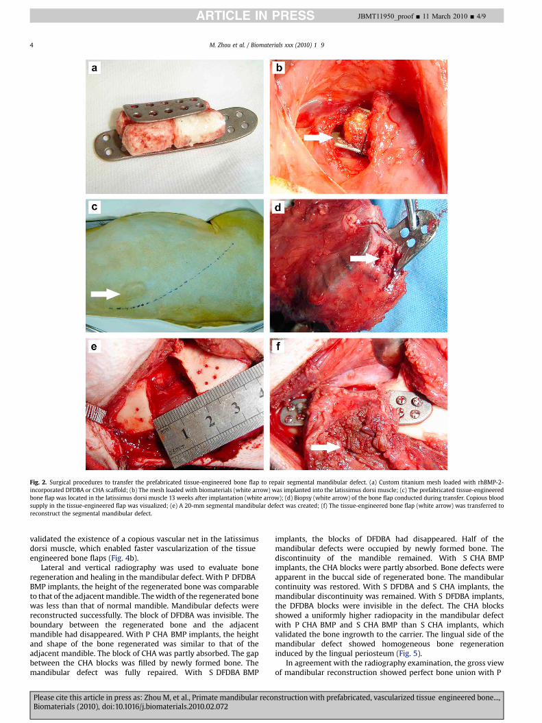
Fig. 2. Surgical procedures to transfer the prefabricated tissue-engineered bone flap to repair segmental mandibular defect. (a) Custom titanium mesh loaded with rhBMP-2-incorporated DFDBA or CHA scaffold; (b) The mesh loaded with biomaterials (white arrow) was implanted into the latissimus dorsi muscle; (c) The prefabricated tissue-engineeredbone flap was located in the latissimus dorsi muscle 13 weeks after implantation (white arrow); (d) Biopsy (white arrow) of the bone flap conducted during transfer. Copious bloodsupply in the tissue-engineered flap was visualized; (e) A 20-mm segmental mandibular defect was created; (f) The tissue-engineered bone flap (white arrow) was transferred toreconstruct the segmental mandibular defect.
M. Zhou et al. / Biomaterials xxx (2010) 1 94
ARTICLE IN PRESS JBMT11950_proof ■ 11 March 2010 ■ 4/9
validated the existence of a copious vascular net in the latissimusdorsi muscle, which enabled faster vascularization of the tissueengineered bone flaps (Fig. 4b).
Lateral and vertical radiography was used to evaluate boneregeneration and healing in the mandibular defect. With P DFDBABMP implants, the height of the regenerated bone was comparableto that of the adjacentmandible. Thewidth of the regenerated bonewas less than that of normal mandible. Mandibular defects werereconstructed successfully. The block of DFDBA was invisible. Theboundary between the regenerated bone and the adjacentmandible had disappeared. With P CHA BMP implants, the heightand shape of the bone regenerated was similar to that of theadjacent mandible. The block of CHA was partly absorbed. The gapbetween the CHA blocks was filled by newly formed bone. Themandibular defect was fully repaired. With S DFDBA BMP
Please cite this article in press as: Zhou M, et al., Primate mandibular recoBiomaterials (2010), doi:10.1016/j.biomaterials.2010.02.072
implants, the blocks of DFDBA had disappeared. Half of themandibular defects were occupied by newly formed bone. Thediscontinuity of the mandible remained. With S CHA BMPimplants, the CHA blocks were partly absorbed. Bone defects wereapparent in the buccal side of regenerated bone. The mandibularcontinuity was restored. With S DFDBA and S CHA implants, themandibular discontinuity was remained. With S DFDBA implants,the DFDBA blocks were invisible in the defect. The CHA blocksshowed a uniformly higher radiopacity in the mandibular defectwith P CHA BMP and S CHA BMP than S CHA implants, whichvalidated the bone ingrowth to the carrier. The lingual side of themandibular defect showed homogeneous bone regenerationinduced by the lingual periosteum (Fig. 5).
In agreement with the radiography examination, the gross viewof mandibular reconstruction showed perfect bone union with P
nstructionwith prefabricated, vascularized tissue engineered bone...,
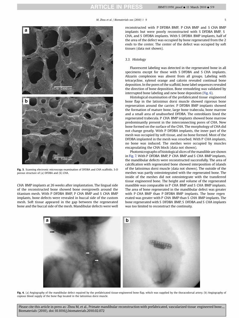
Fig. 3. Scanning electronic microscopy examination of DFDBA and CHA scaffolds. 3-Dporous structure of (a) DFDBA and (b) CHA.
M. Zhou et al. / Biomaterials xxx (2010) 1 9 5
ARTICLE IN PRESS JBMT11950_proof ■ 11 March 2010 ■ 5/9
CHA BMP implants at 26 weeks after implantation. The lingual sideof the reconstructed bone showed bone overgrowth around thetitanium mesh. With P DFDBA BMP, P CHA BMP and S CHA BMPimplants, bone defects were revealed in buccal side of the custommesh. Soft tissue appeared in the gap between the regeneratedbone and the buccal side of themesh. Mandibular defects werewell
Fig. 4. (a) Angiography of the mandibular defect repaired by the prefabricated tissue-engincopious blood supply of the bone flap located in the latissimus dorsi muscle.
Please cite this article in press as: Zhou M, et al., Primate mandibular recoBiomaterials (2010), doi:10.1016/j.biomaterials.2010.02.072
reconstructed with P DFDBA BMP, P CHA BMP and S CHA BMPimplants but were poorly reconstructed with S DFDBA BMP, SCHA, and S DFDBA implants. With S DFDBA BMP implants, half ofthe area of the defect was occupied by bone regenerated from the 2ends to the center. The center of the defect was occupied by softtissues (data not shown).
3.3. Histology
Fluorescent labeling was detected in the regenerated bone in allspecimens except for those with S DFDBA and S CHA implants.Alizarin complexion was absent from all groups. Labeling withtetracycline, xylenol orange and calcein revealed continual bonedeposition. In the pores of the scaffold, bone label sequences revealedthe direction of bone deposition. Bone remodeling was validated byinterrupted bone labeling and new bone deposition (Fig. 6).
Histological examination of the prefabricated tissue engineeredbone flap in the latissimus dorsi muscle showed rigorous boneregeneration around the carrier. P DFDBA BMP implants showedthe formation of mature bone, large bone trabecula, bone marrowand a small area of unabsorbed DFDBA. The osteoblasts lined theregenerated trabecula. P CHA BMP implants showed bone marrowpredominantly present in the interconnecting pores of CHA. Newbone formed on the surface of the CHA. The morphology of CHA didnot change greatly. With P DFDBA implants, the inner part of themesh was occupied by soft tissue, and no bone formed. Most of theDFDBA implanted in the mesh was resorbed. With P CHA implants,no bone was induced. The meshes were occupied by musclesencapsulating the CHA block (data not shown).
Photomicrographsof histological slices of themandible are shownin Fig. 7. With P DFDBA BMP, P CHA BMP and S CHA BMP implants,the mandibular defects were reconstructed successfully. The area ofcalcification with regenerated bone showed interposition of islandsof the latissimus dorsi muscle (data not shown). The outside of themeshes was partly osteointegrated with the regenerated bone. Theinside of the meshes did not osteointegrate with the transferredtissue engineered bone. The height and volume of the regeneratedmandible was comparable in P CHA BMP and S CHA BMP implants.The area of bone regenerated in the mandibular defect was greaterwith P CHA BMP than P DFDBA BMP implants. The bone regenerated was greater with P CHA BMP than S CHA BMP implants. Thebone regeneratedwith S DFDBA BMP, S DFDBA and S CHA implantswas too limited to reconstruct the continuity.
eered bone flap, which was supplied by the thoracodorsal artery. (b) Angiography of
nstructionwith prefabricated, vascularized tissue engineered bone...,
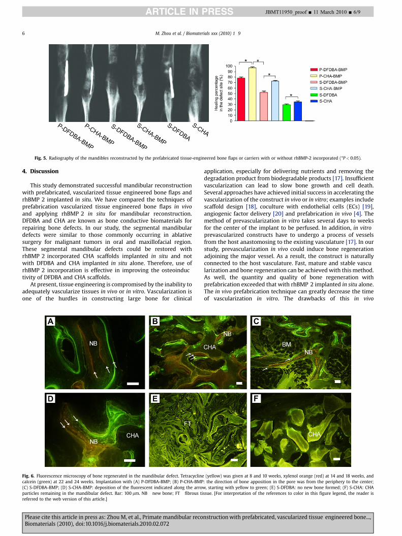
Fig. 5. Radiography of the mandibles reconstructed by the prefabricated tissue-engineered bone flaps or carriers with or without rhBMP-2 incorporated (*P< 0.05).
M. Zhou et al. / Biomaterials xxx (2010) 1 96
ARTICLE IN PRESS JBMT11950_proof ■ 11 March 2010 ■ 6/9
4. Discussion
This study demonstrated successful mandibular reconstructionwith prefabricated, vascularized tissue engineered bone flaps andrhBMP 2 implanted in situ. We have compared the techniques ofprefabrication vascularized tissue engineered bone flaps in vivoand applying rhBMP 2 in situ for mandibular reconstruction.DFDBA and CHA are known as bone conductive biomaterials forrepairing bone defects. In our study, the segmental mandibulardefects were similar to those commonly occurring in ablativesurgery for malignant tumors in oral and maxillofacial region.These segmental mandibular defects could be restored withrhBMP 2 incorporated CHA scaffolds implanted in situ and notwith DFDBA and CHA implanted in situ alone. Therefore, use ofrhBMP 2 incorporation is effective in improving the osteoinductivity of DFDBA and CHA scaffolds.
At present, tissue engineering is compromised by the inability toadequately vascularize tissues in vivo or in vitro. Vascularization isone of the hurdles in constructing large bone for clinical
Fig. 6. Fluorescence microscopy of bone regenerated in the mandibular defect. Tetracyclinecalcein (green) at 22 and 24 weeks. Implantation with (A) P-DFDBA-BMP; (B) P-CHA-BMP(C) S-DFDBA-BMP; (D) S-CHA-BMP: deposition of the fluorescent indicated along the arroparticles remaining in the mandibular defect. Bar: 100 mm. NB new bone; FT fibrous tireferred to the web version of this article.]
Please cite this article in press as: Zhou M, et al., Primate mandibular recoBiomaterials (2010), doi:10.1016/j.biomaterials.2010.02.072
application, especially for delivering nutrients and removing thedegradation product from biodegradable products [17]. Insufficientvascularization can lead to slow bone growth and cell death.Several approaches have achieved initial success in accelerating thevascularization of the construct in vivo or in vitro; examples includescaffold design [18], coculture with endothelial cells (ECs) [19],angiogenic factor delivery [20] and prefabrication in vivo [4]. Themethod of prevascularization in vitro takes several days to weeksfor the center of the implant to be perfused. In addition, in vitroprevascularized constructs have to undergo a process of vesselsfrom the host anastomosing to the existing vasculature [17]. In ourstudy, prevascularization in vivo could induce bone regenerationadjoining the major vessel. As a result, the construct is naturallyconnected to the host vasculature. Fast, mature and stable vascularization and bone regeneration can be achievedwith this method.As well, the quantity and quality of bone regeneration withprefabrication exceeded that with rhBMP 2 implanted in situ alone.The in vivo prefabrication technique can greatly decrease the timeof vascularization in vitro. The drawbacks of this in vivo
(yellow) was given at 8 and 10 weeks, xylenol orange (red) at 14 and 18 weeks, and: the direction of bone apposition in the pore was from the periphery to the center;w, starting with yellow to green; (E) S-DFDBA: no new bone formed; (F) S-CHA: CHAssue. [For interpretation of the references to color in this figure legend, the reader is
nstructionwith prefabricated, vascularized tissue engineered bone...,

Fig. 7. Histological analysis of the bone regenerated in the reconstructed mandible. Implantation with (a) P-DFDBA-BMP: residue of DFDBA surrounded by newly formed bone;(b) P-CHA-BMP: formed woven bone and remaining CHA; (c) S-DFDBA-BMP: fibrous tissues in the central of mandibular defect; (d) S-CHA-BMP; (e) S-DFDBA: mandibular defectmainly occupied by fibrous tissues; (f) S-CHA: scaffold surrounded by fibrous tissues; (g) Histomorphometry of the bone formation in the mandibular defect (*P< 0.05, Bar:500 mm).
M. Zhou et al. / Biomaterials xxx (2010) 1 9 7
ARTICLE IN PRESS JBMT11950_proof ■ 11 March 2010 ■ 7/9
prefabrication technique were the inconvenience of two surgicalinterventions, a rather longer period in ectopic place and donor sitemorbidity in sacrificing the attached muscle [21].
rhBMPs belong to the superfamily of transforming growth factorb and play a important role in embryonic development, includingbrain and bone formation [22,23]. rhBMP 2 induces bone formationby stimulating mesenchymal stem cells (MSCs) differentiation toosteoblasts. Several studies also revealed successful reconstructionof mandibular defects by rhBMP 2 implanted in situ [8,24]. Martineet al. showed that BMPs stimulate angiogenesis through theproduction of vascular endothelial growth factor A by osteoblasts[25]. In our study, we combined the strategy of vascularization withrhBMP 2 delivery and implantation with prefabricated bone flaps invivo to accelerate the vascularization and bone regeneration of thetissue engineered bone. rhBMP 2 accelerated the process of capillary ingrowth from the muscle to the scaffold. The pedicle of theprevascularized tissue engineered bone flap enabled amore invasivesurgical procedure and stable blood supply than with commonsurgical anastomosis in free transplantation [4,26].
rhBMPs do not remain at therapeutic sites because of rapiddiffusion. Different slow releasing systems and carriers weredeveloped to control the dispersion of BMPs from the implant site[14]. An ideal carrier for rhBMPs should be able to maintain a spaceor volume for bone regeneration and should be biocompatible,absorbable, cost effective, and easily manufactured. However, nofavorable carrier has been developed. DFDBA has been widelystudied as a carrier for BMPs [27]. CHA is a porous calciumcarbonate scaffold coated by hydroxyapatite. The main componentof coral is calcium carbonate. The hydroxyapatite coating of CHAcan reduce the resorption of the biomaterials. Therefore, CHA isa good osteoconductive scaffold for bone growth. The porousstructure of DFDBA and CHA provides an ideal site for MSCs toadhere, proliferate and differentiate. We used gelatin to maintainthe slow release of the rhBMP 2 in vivo. CHA showed a better slowrelease property than did DFDBA. The possible immunologicrejection and quick resorption of DFDBA may impede the slowrelease of rhBMP 2. Therefore, as a carrier for rhBMP 2, CHA wasmore stable and effective than DFDBA.
The main component of DFDBA is insoluble, highly cross linkedtype I collagen. By defatting and demineralization, natural BMPadhering to the surface of collagen is exposed, which increasesthe osteoinduction of DFDBA. Some studies showed that the
Please cite this article in press as: Zhou M, et al., Primate mandibular recoBiomaterials (2010), doi:10.1016/j.biomaterials.2010.02.072
osteoinduction of BMP incorporated DFDBA was more stable, effective, and longer than that of DFDBA alone [28]. Aspenberg et al.reported onosteoinduction of DFDBA in rat,mouse and rabbit but notin monkey, other primates and even humans [28e30]. Paul et al.found no osteoinduction of DFDBA in human non orthotopic places[31]. DFDBA from young and adult monkeys showed no osteoinductive property in adult monkey muscle, but, rather, induced boneformation in athymic rat [30]. In our studies, most DFDBA implantedin latissimus dorsi muscle andmandibular defects was resorbed, andno bone regenerationwas observed in the latissimus dorsi muscle. Incontrast to the results observed in small experimental animals [30],the osteoinductivity of DFDBA is decreased in higher species.
We used custom titaniummeshes to shape the regenerated bone.The custom tissue engineered bone flap could be shaped to themandibular defects for better 3 D outcome. The flanges of the custommesh were used to fix the regenerated bone to the stump of themandible. The hole in the mesh engaged the growth of blood vesselfrom the latissimus dorsi muscle to the regenerated bone. To avoidexposure of the titanium mesh to the oral cavity, the height of thetitaniummeshwas less than thatof the resectedmandible. The lingualsideof thecustomtitaniummeshwas integratedwith theregeneratedbone but not in the buccal side. Injury of the buccal periosteum in themandibular defect may impede the bone regeneration.
Our angiography results showed the blood of the transferredtissue engineered bone flap was supplied by thoracodorsal artery.Bone fluorescence labeling of the transferred bone flap validatedthe viability and remodeling of the regenerated bone. The amountof bone regenerated in the reconstructed mandible was larger withimplantation of prefabricated bone flaps thanwith that of rhBMP 2or scaffolds in situ. Terheyden et al. compared direct application ofOP 1 and prefabricated vascularized bone grafts in the latissimusdorsi muscle in miniature pigs with mandibular defects [12]. Theauthors directly implanted OP 1 incorporated BioOss� fixed byabsorbable suture, and the critical size defect was in the mandibular angle, and the internal fixation of the implant differed from ourstudy. The authors found the similar, good results we did with theprefabrication technique. The prefabrication technique may bepreferable because the blood supply ofmuscles is richer thanwherevascular bed is impaired and muscle provides a stable source ofMSCs for cell aggregation and differentiation.
Mandibular defects after ablative surgery of malignant tumor inoral maxillofacial region, especially in the cases with preoperative
nstructionwith prefabricated, vascularized tissue engineered bone...,

M. Zhou et al. / Biomaterials xxx (2010) 1 98
ARTICLE IN PRESS JBMT11950_proof ■ 11 March 2010 ■ 8/9
radiotherapy or advanced recurrent carcinoma, usually showcompromised blood supply, short of soft tissues and tissue scarring.A successful mandibular reconstruction restores both the shapeand function of the mandible, including the contour of the jaw andits surrounding soft tissues, which minimizes the aesthetic deformity and restores mastication. A free composite bone flap is usuallyapplied in these cases. However, application of this flap iscompromised in some patients because of poor anatomic conditionin the donor site and radiotherapy, for example. The prefabricatedtissue engineered bone flap may be a choice for these compositemandibular defects. In our study, the prefabricated tissue engineered bone flap showed more stable bone regeneration than withscaffold and rhBMP 2 application alone. When necessary,a composite musculocutaneous flap with tissue engineered bonemay be designed to reconstruct a large, mandibular defect with softtissue loss. This technique can be used as an alternative to vascularized autologous bone flap.
We used a primate model to apply the 2 techniques of prefabricated tissue engineered bone flaps and applying rhBMP 2 insitu to repair segmental, mandibular critical size defects. Fromanimal experiments reported by Boyne [32] and Seto et al. [33], thewidth of the mandibular “critical size” defect in the Rhesusmonkey was 20 mm. Therefore, we did not include a control groupin our study. The sample size in each group was limited because ofthe expensive animal model, but our study provides a basis forfuture study and clinical application.
5. Conclusions
We validated that a prefabricated tissue engineered bone flapcan be used with rhBMP 2 incorporated DFDBA and CHA scaffolds implanted in Rhesus monkey latissimus dorsi muscle formandibular reconstruction. The pedicled prefabricated bone flapsshowed better bone regeneration than did rhBMP 2 incorporatedDFDBA and CHA scaffolds alone in mandibular reconstruction.The segmental mandibular defects could not be repaired withDFDBA and CHA implanted without rhBMP 2 in situ. Our resultsindicate the feasibility of the use of prefabricated tissue engineered bone flap for repair of large, compound mandibulardefects in the clinic.
Acknowledgements
This project was financially supported by the Beijing MunicipalCommission of Science and Technology (D090600704040291). Wethank the Ear, Nose and Throat Research Institute, Department ofStomatology, Department of Nuclear Medicine and Animal Centerof Chinese PLA General Hospital for their generous help. We thankJIMAFEI Science and Technology Development Co. Ltd. for manufacture custom titanium meshes. We thank OsteoRad BiomaterialCo. Ltd. (China) for manufacture the DFDBA used in the study.
Appendix
Figures with essential colour discrimination. Most of the figuresin this article have parts that are difficult to interpret in black andwhite. The full colour images can be found in the on line version, atdoi:10.1016/j.biomaterials.2010.02.072.
References
[1] Goh BT, Lee S, Tideman H, Stoelinga PJ. Mandibular reconstruction in adults:a review. Int J Oral Maxillofac Surg 2008;37(7):597 605.
[2] Spector JA, Warren SM, Singh SP, McCarthy JG, Siebert JW. Marriage of hardand soft tissues of the face revisited: when distraction meets microsurgery.Ann Plast Surg 2007;59(1):1 5.
Please cite this article in press as: Zhou M, et al., Primate mandibular recoBiomaterials (2010), doi:10.1016/j.biomaterials.2010.02.072
[3] Warnke PH, Wiltfang J, Springer I, Acil Y, Bolte H, Kosmahl M, et al. Man asliving bioreactor: fate of an exogenously prepared customized tissue-engi-neered mandible. Biomaterials 2006;27(17):3163 7.
[4] Warnke PH, Springer IN, Wiltfang J, Acil Y, Eufinger H, Wehmoller M, et al.Growth and transplantation of a custom vascularised bone graft in a man.Lancet 2004;364(9436):766 70.
[5] Yuan J, Cui L, Zhang WJ, Liu W, Cao Y. Repair of canine mandibular bonedefects with bone marrow stromal cells and porous beta-tricalcium phos-phate. Biomaterials 2007;28(6):1005 13.
[6] Bessa PC, Casal M, Reis RL. Bone morphogenetic proteins in tissue engineering:the road from the laboratory to the clinic, part I (basic concepts). J Tissue EngRegen Med 2008;2(1):1 13.
[7] Bessa PC, Casal M, Reis RL. Bone morphogenetic proteins in tissue engineering:the road from laboratory to clinic, part II (BMP delivery). J Tissue Eng RegenMed 2008;2(2 3):81 96.
[8] Herford AS, Boyne PJ. Reconstruction of mandibular continuity defects withbone morphogenetic protein-2 (rhBMP-2). J Oral Maxillofac Surg 2008;66(4):616 24.
[9] Clokie CM, Sandor GK. Reconstruction of 10 major mandibular defectsusing bioimplants containing BMP-7. J Can Dent Assoc 2008;74(1):67 72.
[10] Heliotis M, Lavery KM, Ripamonti U, Tsiridis E, di Silvio L. Transformation ofa prefabricated hydroxyapatite/osteogenic protein-1 implant into a vascu-larised pedicled bone flap in the human chest. Int J Oral Maxillofac Surg2006;35(3):265 9.
[11] Seto I, Tachikawa N, Mori M, Hoshino S, Marukawa E, Asahina I, et al. Resto-ration of occlusal function using osseointegrated implants in the caninemandible reconstructed by rhBMP-2. Clin Oral Implants Res 2002;13(5):536 41.
[12] Terheyden H, Jepsen S, Rueger DR. Mandibular reconstruction in miniaturepigs with prefabricated vascularized bone grafts using recombinant humanosteogenic protein-1: a preliminary study. Int J Oral Maxillofac Surg 1999;28(6):461 3.
[13] Terheyden H, Knak C, Jepsen S, Palmie S, Rueger DR. Mandibular reconstruc-tion with a prefabricated vascularized bone graft using recombinant humanosteogenic protein-1: an experimental study in miniature pigs. Part I:prefabrication. Int J Oral Maxillofac Surg 2001;30(5):373 9.
[14] Seeherman H, Wozney JM. Delivery of bone morphogenetic proteins fororthopedic tissue regeneration. Cytokine Growth Factor Rev 2005;16(3):329 45.
[15] Li RH, Wozney JM. Delivering on the promise of bone morphogenetic proteins.Trends Biotechnol 2001;19(7):255 65.
[16] Hahn M, Vogel M, Delling G. Undecalcified preparation of bone tissue: reportof technical experience and development of new methods. Virchows Arch APathol Anat Histopathol 1991;418(1):1 7.
[17] Rouwkema J, Rivron NC, van Blitterswijk CA. Vascularization in tissue engi-neering. Trends Biotechnol 2008;26(8):434 41.
[18] Hollister SJ. Porous scaffold design for tissue engineering. Nat Mater 2005;4(7):518 24.
[19] Zhou J, Lin H, Fang T, Li X, Dai W, Uemura T, et al. The repair of large segmentalbone defects in the rabbit with vascularized tissue engineered bone. Bioma-terials 2010;31(6):1171 9.
[20] Richardson TP, Peters MC, Ennett AB, Mooney DJ. Polymeric system for dualgrowth factor delivery. Nat Biotechnol 2001;19(11):1029 34.
[21] Santos MI, Reis RL. Vascularization in bone tissue engineering: physiology,current strategies, major hurdles and future challenges. Macromol Biosci2010;10(1):12 27.
[22] Celeste AJ, Iannazzi JA, Taylor RC, Hewick RM, Rosen V, Wang EA, et al.Identification of transforming growth factor beta family members present inbone-inductive protein purified from bovine bone. Proc Natl Acad Sci USA1990;87(24):9843 7.
[23] Mikulski AJ, Urist MR. Collagenase-released non-collagenous proteins ofcortical bone matrix. Prep Biochem 1977;7(5):357 81.
[24] Boyne PJ. Application of bone morphogenetic proteins in the treatment ofclinical oral and maxillofacial osseous defects. J Bone Joint Surg Am 2001;83-A(Suppl. 1 (Pt 2)):S146 50.
[25] Deckers MM, van Bezooijen RL, van der Horst G, Hoogendam J, van Der Bent C,Papapoulos SE, et al. Bone morphogenetic proteins stimulate angiogenesisthrough osteoblast-derived vascular endothelial growth factor A. Endocri-nology 2002;143(4):1545 53.
[26] Terheyden H, Warnke P, Dunsche A, Jepsen S, Brenner W, Palmie S, et al.Mandibular reconstruction with prefabricated vascularized bone grafts usingrecombinant human osteogenic protein-1: an experimental study in minia-ture pigs. Part II: transplantation. Int J Oral Maxillofac Surg 2001;30(6):469 78.
[27] Chen B, Lin H, Wang J, Zhao Y, Wang B, Zhao W, et al. Homogeneous osteo-genesis and bone regeneration by demineralized bone matrix loading withcollagen-targeting bone morphogenetic protein-2. Biomaterials 2007;28(6):1027 35.
[28] Aspenberg P, Wang E, Thorngren KG. Bone morphogenetic protein inducesbone in the squirrel monkey, but bone matrix does not. Acta Orthop Scand1992;63(6):619 22.
[29] Aspenberg P, Turek T. BMP-2 for intramuscular bone induction: effect insquirrel monkeys is dependent on implantation site. Acta Orthop Scand 1996;67(1):3 6.
nstructionwith prefabricated, vascularized tissue engineered bone...,

M. Zhou et al. / Biomaterials xxx (2010) 1 9 9
ARTICLE IN PRESS JBMT11950_proof ■ 11 March 2010 ■ 9/9
[30] Aspenberg P, Lohmander LS, Thorngren KG. Monkey bone matrix inducesbone formation in the athymic rat, but not in adult monkeys. J Orthop Res1991;9(1):20 5.
[31] Paul BF, Horning GM, Hellstein JW, Schafer DR. The osteoinductive potential ofdemineralized freeze-dried bone allograft in human non-orthotopic sites:a pilot study. J Periodontol 2001;72(8):1064 8.
Please cite this article in press as: Zhou M, et al., Primate mandibular recoBiomaterials (2010), doi:10.1016/j.biomaterials.2010.02.072
[32] Boyne PJ, Salina S, Nakamura A, Audia F, Shabahang S. Bone regeneration usingrhBMP-2 induction in hemimandibulectomy type defects of elderly sub-human primates. Cell Tissue Bank 2006;7(1):1 10.
[33] Seto I, Asahina I, OdaM, Enomoto S. Reconstruction of the primatemandiblewitha combination graft of recombinant human bone morphogenetic protein-2 andbone marrow. J Oral Maxillofac Surg 2001;59(1):53 61 [discussion 2 3].
nstructionwith prefabricated, vascularized tissue engineered bone...,