priming prevent nephrotoxic acute renal failure through ... · acute renal failure (arf) seen in...
TRANSCRIPT
Original article | artigO Original
161
AuthorsFernanda Duarte 1
Edson Andrade Pessoa 1
Luciana Aparecida Reis 1
Nestor Schor 1
Fernanda Teixeira Borges 1,2
1 Universidade Federal de São Paulo.2 Universidade Cruzeiro do Sul.
Submitted on: 09/29/2015.Approved on: 01/25/2016.
Correspondence to:Fernanda Teixeira Borges.Universidade Federal de São Paulo.Rua Botucatu, nº 740, São Paulo, Brasil.CEP: 04023-900E-mail: [email protected], CAPES, CNPq, FINEP, FOR.
Priming prevent nephrotoxic acute renal failure through stimulation of antioxidant defense mechanism
Priming previne a insuficiência renal aguda nefrotóxica através da estimulação do mecanismo de defesa antioxidante
Introdução: Priming é um mecanismo de proteção induzida pela exposição anterior de uma célula ou órgão a baixas ou mesmas concentrações de uma substância tóxica. Objetivo: analisar o mecanismo de priming induzido pela exposição a gentamicina em células tubulares proximais humanas e na insuficiência renal aguda (IRA). Métodos: Células tubulares foram expostos a 2 mM de gentamicina durante 24 horas, enquanto ratos Wistar foram expostas a 40 mg/kg du-rante 3 dias. Depois de uma semana, as cé-lulas foram expostas à mesma concentração durante 24h e os ratos durante dez dias. Os animais condicionados foram comparados com ratos controle e tratados com genta-micina durante 10 dias. Foram analisados parâmetros bioquímicos, o estresse oxida-tivo foi analisado por hidroperóxidos e pro-teínas carboniladas urinárias, enquanto a de-fesa antioxidante foi estudada pela atividade antioxidante do plasma e imunomarcação e atividade da catalase, superóxido dismutase, heme oxigenase-1 (HO-1) nos rins. Necrose, apoptose, proliferação e expressão da en-dotelina-1 (ET-1) e HO-1 foram estudadas em células. Resultados: o condicionamento dos animais inibiu o aumento da creatinina, ureia, excreção urinária de sódio e de pro-teína induzida por gentamicina. Bosentana, antagonista do receptor ET-1, e hemin, in-dutor de HO-1, potencializaram a inibição. O mecanismo de proteção foi mediado pela indução de enzimas antioxidantes HO-1, catalase e SOD atividade e redução do es-tresse oxidativo. O condicionamento inibiu a morte celular e induziu a proliferação via produção de ET-1. Conclusão: o mecanismo de condicionamento é persistente e multi-factorial, o estímulo da defesa antioxidante poderia mimetizar o processo de condiciona-mento e impedir a IRA.
Resumo
Palavras-chave: antioxidantes; estresse oxidativo; gentamicinas; lesão renal agu-da; priming de repetição.
Introduction: Priming is the mechanism of protection induced by a previous exposition of a cell or organ to low or equal concen-trations of a toxic substance. Objective: To analyze the mechanism of priming induced by the previous exposition to gentamicin in human proximal tubular cells and nephro-toxic acute renal failure (ARF). Methods: Wistar rats and tubular cells were exposed to gentamicin 2mM during 24h or 40 mg/kg during 3 days and after one rest week were exposed to the same concentration during 24h in cells and additional ten days in rats. The primed animals were compared to control rats receiving vehicle and GEN-TA animals treated with the gentamicin during the same period. Biochemical para-meters were analyzed. The oxidative stress was analyzed by urinary hydroperoxides and carbonylated protein while antioxidant defense was studied by antioxidant activity of the plasma (FRAP), catalase, superoxi-de dismutase, heme-oxygenase 1 (HO-1) immunostaining and enzymatic activity in kidney. Necrosis, apoptosis, proliferation, endothelin 1 (ET-1) and HO-1 expression were studied in cells. Results: Priming of the animals inhibited the increase in creatinine, urea, sodium excretion and urinary protein induced by gentamicin. Bosentan, ET-1 receptor antagonist, and hemin, HO-1 in-ducer, potentiate the inhibition. The me-chanism of protection was mediated by induction of the antioxidant enzymes HO-1, catalase and SOD activity and oxidati-ve stress reduction. Priming inhibited cell death and induced proliferation through ET-1 production. Conclusion: Priming is a persistent and multifactorial mechanism, the stimulation of the antioxidant defense could mimics partially the priming process and prevent the ARF.
AbstRAct
Keywords: acute renal failure; antioxi-dants; gentamicins; oxidative stress; rep-etition priming.DOI: 10.5935/0101-2800.20160025
J Bras Nefrol 2016;38(2):161-172
Priming prevent nephrotoxic ARF through antioxidant defense
162
IntRoductIon
A cell or tissue subject to sub-lethal stimuli initiates an adaptive response that confers protection against another stimulus with the same or higher intensity. Some authors already described it and called priming or auto protection. Tubular cells primed by exposition to a sub-lethal dose of S-(1,2-dichlorovinyl) L-cysteine are protected from subsequent lethal dose,1-3 mainly through cellular proliferation and kidney repair.4 Mice primed with subchronic exposure to chloroform and challenged with lethal dose showed 100% surviving and an increase in the antioxidant capacity.5
A similar mechanism of protection called pre-conditioning was proposed for the injury induced by hypoxia. The hypoxic preconditioning protects the organ against another ischemic insult.6 It was observed in different organs like kidney7 and heart.8 The mechanism of preconditioning involves the modulation of antioxidant capacity, inhibition of the oxidative stress, apoptosis and improvement of the blood flow to organs.7,9
Gentamicin (Genta) is an intravenous aminoglyco-side antibiotic widely used in intensive care units. Its main side effect is the ototoxicity and nephrotoxicity that impairs its use.
Genta nephrotoxicity is a well-known cause of acute renal failure (ARF) seen in 10-20% of patients receiving the drug.10 Gentamicin nephrotoxicity is characterized by proximal tubular necrosis as a consequence of cellular drug accumulation,11 marked decreases in the glomerular filtration rate (GFR), ultrafiltration coefficient (Kf) and renal blood flow.12 Genta stimulates the oxidative stress and the formation of free radicals in the kidney.13 Furthermore, it induces a significant inflammatory process that contributes to amplify its cytotoxicity.
Risk factors such as dehydration, concomitant use of furosemide, contrast agents, aging may increase the risk of nephrotoxic ARF.14
Several strategies were investigated to treat or prevent gentamicin nephrotoxicity in animal models, mainly through natural antioxidant substances, but still poorly used in the clinical practice.15,16
It is not reasonable to suggest that antibiotic priming could treat or prevent nephrotoxic ARF since it increases the risk of resistance, but understanding the mechanism involved in priming could stimulate new strategies to treat or prevent it and maybe increase the use of nephrotoxic antibiotics, especially in Intensive Care Units.
The objective of the present study was to determinate the mechanism of protection induced by priming human proximal tubular cells and rat kidney with gentamicin. We observed that priming the cells with gentamicin inhibited necrosis and apoptosis, increased the cellular proliferation. These effects were potentiated by HO-1 induction with HEMIN and endothelin-1 receptor blockade with bosentan.
In vivo, priming the rat kidney with gentamicin prevented the ARF, inhibit oxidative damage to lipids and proteins in renal tissue. There was an increase in the activity of antioxidant enzymes superoxide dismutase, catalase and HO-1 (HSP 32) expression and activity in the kidney.
mAteRIAls And methods
In vItro experiments
Cell Culture: human proximal tubular cells (HK-2) were grown in Dulbecco’s Modified Eagle Medium/F12 nutrient mixture (DMEM/F12, GIBCO/Life Technologies, USA) with 10% of fetal bovine serum (FBS, Gibco/Life Technologies), NaHCO3 (24 mM), HEPES (10 mM) and penicillin/streptomycin (50,000 U/L) (Gibco/Life Technologies).
Experimental groups: at semi-confluence stage, the medium was replaced by DMEM without FBS and the cells were treated according to the following groups (4 cultures per group): Control (CTL): cells were exposed to DMEM during 24 hours. Genta: cells were treated with gentamicin (2 mM) during 24 hours. Priming (PI): cells were primed with gentamicin (2 mM) during 24 hours and after eight days (PI-8d) or sixteen days (PI-16d) were stimulated again with gentamicin (2 mM) during 24 hours. PI + HEMIN: cells primed with gentamicin (2 mM) and concomitantly exposed to HEMIN (HO-1 inducer, 0.05 or 25 µM), (Sigma-Aldrich, MO, USA) during 24 hours and after eight days were stimulated with gentamicin (2 mM) and HEMIN during 24 hours. PI + ZNPP: cells were primed with gentamicin (2 mM) and concomitantly exposed to ZNPP (Zinc protoporphyrin, HO-1 inhibitor, 10 µM), (Sigma-Aldrich, MO, USA) during 24 hours and after eight days were treated again with gentamicin (2 mM) and ZNPP during 24 hours. Genta + BOS: cells were treat with gentamicin (2 mM) and Bosentan (unspecific endothelin-1 antagonist, 10 nM), (Actelion, Allschwil, Swiss) during 24 hours. PI + BOS: cells were treated with gentamicin (2 mM) and Bosentan (endothelin-1
J Bras Nefrol 2016;38(2):161-172
Priming prevent nephrotoxic ARF through antioxidant defense
163
antagonist, 10 nM) during 24 hours, and after eight days were treated again with gentamicin (2 mM) and Bosentan during 24 hours.
Cellular viability experiments: cell viability was determined by the exclusion of the fluorescent dyes (Sigma-Aldrich, USA).17 Cell suspensions was incubated with 0.3 µL acridine orange/ethidium bromide solution (100 µg/mL). Cells emitting green fluorescence (ethidium bromide) were considered viable, and those emitting red fluorescence (acridine orange) were considered nonviable (necrotic). Cell suspensions were observed under a fluorescent microscope at 400X magnification, and 200 cells per culture flasks were count. Nonviable cells were reported as a percentage of the total cells counted.
Apoptosis: the number of apoptotic cells was determined using the HOE 33342 [Bisbenzimide HOE 33342 (2’-[4-ethoxyphenyl]-5-[4-methyl-1--piperazinyl]-2,5’-bi-1H-benzimidazole) trihydro-chloride] (Sigma-Aldrich, USA) dye. Cell suspensions were incubated with 10 µL HOE 33342 (1.0 mM) so-lution for 15 min for chromatin staining.18 Cells were observed under the light microscope. Blue cells were considered to be non-apoptotic, and cells with con-densed chromatin were considered to be apoptotic. At least 100 cells per culture flask were counted, and the results are reported as the percentage of apoptotic cells.
Proliferation assay: the tetrazolium MTT (3-(4, 5-dimethylthiazolyl-2)-2,5-diphenyltetrazolium bro-mide) (Sigma-Aldrich, USA) is reduced by live cells resulting intracellular purple formazan that is solu-bilized and quantified by spectrophotometric. One thousand cells were seeded in 96 wells plate and after 24h submitted to the respective protocols. During 4 days, a solution of MTT (5mg/ml) was added to the well plate and incubated for 3h. After this period, 100 µl of Dimethyl Sulfoxide (DMSO) was added to solu-bilize MTT and the absorbance read at 540 nm. The results were express as arbitrary units.
Western Blot: kidney cortex were homogenized with 200 µl of RIPA buffer. Forty micrograms of protein were separated by 10% sodium dodecyl sulfate polyacrylamide gel electrophoresis and electrophoretically transferred to PVDF membrane by using a Trans-Blott SD Semi-Dry Electrophoretic Transfer Cell (BIO-RAD, Hercules, CA).
Nonspecific binding sites were blocked wi-th 5% of bovine serum albumin in TBS buffer.
Immunoblots were incubated overnight at 4 °C with following primary antibodies: endothelin-1 (1:500, ABCAM, Cambridge, UK), tubulin (1:1000), GAPDH (1:1000) (Cell signaling, MA, USA) and HO-1 (1:500, Enzo Life Science, USA). After washing, membranes were incubated for 2 h at 4°C in secondary horseradish peroxidase-con-jugated antibody (1:10000, Merk Millipore, MA, USA). Specific protein bands were visualized using the Immobilon Chemiluminescent reagents (Merk Millipore, MA, USA).
Real-Time PCR: Total RNA was purified using a TRIZOL kit (Life Technologies, Gaithersburg, MD, USA). Two micrograms of total RNA were treat with DNase (RQ1 RNase-Free DNase; Promega, Madison, WI, USA). Reverse transcribed by High Capacity cDNA Reverse Transcription kit (Life Technologies). Real-time amplification was obtained using a GeneAmp 7500/ABI Prism (Applied Biosystems, Foster City, CA, USA) performed with primers selective for pre-pro endothelin (ppET-1), Heme-oxygenase 1 (HO-1) and housekeeping gene β-actin. The primer sequences were ppET-1 (sense: 5`-GAG AAA CCC ACT CCC AGT CCG GGA-3` and antisense: 5`-GAT GTC CAG GTG GCA GAA GTC CTA-3`), β-actin (sense: 5’- CCT CTA TGC CAA CAC AGT GC - 3’ and antisense: 5’ - ACA TCT GCT GGA AGG TGG AC - 3’) and HO-1 (sense: 5’ AGA GGG AAT TCT CTT GGC TGG CTT - 3’ and antisense: 5’ ATG CCA TAG GCT CCT TCC TCC TTT-3’). Real-time PCR product accumulation was monitored using SYBR Green I intercalating dye (Life Technologies).
For graphic representation, the fold variation was then determined using the 2-(ΔΔCt) method according to published protocols19 and manufacturer recommendations.
Bilirubin concentration: Bilirubin levels were determining using diazotized sulfanilic acid, forming azobilirubin colored.20 The compound formed was measured spectrophotometric in 560 nm. The results were express as µg/mg of protein.
In vIvo experiments
Animal Ethics: the experimental protocol was approved by the ethics committee of the Universidade Federal de São Paulo (0132/12). Each experimental group was composed of three animals and each experimental group was repeated three times.
J Bras Nefrol 2016;38(2):161-172
Priming prevent nephrotoxic ARF through antioxidant defense
164
Experimental groups: Male Wistar rats weighing 200-250 g were divided into three groups: control (CTL): rats that received intraperitoneal (IP) injections of PBS for 10 days. Genta: rats that received IP injections of gentamicin (40 mg/kg of body weight, GARAMYCIN, Schering-Plough, USA) for 10 days. Primed (PI): animals primed with IP injections of gentamicin (40 mg/kg of body weight) for three days and after one week, received injections of gentamicin for more 10 days. PI + HEMIN: animals primed with IP injections of gentamicin and HEMIN (0.1 mg/kg of body weight) for three days and after one week, received injections of gentamicin and HEMIN for more 10 days. PI + BOS: animals primed with IP injections of gentamicin and Bosentan (10 nM) for three days and after one week, received up injections of gentamicin and Bosentan for more 10 days.
At 1 (BASELINE) and 10 days (Pos-T) after the beginning of experimental protocols, blood samples were collected from the lateral tail vein, and the animals were maintained in metabolic cages for 24 h for urine collection. The animals were killed 20 days after the beginning of the experimental protocol, both the right and left kidneys were removed for histologic analysis.
Biochemical analysis: The levels of blood creatinine, urea, and urine protein were assayed spectrophotometrically according to standard procedures using commercially available diagnostic kits (Labtest Diagnostica, Minas Gerais, Brazil). Creatinine was determined by a colorimetric method based on the Jafé reaction.21 Urea was determined using colorimetric assay based on urease activity. Urinary protein was determined using a colorimetric method based on pyrogallol red-molybdate. The results were expressed in mg/dl for creatinine and urea and mg/24 h for urine protein. Urine sodium concentrations were determined with a Micronal B462 flame photometer (São Paulo, Brazil). Sodium fraction excretion (FENa) was express as the percentage.
Immunohistochemistry: The paraffin-embedded tissues were cut into 4-µm thick sections on a rotary microtome (Leica Microsystems, Herlev, Denmark). The kidney slices were then deparaffinized and rehydrated. Kidney sections were boiled in a target retrieval solution (1 mmol/l Tris, pH 9.0, with 0.5 mM EGTA) during 10 min for antigen exposition. Nonspecific binding was prevented by incubating the sections in PBS containing 1% BSA, 0.05% saponin,
and 0.2% gelatin. Endogenous peroxidase activity was blocked with 5% H2O2 in absolute methanol for 10 min at room temperature. The sections were incubated with primary antibodies for heme-oxygenase 1 (HO-1, Stressgen, Assay Designs, Michigan, USA), endothelin-1, catalase and superoxide dismutase 1 (Abcam, Cambridge, UK) at a dilution of 1:200 for both, overnight at 4°C. After washing, the sections were incubated with horseradish peroxidase-labelled polymer conjugated to secondary antibody (Dako, Denmark) for 1 h at room temperature. The sites of antibody-antigen reactions were visualized with 0.5% 3,3′-diaminobenzidine tetrachloride (DAKO, Denmark) dissolved in distilled water with 0.1% H2O2.
Ten sections per animal along the kidney cortex were analyzed and labeling (staining light to dark brown) was count. Values obtained were express as arbitrary units.
Urinary hydroperoxide: Urinary levels of hydroperoxides were measured by ferrous oxidation with the Xylenol Orange, FOX2 assay for lipid hydroperoxide.22 Briefly, 100-µl aliquots of urine were transferred into microcentrifuge vials with the addition of 900 µl FOX2 solutions composed of xylenol orange (100 µM), Butylated hydroxytoluene (4.4 mM), sulfuric acid (25 mM) and ammonium ferrous sulfate (250 µM). After incubation, the vials were centrifuged at 18,200 × g at 25 °C for 10 min. The absorbance of the supernatant was then determined at 560 nm. The lipid hydroperoxide content in the urine samples was determined according to molar coefficient (4.3 104 M-1cm-1). The results were express as 10-7 M/mg creatinine.
Superoxide Dismutase activity: the samples were added to 50 mM of TrisHCl buffer (pH 8.2), 1 mM EDTA and 2mM of pyrogallol in HCl (10 mM). An increase in absorbance was recorded at 420 nm for 3 min in the spectrophotometer. One unit of enzyme activity is the rate that inhibits 50% of pyrogallol autoxidation as determined by the change in absorbance/min at 420 nm. The protein content of lysate was estimated and the activity of SOD was express as units/mg protein.
Catalase activity: the activity of catalase determined in kidney cortex samples according to the previous protocol.23 The kidney cortex was lysed and it was added to a cuvette containing phosphate buffer (pH 7.0) and H2O2 (30 mM). Catalase
J Bras Nefrol 2016;38(2):161-172
Priming prevent nephrotoxic ARF through antioxidant defense
165
activity was measured at 240 nm for 1 min using a spectrophotometer. The molar extinction coefficient of H2O2, 43.6 M cm-1 was used to determine the catalase activity. One unit of activity was considered equal to one micromole of H2O2 degraded per minute and is express as units per milligram of protein.
Reducing power analyzes: the reducing power of plasma was evaluated according to previous protocols.24 Plasma samples were mixed with FRAP reagent (300 mM acetate buffer, pH 3.6, 2, 4, 6-tripyridyl-s-triazine (TPTZ, 10mM) in 40mM of HCl and FeCl3.6H2O (20 mM)). The mixture was incubated at 37°C for 5 min and the upper layer of the solution was analyzed at 595 nm against a blank sample. The absorbance was compared to an acid ascorbic standard curve and expressed as mM.
Protein Carbonyl Measurement: Protein carbonyl determination was carried out in accordance with the previous reports.25 Kidney protein extract were resuspended in 1ml of 10 mM 2, 4-dinitrophenylhydrazine (DNPH) in 2 M HCl for 1h. Samples were precipitated with trichloroacetic acid (50%) and centrifuged at 11, 000 × g for 5 minutes. After that, they were wash three times with 1 mL of ethanol-ethyl acetate (1:1; v/v) to remove the residual DNPH reagent. The final precipitates were dissolved in 6 N guanidine hydrochloride solutions (1 mL) and the protein carbonyl content was determined by measuring the absorbance of the protein 2, 4 dinitrophenylhydrazone derivatives at 365 nm, using a molar absorption coefficient of 22, 000 M-1 cm-1. Results were express as 10-7 M/mg creatinine.
Lipid peroxidation: Lipid peroxidation was measured by quantifying TBARS (thiobarbituric acid reactive substances assay). The reactive substances combine with thiobarbituric acid, forming a red compound. Malondialdehyde was used as a standard curve, and the results are express as mM of MDA/mg protein. Urine samples were added to a solution of 0.375% thiobarbituric acid, 15% trichloroacetic acid and 0.25 N HCl (Sigma, Saint Louis, USA), after which the samples were continually agitated while being heated to 95°C for 20 min and then allowed to cool to room temperature. The absorbance was spectrophotometrically determined at 535 nm.26 The protein level in the urine samples was assessed as previously described.
statistical analysis
The results are expressed as a mean ± SE. The data were analyzed by one-way analysis of variance
(ANOVA) followed by Tukey tests and p < 0.05 was considered statistically significant.
Results
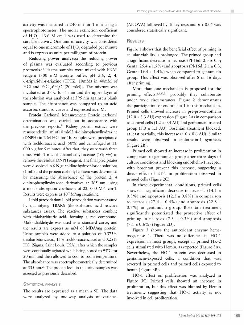
Figure 1 shows that the beneficial effect of priming in cellular viability is prolonged. The primed group had a significant decrease in necrosis (PI-16d: 2.3 ± 0.3; Genta: 25.4 ± 1.5%) and apoptosis (PI-16d: 2.1 ± 0.3; Genta: 19.4 ± 1.4%) when compared to gentamicin group. This effect was observed after 8 or 16 days after priming.
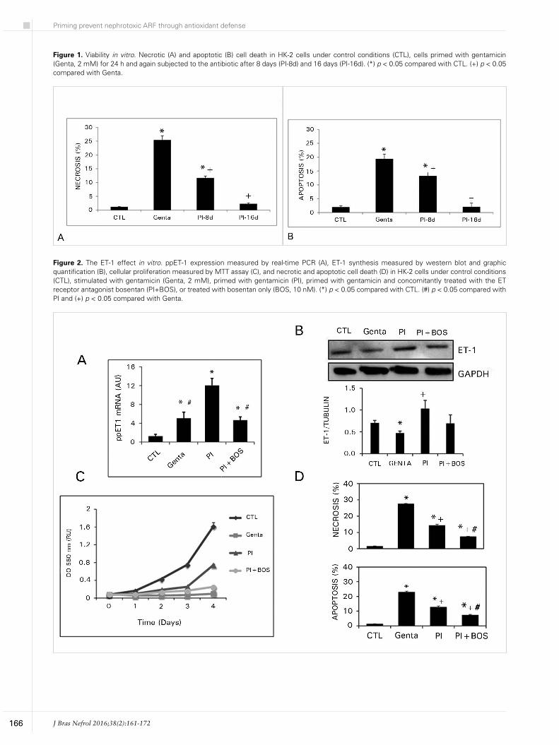
More than one mechanism is proposed for the priming effects;1,4,27,28 probably they collaborate under toxic circumstances. Figure 2 demonstrates the participation of endothelin-1 in this mechanism. Primed cells showed increase in pre-pro-endothelin (12.0 ± 3.3 AU) expression (Figure 2A) in comparison to control cells (1.2 ± 0.4 AU) and gentamicin treated group (5.0 ± 1.3 AU). Bosentan treatment blocked, at least partially, this increase (4.6 ± 0.6 AU). Similar results were observed in endothelin-1 synthesis (Figure 2B).
Primed cell showed an increase in proliferation in comparison to gentamicin group after three days of culture conditions and blocking endothelin-1 receptor with bosentan prevent this increase, suggesting a direct effect of ET-1 in proliferation observed in primed cells (Figure 2C).
In these experimental conditions, primed cells showed a significant decrease in necrosis (14.1 ± 0.8%) and apoptosis (12.5 ± 0.8%) in comparison to necrosis (27.4 ± 0.4%) and apoptosis (22.8 ± 0.7%) in gentamicin group. Bosentan treatment significantly potentiated the protective effect of priming in necrosis (7.3 ± 0.3%) and apoptosis (7.1 ± 0.6%) (Figure 2D).
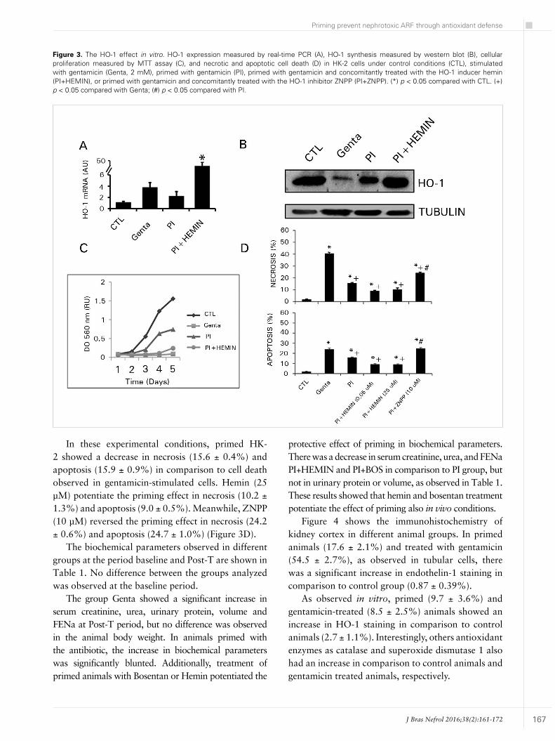
Figure 3 shows the antioxidant enzyme heme-oxygenase 1. There was no difference in HO-1 expression in most groups, except in primed HK-2 cells stimulated with Hemin, as expected (Figure 3A). Nevertheless, the HO-1 protein was decreased in gentamicin-exposed cells, a condition that was reverted in primed cells and primed cells exposed to hemin (Figure 3B).
HO-1 effect on proliferation was analyzed in Figure 3C. Primed cells showed an increase in proliferation, but this effect was blunted by Hemin treatment, suggesting that HO-1 activity is not involved in cell proliferation.
J Bras Nefrol 2016;38(2):161-172
Priming prevent nephrotoxic ARF through antioxidant defense
166
Figure 1. Viability in vitro. Necrotic (A) and apoptotic (B) cell death in HK-2 cells under control conditions (CTL), cells primed with gentamicin (Genta, 2 mM) for 24 h and again subjected to the antibiotic after 8 days (PI-8d) and 16 days (PI-16d). (*) p < 0.05 compared with CTL. (+) p < 0.05 compared with Genta.
Figure 2. The ET-1 effect in vitro. ppET-1 expression measured by real-time PCR (A), ET-1 synthesis measured by western blot and graphic quantification (B), cellular proliferation measured by MTT assay (C), and necrotic and apoptotic cell death (D) in HK-2 cells under control conditions (CTL), stimulated with gentamicin (Genta, 2 mM), primed with gentamicin (PI), primed with gentamicin and concomitantly treated with the ET receptor antagonist bosentan (PI+BOS), or treated with bosentan only (BOS, 10 nM). (*) p < 0.05 compared with CTL. (#) p < 0.05 compared with PI and (+) p < 0.05 compared with Genta.
J Bras Nefrol 2016;38(2):161-172
Priming prevent nephrotoxic ARF through antioxidant defense
167
Figure 3. The HO-1 effect in vitro. HO-1 expression measured by real-time PCR (A), HO-1 synthesis measured by western blot (B), cellular proliferation measured by MTT assay (C), and necrotic and apoptotic cell death (D) in HK-2 cells under control conditions (CTL), stimulated with gentamicin (Genta, 2 mM), primed with gentamicin (PI), primed with gentamicin and concomitantly treated with the HO-1 inducer hemin (PI+HEMIN), or primed with gentamicin and concomitantly treated with the HO-1 inhibitor ZNPP (PI+ZNPP). (*) p < 0.05 compared with CTL. (+) p < 0.05 compared with Genta; (#) p < 0.05 compared with PI.
In these experimental conditions, primed HK-2 showed a decrease in necrosis (15.6 ± 0.4%) and apoptosis (15.9 ± 0.9%) in comparison to cell death observed in gentamicin-stimulated cells. Hemin (25 µM) potentiate the priming effect in necrosis (10.2 ± 1.3%) and apoptosis (9.0 ± 0.5%). Meanwhile, ZNPP (10 µM) reversed the priming effect in necrosis (24.2 ± 0.6%) and apoptosis (24.7 ± 1.0%) (Figure 3D).
The biochemical parameters observed in different groups at the period baseline and Post-T are shown in Table 1. No difference between the groups analyzed was observed at the baseline period.
The group Genta showed a significant increase in serum creatinine, urea, urinary protein, volume and FENa at Post-T period, but no difference was observed in the animal body weight. In animals primed with the antibiotic, the increase in biochemical parameters was significantly blunted. Additionally, treatment of primed animals with Bosentan or Hemin potentiated the
protective effect of priming in biochemical parameters. There was a decrease in serum creatinine, urea, and FENa PI+HEMIN and PI+BOS in comparison to PI group, but not in urinary protein or volume, as observed in Table 1. These results showed that hemin and bosentan treatment potentiate the effect of priming also in vivo conditions.
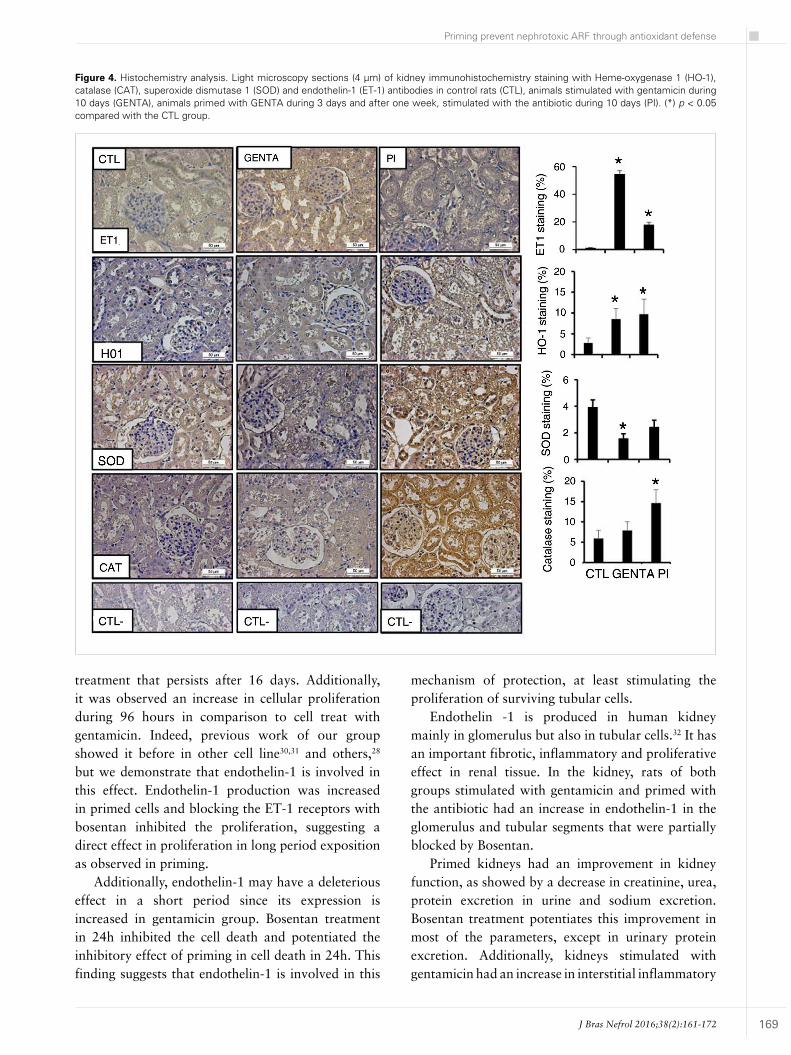
Figure 4 shows the immunohistochemistry of kidney cortex in different animal groups. In primed animals (17.6 ± 2.1%) and treated with gentamicin (54.5 ± 2.7%), as observed in tubular cells, there was a significant increase in endothelin-1 staining in comparison to control group (0.87 ± 0.39%).
As observed in vitro, primed (9.7 ± 3.6%) and gentamicin-treated (8.5 ± 2.5%) animals showed an increase in HO-1 staining in comparison to control animals (2.7 ± 1.1%). Interestingly, others antioxidant enzymes as catalase and superoxide dismutase 1 also had an increase in comparison to control animals and gentamicin treated animals, respectively.

J Bras Nefrol 2016;38(2):161-172
Priming prevent nephrotoxic ARF through antioxidant defense
168
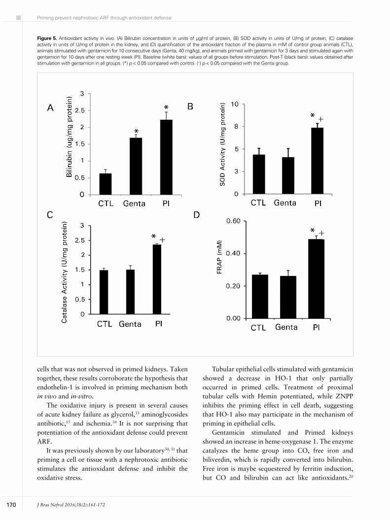
Heme-oxygenase degrades the heme group into CO, free iron and biliverdin, which is rapidly converted in bilirubin by bilirubin reductase.20 We analyzed indirectly the HO-1 activity by the bilirubin content in the extract of cortex kidney (Figure 5A). The bilirubin content in kidney cortex increased in Genta (1.69 ± 0.16 µg/mg protein) and primed (2.22 ± 0.22 µg/mg protein) groups in comparison to control animals (0.63 ± 0.11 µg/mg protein) group.
In Figure 5B and C is shown other antioxidant enzyme activities. There was a significant increase in catalase activity in animals primed (2.35 ± 0.03 U/mg of protein) in comparison to control (1.49 ± 0.06 U/mg of protein) and animals treated with gentamicin (1.5 ± 0.1 U/mg of protein) (Figure 5B). The superoxide dismutase activity also increased only in animals primed (PI: 7.33 ± 0.23 U/mg protein) in comparison to animals treated with the antibiotic and control groups (Genta: 4.06 ± 0.49; CTL: 4.34 ± 0.71 U/mg protein). Additionally, the antioxidant fraction of the plasma showed increase in primed animals (0.48 ± 0.02 mM) in comparison to Genta (0.26 ± 0.03 mM) and control (0.26 ± 0.01 mM) (Figure 5D). Indeed, these results reinforce that priming event stimulates antioxidant defense in the kidney.
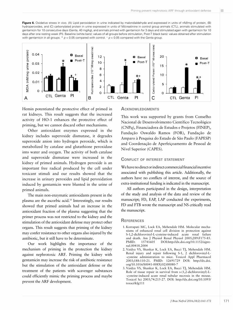
Figure 6 shows the oxidative stress analyzed by urinary peroxides and carbonylated proteins in animal groups.
There was no significant difference between the groups at the baseline period, but the urinary peroxides (Genta: 9.0 ± 1.2; CTL: 2.1 ± 0.05 10-7 M/mg of creatinine) and carbonylated protein (Genta:
14.1 ± 0.6; CTL: 3.3 ± 0.5 10-7 M/mg of creatinine) significantly increased at the post-Genta period. Priming of the animals with gentamicin prevented the increase in urinary peroxides (PI: 4.5 ± 0.4 10-7 M/mg of creatinine) and carbonylated protein (PI: 7.2 ± 0.9 10-7 M/mg of creatinine) induced by the antibiotic (Figure 2A and 2B). Lipid peroxidation showed the same pattern. At the post-T period, there was an increase in Genta group in comparison to control. In primed animals, the lipid peroxidation was blunted.
dIscussIon
The study of the mechanism of priming in a cell or tissue can help to prevent or treat the toxic effect of aminoglycosides antibiotics and others nephrotoxic agents. Several studies showed the beneficial effect of the stimulation of antioxidant defense to treat ARF,15,16,29 but until today it was not enough explored in clinical practice. Our study showed that the mechanism of priming is persistent and multifactorial through the induction of the antioxidant enzymes, increase in the antioxidant fraction of the plasma, inhibition of the oxidative stress, inhibition of the production of endothelin-1,22,25 and probably others mechanism that still have to be determinate. Maybe a strategy based on one approach only could not be enough.
Both tubular cells and rat kidneys were resistant to gentamicin toxicity. In human proximal tubular cells primed with gentamicin, we observed a significant decrease in necrosis and apoptosis after 24 hours
CTL Genta PI PI + HEMIN PI + BOS
Baseline Pos-T Baseline Pos-T Baseline Pos-T Baseline Pos-T Baseline Pos-T
Cr (mg/dL)0.39 ± 0.08
0.45 ± 0.07
0.34 ± 0.01
5.09 ± 0.34*
0.31 ± 0.04
1.55 ± 0.35#
0.31 ± 0.04
0.48 ± 0.03# +
0.53 ± 0.08
0.51 ± 0.04# +
U (mg/dL)35.32 ±
2.4334.28 ±
2.5234.99 ±
3.22144.25 ±
1.5*35.45 ±
2.57101.33 ±
2.7#
35.42 ± 2.53
45.81 ± 1.62# +
42.03 ± 6.19
46.53 ± 3.46# +
uP (mg/24hs)
7.42 ± 0.41
6.81 ± 0.61
9.11 ± 1.12
34.29 ± 3.44*
4.21 ± 2.32
10.20 ± 2.01#
7.2 ± 1.6
17.0 ± 0.9#
7.24 ± 1.64
17.01 ± 0.91#
FENa (%)0.43 ± 0.18
0.50 ± 0.11
0.31 ± 0.05
2.97 ± 0.61*
0.22 ± 0.03
1.46 ± 0.33#
0.21 ± 0.06
0.71 ± 0.01# +
0.30 ± 0.05
0.56 ± 0.08# +
Uv (ml)8.15 ± 0.61
8.55 ± 0.73
12.12 ± 1.54
21 ± 2.35*
5.13 ± 2.30
7.81 ± 1.11#
8.82 ± 0.71
13.30 ± 0.82#
7.82 ± 1.74
9.62 ± 2.42#
BW (g)224 ± 0.51
245 ± 0.62
224 ± 0.87
241 ± 0.7
233 ± 0.54
251 ± 0.59
226.6 ± 0.57
248.3 ± 0.45
226.6 ± 0.41
246.3 ± 0.74
* p < 0.05 vs. CTL; # p < 0.05 vs. Genta + p < 0,05 vs. PI; Data were expressed as the mean ± SEM. The experimental groups were compared via one-way analysis of variance (ANOVA) followed by Tukey tests. The significance level for a null hypothesis was set at 5% (p < 0.05). BW (body weight), Uv (urinary volume), Cr (serum creatinine), U (serum urea), FENa (fractional sodium excretion), were analyzed in CTL, Genta, PI, PI + HEMIN and PI + BOS groups.
tAble 1 physiological parameters
J Bras Nefrol 2016;38(2):161-172
Priming prevent nephrotoxic ARF through antioxidant defense
169
Figure 4. Histochemistry analysis. Light microscopy sections (4 µm) of kidney immunohistochemistry staining with Heme-oxygenase 1 (HO-1), catalase (CAT), superoxide dismutase 1 (SOD) and endothelin-1 (ET-1) antibodies in control rats (CTL), animals stimulated with gentamicin during 10 days (GENTA), animals primed with GENTA during 3 days and after one week, stimulated with the antibiotic during 10 days (PI). (*) p < 0.05 compared with the CTL group.
treatment that persists after 16 days. Additionally, it was observed an increase in cellular proliferation during 96 hours in comparison to cell treat with gentamicin. Indeed, previous work of our group showed it before in other cell line30,31 and others,28 but we demonstrate that endothelin-1 is involved in this effect. Endothelin-1 production was increased in primed cells and blocking the ET-1 receptors with bosentan inhibited the proliferation, suggesting a direct effect in proliferation in long period exposition as observed in priming.
Additionally, endothelin-1 may have a deleterious effect in a short period since its expression is increased in gentamicin group. Bosentan treatment in 24h inhibited the cell death and potentiated the inhibitory effect of priming in cell death in 24h. This finding suggests that endothelin-1 is involved in this
mechanism of protection, at least stimulating the proliferation of surviving tubular cells.
Endothelin -1 is produced in human kidney mainly in glomerulus but also in tubular cells.32 It has an important fibrotic, inflammatory and proliferative effect in renal tissue. In the kidney, rats of both groups stimulated with gentamicin and primed with the antibiotic had an increase in endothelin-1 in the glomerulus and tubular segments that were partially blocked by Bosentan.
Primed kidneys had an improvement in kidney function, as showed by a decrease in creatinine, urea, protein excretion in urine and sodium excretion. Bosentan treatment potentiates this improvement in most of the parameters, except in urinary protein excretion. Additionally, kidneys stimulated with gentamicin had an increase in interstitial inflammatory
J Bras Nefrol 2016;38(2):161-172
Priming prevent nephrotoxic ARF through antioxidant defense
170
Figure 5. Antioxidant activity in vivo. (A) Bilirubin concentration in units of µg/ml of protein, (B) SOD activity in units of U/mg of protein, (C) catalase activity in units of U/mg of protein in the kidney, and (D) quantification of the antioxidant fraction of the plasma in mM of control group animals (CTL), animals stimulated with gentamicin for 10 consecutive days (Genta, 40 mg/kg), and animals primed with gentamicin for 3 days and stimulated again with gentamicin for 10 days after one resting week (PI). Baseline (white bars): values of all groups before stimulation, Post-T (black bars): values obtained after stimulation with gentamicin in all groups. (*) p < 0.05 compared with control. (+) p < 0.05 compared with the Genta group.
cells that was not observed in primed kidneys. Taken together, these results corroborate the hypothesis that endothelin-1 is involved in priming mechanism both in vivo and in-vitro.
The oxidative injury is present in several causes of acute kidney failure as glycerol,33 aminoglycosides antibiotic,15 and ischemia.34 It is not surprising that potentiation of the antioxidant defense could prevent ARF.
It was previously shown by our laboratory30, 31 that priming a cell or tissue with a nephrotoxic antibiotic stimulates the antioxidant defense and inhibit the oxidative stress.
Tubular epithelial cells stimulated with gentamicin showed a decrease in HO-1 that only partially occurred in primed cells. Treatment of proximal tubular cells with Hemin potentiated, while ZNPP inhibits the priming effect in cell death, suggesting that HO-1 also may participate in the mechanism of priming in epithelial cells.
Gentamicin stimulated and Primed kidneys showed an increase in heme-oxygenase 1. The enzyme catalyzes the heme group into CO, free iron and biliverdin, which is rapidly converted into bilirubin. Free iron is maybe sequestered by ferritin induction, but CO and bilirubin can act like antioxidants.20
J Bras Nefrol 2016;38(2):161-172
Priming prevent nephrotoxic ARF through antioxidant defense
171
Figure 6. Oxidative stress in vivo. (A) Lipid peroxidation in urine indicated by malondialdehyde and expressed in units of nM/mg of protein, (B) hydroperoxides, and (C) carbonylated protein in urine expressed in units of M/creatinine in control group animals (CTL), animals stimulated with gentamicin for 10 consecutive days (Genta, 40 mg/kg), and animals primed with gentamicin for 3 days and stimulated again with gentamicin for 10 days after one resting week (PI). Baseline (white bars): values of all groups before stimulation, Post-T (black bars): values obtained after stimulation with gentamicin in all groups. * p < 0.05 compared with control. + p < 0.05 compared with the Genta group.
Hemin potentiated the protective effect of primed in rat kidneys. This result suggests that the increased activity of HO-1 enhances the protective effect of priming, but we cannot discard other mechanisms.
Other antioxidant enzymes expressed in the kidney includes superoxide dismutase, it degrades superoxide anion into hydrogen peroxide, which is metabolized by catalase and glutathione peroxidase into water and oxygen. The activity of both catalase and superoxide dismutase were increased in the kidney of primed animals. Hydrogen peroxide is an important free radical produced by the cell under toxicant stimuli and our results showed that the increase in urinary peroxides and lipid peroxidation induced by gentamicin were blunted in the urine of primed animals.
The main non-enzymatic antioxidants present in the plasma are the ascorbic acid.35 Interestingly, our results showed that primed animals had an increase in the antioxidant fraction of the plasma suggesting that the primer process was not restricted to the kidney and the stimulation of the antioxidant defense may protect other organs. This result suggests that priming of the kidney may confer resistance to other organs also injured by the antibiotic, but it still have to be determinate.
Our work highlights the importance of the mechanism of priming in the protection the kidney against nephrotoxic ARF. Priming the kidney with gentamicin may increase the risk of antibiotic resistance but the stimulation of the antioxidant defense or the treatment of the patients with scavenger substances could efficiently mimic the priming process and maybe prevent the ARF development.
Acknowledgments
This work was supported by grants from Conselho Nacional de Desenvolvimento Científico Tecnológico (CNPq), Financiadora de Estudos e Projetos (FINEP), Fundação Oswaldo Ramos (FOR), Fundação de Amparo à Pesquisa do Estado de São Paulo (FAPESP) and Coordenação de Aperfeiçoamento de Pessoal de Nível Superior (CAPES).
conflIct of InteRest stAtement
We have no direct or indirect commercial financial incentive associated with publishing this article. Additionally, the authors have no conflicts of interest, and the source of extra-institutional funding is indicated in the manuscript.
All authors participated in the design, interpretation of the study and analysis of the data and review of the manuscript; FD, EAP, LAP conducted the experiments, FD and FTB wrote the manuscript and NS critically read the manuscript.
RefeRences
1. Korrapati MC, Lock EA, Mehendale HM. Molecular mecha-nisms of enhanced renal cell division in protection against S-1,2-dichlorovinyl-L-cysteine-induced acute renal failure and death. Am J Physiol Renal Physiol 2005;289:F175-85. PMID: 15741605 DOI:http://dx.doi.org/10.1152/ajpre-nal.00418.2004
2. Vaidya VS, Shankar K, Lock EA, Bucci TJ, Mehendale HM. Renal injury and repair following S-1, 2 dichlorovinyl-L--cysteine administration to mice. Toxicol Appl Pharmacol 2003;188:110-21. PMID: 12691729 DOI: http://dx.doi.org/10.1016/S0041-008X(02)00080-7
3. Vaidya VS, Shankar K, Lock EA, Bucci TJ, Mehendale HM. Role of tissue repair in survival from s-(1,2-dichlorovinyl)-L--cysteine-induced acute renal tubular necrosis in the mouse. Toxicol Sci 2003;74:215-27. DOI: http://dx.doi.org/10.1093/toxsci/kfg111
J Bras Nefrol 2016;38(2):161-172
Priming prevent nephrotoxic ARF through antioxidant defense
172
4. Korrapati MC, Chilakapati J, Lock EA, Latendresse JR, War-britton A, Mehendale HM. Preplaced cell division: a critical mechanism of autoprotection against S-1,2-dichlorovinyl-L--cysteine-induced acute renal failure and death in mice. Am J Physiol Renal Physiol 2006;291:F439-55. PMID: 16495211 DOI: http://dx.doi.org/10.1152/ajprenal.00384.2005
5. Philip BK, Anand SS, Palkar PS, Mumtaz MM, Latendresse JR, Mehendale HM. Subchronic chloroform priming protects mice from a subsequently administered lethal dose of chloroform. Toxicol Appl Pharmacol 2006;216:108-21. PMID: 16815507 DOI:http://dx.doi.org/10.1016/j.taap.2006.04.012
6. Wever KE, Menting TP, Rovers M, van der Vliet JA, Rongen GA, Masereeuw R, et al. Ischemic preconditioning in the animal kidney, a systematic review and meta-analysis. PLoS One 2012;7:e32296. DOI: http://dx.doi.org/10.1371/journal.pone.0032296
7. Yuan Q, Hong S, Han S, Zeng L, Liu F, Ding G, et al. Pre-conditioning with physiological levels of ethanol protect kid-ney against ischemia/reperfusion injury by modulating oxi-dative stress. PLoS One 2011;6:e25811. DOI: http://dx.doi.org/10.1371/journal.pone.0025811
8. Zhu SB, Liu Y, Zhu Y, Yin GL, Wang RP, Zhang Y, et al. Re-mote preconditioning, perconditioning, and postconditioning: a comparative study of their cardio-protective properties in rat models. Clinics (Sao Paulo) 2013;68:263-8. DOI:http://dx.doi.org/10.6061/clinics/2013(02)OA22
9. Thuret R, Saint Yves T, Tillou X, Chatauret N, Thuillier R, Barrou B, et al. Ischemic pre- and post-conditioning: current clinical applications. Prog Urol 2014;24:S56-61. DOI: http://dx.doi.org/10.1016/S1166-7087(14)70065-X
10. Ali BH. Gentamicin nephrotoxicity in humans and animals: some recent research. Gen Pharmacol 1995;26:1477-87. DOI:http://dx.doi.org/10.1016/0306-3623(95)00049-6
11. Bennett WM. Comparison of cyclosporine nephrotoxicity with aminoglycoside nephrotoxicity. Clin Nephrol 1986;25:S126-9. PMID:3519022 DOI: http://dx.doi.org/10.1016/S0272-6386(86)80100-7
12. Schor N, Ichikawa I, Rennke HG, Troy JL, Brenner BM. Pa-thophysiology of altered glomerular function in aminoglycosi-de-treated rats. Kidney Int 1981;19:288-96. PMID: 7014984 DOI: http://dx.doi.org/10.1038/ki.1981.19
13. Cuzzocrea S, Mazzon E, Dugo L, Serraino I, Di Paola R, Britti D, et al. A role for superoxide in gentamicin-mediated nephropa-thy in rats. Eur J Pharmacol 2002;450:67-76. PMID: 12176111 DOI: http://dx.doi.org/10.1016/S0014-2999(02)01749-1
14. Krüger B, Benck U, Singer T, Krämer BK. Drug-induced impairment of renal function. Dtsch Med Wochenschr 2012;137:1873-7. PMID:22971974
15. Naidu MU, Shifow AA, Kumar KV, Ratnakar KS. Ginkgo biloba extract ameliorates gentamicin-induced nephrotoxici-ty in rats. Phytomedicine 2000;7:191-7. DOI: http://dx.doi.org/10.1016/S0944-7113(00)80003-3
16. Farombi EO, Ekor M. Curcumin attenuates gentamicin--induced renal oxidative damage in rats. Food Chem Toxi-col 2006;44:1443-8. PMID: 16814915 DOI: http://dx.doi.org/10.1016/j.fct.2006.05.005
17. Coico R. In vitro assays for mouse B and T cell function. Rela-ted isolation procedures and functional assays. In: Coligan JE, Kruisbeek AM, Margulies DH, Shevach EM, Strober W, ed. Current protocols in immunology. New York: John Wiley & Sons; 1995. p.1-33.
18. Filatov MV, Varfolomeeva EY. Active dissociation of Hoe-chst 33342 from DNA in living mammalian cells. Mutat Res 1995;327:209-15. PMID: 7870089 DOI: http://dx.doi.org/10.1016/0027-5107(94)00189-C
19. Livak KJ, Schmittgen TD. Analysis of relative gene expression data using real-time quantitative PCR and the 2(-Delta Delta C(T)) Method. Methods 2001;25:402-8. DOI: http://dx.doi.org/10.1006/meth.2001.1262
20. Martinek RG. Improved micro-method for determination of se-rum bilirubin. Clin Chim Acta 1966;13:161-70. PMID: 5941734 DOI:http://dx.doi.org/10.1016/0009-8981(66)90290-7
21. Taussky HH. A microcolorimetric determination of creatine in urine by the Jaffe reaction. J Biol Chem 1954;208:853-61. PMID:13174594
22. Nourooz-Zadeh J, Tajaddini-Sarmadi J, Wolff SP. Measure-ment of plasma hydroperoxide concentrations by the ferrous oxidation-xylenol orange assay in conjunction with triphenyl-phosphine. Anal Biochem 1994;220:403-9. PMID: 7978285 DOI:http://dx.doi.org/10.1006/abio.1994.1357
23. Aebi H. Catalase. In: Packer L, ed. Methods in Enzymology. Orlando: Academic Press; 1984. p.121-6.
24. Benzie IF, Strain JJ. Ferric reducing/antioxidant power assay: direct measure of total antioxidant activity of biological fluids and modified version for simultaneous measurement of total antioxidant power and ascorbic acid concentration. Methods Enzymol 1999;299:15-27. PMID: 9916193
25. Levine RL. Carbonyl modified proteins in cellular regula-tion, aging, and disease. Free Radic Biol Med 2002;32:790-6. DOI:http://dx.doi.org/10.1016/S0891-5849(02)00765-7
26. Naziroğlu M, Cay M. Protective role of intraperitoneally ad-ministered vitamin E and selenium on the antioxidative defen-se mechanisms in rats with diabetes induced by streptozoto-cin. Biol Trace Elem Res 2001;79:149-59. DOI:http://dx.doi.org/10.1385/BTER:79:2:149
27. Li Y, Kloner RA. Cardioprotective effects of ischaemic pre-conditioning are not mediated by prostanoids. Cardiovasc Res 1992;26:226-31. DOI: http://dx.doi.org/10.1093/cvr/26.3.226
28. Chang CZ, Wu SC, Lin CL, Hwang SL, Howng SL, Kwan AL. Atorvastatin preconditioning attenuates the production of en-dothelin-1 and prevents experimental vasospasm in rats. Acta Neurochir (Wien) 2010;152:1399-406. DOI: http://dx.doi.org/10.1007/s00701-010-0652-3
29. Mitchell T, Rotaru D, Saba H, Smith RA, Murphy MP, Mac-Millan-Crow LA. The mitochondria-targeted antioxidant mi-toquinone protects against cold storage injury of renal tubular cells and rat kidneys. J Pharmacol Exp Ther 2011;336:682-92. PMID: 21159749 DOI:http://dx.doi.org/10.1124/jpet.110.176743
30. Pessoa EA, Convento MB, Silva RG, Oliveira AS, Borges FT, Schor N. Gentamicin-induced preconditioning of proximal tubular LLC-PK1 cells stimulates nitric oxide production but not the synthesis of heat shock protein. Braz J Med Biol Res 2009;42:614-20. PMID:19466282 DOI: http://dx.doi.org/10.1590/S0100-879X2009005000005
31. Pessoa EA, Convento MB, Ribas OS, Tristão VR, Reis LA, Borges FT, et al. Preconditioning induced by gentamicin pro-tects against acute kidney injury: the role of prostaglandins but not nitric oxide. Toxicol Appl Pharmacol 2011;253:1-6. DOI:http://dx.doi.org/10.1016/j.taap.2011.02.022
32. Kohan DE. Endothelins in the normal and diseased kidney. Am J Kidney Dis 1997;29:2-26. PMID: 9002526 DOI:http://dx.doi.org/10.1016/S0272-6386(97)90004-4
33. Martim EC, Pinto CF, Watanabe M, Vattimo Mde F. Acute kidney injury by glycerol: antioxidant effect of Vitis vinifera L. Rev Bras Ter Intensiva 2007;19:292-6. DOI: http://dx.doi.org/10.1590/S0103-507X2007000300004
34. Dare AJ, Bolton EA, Pettigrew GJ, Bradley JA, Saeb-Parsy K, Murphy MP. Protection against renal ischemia-reperfusion in-jury in vivo by the mitochondria targeted antioxidant MitoQ. Redox Biol 2015;5:163-8. DOI: http://dx.doi.org/10.1016/j.redox.2015.04.008
35. Gasparetto C, Malinverno A, Culacciati D, Gritti D, Prosperini PG, Specchia G, et al. Antioxidant vitamins reduce oxidative stress and ventricular remodeling in patients with acute myocar-dial infarction. Int J Immunopathol Pharmacol 2005;18:487-96.