principals of conventional positive pressure mechanical ventilation
TRANSCRIPT

PRINCIPALS OF CONVENTIONAL PPVSergey Shushunov, MD
I have no commercial interests related to this presentation
RESPIRATTORY SYSTEM FUNCTIONRemoval of carbon dioxide from bloodOxygenation of blood

RESPIRATTORY SYSTEM FAILUREInability to remove carbon dioxideInability to oxygenate bloodBoth

RESPIRATORY SYSTEMORGAN / SYSTEM FUNCTION ETIOLOGY EXAMPLES
CNS Control head injury, CNS infection, seizures, drugs, toxins, liver and kidney failure, hypoglycemia, metabolic
PNS trauma, neuropathy, toxins, metabolic, drugs
Skeletal Mechanical trauma, deformities
Muscular atrophy, myopathy, myolysis, NMB, electrolyte imbalance, tetanus
Respiratory upper airways Gas exchange
croup syndrome, foreign body, tumor, mass, laryngotracheomalacia, trauma, peripheral neuropathy
Respiratory lower airways
RAD, asthma, COPD, bronchiolitis,
Respiratory alveolar space
pneumonia, ARDS, NRDS, pulmonary edema, aspiration, pulmonary contusion/hemorrhage, fibrosis
Gas delivery Gas delivery CHF, PHT, PE, pneumothorax, cardiac tamponade

GAS EXCHANGE
Takes place in alveolar space Depends on:• Partial pressure gradient• Alveolar surface area (mean of I and E surface area)• Alveolar wall thickness• Exposure time• Gas diffusion coefficient (physical property of a gas independent
from any intervention)

CO2 REMOVAL
Depends on • Partial pressure gradient across alveolar-
capillary membrane • CO2 has 200 times higher than O2 diffusion
coefficient and 20 times higher diffusion ability therefore alveolar surface area, wall thickness and exposure time are less relevant

CO2 REMOVAL
• diffusion across alveolar-capillary membrane takes place during inspiration.
• Alveoli don’t have to remain opened during expiration for optimal CO2 removal
• CO2 removal is decreased in:
Hypoventilation (control, mechanical or extreme small airway disease) Increased dead space
• Alveolar disease does not usually lead to CO2 retention
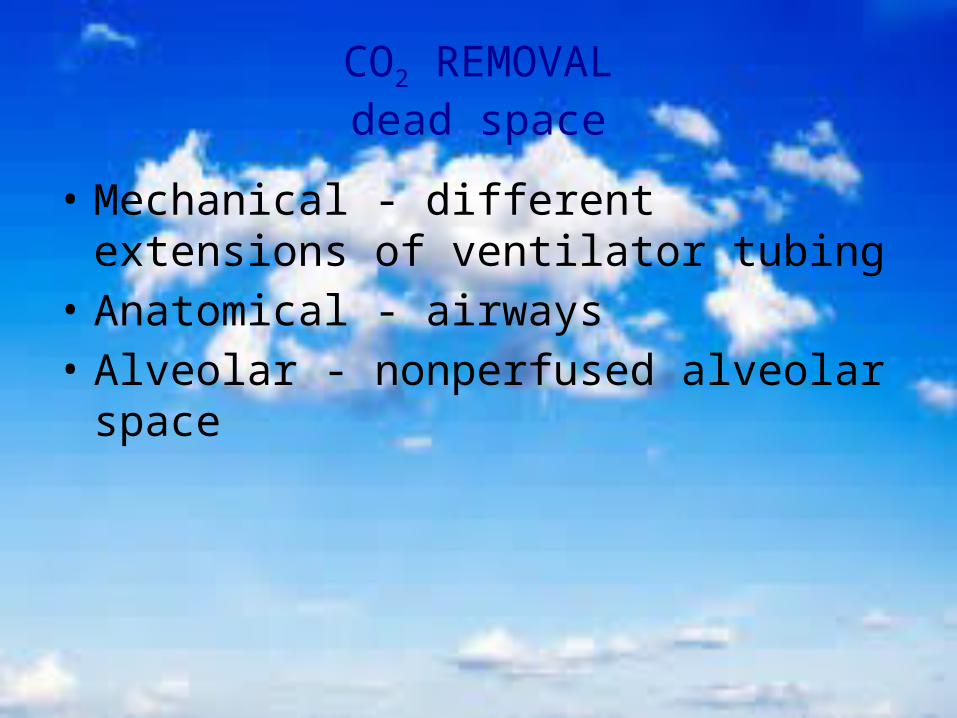
CO2 REMOVALdead space
• Mechanical - different extensions of ventilator tubing
• Anatomical - airways• Alveolar - nonperfused alveolar space

CO2 REMOVAL
• Direct relationship with MV• MV is required to maintain
desired PP gradient • MV (PaCO2)is controlled by
TV (limited by over-distension) RR (limited by auto PEEP)

CO2 REMOVALauto PEEP
• May lead to dynamic hyperinflation
• Complications include barotrauma and hypovolemic shock

OXYGENATION
Depends on• Partial pressure gradient• Alveolar surface area • Alveolar-capillary membrane thickness• Diffusion exposure time• Oxygenation is decreased in alveolar disease large airway disease small airway disease

OXYGENATION
• Normally O2 diffusion across alveolar-capillary membrane takes place during inspiration and expiration.
• Alveolar collapse during expiration shortens diffusion exposure time
• Alveoli must remain opened during expiration for best oxygenation

OXYGENATION
• Direct relationship with FiO2 (limited by 100%)
• Direct relationship with Mean Airway Pressure

CONTROL OF MEAN AIRWAY PRESSURE
Pressure
Time
PEEP
PIP
Increase I. Time
Increase PEEP
Increase PIP (TV)
I. Time E. Time
Increase % Rise (flow)
%Rise
I. Time E. Time
% Rise (flow) PIP – limited by over distention I. Time - limited by auto PEEP PEEP – limited by over distention

ALVEOLAR DISEASE
• Accumulation of intra and extra alveolar fluid• Thickening of alveolar-capillary membrane• Alveolar collapse during expiration

EFFECT OF POSITIVE AIRWAY PRESSUREredistribution of intra alveolar fluid

EFFECT OF POSITIVE AIRWAY PRESSUREincrease of alveolar surface area reduction of thickness of alveolar-capillary membrane

RESPIRATORY FAILURE
Categories• Neither alveolar nor small airway disease
• Small airway disease • Alveolar disease• Mixed small airway and alveolar disease

SELECTION OF INITIAL VENTILATOR SETTINGS
• Reasonable guess• Patient’s age and size• Cause of respiratory failure• Degree of respiratory failure

PPV SETTINGS
• Mode of PPV• PEEP• FiO2
• TV / PIP• Respiratory Rate• I. Time

VENTILATION MODE SELECTION
• Category (or etiology) of respiratory failure• Degree of illness• Respiratory effort

MODES OF CONVENTIONAL PPV(BREATH DELIVERY)
• Controlled (Assist Controlled) PC or VC Patients with no respiratory drive (NMB, anesthesia, coma) Weaning is not imminent
• IMV (SIMV) PC or VC All modern ventilation offer only SIMV
Patients with or without respiratory drive
• Support (PS or VS) Patient with good respiratory drive Moderate degree of respiratory failure
• Mixed (usually SIMV + PS or VS) Patients with good respiratory drive Preferred weaning mode

CMV (AC) vs. IMV (SIMV) CMV IMV
Mandatory and spontaneous breaths have identical TV/PIP and I. Time
Breaths vary in volume and I. Time between mandatory and spontaneous
Hard to wean by decreasing rate Easy to wean: patient takes “own” breaths with or without PS/VS
Minimal work of breathing Potential for increased work of breathing with insufficient PS/VS
Feels unnatural. Patient/ventilator asynchrony is possible in awake patients and may need sedation +/- NMB
Feels more natural. Easier to synchronize, requires less sedation and less likely NMB
Significant hyperventilation in agitated patients is possible
Significant hyperventilation in agitated patients is less likely
In heavily sedated, comatose and paralyzed patients CMV and IMV work identically

CMV (AC) vs. IMV (SIMV)Assist control IMV

INITIAL PEEP SELECTION
Etiology of respiratory failure Zero in small airway disease Low (physiological) in no small airway disease / no alveolar disease High in alveolar disease Tricky in mixed small airway disease / alveolar disease (bronchiolitis)
Degree of hypoxemia (in alveolar disease)
Additional pathology Increased ICP - lower Pulmonary hypertension - lower Increased intra-abdominal pressure - higher
FiO2 0.3 0.4 0.5 0.6 0.7 0.8 0.9 1.0
PEEP 5-6 6-8 8-10 10-12 12-14 14-16 16-18 20-24

PEEP SELECTION
LIMITATIONS• Barotrauma• Pulmonary hypertension• Increase of anatomical
dead space (CO2)• Decrease of CO

TIDAL VOLUME / PIP SELECTION
• Pressure vs. Volume• TV is age independent• Physiological - patients wit neither small airway
nor alveolar disease or patients with isolated small airway disease
• Small – patients with alveolar disease • Selecting TV based on ml/kg may be misleading• Chest rise

PRESSURE vs. VOLUME PRESSUE VOLUME
TV changes as patient’s compliance and airway resistance changes
PIP changes as patient’s compliance and airway resistance changes
Increase of airway resistance, or decrease of compliance will lead to decrease of TV and hypoventilation
Increase of airway resistance or decrease of compliance will lead to increase of PIP and potentially to volutrauma and barotrauma, but not to hypoventilation

PEAK INSPIRATORY PRESSURE
• Changes of PIP in VC mode is an important parameter which can be used as the “sixth” vital sign
• Elevation of PIP while patient receives constant TV always indicates a problem, including mucus accumulation, pulmonary edema, infiltrate, atelectasis, pneumothorax, bronchospasm, artificial airway obstruction
• Decrease of the PIP while patient receives constant TV always indicate clinical improvement, including resolution of atelectasis, bronchospasm, infiltrate or pulmonary edema

RESPIRATION RATE SELECTIONmain principals
• Respiratory drive and effort• Desired pCO2
• BMR (BMR and CO2 production is elevated in sepsis, trauma, burns, etc.)
• Age and weight
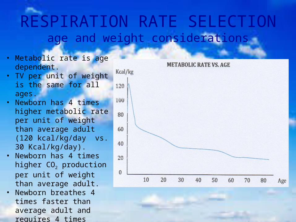
RESPIRATION RATE SELECTIONage and weight considerations
• Metabolic rate is age dependent.
• TV per unit of weight is the same for all ages.
• Newborn has 4 times higher metabolic rate per unit of weight than average adult (120 kcal/kg/day vs. 30 Kcal/kg/day).
• Newborn has 4 times higher CO2 production per unit of weight than average adult.
• Newborn breathes 4 times faster than average adult and requires 4 times faster RR.
• Use ideal body weight.

INSPIRATORY TIME SELECTION
• Respiratory Rate• Desired Expiratory Time to Inspiratory Time ratio• Presence of small airway disease• Targeted Mpaw

INSPIRATORY TIME SELECTION
• I. Time, E. Time and RR are interlinked• Example: RR of 10/min makes respiratory cycle 6 seconds I. Time set at 1 second makes I:E ratio of 1:5 Changing I. Time to 1.5 seconds makes I:E ratio of 1:3 (1.5:4.5 sec) Changing RR to 15/min (resp. cycle of 4 sec.) makes I:E ration of 1:3

I.TIME, RR and I:E RATIO SELECTION
Wrong selection of I. Time/RR/I:E ration can lead to auto-peep and air trapping

COMPLICTIONS OF AUTO PEEP
• CO2 retention• Barotrauma• Decreased CO and shock

SUGGETSTED INITIAL VENTILATORSETINGS
NO ALVEAOLAR, NO SMALL AIRWAY DISEASE• Low FiO2 • Usual TV• Age appropriate rate• Match ventilator I. Time to patient’s I. Time• I:E ratio 1:3• Physiologic PEEP

SUGGETSTED INITIAL VENTILATORSETINGS
NO ALVEAOLAR, NO SMALL AIRWAY DISEASE
AGE 0-12 M 1-5 Y 6-12 Y ADULT
TV 10-12 8-12 8-12 6-10
RATE 30-40 25-40 15-25 8-15
I. TIME 0.3-0.5 0.5-0.7 0.7-0.9 0.9-1.2

SUGGETSTED INITIAL VENTILATORSETINGS
SMALL AIRWAY DISEASE• High FiO2 • Usual TV• Slow rate• Age appropriate I. Time• I:E ratio 4:1 and longer depending on auto PEEP• 0 PEEP

SUGGETSTED INITIAL VENTILATORSETINGS
SMALL AIRWAY DISEASE
AGE 0-12 M 1-5 Y 6-12 Y ADULT
TV 10-12 8-12 8-12 6-10
RATE 20-30 15-25 10-20 6-8
I. TIME 0.5 0.5-0.7 0.7-0.9 0.9-1.2

VENTILATOR SETINGS ADJUSTMENTS
SMALL AIRWAY DISESE• To improve oxygenation Increase flow or decrease rise time Increase PEEP cautiously to low levels Increase TV/PIP cautiously (PIP reflects airway resistance
rather than alveolar compliance)
• To improve CO2 removal Increase PEEP to 5-10 cm (slowly) Increase E. Time Increase TV/PIP

SUGGETSTED INITIAL VENTILATORSETINGS
ALVEOLAR DISEASE• High FiO2 • Small TV• Age appropriate rate• Age appropriate I. Time • I:E ratio 1:<3 • High PEEP

SUGGETSTED INITIAL VENTILATORSETINGS
ALVEOLAR DISEASE
AGE 0-12 M 1-5 Y 6-12 Y ADULT
TV 6-12 6-10 4-10 4-8
RATE 30-40 15-25 15-25 8-15
I. TIME 0.3-0.5 0.5-0.7 0.7-0.9 0.9-1.2

VENTILATOR SETINGS ADJUSTMENTS
ALVEOLAR DISEASE• To improve oxygenation Increase I. Time (Decrease I:E ratio) Increase PEEP to 30 cm, try recruitment maneuver with PEEP
of 40 cm, aim to keep FiO2 < 0.6
Increase TV/PIP (the least desirable option) for small chest rise
• To improve CO2 removal Increase RR Increase TV/PIP

PERMISSIVE HYPERCARBIA(ignored CO2 retention)
• Results from predictably low MV Decreased RR in small airway disease Decreased TV in alveolar disease
• Sedation to overcome hypercarbic drive (35-45 < pCO2 < 80-100)• Well tolerated by most patients• Main limitation is arterial pH• Recommended to limit: pCO2 - 70, pH - 7.2
• Reports of pCO2 in excess of 200 • Increased ICP• Pulmonary hypertension

WEANING PROCESS• Consider using SIMV + PS• D/C NMB and wean sedation• Wean FiO2 to < 0.4• Wean PEEP to 3-5 cm (higher in obese patients)• Wean RR to < ½ of “normal”• Wean PS to 5-10 cm• Monitor Work of breathing (RR, use of accessory muscles) HbO2Sat or PaO2
ET CO2 or PCO2