principles of measurement: arterial blood gas, and …...principles of measurement - arterial blood...
TRANSCRIPT
Part I Anaesthesia Refresher Course – 2018 University of Cape Town 1
Principles of Measurement Arterial blood gas, and electrodes
Dr Dean Nolte Nelson Mandela Children’s Hospital
The measurement of arterial blood gas (ABG) is common in clinical practice, and is essentially in the toolkit of “Point of Care” assessments. Sensors to measure blood gases, electrolytes and metabolites are easy-to-use, automated, and low maintenance: ideal for rapid, reliable, reproducible measurements. As anaesthetists we need to understand:
1. The need for the ABG sampling (what information do we hope to gain from the result. The adage holds true: ”never do a test without knowing what you are going to do with the result”
2. The basics of electrochemistry (including the definitions anode & cathode, reduction & oxidation)
3. What is going to be measured (and how), and what is calculated (and why / purpose of these values).
This is not only to pass Part I Physics, but ensures that we can request the correct information and helps with future development in this field. The most important reasons for blood gas assessment and analysis include:
1. O2: diagnostic and therapeutic reasons 2. CO2: respiratory adequacy 3. Acid-base status 4. Electrolytes (K
+, Na
+, Ca
2+) and metabolites (bilirubin, glucose, lactate)
The ABG is thus the cornerstone of measurement of respiratory and metabolic disorders.
Best arterial blood gas measurement:
The volume required for measurement by the ABG machine varies from 0.65 – 0.15 ml. The lower the volume, the fewer the number of analytes measured (with decreased likelihood of co-oximetry)
Basics / Definitions of Electrochemistry
1. Electrical potential (V) Definition: Work per unit charge between two points (i.e. the work expended in moving positive charge). Electrical potential is thus the work done in moving a positive charge from 0 (a reference point) to a point of determination. It has a positive gradient, which will indicate the magnitude and direction of the driving force (necessitating charge movement). It is established when one metal is immersed into solution causing metal atoms (from an electrode) to move into the surrounding solution. Thus, the surface of the metal will have a net positive charge, and the solution a net negative charge. This charge is constant dependent on the nature of the immersed metal, the nature and concentration of the solution, and the temperature (if it is maintained). Two metals within the same solution will establish a predictable potential difference (V) between them.
Draw blood from an artery, intermittently, using a small needle (22 – 25 gauge) and syringe (heparinised to prevent blood clot formation)
o (OR from an indwelling catheter)
Aseptic technique
Prompt analysis (within several minutes) o OR put on ice ASAP and transport to lab (can be delayed by up to 1 hour)
Put a stopper on the syringe IMMEDIATELY (*prevents arterial blood from interacting with the O2 in air.
Rapid analysis / ice ensures that there is not a rapid decline in the PaO2 due to the metabolism that continues to take place by the erythrocytes, white blood cells, etc.
Principles of measurement - Arterial blood gas, and electrodes Dr D Nolte
1 - 2
2. Electric current This refers to the flow of charge, measured in Ampère’s (intensity of flow). This is the S.I. unit of electric current. Current flows from negative to positive; electron flow is from negative to positive. Flow can be in either direction, or both simultaneously.
3. Electro-chemical cell Two electrodes are found suspended in an electrochemical cell/ solution. Thus each electrode is a single metal conductor within the electrolyte solution. The two sides are separated by a semi-permeable membrane. The standard electrode In order to measure electrode potential, we have to be able to start from a reference point. This is the standardized half-cell potential. We make use of a hydrogen electrode, which is assumed to have an electrical potential of 0mV.
Hydrogen is at a partial pressure of 1 atmosphere, and is bubbled through a hydrogen-containing solution. The temperature is 25 °C; [H
+] is 1 mol/l; pH = 0.
Platinum is used as the catalyst.
This is essentially very impractical for clinical use. In modern practice: reference electrode, with known half-potential (when compared to the “standard, hydrogen electrode”) is used. The most commonly used reference electrodes in clinical practise are Ag/AgCl, and the Hg/HgCl2 (in a KCl solution). The latter is the calomel electrode.
The usual reference electrode employed is a Ag/AgCl half-cell, in a solution of 4M sodium formate adjusted to a pH of 5.5. a triple membrane protects the electrode. Inner: limits diffusion in and out of the electrolyte solution Middle: reduces protein interference (blood) Outer: protects inner electrolyte solution (4M sodium formate) from rinsing contamination,
and from diffusing out.
Convention dictates that the reference electrode is on the LEFT, and the measuring electrode is on the RIGHT. Electrode types
a) First kind: metal surrounded by the electrolyte solution. b) Second kind: metal surrounded by its own salt (increases ionic exchange), and then
suspended in the electrolyte solution containing the anion of the metal salt.
4. I.S.M. Membrane potentials result due to selective permeability to anions and cations. The membrane essentially separates the solutions from one another. Membrane potential = Right – Left. If right (measuring) is > Left (reference) then a POSITIVE membrane potential exists. It is VITAL to note that this does represent actual flow of charge, but instead potential difference.
Marked cation activity on the right binds to the membrane positive charge at the membrane cation dissociation on the opposite side (left, reference) of the semi-permeable membrane. The membrane remains “permeable” until equilibrium is reached.
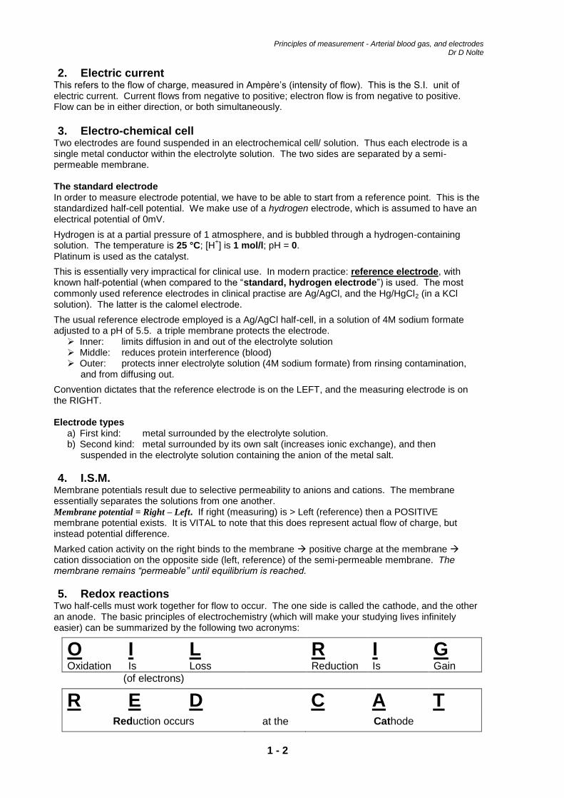
5. Redox reactions Two half-cells must work together for flow to occur. The one side is called the cathode, and the other an anode. The basic principles of electrochemistry (which will make your studying lives infinitely easier) can be summarized by the following two acronyms:
O I L R I G Oxidation Is Loss Reduction Is Gain
(of electrons)
R E D C A T Reduction occurs at the Cathode
Principles of measurement - Arterial blood gas, and electrodes Dr D Nolte
1 - 3
6. Compensatory Mechanisms Some ABG machines require input of FIO2 and temperature to accurately perform / calculate results on ABG testing. This allows for temperature compensation.
O2 / pH / CO2 are all affected by changes in temperature. Temperature calibration is standardized to a patient with a temperature of 37°C.
An increase in temperature change in volume of dissolved gas (in the serum, and within the erythrocytes). This is understood by the application of the “Ideal Gas Equation”:
PV = nRT (where V is proportional to T).
Increasing temperatures increasing vapour pressure (H2O) reduced paO2 in the measured blood stream.
pKa (dissociation constant) is directly related to temperature; temperature changes the chemical
reactions of the buffering solutions (i.e. temperature can or rate of change of the reactions)
Blood Gas Parameters, and Measurement
Measured / Determined Physically Calculated
1. pH 2. PaCO2 Actual / Standard HCO3-
3. PaO2 4. Electrolytes Base Excess (SBE) 5. Metabolites (Lactate, Glucose) 6. Haemoglobin, Bilirubin
The focus of this lecture will be the physical measurement of blood gas values. The calculated values will be dealt with elsewhere in this course.
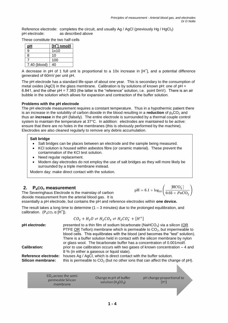
1. Measurement of pH
pH = -= log [H+]
The pH electrode is a “glass” electrode consisting of a 3-dimensional latticework of a central Silicon atom surrounded by 4 Oxygen atoms. There is incorporation of various metal oxides (Ca
2+, Na
+) into
the glass membrane, allowing for variant sensitivity of the electrode. The metal oxides lose electrons to the incorporated oxygen molecules and thus become cations. Charge is displaced across the membrane resulting in the “flow” of current across the glass.
The one side of the glass is exposed to a buffer of KNOWN pH (“reference” electrode); the other side is exposed to blood (“test” solution). The glass membrane is then a partition with differing [H
+] on
either side, establishing a potential difference via the concentration gradient. The pH remains constant despite the change in [H
+] due to the action of the inner buffer solution.
The reference electrode will maintain a constant electrical potential despite changes in pH. The potential difference is measured, and changed to a direct reading of [H
+] which is then converted
into a determinable pH value.
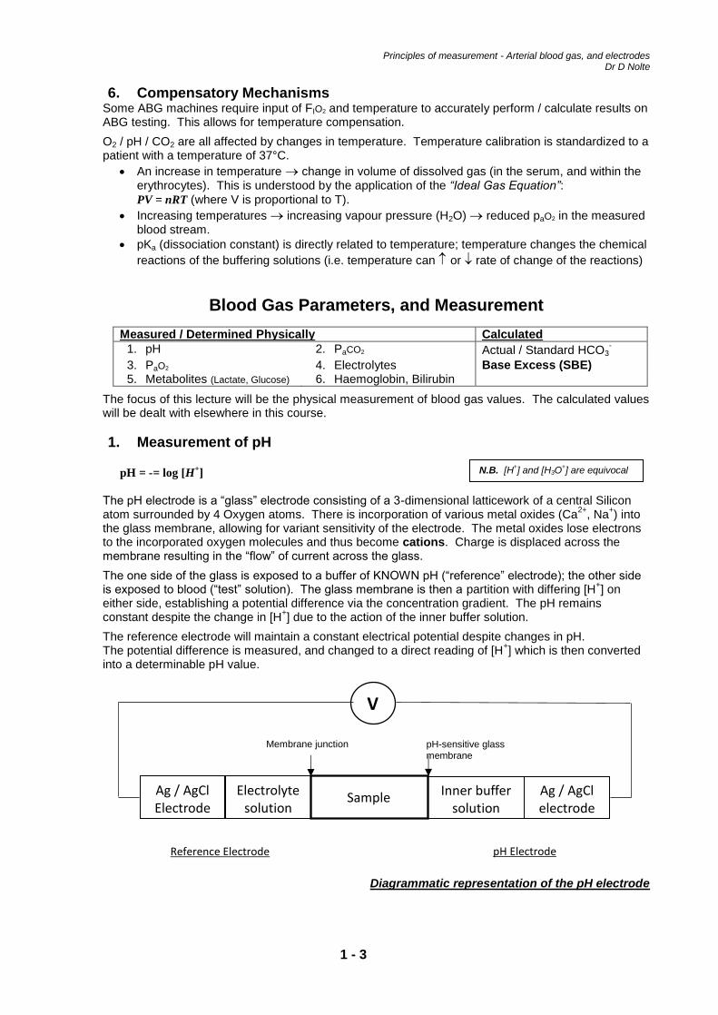
Diagrammatic representation of the pH electrode
Ag / AgCl Electrode
Electrolyte solution
Ag / AgCl electrode
Sample Inner buffer
solution
Reference Electrode pH Electrode
Membrane junction pH-sensitive glass membrane
V
N.B. [H+] and [H3O
+] are equivocal
Principles of measurement - Arterial blood gas, and electrodes Dr D Nolte
1 - 4
Reference electrode: completes the circuit, and usually Ag / AgCl (previously Hg / HgCl2) pH electrode: as described above
These constitute the two half-cells
pH [H+] nmol/l
9 1x10
8 10
7 100
7.40 (blood) 40
A decrease in pH of 1 full unit is proportional to a 10x increase in [H+], and a potential difference
generated of 60mV per unit pH.
The pH electrode has a standard life-span of about one year. This is secondary to the consumption of metal oxides (AgCl) in the glass membrane. Calibration is by solutions of known pH: one of pH = 6.841, and the other pH = 7.383 (the latter is the “reference” solution, i.e. point 0mV). There is an air bubble in the solution which allows for expansion and contraction of the buffer solution. Problems with the pH electrode The pH electrode measurement requires a constant temperature. Thus in a hypothermic patient there is an increase in the solubility of carbon dioxide in the blood resulting in a reduction of paCO2 and thus an increase in the pH (falsely). The entire electrode is surrounded by a thermal couple control system to maintain the temperature at 37°C. In addition: electrodes are maintained to be active: ensure that there are no holes in the membranes (this is obviously performed by the machine). Electrodes are also cleaned regularly to remove any debris accumulation.
2. PaCO2 measurement The Severinghaus Electrode is the mainstay of carbon dioxide measurement from the arterial blood gas. It is essentially a pH electrode, but contains the pH and reference electrodes within one device.
The result takes a long time to determine (1 – 3 minutes) due to the prolonged equilibration, and calibration. (PaCO2 α [H
+]).
𝐶𝑂2 +𝐻2𝑂 ⇌ 𝐻2𝐶𝑂3 ⇌ 𝐻2𝐶𝑂3− + [𝐻+]
pH electrode: presented to a thin film of sodium bicarbonate (NaHCO3) via a silicon (OR PTFE OR Teflon) membrane which is permeable to CO2, but impermeable to blood cells. This equilibrates with the blood (and becomes the “test” solution). There is a buffer solution held in contact with the silicon membrane by nylon or glass wool. The bicarbonate buffer has a concentration of 0.001mol/l.
Calibration: prior to use calibration occurs with two gases of known concentration – 4 and 8 % (in either a gaseous or liquid state).
Reference electrode: houses Ag / AgCl, which is direct contact with the buffer solution. Silicon membrane: this is permeable to CO2 (but no other ions that can affect the change of pH).
Salt bridge
Salt bridges can be places between an electrode and the sample being measured.
KCl solution is housed within asbestos fibre (or ceramic material). These prevent the contamination of the KCl test solution.
Need regular replacement.
Modern day electrodes do not employ the use of salt bridges as they will more likely be surrounded by a triple membrane instead.
Modern day: make direct contact with the solution.
Principles of measurement - Arterial blood gas, and electrodes Dr D Nolte
1 - 5
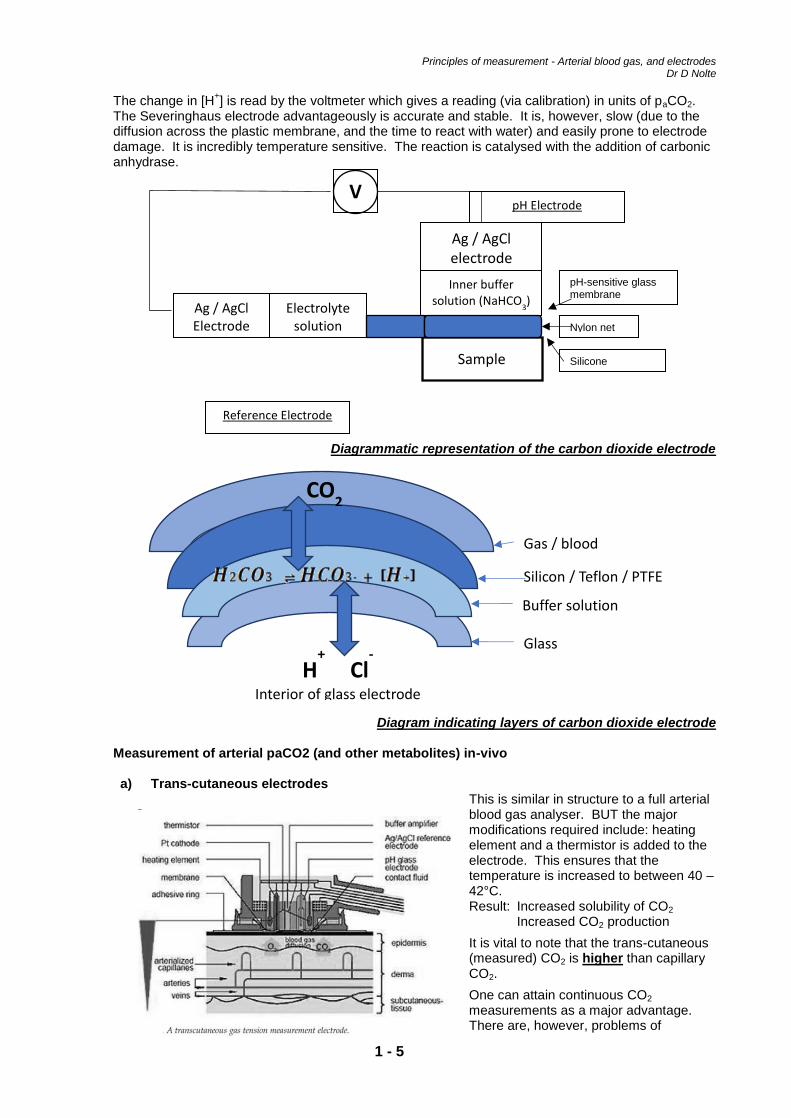
The change in [H+] is read by the voltmeter which gives a reading (via calibration) in units of paCO2.
The Severinghaus electrode advantageously is accurate and stable. It is, however, slow (due to the diffusion across the plastic membrane, and the time to react with water) and easily prone to electrode damage. It is incredibly temperature sensitive. The reaction is catalysed with the addition of carbonic anhydrase.
Diagrammatic representation of the carbon dioxide electrode
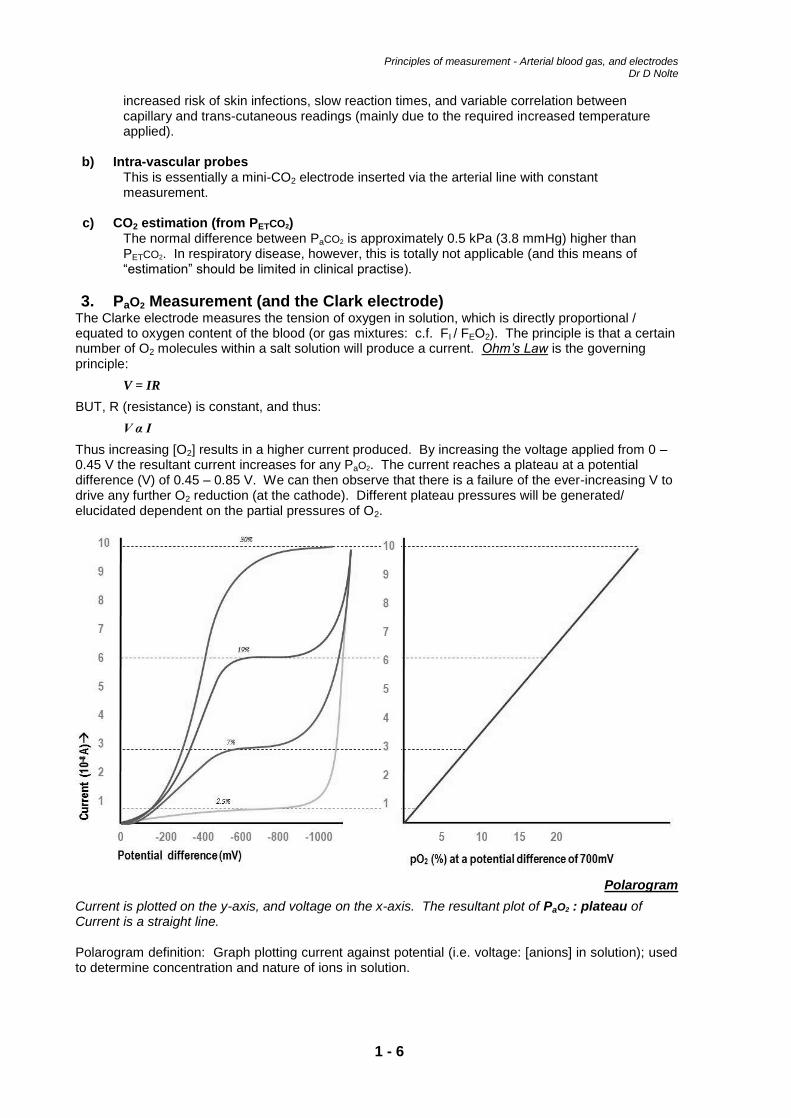
Diagram indicating layers of carbon dioxide electrode Measurement of arterial paCO2 (and other metabolites) in-vivo a) Trans-cutaneous electrodes
This is similar in structure to a full arterial blood gas analyser. BUT the major modifications required include: heating element and a thermistor is added to the electrode. This ensures that the temperature is increased to between 40 – 42°C. Result: Increased solubility of CO2 Increased CO2 production
It is vital to note that the trans-cutaneous (measured) CO2 is higher than capillary CO2.
One can attain continuous CO2 measurements as a major advantage. There are, however, problems of
H+ Cl
-
Interior of glass electrode
Gas / blood
Silicon / Teflon / PTFE
Buffer solution
Glass Membrane
CO2
Ag / AgCl Electrode
Electrolyte solution
Ag / AgCl electrode
Sample
Inner buffer solution (NaHCO
3)
Reference Electrode
pH Electrode
pH-sensitive glass membrane
Nylon net
V
Silicone membrane
Principles of measurement - Arterial blood gas, and electrodes Dr D Nolte
1 - 6
increased risk of skin infections, slow reaction times, and variable correlation between capillary and trans-cutaneous readings (mainly due to the required increased temperature applied).
b) Intra-vascular probes This is essentially a mini-CO2 electrode inserted via the arterial line with constant measurement.
c) CO2 estimation (from PETCO2) The normal difference between PaCO2 is approximately 0.5 kPa (3.8 mmHg) higher than PETCO2. In respiratory disease, however, this is totally not applicable (and this means of “estimation” should be limited in clinical practise).
3. PaO2 Measurement (and the Clark electrode) The Clarke electrode measures the tension of oxygen in solution, which is directly proportional / equated to oxygen content of the blood (or gas mixtures: c.f. FI / FEO2). The principle is that a certain number of O2 molecules within a salt solution will produce a current. Ohm’s Law is the governing principle:
V = IR
BUT, R (resistance) is constant, and thus:
V α I
Thus increasing [O2] results in a higher current produced. By increasing the voltage applied from 0 – 0.45 V the resultant current increases for any PaO2. The current reaches a plateau at a potential difference (V) of 0.45 – 0.85 V. We can then observe that there is a failure of the ever-increasing V to drive any further O2 reduction (at the cathode). Different plateau pressures will be generated/ elucidated dependent on the partial pressures of O2.
Polarogram
Current is plotted on the y-axis, and voltage on the x-axis. The resultant plot of PaO2 : plateau of Current is a straight line. Polarogram definition: Graph plotting current against potential (i.e. voltage: [anions] in solution); used to determine concentration and nature of ions in solution.
Principles of measurement - Arterial blood gas, and electrodes Dr D Nolte
1 - 7
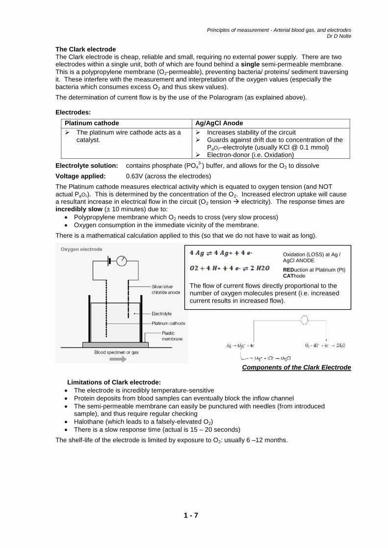
The Clark electrode The Clark electrode is cheap, reliable and small, requiring no external power supply. There are two electrodes within a single unit, both of which are found behind a single semi-permeable membrane. This is a polypropylene membrane (O2-permeable), preventing bacteria/ proteins/ sediment traversing it. These interfere with the measurement and interpretation of the oxygen values (especially the bacteria which consumes excess O2 and thus skew values).
The determination of current flow is by the use of the Polarogram (as explained above). Electrodes:
Platinum cathode Ag/AgCl Anode
The platinum wire cathode acts as a catalyst.
Increases stability of the circuit Guards against drift due to concentration of the
PaO2–electrolyte (usually KCl @ 0.1 mmol) Electron-donor (i.e. Oxidation)
Electrolyte solution: contains phosphate (PO43-
) buffer, and allows for the O2 to dissolve
Voltage applied: 0.63V (across the electrodes)
The Platinum cathode measures electrical activity which is equated to oxygen tension (and NOT actual PaO2). This is determined by the concentration of the O2. Increased electron uptake will cause a resultant increase in electrical flow in the circuit (O2 tension electricity). The response times are incredibly slow (± 10 minutes) due to:
Polypropylene membrane which O2 needs to cross (very slow process)
Oxygen consumption in the immediate vicinity of the membrane.
There is a mathematical calculation applied to this (so that we do not have to wait as long).
Components of the Clark Electrode Limitations of Clark electrode:
The electrode is incredibly temperature-sensitive
Protein deposits from blood samples can eventually block the inflow channel
The semi-permeable membrane can easily be punctured with needles (from introduced sample), and thus require regular checking
Halothane (which leads to a falsely-elevated O2)
There is a slow response time (actual is 15 – 20 seconds)
The shelf-life of the electrode is limited by exposure to O2: usually 6 –12 months.
Oxidation (LOSS) at Ag / AgCl ANODE
REDuction at Platinum (Pt) CAThode
The flow of current flows directly proportional to the number of oxygen molecules present (i.e. increased current results in increased flow).
Principles of measurement - Arterial blood gas, and electrodes Dr D Nolte
1 - 8
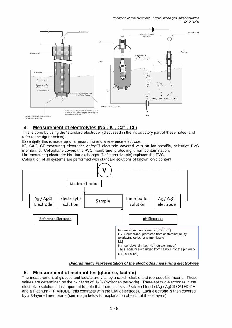
4. Measurement of electrolytes (Na+, K+, Ca2+, Cl-) This is done by using the “standard electrode” (discussed in the introductory part of these notes, and refer to the figure below). Essentially this is made up of a measuring and a reference electrode. K
+, Ca
2+, Cl
- measuring electrode: Ag/AgCl electrode covered with an ion-specific, selective PVC
membrane. Cellophane covers this PVC membrane, protecting it from contamination. Na
+ measuring electrode: Na
+-ion exchanger (Na
+-sensitive pin) replaces the PVC.
Calibration of all systems are performed with standard solutions of known ionic content.
Diagrammatic representation of the electrodes measuring electrolytes
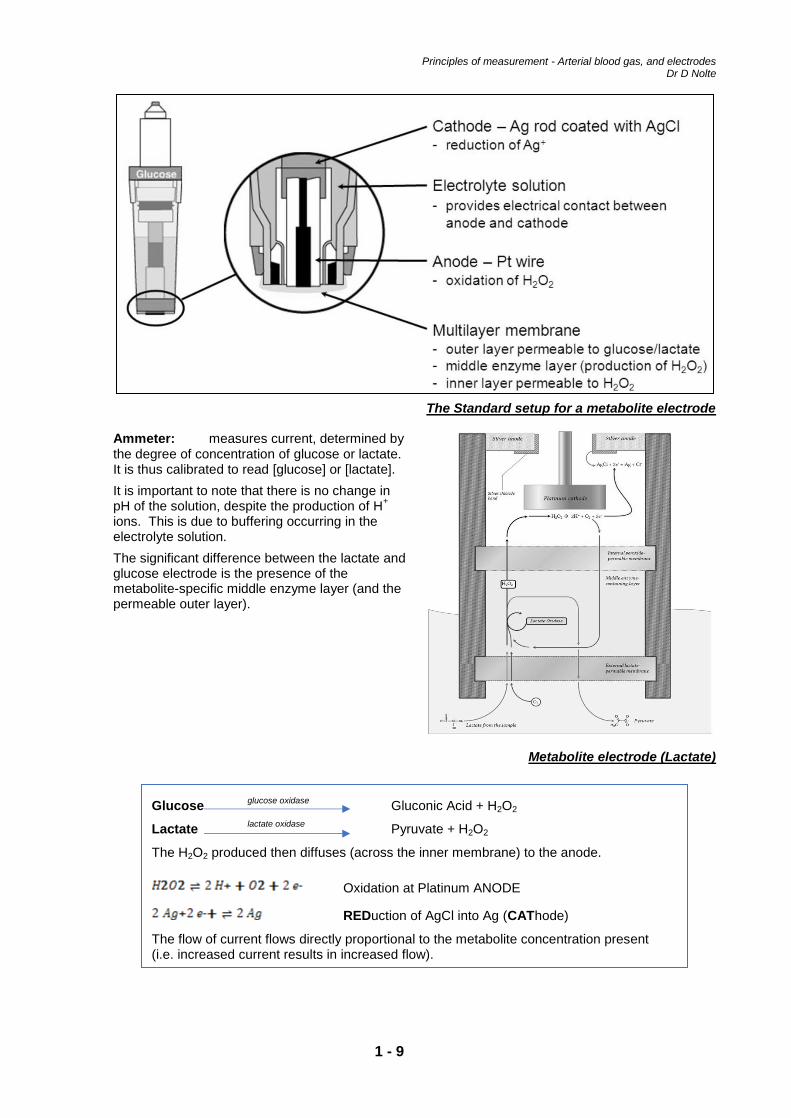
5. Measurement of metabolites (glucose, lactate) The measurement of glucose and lactate are vital by a rapid, reliable and reproducible means. These values are determined by the oxidation of H2O2 (hydrogen peroxide). There are two electrodes in the electrolyte solution. It is important to note that there is a silver/ silver chloride (Ag / AgCl) CATHODE and a Platinum (Pt) ANODE (this contrasts with the Clark electrode). Each electrode is then covered by a 3-layered membrane (see image below for explanation of each of these layers).
Ag / AgCl Electrode
Electrolyte solution
Ag / AgCl electrode
Sample Inner buffer
solution
Reference Electrode pH Electrode
Membrane junction
Ion-sensitive membrane (K+
, Ca2+
, Cl-
) PVC Membrane, protected from contamination by overlaying cellophane membrane OR Na
+
-sensitive pin (i.e. Na+
-ion-exchanger) Thus, sodium exchanged from sample into the pin (very
Na+
, sensitive)
V
Principles of measurement - Arterial blood gas, and electrodes Dr D Nolte
1 - 9
The Standard setup for a metabolite electrode Ammeter: measures current, determined by the degree of concentration of glucose or lactate. It is thus calibrated to read [glucose] or [lactate].
It is important to note that there is no change in pH of the solution, despite the production of H
+
ions. This is due to buffering occurring in the electrolyte solution.
The significant difference between the lactate and glucose electrode is the presence of the metabolite-specific middle enzyme layer (and the permeable outer layer).
Metabolite electrode (Lactate)
Glucose glucose oxidase
Gluconic Acid + H2O2
Lactate lactate oxidase
Pyruvate + H2O2
The H2O2 produced then diffuses (across the inner membrane) to the anode.
Oxidation at Platinum ANODE
REDuction of AgCl into Ag (CAThode)
The flow of current flows directly proportional to the metabolite concentration present (i.e. increased current results in increased flow).
Principles of measurement - Arterial blood gas, and electrodes Dr D Nolte
1 - 10
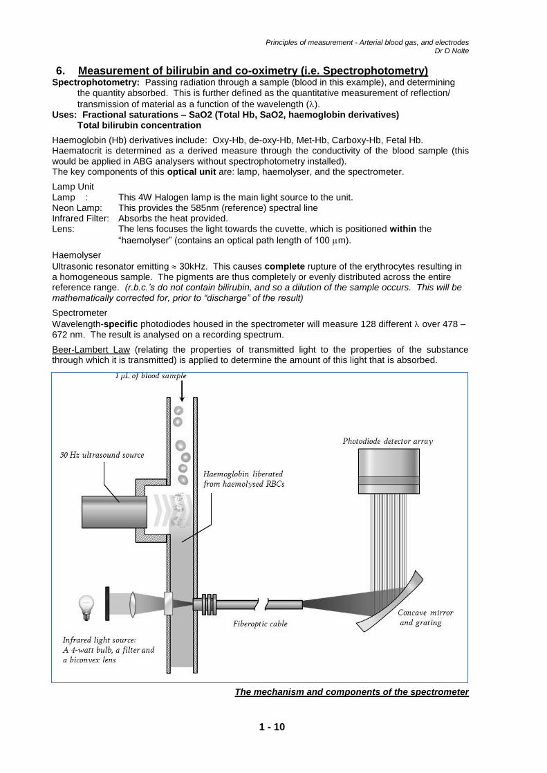
6. Measurement of bilirubin and co-oximetry (i.e. Spectrophotometry) Spectrophotometry: Passing radiation through a sample (blood in this example), and determining
the quantity absorbed. This is further defined as the quantitative measurement of reflection/
transmission of material as a function of the wavelength (). Uses: Fractional saturations – SaO2 (Total Hb, SaO2, haemoglobin derivatives)
Total bilirubin concentration
Haemoglobin (Hb) derivatives include: Oxy-Hb, de-oxy-Hb, Met-Hb, Carboxy-Hb, Fetal Hb. Haematocrit is determined as a derived measure through the conductivity of the blood sample (this would be applied in ABG analysers without spectrophotometry installed). The key components of this optical unit are: lamp, haemolyser, and the spectrometer.
Lamp Unit Lamp : This 4W Halogen lamp is the main light source to the unit. Neon Lamp: This provides the 585nm (reference) spectral line Infrared Filter: Absorbs the heat provided. Lens: The lens focuses the light towards the cuvette, which is positioned within the
“haemolyser” (contains an optical path length of 100 m).
Haemolyser
Ultrasonic resonator emitting 30kHz. This causes complete rupture of the erythrocytes resulting in a homogeneous sample. The pigments are thus completely or evenly distributed across the entire reference range. (r.b.c.’s do not contain bilirubin, and so a dilution of the sample occurs. This will be mathematically corrected for, prior to “discharge” of the result)
Spectrometer
Wavelength-specific photodiodes housed in the spectrometer will measure 128 different over 478 – 672 nm. The result is analysed on a recording spectrum.
Beer-Lambert Law (relating the properties of transmitted light to the properties of the substance through which it is transmitted) is applied to determine the amount of this light that is absorbed.
The mechanism and components of the spectrometer

Principles of measurement - Arterial blood gas, and electrodes Dr D Nolte
1 - 11
The absorption spectra of the haemoglobin species Turbidity will significantly affect the accuracy of the specimen measured. The maximum, acceptable level is < 0.5%. It occurs predominantly in samples with very high lipid contact (e.g. Hyperlipidaemia)
CarboxyHaemoglobin (HbCO) – Interesting Numbers
Cigarettes Home-life % HbCO
Non-smokers Rural <0.5 %
Non-smoker Urban 2 %
Smokers 5 – 6 %
% of Total HbCO Manifestations
10 % Mild symptoms
30 % Severe symptoms
60 – 70 % Respiratory failure (with death imminent)
80 % Rapid death
Clinical application of measurement of HbCO Neonates have a high percentage HbF (fetal haemoglobin) which has a similar structure to HbCO, and HbCO can thus be falsely elevated on a neonatal arterial blood gas sample.
Principles of measurement - Arterial blood gas, and electrodes Dr D Nolte
1 - 12
Conclusion
Some older references refer to means of measurement that are antiquated, and no longer in use. It is critical to understand the physical principles governing the analysis in our modern day blood gas machines. It is more likely that we will find ourselves more readily utilizing in-line arterial blood gas monitors which are non-invasive, reliable, and reproducible (in the near future). These have been highlighted in these notes, and will be further emphasized in the refresher course itself.
Spot (“Easy Reference”) Questions
1. Why do we measure ABG? 2. Which values on the ABG are measured by the blood gas machine? Which ABG values are
calculated? 3. How is the pH value determined? 4. O2
a. How is PaO2 determined by the blood gas machine? b. What are the alternative names for the O2 electrode?
5. How is CO2 determined in the blood gas machine? 6. HCO3
-
a. How is HCO3- measured?
b. Compare standard vs. actual HCO3-.
c. What causes changes in actual HCO3-? (/)
7. How is co-oximetry used in ABG machine measurements? List some clinical applications of this (including emergency department, operating theatre, intensive care settings).
8. How accurate is the measurement of ABG Haemoglobin vs a “formal” [Hb]? 9. Base Excess
a. Define “base excess”(BE). b. How is BE calculated? What does it determine? c. Define “standard base excess” (SBE). What is its clinical use?
10. Interference with ABG. How do the following impact the results on an ABG: a. Excess Heparin b. No Heparin c. Delay in sample analysis d. Air bubbles e. Temperature
References
1. Recommendations for Standards of Monitoring during Anaesthesia and Recovery. 4th Edition 2007.
2. Davis P, Kenny G. Further techniques of gas and vapour analysis. In: Basic Physics and Measurement in Anaesthesia. 2005; Chapter 20: 219 – 226.
3. Joubert I. Understanding electrodes and blood-gas analysers. UCT Part I Anaesthetic Refresher Course. 2009.
4. Dasgupta, A and Wahed A. Common poisoning’s including heavy metal poisoning. Clinical Chemistry, Immunology and Laboratory Quality Control. 2014: 337 – 351.
5. Davis P, Kenny G. Carbon dioxide measurement. In: Basic Physics and Measurement in Anaesthesia. 2005; Chapter 19: 211 – 218.
6. Collins English Dictionary. Copyright © HarperCollins Publishers
7. Omar S. Electrochemistry (How a Blood Gas Machine Works). University of Witwatersrand Part I Anaesthetics Refresher Course. 2007.
8. Rabi Y, Ambalavanan N. Assisted Ventilation of the Neonate. 6th Edition. 2017.
9. Horecker BL. The absorption spectra of hemoglobin and its derivatives in the visible and near infra-red regions. J. Biol. Chem. 1943, 148:173-183.