prion diseases - basic science · prion diseases - basic science dr. david westaway centre for...
TRANSCRIPT
Prion Diseases - Basic Science
Dr. David WestawayCentre for Prionsand Protein Folding Diseases,
University of [email protected]
Part I: The causative agent and patterns of manifestation
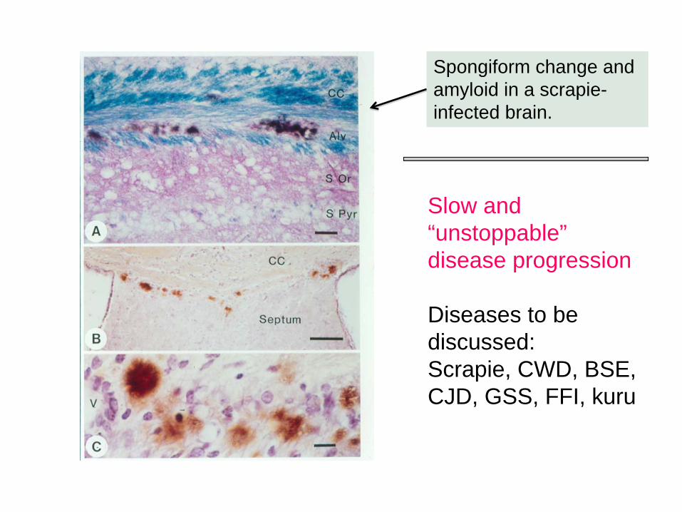
Spongiform change and amyloid in a scrapie- infected brain.
Slow and “unstoppable” disease progression
Diseases to be discussed:Scrapie, CWD, BSE, CJD, GSS, FFI, kuru
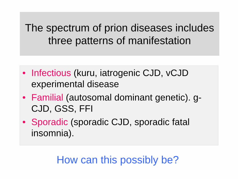
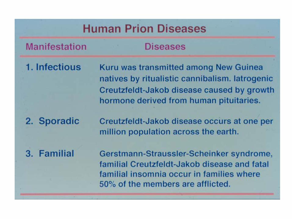
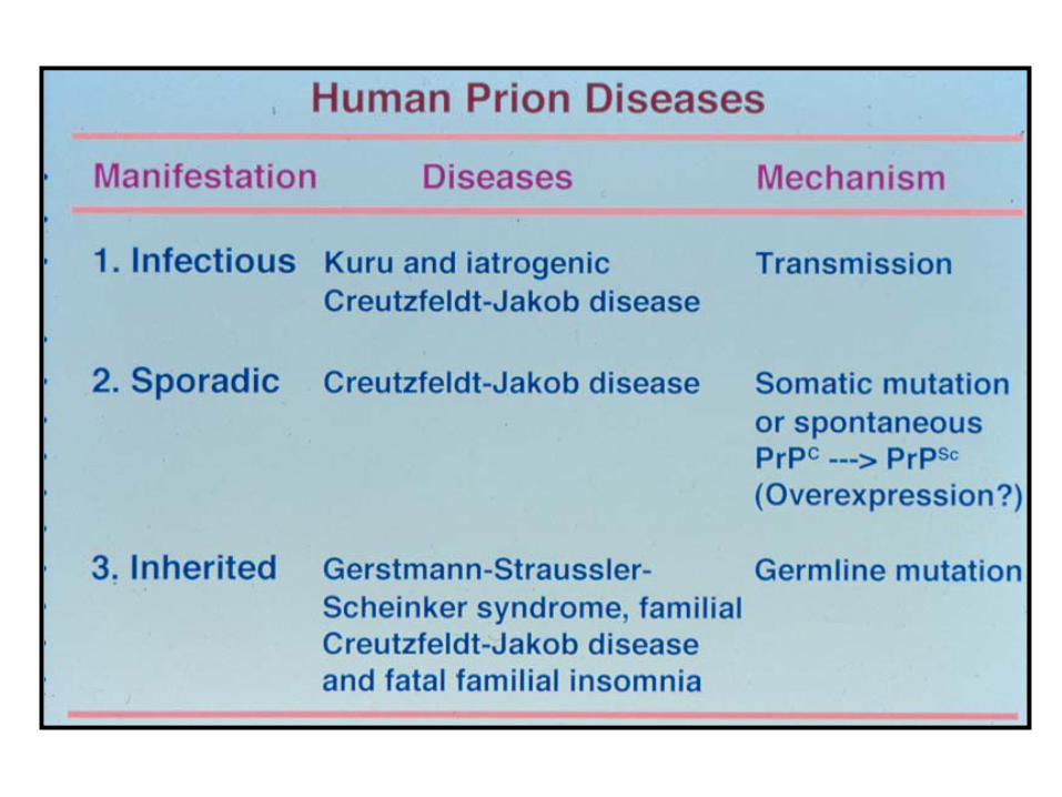
The spectrum of prion diseases includes three patterns of manifestation
• Infectious (kuru, iatrogenic CJD, vCJD experimental disease
• Familial (autosomal dominant genetic). g- CJD, GSS, FFI
• Sporadic (sporadic CJD, sporadic fatal insomnia).
How can this possibly be?
Are diseases like scrapie infectious or genetic?
HB “James” Parry, Univ Oxford: a genetic disease controlled by the recessive “s” gene
Alan Dickinson, Univ Edinburgh: a naturally infectious disease

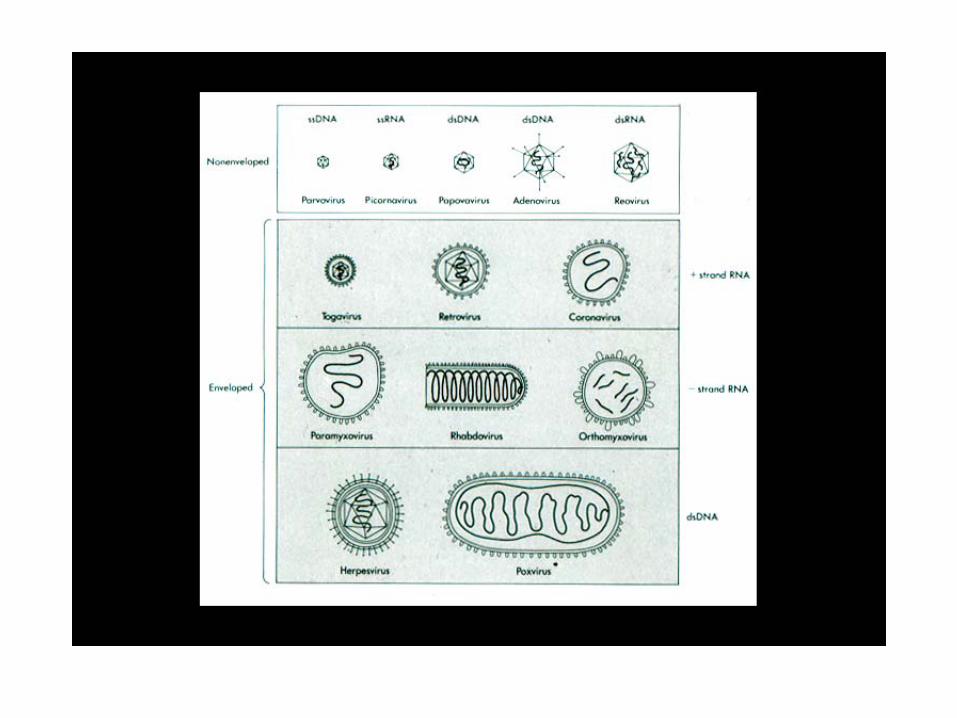
The search for a genomic nucleic acid in the scrapie agent
Studies in the 1960’ and 1970’s when prions were sometimes referred to as “unconventional slow viruses”
Notes on nomenclature:
Unconventional slow virus – meaninglessTSE - inaccurate
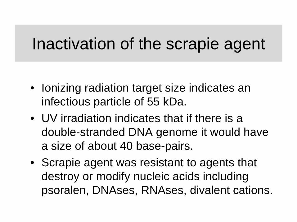
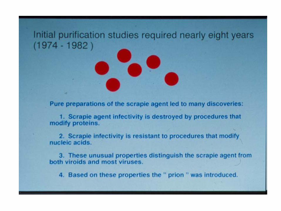
Inactivation of the scrapie agent
• Ionizing radiation target size indicates an infectious particle of 55 kDa.
• UV irradiation indicates that if there is a double-stranded DNA genome it would have a size of about 40 base-pairs.
• Scrapie agent was resistant to agents that destroy or modify nucleic acids including psoralen, DNAses, RNAses, divalent cations.
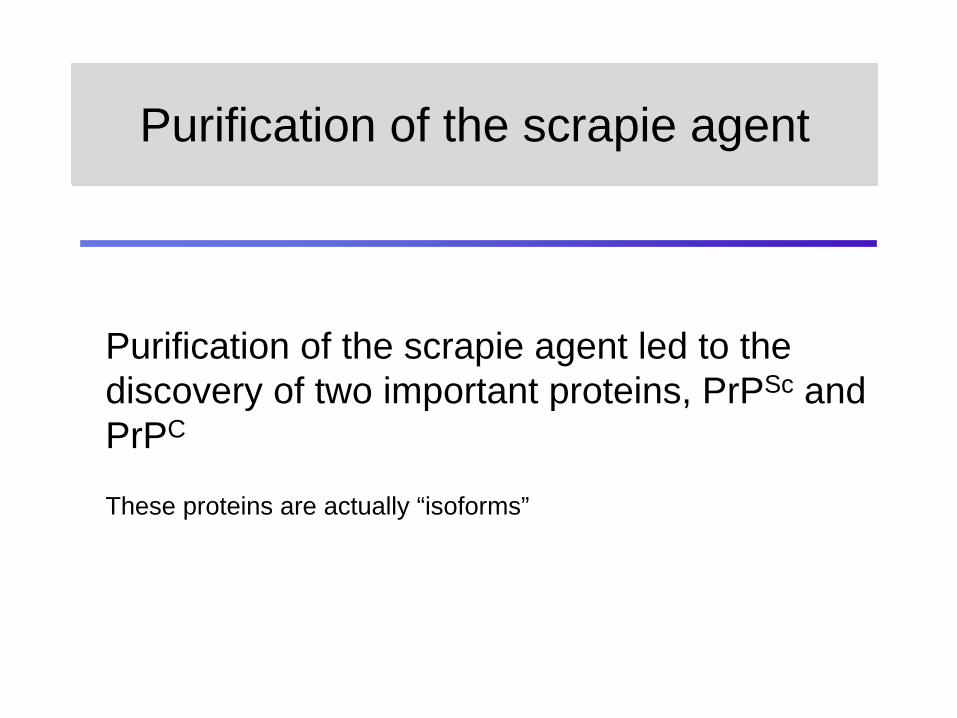
Purification of the scrapie agent led to the discovery of two important proteins, PrPSc and PrPC
These proteins are actually “isoforms”
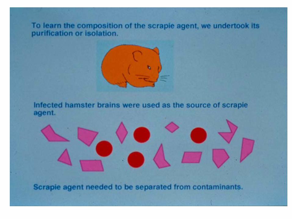
Purification of the scrapie agentPurification of the scrapie agent
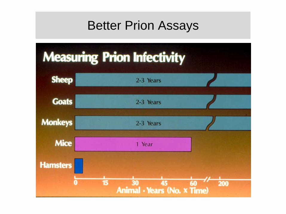
Better Prion AssaysBetter Prion Assays
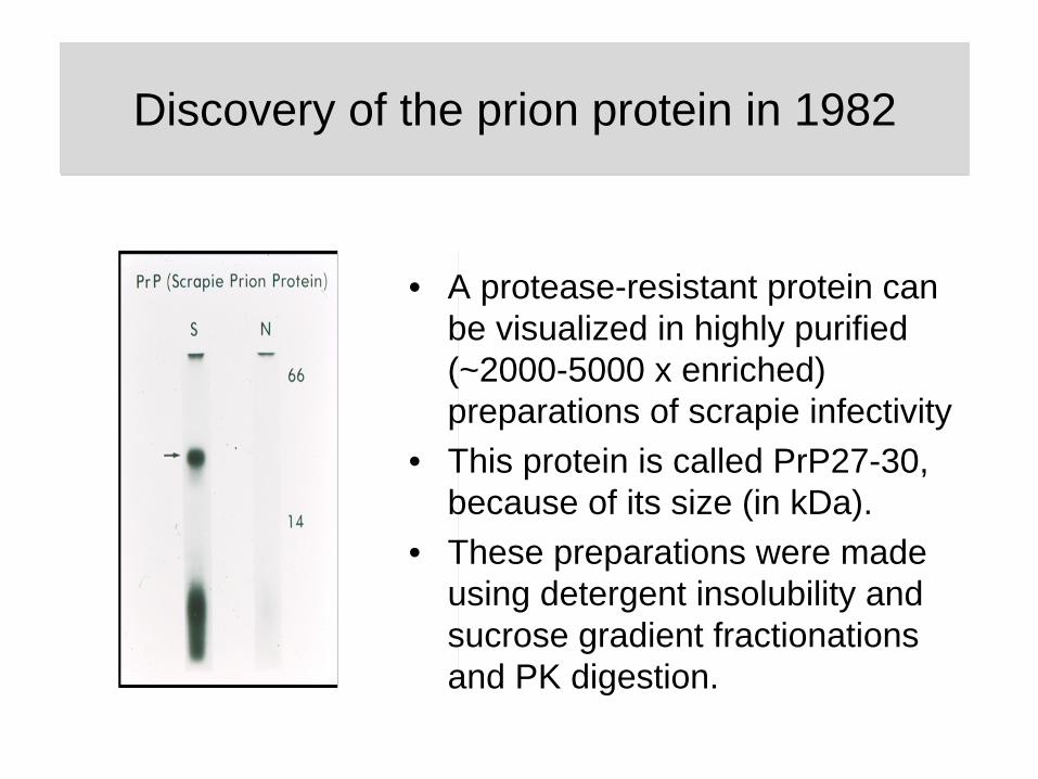
• A protease-resistant protein can be visualized in highly purified (~2000-5000 x enriched) preparations of scrapie infectivity
• This protein is called PrP27-30, because of its size (in kDa).
• These preparations were made using detergent insolubility and sucrose gradient fractionations and PK digestion.
Discovery of the prion protein in 1982Discovery of the prion protein in 1982
PrPC
PrP 27-30
PrPSc derives from the host
Expression, maturation
Proteinase K digestion
PrPSc
5’ open reading frame of an intronless Prnp gene 3’
S-S
CHO CHO
H1 H2 H3
“Conversion”
GPI
Cu domain
Arriving at the “conformational hypothesis” (1)
• PrPSc and PrPC have closely related amino acid sequences (1985).
• The PrP gene has a single uninterrupted coding exon (1986).
• Low resolution structural analysis reveal PrPC is - helical (1992).
PrP encoding gene exon1 254
Arriving at the “conformational hypothesis” (2)
• Low resolution structural analysis reveal PrPSc is enriched in -sheet (1992).
• PrPSc amyloid deposits in scrapie-infected hamsters stain with Congo Red dye (1985).
• Amino acid analysis of proteolytic fragments of PrPSc
(arising from in vitro digestion of purified material) reveal no differences from the predicted sequence of PrPC (1993).
PrP encoding gene exon1 254
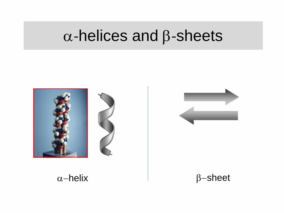
‐helices and ‐sheets
helix sheet
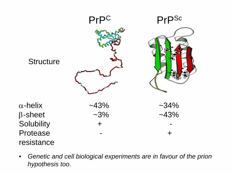
PrPC PrPSc
Structure
-helix ~43% ~34%-sheet ~3% ~43%Solubility + -Protease - +resistance + -
• Genetic and cell biological experiments are in favour of the prion hypothesis too.
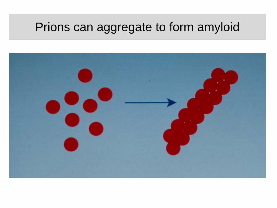
Prions can aggregate to form amyloidPrions can aggregate to form amyloid
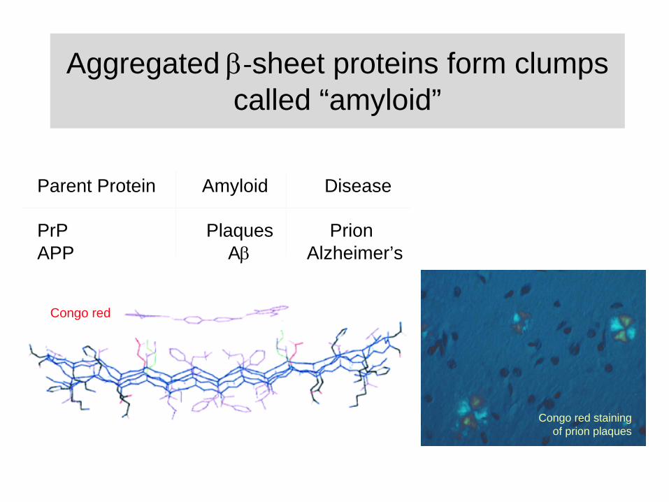
Aggregated ‐sheet proteins form clumps called “amyloid”
Parent Protein Amyloid Disease
PrP Plaques PrionAPP A
Alzheimer’s
Congo red
Congo red staining of prion plaques
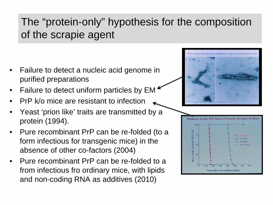
The “protein-only” hypothesis for the composition of the scrapie agent
• Failure to detect a nucleic acid genome in purified preparations
• Failure to detect uniform particles by EM• PrP k/o mice are resistant to infection• Yeast ‘prion like’ traits are transmitted by a
protein (1994).• Pure recombinant PrP can be re-folded (to a
form infectious for transgenic mice) in the absence of other co-factors (2004)
• Pure recombinant PrP can be re-folded to a from infectious fro ordinary mice, with lipids and non-coding RNA as additives (2010)
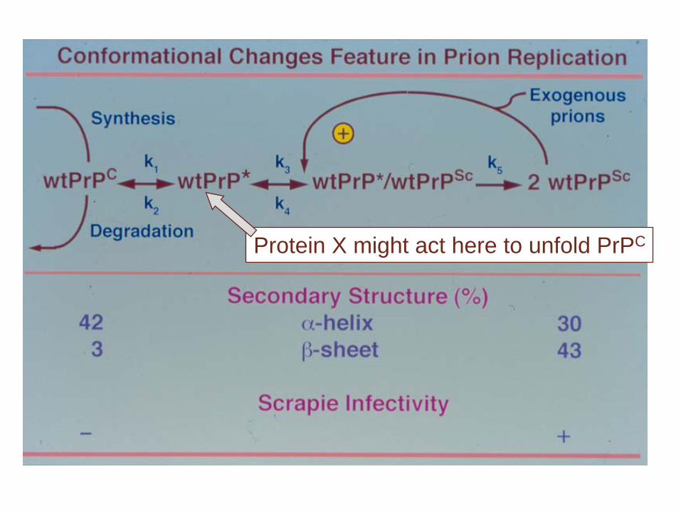
Protein X might act here to unfold PrPC

Heterodimer hypothesis (Prusiner)
• A large energy barrier prevents spontaneous conversion of PrPC to PrPSc.
• PrPC is unfolded by a hypothetical molecular chaperone called protein X. Identity of protein X is unknown
• The replication intermediate is a PrPC /PrPSc
heterodimer (60 kDa).
PrPC PrP-U ? PrPSc
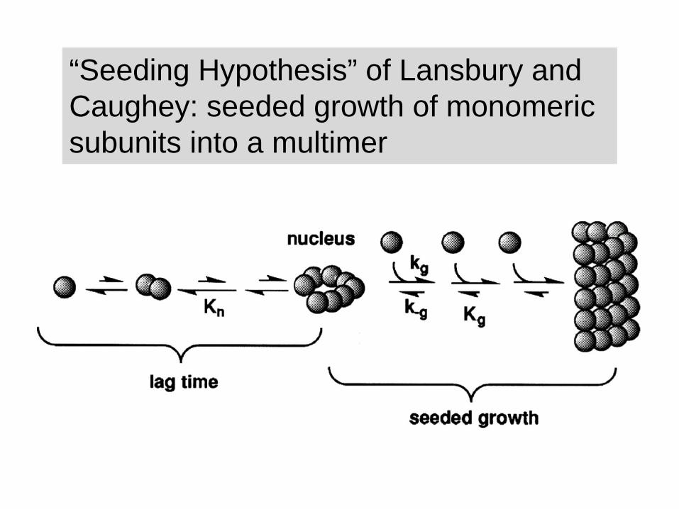
“Seeding Hypothesis” of Lansbury and Caughey: seeded growth of monomeric subunits into a multimer
multimer
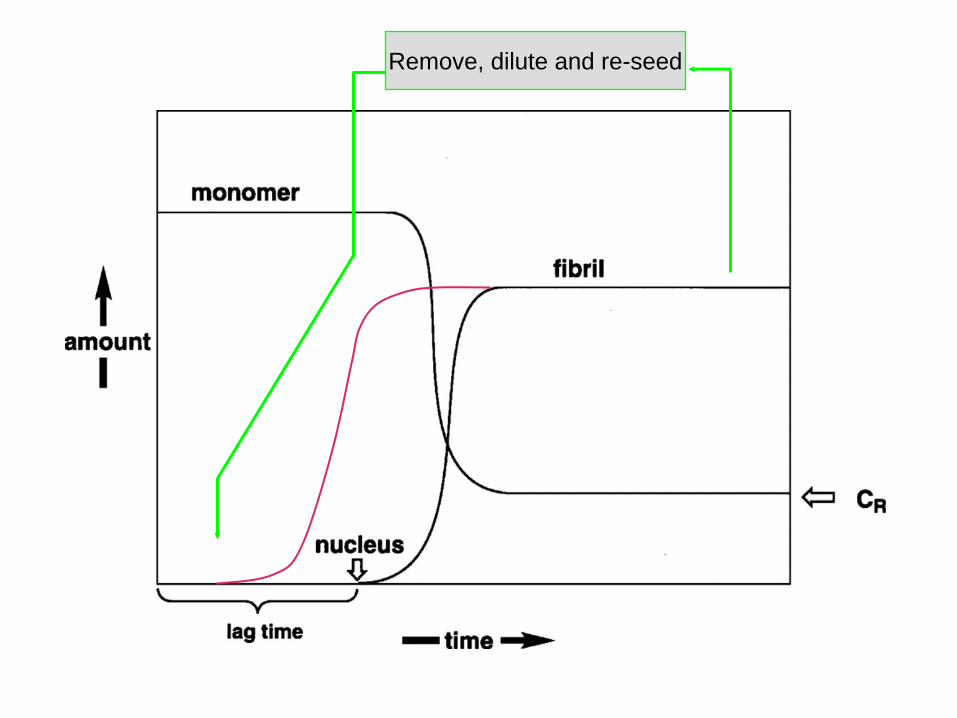
Remove, dilute and re-seed
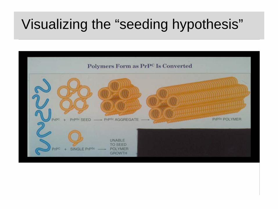
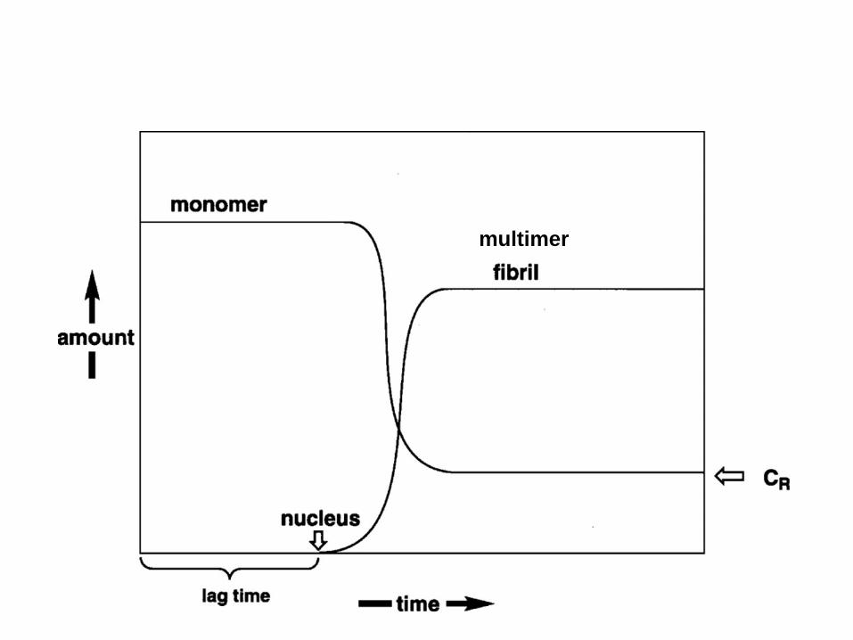
Visualizing the “seeding hypothesis”
Facets of the “Seeding” hypothesis
• There is only a small energy barrier between PrPC
and PrPSc but spontaneous conversion is prevented by a kinetic barrier: conversion is too slow.
• Once a pre-formed seed of PrPSc multimers is made the long lag period is avoided and PrPC to PrPSc
conversion takes place rapidly of the surface of the multimeric PrPSc.
• As multimers get bigger they fragment and thus can create multiple “new” seeds.
• Replication intermediates are big.
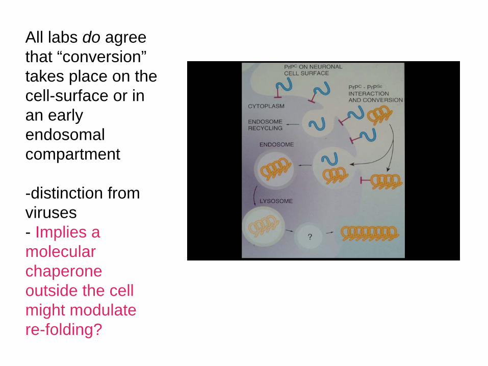
All labs do agree that “conversion” takes place on the cell-surface or in an early endosomal compartment
-distinction from viruses- Implies a molecular chaperone outside the cell might modulate re-folding?
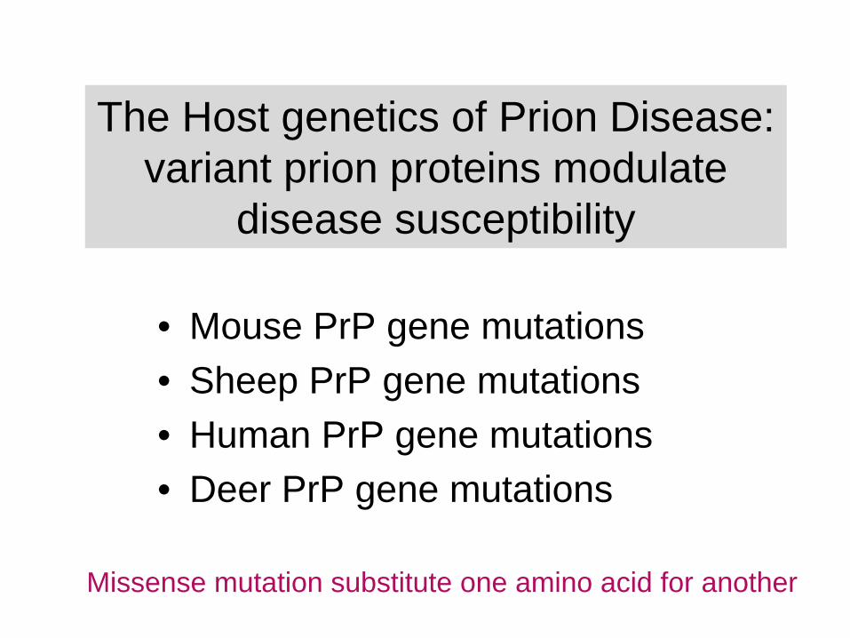
The Host genetics of Prion Disease: variant prion proteins modulate
disease susceptibility
• Mouse PrP gene mutations• Sheep PrP gene mutations• Human PrP gene mutations• Deer PrP gene mutations
Missense mutation substitute one amino acid for another

Methionine (M)129
Valine (V)129
M M M V V V
38% 51% 11%
“Net” genotype for polymorphism affects outcome of infectious, sporadic or familial prion diseases
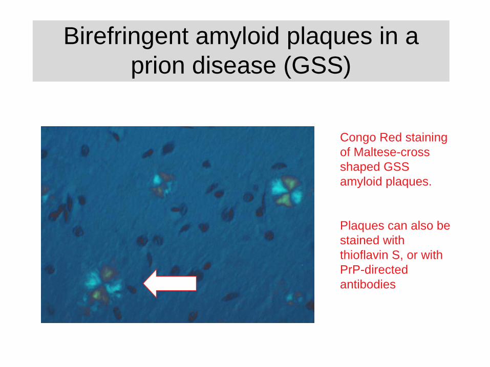
Birefringent amyloid plaques in a prion disease (GSS)
Congo Red staining of Maltese-cross shaped GSS amyloid plaques.
Plaques can also be stained with thioflavin S, or with PrP-directed antibodies
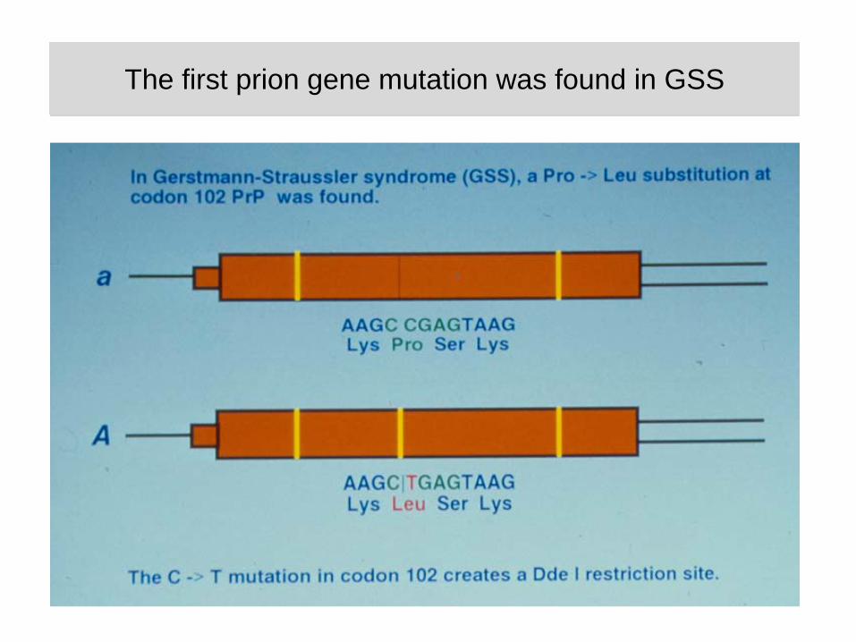
The first prion gene mutation was found in GSSThe first prion gene mutation was found in GSS
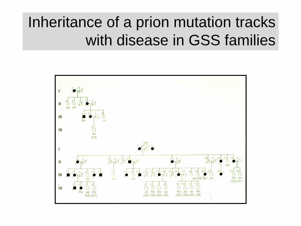
Inheritance of a prion mutation tracks with disease in GSS families
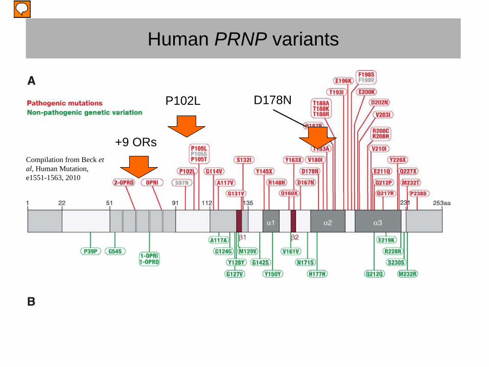
Human PRNP variants
Compilation from Beck et al, Human Mutation, e1551-1563, 2010
+9 ORs
P102L D178N
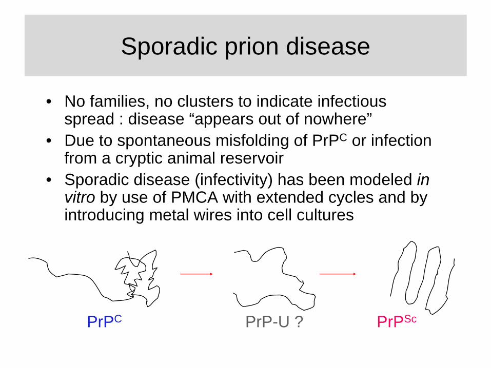
Sporadic prion disease
• No families, no clusters to indicate infectious spread : disease “appears out of nowhere”
• Due to spontaneous misfolding of PrPC or infection from a cryptic animal reservoir
• Sporadic disease (infectivity) has been modeled in vitro by use of PMCA with extended cycles and by introducing metal wires into cell cultures
PrPC PrP-U ? PrPSc
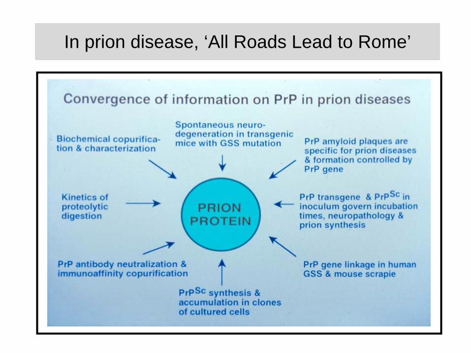
In prion disease, ‘All Roads Lead to Rome’In prion disease, ‘All Roads Lead to Rome’
Some reading
• Prion Biology and Diseases (second edition): Ed, Prusiner S.B. Cold Spring Harbor Laboratory press, Cold Spring Harbor New York, 2004
• The prion's elusive reason for being. Aguzzi A, Baumann F, Bremer J. Annu Rev Neurosci. 2008;31:439-77.
• Discovering DNA encodes Heredity and Prions are Infectious Proteins. Prusiner SB, McCarty M. Annu Rev Genet. 2006;40:25-45.
• A general model of prion strains and their pathogenicity. Collinge J, Clarke AR. Science. 2007 Nov 9;318(5852):930-6.
Assignment - What is going on in the “silent” and symptomatic phases of prion disease ?
• Which events might be important insofar as they might be reliable disease markers?
• Which events might be important insofar as they might be disease targets for small molecule therapy ?
General thoughts
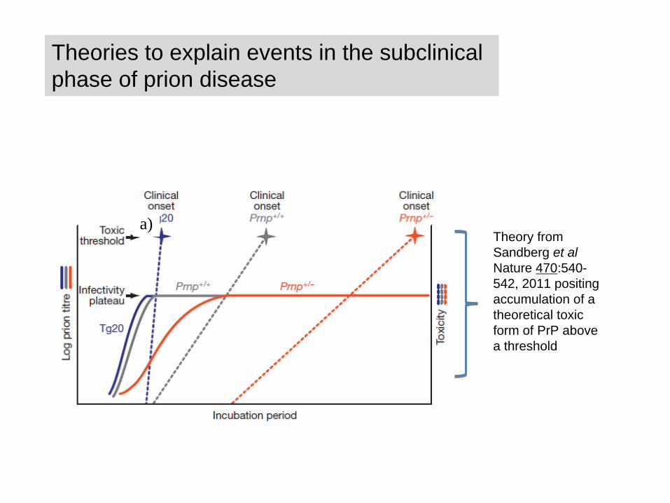
Theories to explain events in the subclinical phase of prion disease
Theory from Sandberg et al Nature 470:540- 542, 2011 positing accumulation of a theoretical toxic form of PrP above a threshold
a)
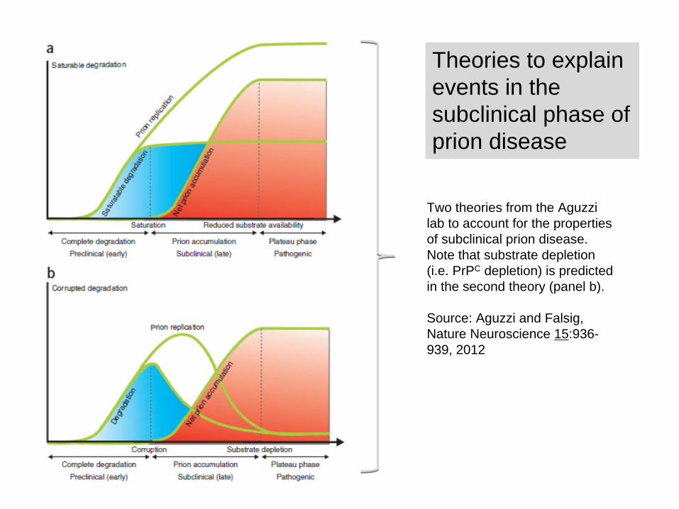
Theories to explain events in the subclinical phase of prion disease
Two theories from the Aguzzi lab to account for the properties of subclinical prion disease. Note that substrate depletion (i.e. PrPC depletion) is predicted in the second theory (panel b).
Source: Aguzzi and Falsig, Nature Neuroscience 15:936- 939, 2012
Reading Assignments
• Sustained translational repression by eIF2a-P mediates prion neurodegeneration
Moreno et al, Nature 485 (7399): 507-511, 2012
• Disease-associated prion protein oligomers inhibit the 26S proteasome.
Kristiansen et al, Molecular Cell 26 (2): 175-188, 2007
Project assignment, group 1
• Critically appraise the paper from the Mallucci group and present a 20 min Powerpoint show to illustrate your critique.
• Also read the paper from the Tabrizi group and the background “review” papers from Collinge and Aguzzi
• At the end of the slide show provide an opinion as to which mechanism (proteasome or unfolded protein response) might be more important and why, or, if they are the about the same, why are they equally important? (1 slide)
• Do the two papers “cover the whole waterfront”, or are there significant gaps where the research might yet advance? (1 slide)
Project assignment, group 2
• Critically appraise the paper from the Tabrizi group and present a 20 min Powerpoint show to illustrate your critique.
• Also read the paper from the Mallucci group and the background “review” papers from Collinge and Aguzzi
• At the end of the slide show provide an opinion as to which mechanism (proteasome or unfolded protein response) might be more important and why, or, if they are the about the same, why are they equally important? (1 slide)
• Do the two papers “cover the whole waterfront”, or are there significant gaps where the research might yet advance? (1 slide)