pärm masteruppsatser iko 2012 - uit
TRANSCRIPT

MASTER THESIS
A literature review on the diagnosis and non-invasive
treatment of small caries lesions
Bo Wold Nilsen and Marius Lund Olsen
Advisor: Catarina Wallman
UNIVERSITETET I TROMSØ

1
Abstract
Diagnosis and treatment of the caries disease and post-treatment follow-up are interconnected
concepts. The aim of the study was to map out the diagnostic methods available for early
caries lesions, the existing non-invasive caries treatment, and in what way the follow-ups of
these treatments are conducted.
The selection of articles was based on a primary search using the PubMed search engine,
including search words associated with the topic. The papers which the primary search
yielded were put through several exclusion rounds to limit the number and heighten the
relevance of the studies to be included. 13 studies were ultimately chosen: 10 clinical studies,
2 systematic reviews and 1 literature review.
The results showed that most of the major diagnostics methods used today, when combined,
have high sensitivity and specificity for identifying dentin caries. However, they do not have
as high specificity and sensitivity for identifying enamel caries.
The treatment modalities examined in this study showed promising results, but most of the
treatments investigated are in need of more (gap filling) research and long-term follow-ups.
The visual tactile method is still the clinician’s most valuable diagnostic tool for identifying
caries in general.
Introduction
Dental caries, herby referred to as caries, is one of the most widespread infectious diseases in
the world1. The caries process causes a destruction of tooth substance. The hard tissues of the
tooth, which includes the enamel, dentine and cementum, will slowly break down due to the
acidic metabolic products produced by specific bacteria in the dental biofilm when

2
metabolizing carbohydrates. The disease manifests itself in several different ways: sub
clinical lesions; sub surface, initial lesions; and manifest caries in cases of a cavity formation.
Depending on the type of the lesion, a suitable treatment should be chosen2.
The cause of caries is multi-factorial, but caries cannot develop in the absence of fermentable
carbohydrates. A causal approach to caries treatment, by risk assessment including diet and
oral hygiene alteration using education and empowerment, could yield a true reduction of the
individual caries prevalence. Yet, vast amounts of resources are spent on symptomatic
treatment of caries. Traditional caries treatment consists of surgical removal of infected
enamel and dentin. The lost tooth-substance is replaced with a biomaterial. In the process of
removing infected tissue, healthy tooth-substance will be lost. Further loss of substance can,
in the lifetime of a tooth or a filling, be expected due to micro leakage, fracture, and/or poor
anatomy and secondary caries. A replacement of the filling will in these cases be necessary3.
In recent years, non-invasive caries treatments have gotten more attention from the scientific
community. This seems to be a result of the drawbacks and limitations of traditional caries
treatment, the advancements in technology and knowledge, and finally, a lower prevalence of
manifest caries lesions.
Today, a spectre of non-invasive caries treatments is available. Many are new, but some have
been available, and tested, for quite some time. Among the non-invasive caries treatments,
there is a large difference in time consumption, costs, proven clinical effect, operator
sensitivity, equipment requirements and the acceptance of the different non-invasive
treatments.

3
In general non-invasive caries treatments are mostly suitable for small carious lesions4.
Cavitation is, in most cases, a contra-indication for a non-invasive treatment. In regard to this,
is it possible to diagnose non-cavitated caries lesions with a high enough sensitivity and
specificity? The visual-tactile examination can assess cavitation of smooth-surfaces, but has
limitations in the diagnosis of caries on proximal surfaces. Radiographs are superior to the
visual-tactile examination in detection of proximal caries, and have therefore been the
diagnostic supplement of choice in the 20th century. There are however limitations and this is
especially true for the diagnosis of non-cavitated caries lesions2. Several new diagnostic tools
have therefore been developed for the improved diagnosis of caries e.g. DIAGNOdent and
FOTI. Can these tools give a precise diagnosis of non-cavitated caries lesions?
When a treatment is performed, there should be a follow-up to assess the outcome. In the case
of fillings, a visual-tactile examination and radiographs are used to check for secondary
and/or residual caries. In case of non-invasive caries treatments, follow-ups should assess the
outcome of these treatments. Has there been a progression of the lesions? Are the diagnosed
caries lesions in an arrested or still in an active phase? It has become apparent that it is very
challenging for the individual practitioner to differentiate between active and non-active
lesions. This is especially true for non-cavitated lesions5. Is it safe to implement a whole array
of new treatment modalities of initial caries lesions, if the means of evaluating the outcome
are questionable or unsatisfactory?
Aim of master thesis and hypothesis
In this literature review different diagnostic tools which are available for caries diagnosis will
be presented. The role of diagnosis in non-invasive caries treatment will be assessed.

4
In addition, non-invasive chairside caries treatments will be reviewed in regards to:
- The treatment: which treatments exist, and are there any scientific evidences for their
efficacy?
- The follow-up: how is the follow-up performed in regards to these treatments?
Hypothesis: Clinical effect, demands for a correct diagnosis and follow-up, varies between
different non-invasive caries treatments.
Material and methods
This paper will be a literature review focused on non-invasive chairside caries treatments,
diagnostic tools, and follow-ups. The diagnostic tools which were selected for this paper were
chosen based on availability and the simplicity of the diagnostic method, as these attributes
are essential for the methods to be useful in a clinical setting. The diagnostic methods which
will be reviewed are:
- Visual-tactile examination
- Dental radiography
- Laser-diagnostics (DIAGNOdent)
- Fiber-Optic Trans-Illumination
The treatments which will be reviewed are:
- Remineralisation products:
o Fluoride products
o Calcium-phosphate remineralisation products.
- Resin based products
o Fissure sealant

5
o Resin sealing
o Resin infiltration
- Other types of treatment
o Ozone-treatment (healozone)
Studies to be considered for inclusion in this literature review were obtained by searching
PubMed, a free database accessing the MEDLINE database. Manual searches were
conducted on PubMeds on-site search engine. No limits were applied to the first search. The
aim of the search was to identify all papers which mentioned the diagnostic methods,
treatment modalities and their follow-up.
It soon became evident that the nomenclature concerning early caries lesions are not
standardized in the scientific literature. Early caries lesion, initial caries lesion, incipient
caries lesion and non-cavitated caries lesion, are all different terms to describe the same
diagnosis. The different search phrases attempted were all listed for future reference I. The
searches included the most common terms used to describe non-cavitated caries lesions. In
total 93 search phrases were used.
The first literature search, using PubMed and the selected search phrases, yielded 6549
papers. 1610 papers on diagnosis, 4512 papers on treatment and 427 papers on follow-upI.
Not all of the search phrases gave results related to dentistry, especially the searches
concerning diagnostics. Search limits were then applied to all the 93 search phrases, by using
the advanced search engine available at PubMed. The limits included were the following:
- Humans
- Clinical trails

6
- Meta-analysis
- RCT
- Review
- All adults: 19+ years
By applying these limits, the number of papers was reduced to 508. 116 papers concerned
diagnostics, 314 concerned treatment and 78 concerned follow-up.
Exclusion criterias were designed in advance of the selection process.
These were as follows:
- Non-chairside treatment
- Medical compromised patients
The list of papers the search process yielded was then manually assessed of relevance by
considering their titles. This was done due to the vast amount of results in spite of the search
limits. In total, 148 papers were found to be relevant based on their titles, but the actual
number was lower due to the fact that some papers appeared several times using different
search phrases.
In the further process of identifying papers to be included in the result section, both authors
read the abstracts and considered their relevance and quality. In cases of disagreement
between the authors in this process, discussions leading to consensus were conducted.

7
The studies which were left after the process mentioned above, were categorized according to
whether the study concerned diagnosis, treatment or follow-up. The list of studies was then
reviewed by a third-party (C.W.) to assess quality and relevance.
Table 1 was made based on the abstracts of the remaining 36 papers II, describing study type,
age range, type of caries, and diagnostic/treatment modalities used in the papers. The table
was used to assess which studies were most suitable for this review. A priority list was made;
if there were studies describing the same subject, the paper highest on the priority list would
be used:
- In vivo > In situ > In vitro
- Meta-analysis > RCT > clinical study (/w control) > clinical study (unspecified)>
review
- Non-cavitated caries lesions > root caries
- Follow-up (more time > less time) > no follow-up
- Published after 2000 > older studies
Based on these priorities, 13 papers were selected to be included in the result section.
Results
Diagnostics of non-cavitated caries lesions
Diagnosis of caries was up until recently concerned with the macroscopic detection of caries.
This was due to a higher prevalence of manifest caries worldwide and the lack of treatment
options other than surgical removal of affected tooth-substance. In modern dentistry, non-

8
invasive treatments of early caries lesions exist, thus making the diagnosis of such lesions
important2.
Non-invasive treatments have a small treatment window in regard to diagnosis; Cavity
formation is in most cases a contraindication for non-invasive treatment. This is a problem, as
the state of carious lesions often is underestimated2.
Detection of caries has changed during the last 30 years, partly due to increased use of
fluoride in different vehicle products. This is especially true for the diagnosis of occlusal
caries. Fluoride inhibits the breakdown of tooth substance. This makes it possible to have an
intact tooth surface, even though it can be completely undermined by caries. This kind of
occlusal caries lesion is most commonly not diagnosed by the visual tactile method, and is
often only discovered in late stages of the disease6.
The visual-tactile examination
The visual-tactile method is the oldest and most commonly used diagnostic method for the
detection of caries. The modern systematic approach to this method is: dry teeth, enough
operating light, assessment of the gingiva, and a careful use of the probe.
The visual-tactile examination is a cheap and available method due to the low equipment
demands. The method has however several limitations: it is difficult to learn and years of
experience are needed to achieve a reliable sensitivity and specificity, which can vary
considerably among clinicians7.
Proximal caries has in particular proven to be very difficult to diagnose using the visual-
tactile method, as 83 % of cavitated proximal caries lesions were not detected using this
method alone7. In regards to this, the site-specific gingival scores seem to correlate with the

9
presence of cavitated proximal lesions. This might be due to the fact that cavitated lesions
retain plaque and food debris, which can cause inflammation of the gingiva. Gingival
inflammation could thus be used as an additional diagnostic indicator for the presence or
absence of cavitation7.
Caries diagnosis based on a visual-tactile examination alone is difficult, and misclassifications
occur to a greater extent among non-cavitated lesions than cavitated ones5.
Concerning occlusal caries, it seems that the probe is not a reliable method for the detection
of caries. Even worse, the probe may sometimes actually cause cavities in the search for
caries5. The probe is, very carefully used, still a suitable tool for the assessment of root caries,
as the texture of the root surface is the main diagnostic criteria for caries, not the color8.
Dental radiography
Dental radiography is electromagnetic radiation, which has wavelengths between 0.01 to 10
nanometers. It is a useful diagnostic method which utilizes the fact that the absorption of the
x-rays differs depending on what type of tissue they penetrate. The difference can be
visualized by the use of different kind of detectors, analog, as well as digital. X-ray is
especially useful for hard tissue diagnostics. Unlike the visual-tactile method, X-rays are
potentially harmful, and should only be used when indicated.
In the context of caries detection, X-ray is a useful tool, but should not be used as the sole
diagnostic tool. This is especially true for the detection of occlusal caries, which is the most
prevalent form of caries in the population9. Proximal dentin lesions are, however, more easily
detected with X-ray compared to the visual-tactile method. Combining the visual-tactile
method with bitewing radiographs seems to yield higher sensitivity and specificity, compared
to either method alone5.

10
Caries lesions situated only in the enamel may not be apparent on radiographs until there is a
loss of mineral of 30 – 40 %. Therefore, the extent of a lesion is often greater than what is
apparent or visible on the radiographs. X-ray is therefore more useful to detect caries lesions
in the dentin compared to the enamel2. This is unfortunate, since one study has shown that in
85 % of cases where an outer dentin lesion can be seen on the radiograph, there is a
macroscopic cavitation7.
The diagnostic values of radiography are dependent on correct handling of the equipment and
the X-rays. Correct angulation of the X-ray tube is necessary to get a correct projection; an
axial-eccentric angulation of only 10o is sufficient to impair diagnosis of cervical root caries.
Phenomena like cervical burn-outs and Mach-band effect will also affect the caries
diagnostics. The Mach-band effect is an optical illusion, which causes an increased perceived
contrast between two surfaces with different luminance. In the clinic this might affect caries
diagnostics, by increasing the number of false-positive results. Correct processing of the
images and viewing conditions are essential for good diagnostics5.
Laser diagnostics
Dental laser diagnostic is a non-ionizing diagnostic method based on the measurement of the
fluorescence of the enamel and dentin. Demineralized enamel has different optical attributes
compared to the healthy enamel. Demineralized enamel can visually be seen as a white spot
lesion. The optical change is caused by increased pore volume in the demineralized enamel.
This change of the optical values can be quantified by a laser fluorescence device and
theoretically it can be used to identify early caries lesions.
An example of such a device is the KaVo DIAGNOdent. It is a small chairside battery-
powered laser fluorescence device. It is used by scanning the area of interest and noting the

11
peak value that the device shows. The value can then be interpreted to decide whether
treatment is needed. The values will vary depending on age, tooth color, staining, and location
of lesions. The recommended cut-off value that yields the highest sensitivity and specificity
can be found in scientific literature2.
The KaVo DIAGNOdent method shows a very high sensitivity for low cut-off values. Cut-off
values are the baseline for which higher values will be interpreted as the presence of caries
lesions. Unfortunately the specificity is poor for low values6. This may be caused by calculus,
fillings and/or stain that will give an artificial high value, which can be interpreted as caries.
Studies do however report excellent intra-examinator reliability and good to excellent inter-
examinator reliability by using the KaVo DIAGNOdent, i.e. getting a correct value does not
seem to be affected by experience. However, the type of carious lesions (proximal/occlusal)
and different kinds of tooth surfaces will also affect the values4.
KaVo DIAGNOdent is a viable tool for the diagnosis of occlusal dentin caries, and studies
even suggest that the KaVo DIAGNOdent combined with the visual-tactile method is a
superior diagnostic option for the diagnosis of fissure caries, compared to the visual-tactile
method supplemented with radiographs5. The KaVo DIAGNOdent is also a useful diagnostic
tool, in combination with the visual-tactile method, for detection of proximal lesions.
Accurate results have however been difficult to obtain in cases of tight contact points, due to
the large tip of the tool. A smaller tip is suggested to help aid better diagnostics2 .
When it comes to the detection of early caries lesions, the reports are more mixed. According
to one study2, the KaVo DIAGNOdent could differentiate between caries up to the EDJ and
dentin caries in proximal lesions by a sensitivity of 0,6 and a specificity of 0,84 given a cut-
off value of 16. This implies that there is no significant difference between dental radiographs

12
and the KaVo DIAGNOdent when it comes to the diagnosis of early proximal caries lesions
in permanent teeth2.
Fiber-optic trans illumination
Fiber-optic trans illumination (FOTI) is a diagnostic tool for the detection of caries, which
utilizes the fact that caries can change the optical properties of the tooth substance. These
optical changes can be seen as shadows, which can be interpreted as caries. No studies solely
concerned with FOTI were found, but a paper evaluating FOTI was mentioned by Huth KC et
al. (2010). In this study, FOTI was described as the least reliable method for caries detection
considered in that specific paper. Elective temporary tooth separation detected 41.4 % more
lesions compared to FOTI, and revealed that 17.5 % of the lesions diagnosed by FOTI were
actually sound enamel when clinically assessed10.
Non-operative treatment in general
Non-cavitated lesions even into the first third and middle part of the dentine can be arrested,
both in the case of occlusal as well as proximal lesions11. A non-surgical treatment approach
will possibly decrease further development of a caries lesion and thus surgical intervention
can be either postponed or unnecessary3. There are two major non-surgical approaches. The
caries lesion can either be given a remineralizing treatment or, it can be arrested by the
application of a resin based solution.
The clinically visible tooth substance is in a constant alternating state of demineralization and
remineralization. Remineralization products, mainly fluoride containing vehicles, have been
manufactured in order to treat non-cavitated lesions before surgical intervention is indicated
by disrupting the imbalance between demineralization and remineralization. The caries

13
development will thereby be slowed down or arrested. These products include both
professionally applied substances and products meant for home treatment. Home treatment
products include dentifrices, rinses, chewing gums and lozenges.
Remineralizing treatment of non-cavitated carious lesions is today being acknowledged by the
profession as an appropriate interventional method being able of arresting lesions and thereby
preventing surgical intervention.
Other treatment options are for example resin based solutions, which can be used in order to
seal early non-cavitated lesions from the oral cavity environment, or to infiltrate these lesions,
resulting in an impediment of further progression of caries.12
In addition there are non-invasive treatment methods other than remineralizing or resin based
ones. Ozone treatment is one of these which will be discussed later in this paper.
Remineralization products
Fluorides
Fluoride has a well proven caries preventive and cariostatic effect. The preventive effect is
through inhibition of the demineralization, and the cariostatic action is through enhancement
of remineralization processes, the latter being the stronger effect5. Fluoride used in the clinic
is delivered to the tooth surface by site specific administration. It has also been added to a
whole array of different products in order to aid the remineralization of tooth substance and
for the prevention of both caries lesion formation and progression. These products range from
home-treatment products like toothpaste, chewing gum, lozenges and rinsing solutions, to
products such as varnishes, professionally applied, and fluoride added to drinking water by
the public health systems.

14
There are only a few clinical studies on fluoride treatment of adults regarding its effect on
primary crown caries5.The same source claims that studies can be biased because the
widespread use of fluorides in the population gives rise to non-effective control groups. In
addition, older studies operated with a DMFT-index not taking into account non-cavitated
lesions. This could cause divergent results regarding the evidence of the efficacy of fluoride
treatment, and thus give an underestimation of caries in both experimental and control
groups5.
There are only a low number of studies on the treatment of non-cavitated lesions in adults1.
Only seven studies were found on this topic, all of which concerned permanent teeth in
children. And, most of the studies were on treatments other than those used in-clinic or a
combination of professionally applied materials and home products. The conclusion was that
the evidence for efficacy of any given method when it comes to arresting or reversing the
progression of non-cavitated lesions was insufficient due to limitations in the evidence base.
Among the limitations mentioned were too few studies, low evidence levels and disagreement
on the diagnostic criteria for non-cavitated lesions. Recommendations for improvement
included further studies with higher quality and evidence level through a gap-filling strategy,
and a consensus regarding diagnostic criteria1.
Comparing Colgate Palmolive DuraphatTM 2, 23 % fluoride varnish with proximal sealing
using a fissure sealant has shown no significant difference in effect. The number of treated
surfaces without progression in the two experimental groups on a 2 year follow-up was almost
identical3.
In one study a Colgate Palmolive DuraphatTM 2, 23 % fluoride varnish group and an 8 %-
stannous fluoride solution (SnF2) group were assessed regarding their ability to arrest root
caries lesions. No obvious differences regarding the efficacy were found between the two

15
groups on the 3, 6, 12 and 18 months follow-up. It was believed that a small number of
subjects could be the reason for this. In this study it was concluded that frequent topical
application of fluoride might be a successful treatment for incipient root caries lesions,
irrespective of the type of fluoride treatment used13. Higher concentrations of fluoride seem to
be needed in order to arrest a root caries lesion compared to an enamel lesion5.
Calcium-phosphate remineralisation products
This is a rather new way of remineralizing tooth surfaces by keeping high levels of calcium
and phosphorus ions in the proximity of the enamel. A chemical gradient favoring net
remineralization of the demineralized tooth substance is created5. For the formation and
incorporation of fluoride apatite to take place, sufficient levels of calcium and phosphate ions
are needed in the saliva and dental plaque, the levels of which will be a limiting factor for the
remineralization. However calcium and phosphorus ions form complexes of low solubility,
which will give a lower degree of remineralization. Hence products were developed to solve
this problem5.
One of the solutions to this problem is to stabilize the complexes through casein
phosphopeptides that bind the calcium and phosphate ions forming CPP-ACP-complexes
(casein phosphopeptide stabilized amorphous calcium phosphate) which release calcium and
phosphorus ions at pH values below 75. The mechanisms of this anticariogenicity have been
thought to be caused by the ACP acting as a buffer on the actions of free calcium and
phosphate, thereby promoting remineralization14. Casein has also been shown to have an
anticariogenic capacity in itself through its ability of being incorporated into plaque thus
preventing subsurface demineralization of the enamel15. The CPP-ACP technology has been
used both in professionally applicated products and products meant for home use.

16
Through the search for calcium-phosphate remineralization products only one single chair
side study was found. It concluded that further evidence is needed in order to decide if the use
of such substances has a clinical effect on carious lesions5.
Resin based products
Fissure sealant
Fissure sealing is a treatment modality for non-cavitated occlusal lesions used on erupting
teeth in young patients and adolescents. Fissure sealants create a mechanical obstacle to
prevent undisturbed plaque formation in the occlusal fissures. The occlusal tooth surfaces are
the most susceptible sites, probably due to their anatomical shapes, which make plaque
removal through tooth-brushing more difficult and besides that, occlusal lesions extending
into dentin are difficult to diagnose16.
Both clinical and radiographic progression of lesions has been confirmed to be significantly
lower in fissure sealed teeth compared to non-sealed teeth. Clinical alterations and an increase
in radiolucency were found in almost 50% of the cases for non-sealed teeth, while clinical
alterations were found in 0% of the sealed teeth on the one year follow-up11. All subjects in
this particular study presented with a moderate to high risk of developing caries. Their age
was not specified other than that the treatment of some of the subjects needed parental
consent. An increase in the occlusal radiolucency was found in about one out of ten subjects
with sealed teeth. In all the lesions showing radiographic progression in the experiment group,
the sealing had been partially or totally lost during the study. The one year retention level of
the sealings with total retention was said to reflect those of similar studies. The importance of
the use of rubber dam was stressed as being of fundamental importance for the longevity of a
fissure sealing. Fissure sealing was found to be an excellent treatment modality for non-

17
cavitated occlusal lesions, and to significantly arrest lesions. It may replace the surgical
treatment for these lesions11.
Resin sealing
Proximal sealing is the use of resin based light curing material for sealing an approximal tooth
surface in case of a non-cavitated lesion in order to to prevent further progression. Bonding is
often used as the material of choice in this kind of treatment. Resin tags of up to 6µm into the
enamel have been demonstrated17.
The method of fissure sealing has proved to be a successful and an effective way for treating
non-cavitated lesions in several clinical studies3 and this principle has also been tested for
proximal surfaces.
The potential of sealants to non-invasively treat early proximal lesions has also been tested.
Ninety-three percent of treated surfaces showed no progression of the baseline radiolucency
in the enamel on a 2 year follow-up3. Only 1.3 % of the lesions progressed to a state which
indicated the need for a restoration. However, no significant difference was found in the effect
of this treatment compared with the control group which received Colgate Palmolive
DuraphatTM 5 % sodium fluoride varnish applications3.
Resin infiltration
Resin infiltration is a new way of treating non-cavitated lesions in which the goal is to fill a
sub surface lesion with low-viscosity resins. Demineralized non-cavitated lesions have
widened enamel pores where resins can infiltrate and subsequently be hardened by the use of
light curing5. It is claimed that this technique will block the diffusion pathways for cariogenic
acids. The method is somewhat in the middle of a non-operative and operative treatment, and

18
might be able to fill this gap, thus avoiding the need of a restoration. The difference between
resin infiltration and the use of a fissure sealant is the formation of a barrier inside the hard
tissue of the tooth and not on the surface. Infiltrated lesions cannot be distinguished
radiographically from non-infiltrated lesions due to the fact that the infiltrants are not
radiopaque 12.
A clinical study assessing the efficacy of resin infiltration in case of a lesion radiographically
extending into the inner half of enamel or the outer third of dentin, has shown that the use of
resin infiltration can stop the lesion progression in 93% of the cases still after 18 months.
Thirty-seven percent of the lesions in the placebo control group showed a progression. This
difference was significant12. Digital subtraction radiography was used when receiving these
values. This is a method in which two radiographs can be digitally superimposed so that a
follow-up image can be subtracted from the baseline image to monitor eventual progression
of a carious lesion. 18 When baseline and follow-up radiographs were assessed manually and
pairwise, there was a borderline significance between the experimental and the placebo
groups. No unwanted side effects were observed during the 18 months, which indicates that
the method is safe to use. Resin infiltration was regarded as being a clinically feasible and
efficacious way of reducing the progression of interproximal lesions. The method was said to
be able to serve as an alternative to the non-operative and operative treatments when
indicated12.
Ozone treatment
In this method ozone gas is directed towards a caries lesion and the surrounding tooth surface.
Ozone has a well-documented antibacterial effect and is claimed to be able to stop the

19
microorganisms responsible for the development of caries, thus arresting a lesion or slowing
down its progression.
However, the effect in the clinical setting has been questioned. There is no conclusive
evidence proving that the degree of effect seen in vitro can be transferred to in vivo
conditions19.
It has been concluded through an in vitro study that ozone treatment may be effective in the
treatment of root caries lesions20. More clinical studies with high evidence levels are however
needed20. A combined treatment of remineralizing products and ozone has been shown to be
able to arrest non-cavitated primary root caries21.
The general quality of studies regarding KaVo HealOzone has been judged to be modest8. The
reasons for this were that many important methodological aspects were not reported. The
conclusion was, that not enough evidence is present from the published RCTs, and therefore
an evaluation of the effectiveness becomes difficult. A conclusion was also made that there is
not enough evidence present to decide whether HealOzone treatment is a cost-effective
additive to the management of occlusal and root caries. Further research based on RCTs is
needed in order to prove this8.
Discussion
Our results uncovered that our method and material section may have some flaws. The
scientific nomenclatures which are used concerning caries lesions are not standardized. This
implies that even though we had this in mind when we attempted our searches, some search
words may have been left out.

20
The list of search words used to find relevant papers might also be missing some words to
completely map out all the diagnostic methods and treatment modalities available. Search
words regarding electrical caries detection and elective tooth separation were for example not
included, although they eventually should have been. In reality, this means that this literature
review does not include every single treatment and diagnostic method, even though a major
part of them are included.
It also seems as if the limits which were applied did not filter out all the studies it should have
filtered out, such as studies concerning children, even though one of the limits applied was the
age of above 18. It is unfortunate that some of these studies even made it through the manual
filtration, as the abstract was the only thing taken into consideration when the decision about
inclusion of papers were made. The assessment of which studies to include in this study might
also be faulty, as the authors have little experience judging the quality of scientific papers.
Concerning the results, it is interesting to note that no diagnostic modality alone seems to be
sufficient when attempting a caries diagnose. This is especially true for the diagnosis of early
caries lesions. None of the diagnostic methods described in this review seem to be 100 %
accurate in diagnosing early caries lesions. This gives rise to problems because the prognosis
of non-invasive treatment seems to depend on a correct diagnosis.
It was surprising that none of the papers concerning the use of radiographs dealt with the use
of digital subtraction radiography as a diagnostic tool for early detection of caries lesions, as it
can be used for both proximal and occlusal lesions22. However, radiographs with nearly
identical projections are needed. This x-ray technique has proved to be a suitable
complementary method of surveying the progression of caries lesions.23 So far, digital
subtraction radiography is not commonly used in clinics. The reasons for this might be that it
is difficult to use, the need for expensive equipment and finally that the method still is not

21
well proven for clinical use. Most of the papers did not mention whether digital or
conventional radiography was used for the diagnosis of caries.
The studies concerned with the use of KaVo DIAGNOdent were abundant. However it can be
questioned if it is such a viable tool as some studies indicate, due to the fact that most studies
have exclusion criteria that remove factors which would negatively influence the diagnostic
results of the KaVo DIAGNOdent, for example fillings, stains, hypoplasia etc. This means,
that the method has mostly been tested on smooth surfaces, which in any case would be easy
to diagnose by the visual tactile method.
Little or no results were found on other diagnostic methods, such as electrical caries detection,
DIFOTI (by Electro-Optical Sciences, Inc.), and QLF (Quantitative Light-induced
Fluorescence by Inspektor Research Systems). This might be due to the design of the material
and method section, but most likely also due to lack of quality studies. This is at least true for
DIFOTI and FOTI, where 13 search words were attempted.
Diagnostic methods/indications which could have been explored further, by being included in
the initial search process, is the association between gingival health and cavitation and
elective tooth separationI. Gingival inflammation could be used as an additional diagnostic
indicator on the presence or absence of cavitation7. Elective tooth separation has shown
promising results10.
Regarding remineralizing treatments, few papers on the topic were found and most of them
did not have placebo control groups. Instead they compared in-chair treatment modalities with
home treatment products or tested a combination of these. In one study it might be that the
fluoride solution used (8 % stannous fluoride) was effective and that the fluoride varnish
(Colgate Palmolive DuraphatTM 2, 23 % fluoride) was not, but this can of course not be

22
clarified because of the particular study model used3. Only one study, a review of different
managements of non-cavitated lesions, compared professionally applied 8 % stannous
fluoride solution with a placebo control group1. In this study the results in the experimental
group showed statistically significant reductions of the number of lesions that progressed.
But, the evidence for efficacy was judged to be low and insufficient for both professionally
applied and home treatment products regarding the management on non-cavitated lesions1.
More studies would be needed in order to increase the evidence base for remineralizing
treatment as a whole. Optimally, the different treatment methods should be compared with
placebo control groups, but due to ethical concerns this would not be achievable. However, it
is worth noticing that a control group not already influenced by fluoride is impossible to
achieve today due to the daily use of fluoride dentifrices and fluoridation of water supplies at
a public health level in many countries. The studies from the reviews concerning fluoride
treatment were mostly conducted on children and adolescents, not adults. This is interesting,
due to the fact that the enamel is not completely mature before the age of mid-twenties. A
more local effect may therefore be expected in adults.
Studies comparing different fluoride treatments used for coronal caries lesions were not found
in the search for papers. Thus the difference in efficacy of these methods could not be
evaluated on the basis of combined therapies. However, as previously described, a clinical
study assessing the different fluoride treatments of initial root caries lesions was included13.
Limitations in evidence base are especially evident concerning the use of casein
phosphopeptides (CPP). No papers reviewing CPP’s effect in clinical trials were found. It
seems that most of the studies on this topic are in situ studies, and more clinical trials are
needed in order to judge the efficacy of treatment of early caries lesions and clinical
relevance.

23
The use of resin sealing and resin infiltration shows very promising results. The fissure
sealing technique has, since its arrival in the 1960’s, proved to be a successful method for
arresting and controlling non-cavitated occlusal lesions, in addition of being an efficient
preventive measure regarding the same kind of lesions. Studies have also shown that
professional cleaning of the tooth substance and ensuring effective moisture control seem to
have a huge impact on the outcome of the treatment11.
A study concerning proximal sealing, not infiltration, was conducted before a proximal resin
infiltration agent was available24. Even though proximal sealing showed good results in
arresting caries lesions the method used in that particular study required two successive visits
within 1-2 days, which seems very time demanding and technique sensitive3. Proximal sealing
might be concerned as a pioneer technique before other methods are available, and hence its
clinical relevance in non-operative treatment can be questioned. However, equal results are
achieved by fluoride varnish treatment in comparative studies with proximal sealing3. In
relation to these results it is a less cost effective treatment in comparison with professionally
applied fluoride.
Resin infiltration using DMG ICON showed very good results compared with a placebo
control group12. The technique uses a method which does not require tooth separation, as the
principle behind the technique used in ICON is a capillary effect. It should however be noted
that the studies concerning ICON were supported by DMG, the producer of ICON. The fact
is, that up to the date when this study was released in 2010, it was the only randomized
control trial on the efficacy of the infiltration technique12. There is a great need for
establishing an evidence base for this novel method through more research and clinical
studies by independent sources judging its relevance and performance.

24
Ozone might be an effective treatment of small carious lesions in combination with other
treatment methods. But, in a systematic review concerning ozone, the cost effectiveness of the
treatment is questioned. The method has proven to be an effective treatment in in-vitro
studies. The low evidence base was highlighted and the reason was that the effectiveness of
ozone treatment could not be evaluated for both root caries and occlusal caries8.
Three out of seven papers concerning treatment, which was included in the result section, did
not present data on follow-ups. Among the four papers which had a follow-up, none had a
follow-up period exceeding 18 monthsII. In regards to new treatment modalities, a follow-up
should be performed to properly assess their long-term efficacy.
In general non-invasive symptomatic caries treatments are mostly suitable for non-cavitated
carious lesions. Cavitation is, in most cases, a contra-indication for a non-invasive treatment.
But all diagnostic tools have difficulties diagnosing caries with a high enough specificity and
sensitivity, especially when it comes to non-cavitated lesions. So far, the new diagnostic tools
must be regarded as being supplemental to the visual tactile method. Even this approach,
which through studies has been shown to be the most accurate, has its limitations when it
comes to seeking out lesions suitable for non-invasive treatment. This justifies the question to
be raised if it is safe to implement a whole array of new treatment modalities of initial caries
if the means of treatment outcome evaluation are questionable or even unsatisfactory.
Non-invasive treatment methods are only symptomatically treating the caries disease. This
means that a pre-treatment evaluation of causal factors, such as diet and oral hygiene, is very
important before attempting any kind of treatment, both operative and non-operative. The use
of a basic fluoride prophylaxis, such as a fluoride dentifrice, might be regarded as a predictor
for successful non-operative treatment; the same might apply for the long term prognosis.

25
Conclusion
Present evidence suggests that there are no diagnostic tools which can be used to precisely
determine if, and when, non-invasive treatment is indicated. The most important basic tool is
still the carefully conducted clinical examination combined with a risk assessment and a
classification of existing carious lesions. Non-invasive treatment shows promising results, but
more gap filling research, with longer and more extensive follow-ups, needs to be conducted
to increase the evidence base.

26
Appendixes
I Appendix 1
II Appendix 2
References
1 Bader JD et al - 2001 - A systematic reviews of selected caries prevention and management methods.- Journal of Dental Education October 1, 2001 vol. 65 no. 10 960-968
2 Huth KC et al – 2010 -In vivo performance of a laser fluorescence device for the approximal detection of caries in permanent molars.- J Dent. 2010 Dec;38(12):1019-26.
3 Gomez SS et al – 2005 - A 2-year clinical evaluation of sealed noncavitated approximal posterior carious lesions in adolescents. - Clinical Oral Investigations Volume 9, Number 4, 239-243
4 Huth KC et al – 2008 - Clinical performance of a new laser fluorescence device for detection of occlusal caries lesions in permanent molars. - J Dent. 2008 Dec;36(12):1033-40. Epub 2008 Oct 18.
5 Rodrigues JA et al – 2000 - Prevention of crown and root caries in adults. - Periodontol 2000. 2011 Feb;55(1):231-49
6 Chu CH et al – 2009 - Clinical diagnosis of fissure caries with conventional and laser-induced fluorescence techniques. - Lasers Med Sci. 2010 May;25(3):355-62.
7 Ratledge DK et al – 2001 - A clinical and microbiological study of approximal carious lesions. Part 1: the
relationship between cavitation, radiographic lesion depth, the site-specific gingival index and the level of
infection of the dentine. - Caries Res. 2001 Jan-Feb;35(1):3-7.
8 Brazzelli M – 2006 - Systematic review of the effectiveness and cost-effectiveness of HealOzone for the
treatment of occlusal pit/fissure caries and root caries. - Health Technol Assess. 2006 May;10(16):iii-iv, ix-80.
9 Ricketts DN et al – 1995 - Operative and microbiological validation of visual, radiographic and electronic
diagnosis of occlusal caries in non-cavitated teeth judged to be in need of operative care. - Br Dent J. 1995
Sep 23;179(6):214-20.
10 C.Deery et al – 2000 – Prevalence of Dental caries in Latvian 11- to 15-year-old children and the
enhanced diagnostic yield of temporary tooth separation, FOTI and electronic caries measurement
(Reference found in Huth KC et al – 2010) - Caries Res. 2000 Jan-Feb;34(1):2-7.

27
11 Borges BC – 2010- Efficacy of a pit and fissure sealant in arresting dentin non-cavitated caries: a 1-year
follow-up, randomized, single-blind, controlled clinical trial. - Am J Dent. 2010 Dec;23(6):311-6
12 Paris S et al – 2010- Resin infiltration of caries lesions: an efficacy randomized trial. - J Dent Res. 2010
Aug;89(8):823-6
13 Fure S et al – 2009 - Evaluation of different fluoride treatments of initial root carious lesions in vivo. -
Oral Health Prev Dent. 2009;7(2):147-54
14 Cochrane NJ, Saranathan S, Cai F, Cross KJ, Reynolds EC. – 2008 Enamel subsurface lesion
remineralization with casein phosphopeptide stabilized solutions of calcium, phosphate and fluoride
Reynolds EC – 1998 Anticariogenic complexes of amorphous calcium phosphate stabilized by casein
phosphopeptides: a review (Reference found in Rodrigues JA et al – 2000) - Caries Res. 2008;42(2):88-97
15 Reynolds EC. – 1987 The prevention of sub-surface demineralization of bovine enamel and change in
plaque composition by casein in an intra-oral model (Reference found in Rodrigues JA et al – 2000) - J
Dent Res. 1987 Jun;66(6):1120-7.
16 Agnes G. et al– 2005 Occlusal caries diagnosis in permanent teeth: An in vitro study. (Reference found in
Borges BC et al – 2010) - Braz Oral Res. 2005 Oct-Dec;19(4):243-8.
17 Gomez SS. et al – 2003, SEM analysis of sealant penetration in proximal incipient non-cavitated caries
lesion (Reference found in Gomez SS et al – 2005) - J.Dent Res 82(spec issue B):256 (Abstr. 1965))
18 Carneiro LS. et al – 2009 In vivo study of pixel grey-measurement in digital subtraction radiography for
monitoring caries remineralization - Dentomaxillofac Radiol. 2009 Feb;38(2):73-8.
19 Azarpazhooh A. et al – 2008 The application of ozone in dentistry: a systematic review of literature
(Reference found in Rodrigues JA et al – 2000) - J Dent. 2008 Feb;36(2):104-16
20 Baysan A. et al – 2000 Antimicrobial effect of a novel ozone-generating device on micro-organisms
associated with primary root caries lesions in vitro; (Reference found in Rodrigues JA et al – 2000) - Caries
Res 2000: 34: 498-501
21 Holmes J. – 2003 - Clinical reversal of root caries using ozone, double-blind, randomised, and controlled 18-month trial - Gerodontology. 2003 Dec;20(2):106-14
22 Neuhaus KW. et al – 2009 Novel lesion detection aids- Monogr Oral Sci. 2009;21:52-62.
28
23 Carneiro LS. et al – 2009 In vivo study of pixel grey-measurement in digital subtraction radiography for
monitoring caries remineralization - Dentomaxillofac Radiol. 2009 Feb;38(2):73-8.
24 Icon – scientific documentation. DMG Dental website. URL: http://www.dmg-
dental.com/downloads/scientific-documentations/

29

Appendix 1 Initial search
Explanation of numbers: The number behind each search paragraph is the number of results that particular search yielded.
“non invasive” AND “early caries”: 2 “non invasive” AND “initial caries”: 0 “non invasive” AND “incipient caries”: 1 “non invasive” AND “small caries lesion”: 2 “Early caries” AND diagnostic: 67 “Initial caries” AND diagnostic: 56 “Non-cavitated” AND diagnostic: 91 “Small caries lesion” AND diagnostic: 71 “Incipient caries” AND treatment: 62 “Early caries” AND treatment: 64 “Initial caries” AND treatment: 71 “Non-cavitated” AND treatment: 63 “Small caries lesion” AND treatment: 40 “incipient caries” AND treatment: 62 “Visual tactile examination”: 15 “Visual tactile” AND diagnostic: 84 “Visual tactile” AND “early caries”: 2 “Visual tactile” AND “incipient caries”: 0 “Visual tactile” AND “initial caries”: 0 Diagnodent: 179 “Laser diagnostics”: 37 “Fiber-Optic Trans-Illumination”: 3 "fiber optic transillumination" AND diagnostic: 22 FOTI AND "diagnostic": 73 FOTI AND “early caries”: 0 FOTI AND “incipient caries”: 0 FOTI AND “non cavitated”: 2 FOTI AND “initial caries”: 0 FOTI AND “small caries lesion”: 0 DIFOTI AND "diagnostic": 3 DIFOTI AND “early caries”: 0 DIFOTI AND “incipient caries”: 0 DIFOTI AND “non cavitated”: 0 DIFOTI AND “initial caries”: 0 DIFOTI AND “small caries lesion”: 0 DIFOTI: 7 "Dental radiography" diagnostic early: 116 "Dental radiography" diagnostic small: 232 “Radiographic examination” AND “early caries”: 1 “Radiographic examination” AND “incipient caries”: 2 “Radiographic examination” AND “initial caries”: 8 “Radiographic examination” AND “non cavitated”: 12 Radiography AND “early caries”: 18 Radiography AND “ initial caries”: 25

Radiography AND ”non cavitated”: 32 Radiography AND ”incipient caries”: 14 Radiography AND “small caries lesion”: quote not found detection AND ”initial caries”: 4 detection AND “early caries”: 31 detection AND “non cavitated”: 36 detection AND “incipient caries”: 10 detection AND “small caries lesion”: quote not found “CPP-ACP” : 96 "CPP-ACP" AND "non cavitated": 3 “amorphous calcium phosphate”: 539 "amorphous calcium phosphate" AND treatment: 99 "calcium phosphate remineralization": 193 “casein phosphopeptide”: 185 "casein phosphopeptide" AND "non cavitated": 2 Fluoride AND “early caries”: 24 fluoride AND "early caries" AND topical: 5 fluoride AND “initial caries”: 46 fluoride AND “non cavitated”: 34 fluoride AND “incipient caries”: 34 fluoride AND remineralization: 934 fluoride AND remineralization AND varnish: 82 “fluoride gel” AND remineralization: 41 “fluoride gel” AND “non cavitated”: 3 "fluoride gel" AND treatment AND caries: 144 Remineralization AND “early caries”: 18 Remineralization AND “initial caries”: 18 remineralization AND "incipient caries": 29 remineralization AND "non cavitated": 17 “resin infiltration”: 98 diagnodent AND diagnostic: 160 diagnodent AND "non cavitated": 18 “laser fluorescence”: 441 “laser fluorescence” AND diagnostic: 282 "laser fluorescence" AND "non cavitated": 14 "laser fluorescence" AND "early caries": 2 "laser fluorescence" AND "incipient caries": 4 "laser fluorescence" AND "initial caries": 5 "laser fluorescence" AND radiography: 54 ozone AND caries: 55 “fissure sealing”: 159 “fissure sealant”: 443 "fissure sealant" AND "non invasive”: 5 "non invasive" AND caries: 78 "non invasive" AND "non cavitated": 6 “Icon infiltration”: 7 "fissure sealing" AND caries: 124 radiograph AND subtraction AND caries: 6

“follow up” AND “remineralisation”: 3 “follow up” AND “remineralisation products”: 0 “follow up” AND “remineralising products”: 1 “follow up” AND “remineralisation treatment”: 2 “follow up” AND “remineralising treatment”: 2 “follow up” AND “remineralizing”: 6 “follow up” AND “remineralising”: 2 “follow up” AND “remineralizing treatment”: 1 “follow up” AND “remineralizing products”: 0 “follow up” AND “fluoride products”: 0 “follow up” AND “fluoride gel”: 33 “follow up” AND “fluoride varnish”: 48 “follow up” AND “fluoride treatment”: 19 “follow up” AND “CPP-ACP”: 4 “follow up” AND “ACP”: 143 “follow up” AND “amorphous calcium phosphate”: 9 “follow up” AND “casein phosphopeptide”: 8 “follow up” AND “resin infiltration”: 2 “follow up” AND “resin infiltrating products”: 0 “follow up” AND “resin infiltration products”: 0 “follow up” AND “resin based products”: 0 “follow up” AND “fissure sealing”: 27 “follow up” AND “fissure sealant”: 80 “follow up” AND “ICON”: 30 “follow up” AND “ICON infiltration”: 0 “follow up” AND “ozone treatment”: 5 “follow up” AND “healozone”: 2
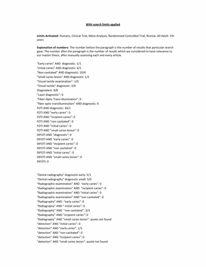
With search limits applied
Limits Activated: Humans, Clinical Trial, Meta-Analysis, Randomized Controlled Trial, Review, All Adult: 19+ years Explanation of numbers: The number before the paragraph is the number of results that particular search gave. The number after the paragraph is the number of results which we considered to have relevance to our master thesis, after manually assessing each and every article. “Early caries” AND diagnostic: 1/1 “Initial caries” AND diagnostic: 6/1 “Non-cavitated” AND diagnostic: 10/4 “Small caries lesion” AND diagnostic: 1/1 “Visual tactile examination”: 1/0 “Visual tactile” diagnostic: 3/0 Diagnodent: 9/8 “Laser diagnostic”: 0 “Fiber-Optic Trans-Illumination”: 0 "fiber optic transillumination" AND diagnostic: 0 FOTI AND diagnostic: 34/1 FOTI AND “early caries”: 0 FOTI AND “incipient caries”: 0 FOTI AND “non cavitated”: 0 FOTI AND “initial caries”: 0 FOTI AND “small caries lesion”: 0 DIFOTI AND "diagnostic": 0 DIFOTI AND “early caries”: 0 DIFOTI AND “incipient caries”: 0 DIFOTI AND “non cavitated”: 0 DIFOTI AND “initial caries”: 0 DIFOTI AND “small caries lesion”: 0 DIFOTI: 0 "Dental radiography" diagnostic early: 5/1 "Dental radiography" diagnostic small: 5/0 “Radiographic examination” AND “early caries”: 0 “Radiographic examination” AND “incipient caries”: 0 “Radiographic examination” AND “initial caries”: 0 “Radiographic examination” AND “non cavitated”: 0 “Radiography” AND “early caries”: 0 “Radiography” AND “ initial caries”: 0 “Radiography” AND ”non cavitated”: 3/3 “Radiography” AND ”incipient caries”: 0 “Radiography” AND “small caries lesion”: quote not found “detection” AND ”initial caries”: 0 “detection” AND “early caries”: 1/1 “detection” AND “non cavitated”: 0 “detection” AND “incipient caries”: 0 “detection” AND “small caries lesion”: quote not found

diagnodent AND diagnostic: 7/4 diagnodent AND "non cavitated": 9/4 “laser fluorescence”: 9/9 “laser fluorescence” AND diagnostic: 8/7 "laser fluorescence" AND "non cavitated": 1/0 "laser fluorescence" AND "early caries": 0 "laser fluorescence" AND "incipient caries": 0/0 "laser fluorescence" AND "initial caries": 1/1 "laser fluorescence" AND radiography: 1/1 Radiograph AND subtraction AND caries: 1/1 “Incipient caries” + treatment: 0 “Early caries” + treatment: 2/1 “Initial caries” + treatment: 6/5 “Non-cavitated” + treatment: 10/4 “Small caries lesion” + treatment: 3/0 “incipient caries” + treatment: 0 “non invasive” AND “early caries”: 0 “non invasive” AND “initial caries”: 0 “non invasive” AND “incipient caries”: 0 “non invasive” AND “small caries lesion”: 0 CPP-ACP: 22/4 “amorphous calcium phosphate”: 20/2 "amorphous calcium phosphate" AND treatment: 20/2 "calcium phosphate remineralization": 21/4 “casein phosphopeptide”: 23/3 "casein phosphopeptide" AND "non cavitated": 0 fluoride AND “early caries”: 1/1 fluoride AND "early caries" AND topical: 1/1 fluoride AND “initial caries”: 5/1(3) fluoride AND “non cavitated”: 7/2 fluoride AND “incipient caries”: 6/6 fluoride AND remineralization: 90/15 fluoride AND remineralization AND varnish: 3/1 “fluoride gel” AND remineralization: 6/2 “fluoride gel” AND “non cavitated”: 2/1 (1) "fluoride gel" AND treatment AND caries: 18/1 Remineralization AND “early caries”: 2/1 Remineralization AND “initial caries”: 3/2 remineralization AND "incipient caries": 1/1 remineralization AND "non cavitated": 4/2 “resin infiltration”: 3/5 ozone AND caries: 4/4 “fissure sealing”: 3/0 “fissure sealant”: 11/5 "fissure sealant" AND "non invasive”: 0 "non invasive" AND caries: 5/2

"non invasive" AND "non cavitated": 0 Icon infiltration: 1/1 "Fissure sealing" AND caries: 1/0 “follow up” AND “remineralisation”: 0 “follow up” AND “remineralisation products”: 0 “follow up” AND “remineralising products”: 1/1 “follow up” AND “remineralisation treatment”: 0 “follow up” AND “remineralising treatment”: 1/1 “follow up” AND “remineralizing”: 2/0 “follow up” AND “remineralising”: 1/1 “follow up” AND “remineralizing treatment”: 0 “follow up” AND “remineralizing products”: 0 “follow up” AND “fluoride products”: 0 “follow up” AND “fluoride gel”: 9/1 (9) “follow up” AND “fluoride varnish”: 7/6 “follow up” AND “fluoride treatment”: 4/2 “follow up” AND “CPP-ACP”: 2/1 (1) “follow up” AND “ACP”: 22/1 (2) “follow up” AND “amorphous calcium phosphate”: 3/1 “follow up” AND “casein phosphopeptide”: 3/2 “follow up” AND “resin infiltration”: 1/0 “follow up” AND “resin infiltrating products”: 0 “follow up” AND “resin infiltration products”: 0 “follow up” AND “resin based products”: “follow up” AND “fissure sealing”: 2/0 “follow up” AND “fissure sealant”: 6/4 “follow up” AND “ICON”: 9/0 “follow up” AND “ICON infiltration”: 0 “follow up” AND “ozone treatment”: 3/0 “follow up” AND “healozone”: 2/0

APPENDIX 2
The papers used in the result section have been written with bold text.
In vitro/ in vivo
Type of study
Age range
Caries type Modality Follow-up Other factors
Diagnosis Heinrich-Weltzien R et al - 2002 Clinical evaluation of visual, radiographic, and laser fluorescence methods for detection of occlusal caries.
In vitro Clinical study 18 years +
Occlusal caries (enamel and dentin)
DIAGNOdent, radiographic, visual - -
Ricketts DN et al – 1995 Operative and microbiological validation of visual, radiographic and electronic diagnosis of occlusal caries in non-cavitated teeth judged to be in need of operative care.
In vivo Clinical study 18 years +
Occlusal caries (non-cavitated)
Visual, radiographic, electronic - -
Winston AE et al – 1998 Caries prevention in the 21st century.
- - 18 years +
- - - -
Ratledge DK et al – 2001 A clinical and microbiological study of approximal carious lesions. Part 1: the relationship between cavitation, radiographic lesion depth, the site-specific gingival index and the level of infection of the dentine.
In vivo Clinical study 18 years +
Approximal caries Visual and radiographic - -
Chu CH et al – 2009 Clinical diagnosis of fissure caries with conventional and laser-induced fluorescence techniques.
In vivo
Clinical study Young adults
Fissure/occlusal caries (enamel/dentin)
Visual, radiographic, DIAGNOdent - -

Huth KC et al – 2008 Clinical performance of a new laser fluorescence device for detection of occlusal caries lesions in permanent molars.
In vivo RCT 18 years +
Occlusal caries (enamel/dentin)
Visual, radiographic, DIAGNOdent 12 month -
Lussi A et al – 2001 Clinical performance of a laser fluorescence device for detection of occlusal caries lesions.
In vivo Clinical study Possible younger patients
Occlusal caries (enamel/dentin)
Visual, radiographic, DIAGNOdent - -
Huth KC et al – 2010 In vivo performance of a laser fluorescence device for the approximal detection of caries in permanent molars.
In vivo RCT 18 years +
Approximal caries (non-cavitated and cavitated)
Visual, radiographic, DIAGNOdent - -
Choksi SK et al – 1994 - Detecting approximal dental caries with transillumination: a clinical evaluation.
In vivo Clinical study 18 years +
Unspecified caries maxillary front
Visual, radiographic, FOTI - -
Treatment Bader JD et al - 2001 A systematic review of selected caries prevention and management methods.
In vivo Systematic review
18+ Alt om non-cavitated er på barn
Non-cavitated caries lesions.
Fluor varnish and other methods (not mentioned in abstract)
- High caries risk
Borges BC – 2010 Efficacy of a pit and fissure sealant in arresting dentin non-cavitated caries: a 1-year follow-up, randomized, single-blind, controlled clinical trial.
In vivo RCT 18 years +
Non-cavitated occlusal dentin caries lesions.
Visual, radiographic, fissure sealant Fluorholdig fissurforsegling
4 months intervals
over 1 year
Moderat to high caries risk
Kugel G – 2009 Treatment modalities for caries management, including a new resin infiltration system.
- Review 18 years +
Non-cavitated caries lesions
Resin infiltration - -
Trairatvorakul C – 2008 Active management of incipient caries and choice of materials.
In vitro
In Vitro study 18 years +
Non-cavitated caries lesions, approximal, posterior teeth
Resin based sealant, F-sealant, F-varnish, GIC
- -

Silva KG – 2010 In situ evaluation of the remineralizing capacity of pit and fissure sealants containing amorphous calcium phosphate and/or fluoride.
In situ In situ study 18 years +
Non-cavitated smooth surface caries lesions
Fissure sealant (with APC) - -
Altenburger MJ et al – 2008 Fluoride uptake and remineralisation of enamel lesions after weekly application of differently concentrated fluoride gels.
In situ In situ study, randomized, double-blind, placebo-controlled.
18 years +
- High concentration fluoride gel (home treatment?)
- -
Altenburger MJ et al – 2009 In situ fluoride retention and remineralization of incipient carious lesions after the application of different concentrations of fluoride.
In situ In situ study, double-blind, placebo-controlled
18 years +
Initial caries lesions High concentration fluoride gel/liquid (home treatment?)
4 weeks -
Fure S et al – 2009 Evaluation of different fluoride treatments of initial root carious lesions in vivo.
In vivo RCT 18 years +
Root caries Topical fluoride treatment 3, 6, 18 months
-
Altenburger MJ et al 2010 The evaluation of fluorescence changes after application of casein phosphopeptides (CPP) and amorphous calcium phosphate (ACP) on early carious lesions.
In vivo Clinical study, investigator blind.
18 years +
Diagnodent values 15 – 20 CPP-ACP (home treatment) 15 days -
Beerens MW – 2010 Effects of casein phosphopeptide amorphous calcium fluoride phosphate paste on white spot lesions and dental plaque after orthodontic treatment: a 3-month follow-up.
In vivo
Clinical study, randomized, double-blind prospective
18 years +
WSL QLF, CPP-ACP (home treatment) 12 weeks -

Beerens MW et al – 2010 Comparison of the remineralization potential of CPP-ACP and CPP-ACP with 900 ppm fluoride on eroded human enamel: An in situ study.
In situ In situ study 18 years +
Chemically demineralized enamel
Fluor CPP-ACP - -
Reynolds EC et al – 2008 Fluoride and casein phosphopeptide-amorphous calcium phosphate.
In situ In situ study, randomized, double blind, cross-over
18 years +
Subsurface lesions Fluor CPP-ACP (home treatment) - -
Rodrigues JA et al – 2000 Prevention of crown and root caries in adults.
- Literature review
18 years +
- Diagnostics, and non-invasiv treatment in general
- -
Llena C et al – 2009 Anticariogenicity of casein phosphopeptide-amorphous calcium phosphate: a review of the literature.
- Literature review
18 years +
WSL CPP-ACP - -
Rehder Neto FC et al – 2009 Potential agents to control enamel caries-like lesions.
In situ In situ study 18 years +
Incipient caries-like lesions
CPP (home treatment) - -
Paris S et al – 2010 Resin infiltration of caries lesions: an efficacy randomized trial.
In vivo RCT Young adults
Interproximal caries lesion with radiographic diagnosis inner half of enamel and outer third part of dentin
Resin infiltration (ICON), subtraction radiography
18 months Different caries risk
Paris S et al – 2010 Inhibition of caries progression by resin infiltration in situ.
In situ In situ study 18 years +
Enamel caries lesions Resin infiltration 100 days -
Wicht MJ et al – 2003 Treatment of root caries lesions with chlorhexidine-containing varnishes and dentin sealants.
In vivo Clinical study 18 years +
Root caries CHX-varnish, sealant 1 and 3 months
-
Simonsen RJ et al – 2011 A review of the clinical application and performance of pit and fissure sealants.
-
Literature review
18 years +
Occlusal caries Fissure sealant - -
Nakamura A et al – 2009 - Follow-up Children Caries in general Fissure sealant - -

Long-term follow-up of the effects of a school-based caries preventive programme involving fluoride mouth rinse and targeted fissure sealant: evaluation at 20 years old. Baysan A et al - 2007 Clinical reversal of root caries using ozone: 6-month results
In vivo Clinical study 18 years +
Root caries Electrical Caries Monitor, DIAGNOdent, ozone, sealant
1,3 and 6 months
-
Holmes J. – 2003 Clinical reversal of root caries using ozone, double-blind, randomised, and controlled 18-month trial.
In vivo RCT 18 years +
Root caries Ozone, visual 3, 6, 12 and 18 months
-
Brazzelli M – 2006 Systematic review of the effectiveness and cost-effectiveness of HealOzone for the treatment of occlusal pit/fissure caries and root caries.
- Systematic review
18 years +
Occlusal and root caries Ozone Minimum 6 months
-
Follow-up Gokalp S et al – 2005 Use of laser fluorescence in monitoring the durability and cariostatic effects of fluoride and chlorhexidine varnishes on occlusal caries: a clinical study.
In vivo Clinical study 18 years +
Occlusal caries DIAGNOdent, fluorvarnish 1 mnd og 6 mnd
-
Martignon S et al – 2006 Efficacy of sealing proximal early active lesions: an 18-month clinical study evaluated by conventional and subtraction radiography.
In vivo Clinical study, split-mouth design
15 – 39 years
Approximal caries (radiographic enamel and dentin lesion)
Resin sealant, radiography (substraction), visual.
18 month -
Tan HP et al – 2010 A randomized trial on root caries prevention in elders.
In vivo RCT Elders Root caries Fluoride varnish, CHX varnish 3 years -
Gomez SS et al – 2005 A 2-year clinical evaluation of sealed noncavitated approximal posterior carious lesions in adolescents.
In vivo Clinical study, blind-study, split-mouth, control
Adolescents
Approximal caries (non-cavitated)
Resin infiltration, fluoride varnish 2 years -
