probing pressure, a highly undervalued unit of measure in periodontal probing: a systematic review...
TRANSCRIPT

Review Article
Probing pressure, a highlyundervalued unit of measure inperiodontal probing: a systematicreview on its effect on probingpocket depth
Larsen HC, Barendregt DS, Slot DE, van der Velden U, van der Weijden F. Probingpressure, a highly undervalued unit of measure in periodontal probing: a systematicreview on its effect on probing pocket depth. J Clin Periodontol 2009; 36: 315–322.doi: 10.1111/j.1600-051X.2009.01383.x.
AbstractAim: To investigate the influence of probing pressure on the probing pocket depth(PPD) in diseased and healthy periodontal tissue conditions through a systematicreview. In addition, to facilitate comparison of the study outcomes, an attempt wasmade to provide a correction factor that compensates for the different probingpressures used.
Material and Methods: The MEDLINE-PubMed and Cochrane Central Register ofcontrolled trails (Central) were searched up to June 2008 to indentify appropriatestudies.
Results: The search yielded 3032 titles and abstracts. In total, five papers fulfilled theeligibility criteria. These studies provided data with probing pressures ranging from 51to 995 N/cm2. For the evaluation of the results a distribution was made betweendiseased and healthy/treated sites. The incremental change in PPD in healthy/treatedsites decreased as the pressure increased above 398 N/cm2. In diseased sites, thisphenomenon was already present at pressures above 100 N/cm2. At healthy/treatedsites, a mean increase of PPD of 0.002 mm per increase of 1 N/cm2 in probing pressurecould be calculated whereas at diseased sites this value amounted to 0.004 mm.
Conclusion: The results show that with increasing probing pressure, the PPDincreases. The dimensions of the increase are dependent on the periodontal tissueconditions.
Key words: periodontitis; probing force;probing pocket depth; probing pressure;systematic review
Accepted for publication 06 January 2009
Periodontitis is an inflammatory diseaseof the supporting tissues of the teethresulting in the breakdown of the alveo-lar bone and connective tissue, causing
loss of attachment and pathologicalpocket formation. The depth of thispocket is one of the most importantaspects of the diagnosis and treatmentof periodontitis. For more than a cen-tury, the periodontal probe has beenused to assess the probing depth ofperiodontal pockets (Hefti 1997). J. M.Riggs, an American dentist was the firstto describe the periodontal probe in theliterature (Riggs 1882). Much later, inthe 1920s, the periodontal probe
appeared in Europe and was describedby a German periodontist, Sachs (1929),using a thin 1.3-mm-wide steel blade.Over the years, several different probedesigns have been developed, resultingin a tapered probe tine with a round tip(Ramfjord 1959). At present, this designis still the most popular probe type for aperiodontal examination.
Periodontal probing should be accu-rate and technically simple (Hefti 1997).The current probing methods are subject
Christian Larsen1, Dick S.Barendregt1,2, Dagmar E. Slot2,Ubele Van der Velden2 andFridus Van der Weijden2
1Clinic for Periodontology, Rotterdam,
The Netherlands; 2Department of
Periodontology, Academic Centre of
Dentistry, Amsterdam (ACTA), The
Netherlands
Conflict of interest and source offunding statement
The authors declare that they have noconflict of interests. The study was self-funded by the authors and Academic Cen-tre for Dentistry, Amsterdam.
J Clin Periodontol 2009; 36: 315–322 doi: 10.1111/j.1600-051X.2009.01383.x
315r 2009 John Wiley & Sons A/SJournal compilation r 2009 John Wiley & Sons A/S

to various errors. Among others, thereappears to be a relationship betweenprobing force and pocket penetration(Hassell et al. 1973, Van der Velden1979, Mombelli et al. 1992). The degreeof probe tip penetration into the pocketis also influenced by the presence ofinflammation of the periodontal tissues(Armitage et al. 1977, Van der Velden1980, Fowler et al. 1982, Bulthuis et al.1998). Even with relatively high forces,the probe tip usually fails to reach theconnective tissue attachment in healthysites (Fowler et al. 1982). In inflamedsites the probe tip generally stops,already with minimal probing pressures,at the level of intact connective tissuefibres or may even penetrate beyond(Bulthuis et al. 1998). Also, the probetine shape has an effect on the recordedpocket depth (Atassi et al. 1992, Bare-ndregt et al. 1996). The probe tineshould be small enough to fit into theperiodontal pocket without trauma(Caton et al. 1981). When comparingdifferent probe tine shapes with rela-tively low probing forces (Atassi et al.1992, Barendregt et al. 1996) or higherprobing forces (Barendregt et al. 1996),more shallow pockets were assessed witha tapered tine. This is most likely due tothe tapered shape, that gradually meetsmore resistance when inserted into theperiodontal pocket. Consequently, whenevaluating the influence of probing forceon the recorded probing pocket depth(PPD), the level of periodontal healthand the probe tine diameter are aspectsto be taken into account.
In order to be able to compare theresults of probing studies using variousamounts of probing force, the probediameter should be taken into accountin order to estimate the probing pressureat the tip of the probe. Many publica-tions on clinical studies with PPDs as aparameter for evaluating treatmentresults fail to report sufficient data onthe method used for probing. If studiesreport methodological aspects, the ma-jority of studies report only the probingforce or the dimensions of the conven-tional probe used. Instead of providingeither one, they should report bothbecause it is the pressure at the tip, aresult of probing force and probe dia-meter, that eventually determines probepenetration.
The aim of the present study was toreiterate the influence of probing pres-sure on the PPD in diseased and healthyperiodontal tissue conditions through asystematic review. In addition, to facil-
itate comparison of outcomes of studiesusing different probing pressures, anattempt was made to provide a correc-tion factor (CF) that compensates for theprobing pressure used.
Material and MethodsFocused question
When using a periodontal probe with around tapered probe tine in periodontalpockets, what is the effect of differentprobing pressures on the recorded PPD?
Search strategy
Two internet sources of evidence wereused to search for appropriate papersfulfilling the study purpose: TheNational Library of Medicine, Washing-ton, DC (MEDLINE-PubMed), and theCochrane Central Register of ControlledTrials (Central; Clinical Trials). Thedatabases were searched up to andincluding June 2008 using the followingterms for the search strategy:Problem:([text words] periodontal diseases ORperiodontal disease OR[MeSH terms/all subheadings] ‘‘Perio-dontal Diseases’’)ANDIntervention:([text words] periodontal pressure probeOR pressure-probe OR pressure probeOR probe-diameter OR probe diameterOR probing force OR probing-force ORprobe-force OR probe force OR resis-tance to probing OR probe-penetrationOR probe penetration OR probing resis-tance OR probing-pressure OR probingpressure OR periodontal-probing ORperiodontal probing)ANDOutcome:([text words] Periodontal pocket ORperiodontal pockets OR pockets ORgingival pocket OR gingival pocketsOR probing depth OR probing-depthOR pocket depth OR pocket-depthOR probing-pocket-depth OR probingpocket depth OR[MeSH terms/all subheadings] ‘‘Perio-dontal Pocket’’ OR ‘‘Gingival Pocket’’)
Screening and selection
The papers were screened independentlyby two reviewers (H. C. L. & D. S. B.).At first they were screened by title andabstract. Only papers written in theEnglish language were accepted. Casereports, letters and narrative/historical
reviews were not included in the search.Papers without abstracts whose titlesuggested that they were related to theobjectives of this review were alsoselected so that the full text could bescreened for eligibility. All referencelists of the selected studies werescreened for additional papers. Any dis-agreement between the two reviewerswas resolved after additional discussion.As a second step, after full text reading,papers were selected when they fulfilledthe criteria of the study aim.
Eligibility criteria:
� Randomized-controlled trials (RCTs)� Controlled clinical trials (CCTs)� Conducted on human subjects� Use of a tapered probe tine� Pocket depth recordings with more
than 1 probing force used at thesame site
The following factors were recordedto investigate the heterogeneity of out-come across studies:
� Subjects� Periodontal tissue condition� Sites� Probes� Probing pressures� Extent of probe penetration� Methodological study quality assess-
ment
The following parameters were inves-tigated as proposed in the CochraneHandbook of Systematic reviews (http//www.cochrane-handbook.org accessedon 18 December 2008):
(a) allocation concealment(b) randomization(c) blindness of the examiner or the
patients and(d) loss to follow-up
Data extraction & analyses
From the papers that met the criteria,data were processed for analysis byHCL, DSB & GAW. The mean PPD inrelation to the probing force wasextracted. In addition, the probe tipdiameter was used to calculate themean increase in PPD per increase inN/cm2 probing pressure. Some of thestudies provided standard errors (SE) ofthe mean. If possible, the standarddeviations (SD) in these studies werecalculated by the authors of the present
316 Larsen et al.
r 2009 John Wiley & Sons A/SJournal compilation r 2009 John Wiley & Sons A/S
review based on the sample size. For acorrect analysis the data were dividedinto diseased sites and healthy/treatedsites. In order to compensate the influ-ence of different probing pressureswhen comparing different study out-comes in either diseased or healthy/treated sites, a CF was computedaccording to the following formula:
CF ¼ w1�x1 þ w2�x2 þ � � � þ wn�xn
w1 þ w2 þ � � � þ wn
In this formula is the weight of eachstudy i.e. the number of sites and
�x1 . . . �xn the mean increase in PPD per1 N/cm2 of each study.
Results
Search and selection results
The PubMed search yielded in 2983papers and the Cochrane search yielded857 papers. After extracting thosepapers that were present in bothsearches, 3032 papers remained to bescreened. The screening of the titlesand abstracts initially resulted in 13
full articles. A search of the referencelists of the selected studies resultedin one additional paper (Caton et al.1981). After full-text reading, sevenpapers had to be excluded becauseno data on the relationship betweenprobing force and probing depth werereported (Hassell et al. 1973, Abbaset al. 1982, Mombelli & Graf 1986,McCulloch et al. 1987, Sild et al.1987, Karim et al. 1990, Mombelliet al. 1992). Two papers (Van derVelden 1979, 1980) had to be excludedbecause they reported data obtained
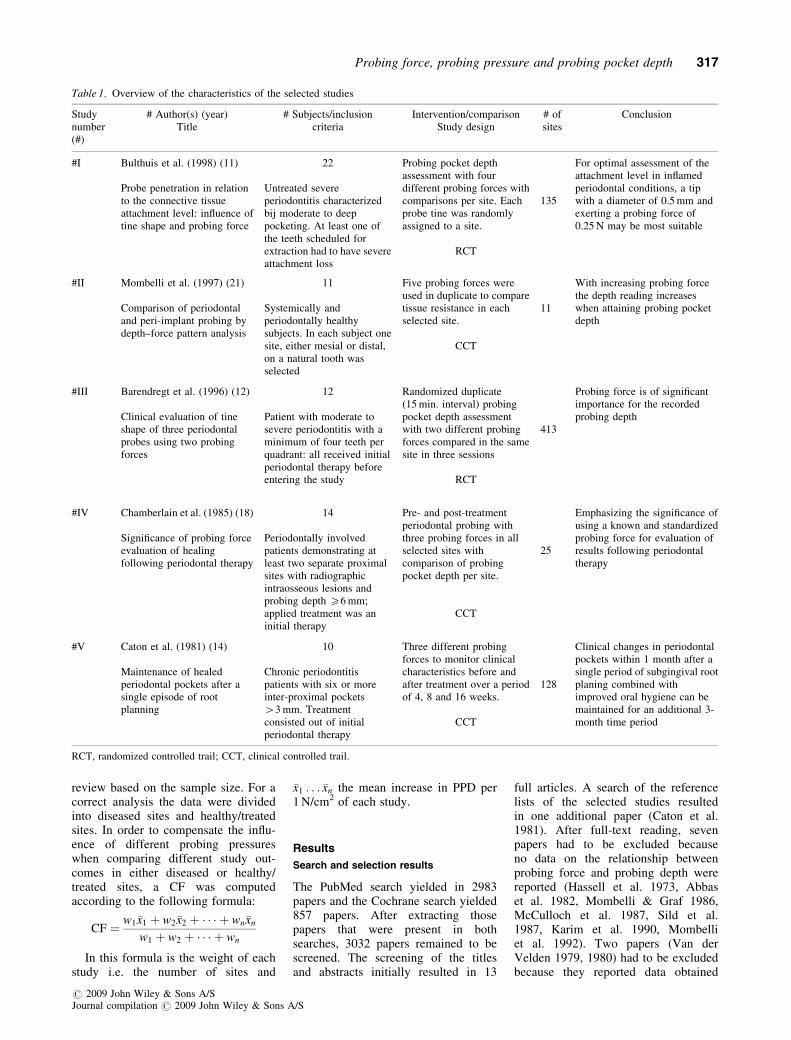
Table 1. Overview of the characteristics of the selected studies
Studynumber(#)
# Author(s) (year)Title
# Subjects/inclusioncriteria
Intervention/comparisonStudy design
# ofsites
Conclusion
#I Bulthuis et al. (1998) (11)
Probe penetration in relationto the connective tissueattachment level: influence oftine shape and probing force
22
Untreated severeperiodontitis characterizedbij moderate to deeppocketing. At least one ofthe teeth scheduled forextraction had to have severeattachment loss
Probing pocket depthassessment with fourdifferent probing forces withcomparisons per site. Eachprobe tine was randomlyassigned to a site.
RCT
135
For optimal assessment of theattachment level in inflamedperiodontal conditions, a tipwith a diameter of 0.5 mm andexerting a probing force of0.25 N may be most suitable
#II Mombelli et al. (1997) (21)
Comparison of periodontaland peri-implant probing bydepth–force pattern analysis
11
Systemically andperiodontally healthysubjects. In each subject onesite, either mesial or distal,on a natural tooth wasselected
Five probing forces wereused in duplicate to comparetissue resistance in eachselected site.
CCT
11
With increasing probing forcethe depth reading increaseswhen attaining probing pocketdepth
#III Barendregt et al. (1996) (12)
Clinical evaluation of tineshape of three periodontalprobes using two probingforces
12
Patient with moderate tosevere periodontitis with aminimum of four teeth perquadrant: all received initialperiodontal therapy beforeentering the study
Randomized duplicate(15 min. interval) probingpocket depth assessmentwith two different probingforces compared in the samesite in three sessions
RCT
413
Probing force is of significantimportance for the recordedprobing depth
#IV Chamberlain et al. (1985) (18)
Significance of probing forceevaluation of healingfollowing periodontal therapy
14
Periodontally involvedpatients demonstrating atleast two separate proximalsites with radiographicintraosseous lesions andprobing depth X6 mm;applied treatment was aninitial therapy
Pre- and post-treatmentperiodontal probing withthree probing forces in allselected sites withcomparison of probingpocket depth per site.
CCT
25
Emphasizing the significance ofusing a known and standardizedprobing force for evaluation ofresults following periodontaltherapy
#V Caton et al. (1981) (14)
Maintenance of healedperiodontal pockets after asingle episode of rootplanning
10
Chronic periodontitispatients with six or moreinter-proximal pockets43 mm. Treatmentconsisted out of initialperiodontal therapy
Three different probingforces to monitor clinicalcharacteristics before andafter treatment over a periodof 4, 8 and 16 weeks.
CCT
128
Clinical changes in periodontalpockets within 1 month after asingle period of subgingival rootplaning combined withimproved oral hygiene can bemaintained for an additional 3-month time period
RCT, randomized controlled trail; CCT, clinical controlled trail.
Probing force, probing pressure and probing pocket depth 317
r 2009 John Wiley & Sons A/SJournal compilation r 2009 John Wiley & Sons A/S

with a parallel probe tine shape. Theremaining five papers that fulfilled theselection criteria were processedfor data extraction (Caton et al. 1981,Chamberlain et al. 1985, Barendregt etal. 1996, Mombelli et al. 1997, Bulthuiset al. 1998). From one selected study(Mombelli et al. 1997), the originalmean data, as assessed around the teeth,were obtained from the author becausethe paper provided only descriptivedata. Also, Barendregt et al. (1996)provided the original mean data, repre-senting measurements with differentprobing pressures assessed at the samesite.
Assessment of heterogeneity
Considerable heterogeneity was observedin the study design, characteristics andoutcome variables i.e. selection criteriaof the studies, number of subjects, num-ber of sites, the number and magnitudeof probing forces/pressures and probetip diameter. Information regarding thestudy characteristics is shown in Tables 1and 2.
Subjects and periodontal tissuecondition
The subjects in the five selected studiesincluded both male and female adultswith diseased and healthy/treated perio-dontal tissues. The number of partici-pants varied per study (range 10–22). As
shown in Table 2, three studies includeddata of diseased tissues (Caton et al.1981, Chamberlain et al. 1985, Bulthuiset al. 1998) while four studies provideddata of healthy/treated sites (Caton et al.1981, Chamberlain et al. 1985, Bare-ndregt et al. 1996, Mombelli et al. 1997).
Sites
A large variation was present in thenumber of sites that were assessed, ran-ging from 11 (Mombelli et al. 1997) to413 (Barendregt et al. 1996) sites.
Probes
All selected studies used probes with aforce control or a probing force indica-tor. In three of the selected studies, aprobe tip diameter at the tip of 0.5 mmwas used (Chamberlain et al. 1985,Barendregt et al. 1996, Bulthuis et al.1998). One study (Mombelli et al. 1997)used a probe tip with a diameter of0.4 mm at the tip and an other study0.35 mm (Caton et al. 1981).
Probing pressures
Barendregt et al. (1996) and Bulthuis etal. (1998) related their results to probingpressure. The remaining studies used pre-sentation probing forces ranging from0.10 to 1.25 N in their data. For thepresent review the probing pressure (N/cm2) in these studies was calculated based
on the probing force and the probe dia-meter. Over the five studies, the probingpressure ranged from 51 to 995 N/cm2.
Extend of probe penetration
In the studies of Bulthuis et al. (1998)and Mombelli et al. (1997), an electro-nic pressure-sensitive probe was used.Bulthuis et al. (1998) used a system(Florida Probes, Florida Probe Com-pany, Gainesville, Florida. USA) with aprecision of 0.1 mm while Mombelliassessed the extent of probe penetrationwith an accuracy of 0.5 mm. The systemof Chamberlain et al. (1985) had cali-brated markings on the probe at eachmillimere and recordings were made tothe nearest 0.5 mm. Barendregt et al.(1996) and Caton et al. (1981) describethat they recorded the probing depth tothe nearest whole millimtre when thepresent pressure was reached.
Study quality
Allocation concealment
Becuase of the study design of theselected studies, allocation concealmentwas not possible. Instead, two otherdesign aspects were investigated: assess-ment of the inflammatory status of theincluded subjects and the study designfeatures (Table 1).
Table 2. Summary of selected study divided into diseased and healthy/treated sites; mean probing pocket depth (PPD) per probing force/pressure(mm) and standard deviation in parenthesis (if available); increase in PPD (mm) calculation for each increase in probing force in relation to thepreceding probing force
Author study number (#) Forces(N)
Diameter(mm)
Pressuren
(N/cm2)PPD
(diseased sites)PPD
increasenPPD (healthy/treated sites)
PPDincreasen
Bulthuis et al. (1998) (# I) 0.10 0.50 51 2.80 (1.88) – – –0.15 76 2.83 (1.81) 0.03 – –0.20 102 3.11 (2.00) 0.28 – –0.25 127 3.14 (2.02) 0.03 – –
Mombelli et al. (1997) (# II) 0.25 0.40 199 – – 3.41 (0.49) –0.50 398 – – 3.92 (0.64) 0.510.75 597 – – 4.08 (0.72) 0.161.00 796 – – 4.16 (0.71) 0.081.25 995 – – 4.21 (0.69) 0.05
Barendregt et al. (1996) (# III) 0.25 0.50 127 – – 2.40 (1.20)n –0.50 255 – – 2.70 (1.40)n 0.3
Chamberlain et al. (1985) (# IV) 0.25 0.50 127 5.1 (1.4) – 4.1 (1.3) –0.50 255 6.1 (1.2) 1.0 4.7 (1.3) 0.60.75 382 6.7 (1.0) 0.6 5.2 (1.3) 0.5
Caton et al. (1981) (# V) 0.15 0.35 155 3.06 (0.44)n – 2.00 (0.41)n –0.25 259 3.60 (0.51)n 0.54 2.36 (0.41)n 0.360.50 520 3.99 (1.80)n 0.39 2.64 (0.47)n 0.28
nCalculated by the author.
318 Larsen et al.
r 2009 John Wiley & Sons A/SJournal compilation r 2009 John Wiley & Sons A/S

Assessment of the inflammatory status
Bulthuis et al. (1998) evaluated theperiodontal condition based on manualprobing to assess the moderate to deeppocketing around the selected teeth.Mombelli et al. (1997) evaluated theperiodontal health based on conven-tional probing and scored a plaque index(Silness & Loe 1964). Barendregt et al(1996) assessed the inflammatory statusbased on manual probing after the initialtherapy. The sites used for this systema-tic review from Chamberlain et al.(1985) originated from the study ofRenvert et al. (1985). They describethe selection of the sites evaluated ashaving o15% plaque, proximalintraosseous lesions and pocket probingdepths X6 mm after initial therapy bymanual probing. Finally, Caton et al.(1981) selected patients referred fortreatment of chronic periodontitis andevaluated the inflammatory status-basedpocket depth and bleeding on probingby manual probing.
Study design
Mombelli et al. (1997) repeated allduplicate probing measurements within1 week. The PPD assessments in thestudy of Barendregt et al. (1996) wereobtained in three sessions with a 1-weekinterval. Per session, the assessmentswere repeated within 15 min. In thestudy of Chamberlain et al. (1985), thePPD was assessed before and 6 monthsafter treatment. The before-treatmentmeasurements were performed at least6 months after oral hygiene instructionand root planing. Because they repre-sented deep residual pockets (meanPPD45.0 mm), they were eligible forthis review as diseased sites. From thestudy of Chamberlain et al. (1985), onlythe data from the root planing groupwere used because this was also thetreatment modality used in the study ofCaton et al. (1981) and Barendregt et al.(1996). Caton et al. (1981) measuredthe PPD at baseline and 4, 8 and 16weeks following root planing. For thisreview, the pocket assessment at base-line and the 16-week assessment wereused.
Randomization
Barendregt et al. (1996) and Bulthuiset al. (1998) provided partial randomi-zation in their RCTs. Barendregt et al.(1996) randomized the order of use of
tine/force combinations over thepatients and sessions. In the study ofBulthuis et al. (1998), the sites to beprobed were randomly allocated to eachprobe tine. Neither study randomizedfor probing pressure. Additionally, inboth studies, the method of randomiza-tion is unclear. In all selected studies,logically, the lowest probing pressurewas used first when measuring the PPD.
Blinding of examiner or patients
In four of the five selected studies, it wasrecognized that blinding of the exami-ners was not possible due to the studydesign and the probes used. Only in thestudy of Bulthuis et al. (1998) was theexaminer blind for all the recordedmeasurements due to the use of theFlorida Probes. Blinding of patientswas not applicable because they werenot actively involved in the study.
Loss to follow-up
In all studies none of the patients/siteswere lost to follow-up during the experi-mental period.
Study outcomes
In Table 2 the results of the five selectedstudies are presented. The probingforces ranged from 0.10 N (Bulthuis etal. 1998) to 1.25 N (Mombelli et al.1997), corresponding to probing pres-sures of 51 and 995 N/cm2. The PPD inthe diseased group ranged from 2.80 mm
(Bulthuis et al. 1998) to 6.7 mm (Cham-berlain et al. 1985) obtained with a 51and a 382 N/cm2 probing pressure,respectively. In the healthy/treated sites,the most shallow PPD was assessed inthe study of Caton et al. (1981), whichamounted to 2.00 mm, assessed with aprobing pressure of 155 N/cm2. Cham-berlain et al. (1985) showed the deepestPPD measured with a probing pressureof 382 N/cm2 (5.2 mm). Because theheterogeneity of the studies (probingpressure) no meta-analysis could beperformed on the pooled data.
In all instances, a higher probingpressure resulted in an increase inPPD. When analysing the data fromthe study of Mombelli et al. (1997), inhealthy sites, the incremental change inPPD decreases as the pressure increasesabove 398 N/cm2. This phenomenonwas also found in the studies of Catonet al. (1981) and Chamberlain et al.(1985) in both diseased and healthy/treated sites for pressures higher than255 and 259 N/cm2, respectively. Withrelatively low probing pressures in dis-eased conditions in the study of Bulthuiset al. (1998), the largest increment inPPD was found when the probing pres-sure increased from 76 to 102 N/cm2.
In Tables 3a and b, the computationsare presented of the CFs for both dis-eased and healthy/treated sites. Thisfactor amounted, in diseased sites, to amean PPD increase of 0.004 mm foreach increase of 1 N/cm2 in probingpressure. For healthy/treated sites, theCF was 0.002 mm (Table 3b).
Table 3a. Calculation of the correction factor in diseased sites; mean increase in probing pocketdepth per 1 N/cm2 as result of mean increase of probing pocket depth (PPD) and probing pressurerelative to the lowest probing pressure and corresponding PPD
Author Study number (#) Increase Weight (wn)(# sites)
wn�xn
pressure(N/cm2)
PPD(mm)
PPD per1 N/cm2
Bulthuis et al. (1998) (# I) 25 0.03 0.001251 0.31 0.00676 0.33 0.004
Mean (�xI) 0.0037 x 135 (wI) 5 0.50
Chamberlain et al. (1985) (# IV) 128 1.0 0.008255 1.6 0.006
Mean (�xIV ) 0.007 x 25 (wIV) 5 0.18
Caton et al. (1981) (# V) 104 0.54 0.005365 0.93 0.0025
Mean (�xV ) 0.0038 x 128 (wV) 5 0.48
Pwn�xn 2.0
Correction factor �x ¼P
wn �xn
wn
2:0
2880.004
The italic is used since these data are the mean data with which the weight is calculated.
Probing force, probing pressure and probing pocket depth 319
r 2009 John Wiley & Sons A/SJournal compilation r 2009 John Wiley & Sons A/S

Discussion
The goals of periodontology can bedefined in terms of keeping teeth forlife, maintaining function, preventingand eliminating pain and discomfort.This can be achieved by aiming for anoptimal healthy periodontium that ischaracterized by the presence of shallowpockets and the absence of inflammation(Van der Velden & Jansen 1981). Theperiodontal probe is an important toolfor the clinical assessment of the perio-dontal status, diagnosis and treatmentplanning. To be able to enter the pocketwith a periodontal probe, a certain forceis needed to overcome the resistance(tonus) of the gingival tissues; not onlythe force applied but also the dimen-sions of the probe tip should be consid-ered (Garnick & Silverstein 2000).
Probing force as such has been recog-nized as an important factor in measur-ing PPD but little attention has beenpaid to the issue of probing pressure.Already in 1950 Miller stated the impor-tance of pressure when probing: ‘‘Gen-tle pressure against the epithelialattachment with the probe passed intothe gingival sulcus, or a periodontalpocket, meets with springy resistanceof the epithelial attachment’’(Miller1950). In the early 1970s, the termpressure was used by Gabathuler &Hassell (1971) in the title of their pub-
lication: ‘‘A pressure-sensitive perio-dontal probe’’, but the paper includedonly probing force data. Two years later,Hassell et al. (1973) first calculatedand published ‘‘light hand pressures’’as proposed by Waerhaug (1952) andGabathuler & Hassell (1971) whichamounted to 20 and 70 ponds/mm2,respectively. Also, in an attempt tostandardize the probing force, Van derVelden & De Vries (1978) introduced‘‘The pressure probe’’ but they also didnot use probing pressures to presenttheir data. Other studies during thesame time period, dealing with the issueof pocket probing with a force-con-trolled probe, all mention the probingforce and probe diameter without trans-lating this to probing pressure (Armitageet al. 1977, Spray et al. 1978, Robinson& Vitek 1979, Van der Velden 1979,Polson et al. 1980, Hancock & Wirthlin1981, Fowler et al. 1982). It was notuntil 1982 that the study results werecompared based on probing pressure(Van der Velden 1982). After this pub-lication, numerous studies evaluatingthe different ‘‘constant-force’’ probesfor the accuracy and reproducibility stillpreferred presenting the data in relationto probing force. Some authorsacknowledged the importance of theuse of probing pressure. Garnick et al.(1989), in a study to evaluate the effectof inflammation and pressure on probe
displacement in beagle dog gingivitis,reported four different probing pressures(in N/cm2 or kPa). In a study of Lang etal. (1991), the title included the termprobing pressure but provided onlyprobing force-related data. At the endof their discussion, however, the con-clusion was related to probing pressure.Later study results based on probingpressures were presented on the influ-ence of probe tine when assessing PPD(Barendregt et al. 1996). In general,probing force still remained the pre-ferred way for interpretation of studyoutcomes. Some authors, however, diduse probing pressures as a unit of mea-sure. For instance, with the introductionof a new probe design in 2004, properprobing pressure data were presentedand discussed in support of the proposedprobe design (Vartoukian et al. 2004).Nevertheless, in a recent study on theprobe penetration in periodontal andperi-implant tissues in dogs, only prob-ing force and tip diameter were reported(Abrahamsson & Soldini 2006). There-fore, it is reasonable to conclude that theaspect of probing pressure has beengreatly undervalued.
A probing pressure is a product of theprobing force (N) relative to the tipdiameter (mm). The pressure exertedby the probe is directly proportional tothe force on the probe and inverselyproportional to the surface area at theprobe tip (Garnick & Silverstein 2000).Because the surface area of a roundprobe is determined by pr2, with r,being the radius of the tip, a reductionin the probe diameter will increase thepressure by a proportional amount, thatis squared. Therefore, a change in tipdiameter has a more profound effect onthe pressure than the actual forceexerted on the probe (Aguero et al.1995). For example, if a force of0.50 N is used on a probe with a dia-meter of 1 mm, the pressure on the tip ofthe tine will be 64 N/cm2. Using thesame force on a tip with a diameter of0.5 mm the pressure will be 255 N/cm2.Van der Velden (1979) found that with aprobing force of 0.75 N in treated resi-dual deep periodontal pockets the probetip is located at the attachment level.These results were obtained with a probediameter of 0.63 mm (241 N/cm2). Usingthe same probing force other authors(Armitage et al. 1977, Spray et al.1978, Robinson & Vitek 1979) observedpenetration into the connective tissue.However, they used a probe diameter of0.35 mm. This tip and force combination
Table 3b. Calculation of the correction factor in healthy/treated sites; mean increase in probingpocket depth per 1 N/cm2 as result of mean increase of probing pocket depth (PPD) and probingpressure relative to the lowest probing pressure and corresponding PPD
Author Study number (#) Increase Weight (wn)(# sites)
wn�xn
Pressure(N/cm2)
PPD(mm)
PPD per1 N/cm2
Mombelli et al. (1997) (# II) 199 0.51 0.0025389 0.67 0.0017579 0.75 0.0012796 0.80 0.001
Mean (�xII) 0.0012 x 11 (wII) 5 0.013
Barendregt et al. (1996) (# III) 128 0.3 0.002Mean (�xIII ) 0.002 x 413 (wIII) 5 0.826
Chamberlain et al. (1985) (# IV) 128 0.6 0.005255 1.1 0.004
Mean (�xIV ) 0.0045 x 25 (wIV) 5 0.113
Caton et al. (1981) (# V) 104 0.36 0.003365 0.64 0.0017
Mean (�xV ) 0.002 x 128 (wV) 5 0.30
Pwn�xn 1.253
Correction factor �x ¼P
wn �xn
wn
1:253
5770.002
320 Larsen et al.
r 2009 John Wiley & Sons A/SJournal compilation r 2009 John Wiley & Sons A/S

delivers a probing pressure at the tip of780 N/cm2, which explains the differencebetween the studies.
Because of the fact that in variousstudies different amounts of probingpressure are used, comparison of forexample treatment results becomes dif-ficult. For instance, Badersten et al.(1984), when evaluating the effect ofnon-surgical periodontal therapy, per-formed their measurements with a prob-ing force of 0.75 N with a tip diameterof 0.5 mm which amounts to a probingpressure of 382 N/cm2. The resultsshowed a mean overall PPD of 3.8 mm12 months after treatment with handinstruments (Badersten et al. 1984). Inthe study of Kaldahl et al. (1988) aprobing force of 0.5 N and a tip diameterof 0.35 mm (519 N/cm2) was used whentesting the effect of four treatment mod-alities. The mean PPD in sites treatedwithin the non-surgical periodontal ther-apy modality was 4.26 mm after 12months (Kaldahl et al. 1988). In orderto be able to compare the probing depthafter treatment of the two studies, theprobing pressure of the Badersten studyshould be adopted to the level that wasused in the Kaldahl study with a corre-sponding mean PPD increase. This canbe achieved using the CF of 0.002 mmincrease per 1 N/cm2 for healthy/treatedsites. Thus, the discrepancy of 137 N/cm2 between the pressures used in thetwo studies times 0.002 is 0.27 mm.Therefore, if in the Badersten study thesame probing pressure was used as inthe Kaldahl study, the probing depthwould have been 4.07 mm. This probingdepth value appears to be in closer rangeof the 4.26 mm as presented by Kaldahlet al. (1988).
It has been described that withincreasing probing force i.e. probingpressure, the recorded probing depthwill increase (Robinson & Vitek 1979,Van der Velden 1979, Barendregt et al.1996), an observation supported by theoutcome of this review. Histologic loca-tions of the probe tip considered to bethe most relevant in periodontal diag-nostics are the base of the periodontalpocket and the most coronal connectivetissue attachment (Aguero et al. 1995).Based on the results of the study ofBulthuis et al. (1998) in diseased sites,the tapered probe (tip diameter 0.5 mm)with a 0.25 N force was on averagelocated at this level. In healthy/treatedsites in humans, even pressures upto 400 N/cm2 left the probe tip coronalto this landmark by a mean of 0.73 mm
(Fowler et al. 1982). One has to bear inmind therefore that in a number ofcases, an over – or underestimationof the true attachment level will stilloccur when assessing the PPD (Listgar-ten 1980, Kalkwarf et al. 1986). A highprobing pressure is deliberately used inbone sounding to determine the actualalveolar bone level in relation to thelocation of the gingival margin or thecemento-enamel junction. The tip of theprobe is pushed through the supra-alveolar connective tissue to make con-tact with the bone (Lindhe et al. 2003).This implies that with a certain probingpressure, the increase of probing depthmay be physically limited by the alveo-lar crest. This may explain why withpressures of more than 796 N/cm2, theincrease in PPD is smaller as comparedwith pressures of 76–597 N/cm2 probingpressure (Table 2). On the other hand, iftoo gentle probing forces are applied,the probe tip may not enter the orifice ofthe pocket (Bulthuis et al. 1998, Bare-ndregt et al. 2006). This could explainwhy almost no difference in PPD isobserved with the very low pressuresbetween 51 and 76 N/cm2.
Periodontal probing registers resis-tance of the tissue to the pressureapplied by the probe. The greater thepressure, the greater the advancement ofthe probe into the tissues (Table 2).However, the advancement depends onthe resistance of the tissue at the sitebeing measured (Garnick & Silverstein2000). With a specific pressure, theprobe will proceed until a reaction pres-sure develops from deformation of tis-sues (Aguero et al. 1995). Tissuepressure that resists probe displacementdepends on the tissue morphologyincluding loss of connective tissueattachment and the severity of tissueinflammation. Accordingly, this tissuepressure will vary (Aguero et al.1995). With treatment, inflammation isreduced and/or tissue attachment isincreased, the resistance to probingpressure is increased and the displace-ment of the probe will be less. Thedifference in probing depth thereforereflects a reduction of inflammationand the response to treatment (Garnick& Silverstein 2000). Based on theresults presented in Tables 3a and b, aclear difference between diseased andhealthy/treated tissue is apparent withrespect to increase of probing depth.The increase in PPD in relation topressure increase (N/cm2) is approxi-mately twice as high in diseased sites.
Conclusion
The results of the present review showthat with increasing probing pressure,the PPD increases. The dimensions ofthe increase are dependent on the perio-dontal tissue conditions. PPD showed amean increase of 0.004 mm per increaseof 1 N/cm2 at diseased sites and0.002 mm at healthy/treated sites. Bothcan be used as a CF for the comparisonof outcomes of studies that have useddifferent probing pressures.
References
Abbas, F., Hart, A. A., Oosting, J. & Van der
Velden, U. (1982) Effect of training and
probing force on the reproducibility of pocket
depth measurements. Journal of Periodontal
Research 17, 226–234.
Abrahamsson, I. & Soldini, C. (2006) Probe
penetration in periodontal and peri-implant
tissues. An experimental study in the beagle
dog. Clinical Oral Implants Research 17,
601–605.
Aguero, A., Garnick, J. J., Keagle, J., Steflik, D.
E. & Thompson, W. O. (1995) Histological
location of a standardized periodontal
probe in man. Journal of Periodontology
66, 184–190.
Armitage, G. C., Svanberg, G. K. & Loe, H.
(1977) Microscopic evaluation of clinical
measurements of connective tissue attach-
ment levels. Journal of Clinical Perio-
dontology 4, 173–190.
Atassi, F., Newman, H. N. & Bulman, J. S.
(1992) Probe tine diameter and probing
depth. Journal of Clinical Periodontology
19, 301–304.
Badersten, A., Nilveus, R. & Egelberg, J. (1984)
Effect of nonsurgical periodontal therapy. II.
Severely advanced periodontitis. Journal of
Clinical Periodontology 11, 63–76.
Barendregt, D. S., Van der Velden, U., Reiker,
J. & Loos, B. G. (1996) Clinical evaluation of
tine shape of 3 periodontal probes using 2
probing forces. Journal of Clinical Perio-
dontology 23, 397–402.
Barendregt, D. S., Van der Velden, U., Timmer-
man, M. F. & Van der Weijden, G. A. (2006)
Comparison of two automated periodontal
probes and two probes with a conventional
readout in periodontal maintenance patients.
Journal of Clinical Periodontology 33, 276–
282.
Bulthuis, H. M., Barendregt, D. S., Timmerman,
M. F., Loos, B. G. & Van der Velden, U.
(1998) Probe penetration in relation to the
connective tissue attachment level: influence
of tine shape and probing force. Journal of
Clinical Periodontology 25, 417–423.
Caton, J., Greenstein, G. & Polson, A. M.
(1981) Depth of periodontal probe penetra-
tion related to clinical and histologic signs of
gingival inflammation. Journal of Perio-
dontology 52, 626–629.
Probing force, probing pressure and probing pocket depth 321
r 2009 John Wiley & Sons A/SJournal compilation r 2009 John Wiley & Sons A/S
Chamberlain, A. D. H., Renvert, S., Garrett, S.,
Nilveus, R. & Egelberg, J. (1985) Signifi-
cance of probing force for evaluation of
healing following periodontal therapy. Jour-
nal of Clinical Periodontology 12, 306–311.
Fowler, C., Garrett, S., Crigger, M. & Egelberg,
J. (1982) Histologic probe position in treated
and untreated human periodontal tissues.
Journal of Clinical Periodontology 9, 373–
385.
Gabathuler, H. & Hassell, T. (1971) A pressure-
sensitive periodontal probe. Helvetica Odon-
tologica Acta 15, 114–117.
Garnick, J. J., Keagle, J. G., Searle, J. R., King,
G. E. & Thompson, W. O. (1989) Gingival
resistance to probing forces. II. The effect of
inflammation and pressure on probe displace-
ment in beagle dog gingivitis. Journal of
Periodontology 60, 498–505.
Garnick, J. J. & Silverstein, L. (2000) Perio-
dontal probing: probe tip diameter. Journal of
Periodontology 71, 96–103.
Hancock, E. B. & Wirthlin, M. R. (1981) The
location of the periodontal probe tip in health
and disease. Journal of Periodontology 52,
124–129.
Hassell, T. M., Germann, M. A. & Saxer, U. P.
(1973) Periodontal probing: interinvestigator
discrepancies and correlations between prob-
ing force and recorded depth. Helvetica
Odontologica Acta 17, 38–42.
Hefti, A. F. (1997) Periodontal probing. Critical
Review Oral Biology and Medicine 8, 336–356.
Kaldahl, W. B., Kalkwarf, K. L., Patil, K. D.,
Dyer, J. K. & Bates, R. E. (1988) Evaluation
of four modalities of periodontal therapy.
Mean probing depth, probing attachment
level and recession changes. Journal of
Periodontology 59, 783–793.
Kalkwarf, K. L., Kaldahl, W. B. & Patil, K. D.
(1986) Comparison of manual and pressure-
controlled periodontal probing. Journal of
Periodontology 57, 467–471.
Karim, M., Birek, P. & McCulloch, C. A.
(1990) Controlled force measurements of
gingival attachment level made with the
Toronto automated probe using electronic
guidance. Journal of Clinical Periodontology
17, 594–600.
Lang, N. P., Nyman, S., Senn, C. & Joss, A.
(1991) Bleeding on probing as it relates to
probing pressure and gingival health. Journal
of Clinical Periodontology 18, 257–261.
Lindhe, J., Lang, N. P. & Karring, T. (2003)
Clinical Periodontology and Implant Dentis-
try, 4th edition, p. 410. Oxford: Blackwell
Munksgaard.
Listgarten, M. A. (1980) Periodontal probing:
what does it mean? Journal of Perio-
dontology 7, 165–176.
McCulloch, C. A., Birek, P. & Hardy, V. (1987)
Comparison of gingival attachment level
measurements with an automated periodontal
probe and a pressure-sensitive probe. Journal
of Periodontal Research 22, 348–352.
Miller, S. C. (1950) Textbook of Periodontia,
3rd edition, p. 44. Philadelphia: The Blakis-
ton Company.
Mombelli, A. & Graf, H. (1986) Depth–force-
patterns in periodontal probing. Journal of
Clinical Periodontology 13, 126–130.
Mombelli, A., Muhle, T., Bragger, U., Lang, N.
P. & Burgin, W. B. (1997) Comparison of
periodontal and peri-implant probing by
depth–force pattern analysis. Clinical Oral
Implants Research 8, 448–454.
Mombelli, A., Muhle, T. & Frigg, R. (1992)
Depth–force patterns of periodontal probing.
Attachment-gain in relation to probing
force. Journal of Clinical Periodontology
19, 295–300.
Polson, A. M., Caton, J. G., Yeaple, R. N. &
Zander, H. A. (1980) Histological determina-
tion of probe tip penetration into gingival
sulcus of humans using an electronic pres-
sure-sensitive probe. Journal of Clinical
Periodontology 7, 479–488.
Ramfjord, S. P. (1959) Indices for prevalence
and incidence of periodontal disease. Journal
Periodontology 30, 51–59.
Renvert, S., Nilveus, R. & Egelberg, J. (1985)
Healing after treatment of periodontal
intraosseous defects. V Effect of root planing
versus flap surgery. Journal Periodontology
12, 619–629.
Riggs, J. M. (1882) Proceedings of the southern
dental association – fourteenth annual ses-
sion. Dental Cosmos 24, 523–527.
Robinson, P. J. & Vitek, R. M. (1979) The
relationship between gingival inflammation
and resistance to probe penetration. Journal
of Periodontal Research 14, 239–243.
Sachs, H. (1929) Die behandlung lockerer Zahnen
nach Younger-Sachs. Preface, pp. 22–23, 212.
Berlin: Berlinersche Verlagsanstalt.
Sild, E., Bernardi, F., Carnevale, G. & Milano,
F. (1987) Computerized periodontal probe
with adjustable pressure. International Jour-
nal of Periodontics and Restorative Dentistry
7, 53–62.
Silness, J. & Loe, H. (1964) Periodontal disease
in Pregnancy. II. Correlation between oral
hygiene and periodontal condition. Acta
Odontologica Scandinavica 22, 121–135.
Spray, J. R., Garnick, J. J., Doles, L. R. &
Klawitter, J. J. (1978) Microscopic demon-
stration of the position of periodontal probes.
Journal of Periodontology 49, 148–152.
Van der Velden, U. (1979) Probing force and
the relationship of the probe tip to the perio-
dontal tissues. Journal of Clinical Perio-
dontology 6, 106–114.
Van der Velden, U. (1980) Influence of perio-
dontal health on probing depth and bleeding
tendency. Journal of Clinical Periodontology
7, 129–139.
Van der Velden, U. (1982) Location of probe tip
in bleeding and non-bleeding pockets with
minimal gingival inflammation. Journal of
Clinical Periodontology 9, 421–427.
Van der Velden, U. & de Vries, J. H. (1978)
Introduction of a new periodontal probe: the
pressure probe. Journal of Clinical Perio-
dontology 5, 188–197.
Van der Velden, U. & Jansen, J. (1981) Micro-
scopic evaluation of pocket depth measure-
ments performed with six different probing
forces in dogs. Journal of Clinical Perio-
dontology 8, 107–116.
Vartoukian, S. R., Palmer, R. M. & Wilson, R.
F. (2004) Evaluation of a new periodontal
probe tip design. A clinical and in vitro study.
Journal of Clinical Periodontology 31, 918–
925.
Waerhaug, J. (1952) The gingival pocket;
anatomy, pathology, deepening and elimi-
nation. Odontolisk Tidskrift 60 (Suppl. 1),
1–186.
Address:
D. S. Barendregt
Department of Periodontology
Academic Centre for Dentistry Amsterdam.
ACTA
Louwesweg 1
1066 EA Amsterdam
The Netherlands
E-mail: [email protected]
Clinical Relevance
Scientific rationale for the study:Many factors such as probing force,tip diameter/shape and periodontalhealth influence probing pocket mea-surements. Of the aforementionedfactors, the probing force seems tobe most important. The majority ofstudies report only probing forcewhile it is the pressure at the tip,the resultant of probing force andprobe diameter, that eventuallydetermines probe penetration. There-
fore, in a systematic review on theinfluence of probing pressure on theprobing pocket measurements (PPD),an attempt was made to obtain acorrection factor (CF) for comparingdata obtained with different probingpressures.Principal findings: The probing pres-sures in the selected studies, all usingtapered probe tines, ranged fromextremely low to very high i.e. 51–995 N/cm2. PPD increased withincreasing probing pressure in both
diseased and healthy/treated sites.The CF compensating for the influ-ence of the used probing pressureused at healthy/treated sites amountedto 0.002 mm per increase of 1 N/cm2
in probing pressure whereas at dis-eased sites this was 0.004 mm.Practical implications: For a bettercomparison of the study outcomesobtained with different probing pres-sures, the CF presented in this studyrepresents a viable tool.
322 Larsen et al.
r 2009 John Wiley & Sons A/SJournal compilation r 2009 John Wiley & Sons A/S