process of growth tio nanotubes by anodization in an organic media · · 2014-08-29process of...
TRANSCRIPT

Process of growth TiO2 nanotubes by anodization in an organic media
I. Zamudio Torres, J. J. Pérez Bueno and Y. Meas Vong
Centro de Investigación y Desarrollo Tecnológico en Electroquímica, S.C., Parque Tecnológico Querétaro-Sanfandila, Sanfandila, Pedro Escobedo, Querétaro, México. CP 76703
Corresponding authors: [email protected]; [email protected]; [email protected]
The aim of this chapter is to show the process for the growth of TiO2 nanotube by anodizing and its features as surface. The Ti anodized nanotubes were obtained by anodizing Ti in organic medium. The process of formation of TiO2 nanotubes on Ti surfaces was established ex-situ by Scanning Electron Microscopy (SEM). At first, the growth of the TiO2 on the surface of the Ti plate occurs because of the interaction of the Ti with O2− ions. Then, an early oxide layer is formed. These anions migrate through the oxide layer to the Ti/TiO2 interface and then react with Ti. Meanwhile, the applied electric field ejects Ti4+ cations from the Ti/TiO2 interface and makes Ti4+ move towards the TiO2/electrolyte interface. The chemical dissolution occurs at the interface of TiO2/electrolyte where TiO2 is dissolved by F− with the help of H+ and the field assisted dissolution occurs at the Ti/TiO2 interface. Simultaneously, with the time of anodizing increased, the current density also increases to reach a maximum value. The increase in current density was caused by the formation of pits and enlargement of the pits on the oxide layer. Beyond this point, current density drops, indicating the growth of the nanotube structure. In this work, TiO2 nanotubes with inner diameter between 90-110 nm and wall sizes of 15-20 nm, after 120 min of anodizing are shown. The presence of TiO2 was confirmed with Energy Dispersive X-ray Spectroscopy (EDS) results. Tests of roughness, and surface energy were carried out too.
Keywords: Anodizing; nanotubes; contact angle; surface energy
1. Introduction
The TiO2 (titanium dioxide or titania) is a compound widely investigated and there are reports of multiple applications including: degradation of organic compounds and bacteria in both water and air, electric energy generation, application prosthesis, among others. The main features of this compound are: its high oxidizing capacity, superhydrophilicity, chemical stability, high durability, transparency to visible light, corrosion resistant and non-toxicity, making it friendly with the environment and with the interaction with the human beings. The titanium dioxide does not absorb light in the visible region (400-700 nm) but disperse it very efficiently. This is the most broadly commercially used characteristic as white basis for paints, toothpastes, cosmetic products, plastics, etc. However, TiO2 absorbs UV (ultra violet radiation, 100-400 nm). This absorption causes the generation of the electro-hole pair (charge carriers), which cause chemical reactions on surfaces. Its photochemical properties have been extensively investigated but not widely used at commercial level. So, the titanium oxide semiconductor or titania (TiO2) is one of the most important photoactive compounds, mainly for its characteristic stability and band gap energy with potential applications, such as: photovoltaic cells, batteries, sensors, optical emitters, photonic crystals, catalysis and phtocatalysis, selective adsorption, ion exchange, ultraviolet blocking, smart surface coatings and as smart functional filling material in fabrics, paints, cosmetics, among others. This kind of compound has been synthesized both with micro- and nano- sizes. However, the latter scale has been predominantly the millstone for research, especially by the acquired properties which differ from those of bulk materials. Among the various forms of achieved morphologies, there are: spheres, rods, sheets, fibers and tubes.
2. Background
2.1 Features of TiO2 nanotubes.
It is proposed for the tubular TiO2 semiconductor nanostructures that could be less recombination of charge carriers electron-hole (e– ‒ h+) caused by shorter necessary diffusion distance towards the target compounds to decompose. Nanotubes have been obtained by different methods, among which are: hydrothermal, sol- gel, assisted electrochemical methods with templates and anodizing methods. The anodizing of Ti is among the most used methods for obtaining nanotubes. It has the lower inconveniences whenever carrying out the process. The main interest in this kind of structure in particular is because it has proved been efficient in photocatalytic applications. The photoconversion efficiency in nanotubular titania surfaces is influence by various causes, among others, may be the manufacturing method, in which the factors are: the current, the electrolyte, concentration, impurities, processing temperature, dissolved oxygen, experimental setup, etc. For the preparation of this compound, the most frequently used methods are the templates and the hydrothermal, which usually obtain a product in powder form. However, thin films and coatings with oriented nanostructure are more
Microscopy: advances in scientific research and education (A. Méndez-Vilas, Ed.)__________________________________________________________________
887© FORMATEX 2014
desirable because it is possible to implement in applications, such as: catalysis, filtration, sensing, photovoltaic cells and electrodes with large surface area [1]–[12]. It is well known that the functional properties of the materials depend on their microstructure. Many types of nano-dimensional materials have been studied extensively [3-5], such as: nanowires, nanoneedles, nanofibers and nanotubes.
2.2 Some methods for obtaining TiO2 nanotubes.
The TiO2 films have been obtained with different morphologies, such as: nanorods [13]–[17], nanowires [18]–[20], and nanotubes [21]–[27]. These arrangements are of great interest since they have optical and electronic properties considered unique. There are different methods for preparing TiO2 nanotubes layers, such as: the sol-gel method using gels as templates [28], hydrothermal predeposits of TiO2 [29], [30], microwave-assisted hydrothermal method [31], sonochemical [28], microemulsion [32], solvothermal [33], atomic layer deposition (ALD) growth method without template, ionic liquids [34], anodized Ti [35], [36], among others.
3. Anodizing of Ti
3.1 Some aspects of anodizing of Ti.
No method used for obtaining TiO2 nanotubes offers much control over the dimensions of these as does the anodized titanium (Ti) in fluorine -based electrolytes [37]–[39]. In 2001, Gong et al.[40] first developed titania nanotube arrays self-organized on a Ti substrate by potentiostatic anodic oxidation using a solution of HF as electrolyte and pure Ti plates as substrates, which led to the rise of research of TiO2 nanotube arrangements. Their special nano-architecture provides a large internal surface without decrease in geometric and structural order [25,26]. The orientation of these nanostructures make them excellent electron percolation pathways for charge transfer between the interfaces, and offers better properties for applications in fields, such as: solar cells, catalysis, photolysis of water, hydrogen sensors, among others. The anodizing process of Ti have influence during the growth of TiO2 nanotubes the following main parameters: electrolyte composition, voltage, and time. Grimes et al. [17] reported the development of pores and holes in the surface of the titanium plates in the formation of nanotubes process. They proposed that the tube structure was the result of the depth and spread of small pores. Schmuki et al. [41] found that the low acidity in the bottom of the pore helps sculpting the pore into a tubular structure. However, it is not clear the detailed process of how the small pores become large pores and eventually evolve into a tubular structure. The relationship between the pore diameter, the wall thickness and the anodizing voltage is related to a process of forming the array of TiO2 nanotubes in the titanium surface [42], [43].
3.2 Electrolytic bath
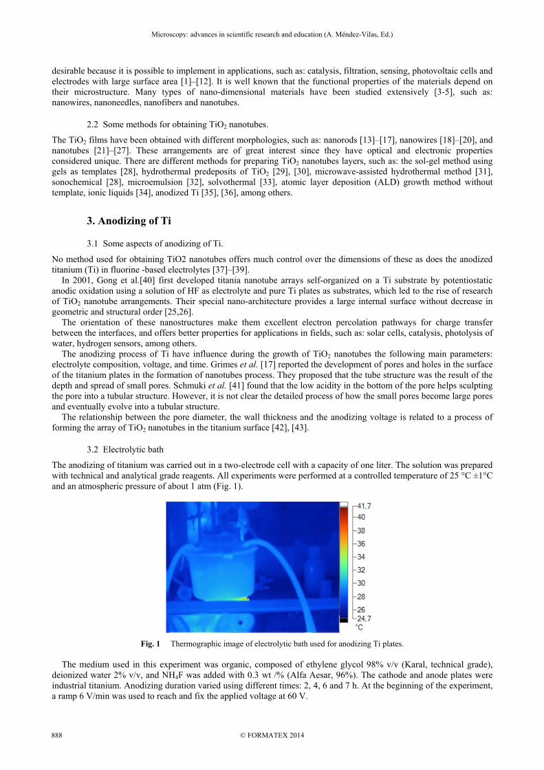
The anodizing of titanium was carried out in a two-electrode cell with a capacity of one liter. The solution was prepared with technical and analytical grade reagents. All experiments were performed at a controlled temperature of 25 °C ±1°C and an atmospheric pressure of about 1 atm (Fig. 1).
Fig. 1 Thermographic image of electrolytic bath used for anodizing Ti plates. The medium used in this experiment was organic, composed of ethylene glycol 98% v/v (Karal, technical grade), deionized water 2% v/v, and NH4F was added with 0.3 wt /% (Alfa Aesar, 96%). The cathode and anode plates were industrial titanium. Anodizing duration varied using different times: 2, 4, 6 and 7 h. At the beginning of the experiment, a ramp 6 V/min was used to reach and fix the applied voltage at 60 V.
Microscopy: advances in scientific research and education (A. Méndez-Vilas, Ed.)__________________________________________________________________
© FORMATEX 2014888

3.3 Roughness in the Ti plates.
The Ti plates used for producing nanotubes were industrial grade, which had 98 wt/% of Ti. These plates were pretreated by sandblasting technique. Roughness was measured by optical profilometry in order to analyze the surface texture and topography of the samples. Figure 1 shows the topographic image of a sample with Ti sandblasting treatment with an average roughness (Ra) of 1.8 ± 0.15 µm. Figure 2 shows the surface topography of anodized Ti with TiO2 nanotubes with an average roughness (Ra) of 1.7 ± 0.18 µm. The roughness did not show a signifying change with the anodizing process but the TiO2 nanotubes shown a better mechanical strength avoiding the treated layer detachment.
Fig. 2 Optical profilometry of Ti with sandblasting.
Fig. 3 Optical profilometry of TiO2 nanotubes.
3.4 Growth mechanism of TiO2 nanotubes.
1. In a first stage, the formation of a compact layer of amorphous titanium dioxide occurs. It was formed due to interaction of the metal with O-2 or OH- ions under influence of the electric field (reactions 1 and 2). Figure 4 show changes with a crust form at microscopic level. Ti → Ti 4 (1) Ti 2H O → TiO 4H (2)
Fig. 4 First step in the formation of TiO2 nanotubes, TiO2 layer.
Fig. 5 Formation of pits in the TiO2 layer, indicated with circle.
2. Once formed the TiO2 layer on the anode, it reacts with fluorine ions with the help of the electric field forming the [TiF6]-2 complex, resulting in fractures on the metal surface. TiO 6F 4H → TiF 2H O (3) 3. The fractures become larger pores and pore density increases. Subsequently, the pores spread evenly over the surface (Fig. 5). 4. As soon as the oxide growth rate on the metal-oxide interface and the ratio of oxide dissolution at the interface oxide-solution are finally equal, the thickness of the barrier layer remains unchanged, at the same time that it displaces deeper into the metal with the growth of the length of the pore.
Microscopy: advances in scientific research and education (A. Méndez-Vilas, Ed.)__________________________________________________________________
889© FORMATEX 2014

5. The nanotube length increases until the rate of electrochemical oxidation is equal to the rate of chemical dissolution in the upper surface of the nanotubes. Thus, a self-organized porous layer was obtained. In the case, that a sandblasting was used as pretreatment, an uneven distribution of the nanotubes layer was obtained (Fig. 6).
Fig. 6 The last step consists in the spreading of pores and formation of TiO2 nanotubes.
0 120 240 360 480 1800 3600 5400 72000
10
20
30
40
50
60
Voltage Current density
Time (s)
Vo
ltag
e (V
)
0.000
0.005
0.010
0.015
0.020
Cu
rren
t d
ensi
ty (
A/c
m2 )
Fig. 7 Voltage ramp used and density current. The formation of TiO2 is limited by the chemical oxidation
At the time that the anodizing time increases, the current density proportionally increases, reaching a maximum value of 23mA/cm2 (Fig. 7). The increased current density is caused by the formation of pits and holes extending into the oxide layer. Following this, the current density decreases, which indicate the growth of the TiO2 nanotubes structures, which were formed after 120 min of anodizing. The inner diameter reached was between 90-110 nm in size and walls were around 15 to 20 nm (Fig. 6) in thickness. Analysis of Energy Dispersive Spectroscopy X-ray (EDX) (Fig. 8) shows the presence of F. The presence of Al is attributable to sandblast. Cl, K, and Si are discarded in these analyses. This analysis confirms the formation of TiO2.
Fig. 8 EDX analysis shows the presence of F in the nanostructures.
3.5 Wettability in TiO2 nanotubes.
Table 1 shows the data obtained for the contact angles measurements. The TiO2 nanotubes generated on Ti surfaces were shown to be hydrophilic. It can be seen that by measuring the contact angle with distilled water, the surface of Ti metal alone has a contact angle of 85.3°, while after anodizing, this result changes dramatically, showing a value of 9.8°. Also, the measurements done included assessment with canola oil, where it is possible to observe that the TiO2 tubular nanostructures remarkably change the contact angle. The purpose of measuring with canola oil was to extend the study for including the surface energy measurements. Results about surface energy are shown in the table 1, this results was determined by Zisman method [44]. The Ti plates anodized are highly hydrophilic and have higher level of wettability, which is clearly defined by the surface free energy.
Microscopy: advances in scientific research and education (A. Méndez-Vilas, Ed.)__________________________________________________________________
© FORMATEX 2014890

Table 1 Contact angle measurements of Ti and TiO2 nanotubes surfaces.
4. Conclusions
The TiO2 nanotubes were obtained by using pretreated Ti surfaces through the sandblasting technique. These rough surfaces were anodized into an organic bath. Whenever sandblasting was used as pretreatment, an uneven distribution of the nanotubes layer was obtained. The roughness did not show a signifying change with the anodizing process but the TiO2 nanotubes shown a better mechanical strength avoiding the treated layer detachment. Through SEM imaging, the steps that occur during the anodizing process, forming a first layer of titanium oxide on the surface of Ti, then under influence of the electric field and the presence of fluorine chemical attack occurs is established, which causes fractures and pores subsequently grow into nanotubes by the effect of the electric field. It was also possible to establish the influence of the current during this process. It was observed that the increase in current density generated by the formation of pores and widening of these in the layer oxide. Following this, the low current density and is stable, indicating the growth of TiO2 nanotube structure.
Acknowledgements The authors gratefully acknowledge the financial support from the Mexican Council for Science and Technology (CONACyT, Grant CB-2009-01 133157). Also, the first author acknowledges CONACyT for his graduate fellowship. Especial thanks to José Eleazar Urbina Alvarez for his valuable help in obtaining the SEM images, and Dr. Francisco Espinoza for his assistance in obtaining the images of optical profilometry.
References
[1] R. Liu, W. Yang, L. Qiang, and H. Liu, “Conveniently fabricated heterojunction ZnO/TiO2 electrodes using TiO2 nanotube arrays for dye-sensitized solar cells,” vol. 220, pp. 153–159, 2012.
[2] Y. B. XIE and X. Z. LI, “Preparation and characterization of TiO2/Ti film electrodes by anodization at low voltage for photoelectrocatalytic application,” J. Appl. Electrochem., vol. 36, no. 6, pp. 663–668, Feb. 2006.
[3] F. Mole, J. Wang, D. a Clayton, C. Xu, and S. Pan, “Highly conductive nanostructured C-TiO2 electrodes with enhanced electrochemical stability and double layer charge storage capacitance.,” Langmuir, vol. 28, no. 28, pp. 10610–9, Jul. 2012.
[4] H. Wang, C. T. Yip, K. Y. Cheung, A. B. Djurišić, M. H. Xie, Y. H. Leung, and W. K. Chan, “Titania-nanotube-array-based photovoltaic cells,” Appl. Phys. Lett., vol. 89, no. 2, p. 023508, 2006.
[5] S. V NAIR, A. BALAKRISHNAN, K. R. V SUBRAMANIAN, A. M. ANU, A. M. ASHA, and B. DEEPIKA, “Effect of TiO2 nanotube length and lateral tubular spacing on photovoltaic properties of back illuminated dye sensitized solar cell,” Bull. Mater. Sci., vol. 35, no. 4, pp. 489–493, Sep. 2012.
[6] Y. Kwon, H. Kim, S. Lee, I.-J. Chin, T.-Y. Seong, W. I. Lee, and C. Lee, “Enhanced ethanol sensing properties of TiO2 nanotube sensors,” Sensors Actuators B Chem., vol. 173, pp. 441–446, Oct. 2012.
[7] N. Kılınç, E. Şennik, and Z. Z. Öztürk, “Fabrication of TiO2 nanotubes by anodization of Ti thin films for VOC sensing,” Thin Solid Films, vol. 520, no. 3, pp. 953–958, Nov. 2011.
[8] P. M. Perillo and D. F. Rodríguez, “Sensors and Actuators B : Chemical The gas sensing properties at room temperature of TiO2 nanotubes by anodization,” vol. 172, pp. 639–643, 2012.
[9] “Fabrication of TiO2 nanotubes by anodization of Ti thin films for VOC sensing.pdf.” . [10] R. Liu, W.-D. Yang, and L.-S. Qiang, “Enhanced efficiency for dye-sensitized solar cells using a surface-treated photo-anode,”
J. Power Sources, vol. 199, pp. 418–425, Feb. 2012. [11] P. Roy, D. Kim, I. Paramasivam, and P. Schmuki, “Improved efficiency of TiO2 nanotubes in dye sensitized solar cells by
decoration with TiO2 nanoparticles,” Electrochem. commun., vol. 11, no. 5, pp. 1001–1004, May 2009.
Microscopy: advances in scientific research and education (A. Méndez-Vilas, Ed.)__________________________________________________________________
891© FORMATEX 2014

[12] M. Sun and X. Cui, “Needle-shaped 3D dye-sensitized solar cells using anodized Ti wire and Pt nanoparticle/carbon fiber electrodes,” J. Power Sources, vol. 223, pp. 74–78, Feb. 2013.
[13] Y. Li, P. Lu, M. Jiang, R. Dhakal, P. Thapaliya, Z. Peng, B. Jha, and X. Yan, “Femtosecond Time-Resolved Fluorescence Study of TiO2 -Coated ZnO Nanorods/P3HT Photovoltaic Films,” J. Phys. Chem. C, vol. 116, no. 48, pp. 25248–25256, Dec. 2012.
[14] A. Hu, H. Li, Z. Jia, and Z. Xia, “TiO2 nanorods branched on fast-synthesized large clearance TiO2 nanotube arrays for dye-sensitized solar cells,” J. Solid State Chem., vol. 184, no. 11, pp. 2936–2940, Nov. 2011.
[15] J.-H. Yoon, S.-R. Jang, R. Vittal, J. Lee, and K.-J. Kim, “TiO2 nanorods as additive to TiO2 film for improvement in the performance of dye-sensitized solar cells,” J. Photochem. Photobiol. A Chem., vol. 180, no. 1–2, pp. 184–188, May 2006.
[16] S. Limmer, T. Chou, and G. Cao, “A study on the growth of TiO2 nanorods using sol electrophoresis,” J. Mater. Sci., vol. 9, pp. 895–901, 2004.
[17] S. Huang, C. Ning, W. Peng, and H. Dong, “Anodic formation of Ti nanorods with periodic length,” Electrochem. commun., vol. 17, pp. 14–17, Apr. 2012.
[18] C. Xue, F. Zhang, S. Chen, Y. Yin, and C. Lin, “Tailoring the surface morphology of TiO2 nanotube arrays connected with nanowires by anodization,” Mater. Sci. Semicond. Process., vol. 14, no. 2, pp. 157–163, Jun. 2011.
[19] S.-L. Chou, J.-Z. Wang, S.-Y. Chew, H.-K. Liu, and S.-X. Dou, “Electrodeposition of MnO2 nanowires on carbon nanotube paper as free-standing, flexible electrode for supercapacitors,” Electrochem. commun., vol. 10, no. 11, pp. 1724–1727, Nov. 2008.
[20] Z. Miao, D. Xu, J. Ouyang, G. Guo, X. Zhao, and Y. Tang, “Electrochemically Induced Sol−Gel Preparation of Single-Crystalline TiO2 Nanowires,” Nano Lett., vol. 2, no. 7, pp. 717–720, Jul. 2002.
[21] F. Schmidt-Stein, S. Thiemann, S. Berger, R. Hahn, and P. Schmuki, “Mechanical properties of anatase and semi-metallic TiO2 nanotubes,” Acta Mater., vol. 58, no. 19, pp. 6317–6323, Nov. 2010.
[22] Y. Su and Y. Deng, “Effect of structure on the photocatalytic activity of Pt-doped TiO2 nanotubes,” Appl. Surf. Sci., vol. 257, no. 23, pp. 9791–9795, Sep. 2011.
[23] I. Hanzu, V. Hornebecq, T. Djenizian, and P. Knauth, “In situ study of electrochromic properties of self-assembled TiO2 nanotubes,” Comptes Rendus Chim., vol. 16, no. 1, pp. 96–102, Jan. 2013.
[24] a Kodama, S. Bauer, a Komatsu, H. Asoh, S. Ono, and P. Schmuki, “Bioactivation of titanium surfaces using coatings of TiO2 nanotubes rapidly pre-loaded with synthetic hydroxyapatite.,” Acta Biomater., vol. 5, no. 6, pp. 2322–30, Jul. 2009.
[25] X. Wang, S. Zhang, and L. Sun, “A Two-step anodization to grow high-aspect-ratio TiO2 nanotubes,” Thin Solid Films, vol. 519, no. 15, pp. 4694–4698, May 2011.
[26] Y. Zhang, W. Fu, H. Yang, S. Liu, P. Sun, M. Yuan, D. Ma, W. Zhao, Y. Sui, M. Li, and Y. Li, “Synthesis and characterization of P-doped TiO2 nanotubes,” Thin Solid Films, vol. 518, no. 1, pp. 99–103, Nov. 2009.
[27] M. Zlamal, J. Macak, P. Schmuki, and J. Krysa, “Electrochemically assisted photocatalysis on self-organized TiO2 nanotubes,” Electrochem. commun., vol. 9, no. 12, pp. 2822–2826, Dec. 2007.
[28] H. Arami, M. Mazloumi, R. Khalifehzadeh, and S. K. Sadrnezhaad, “Sonochemical preparation of TiO2 nanoparticles,” Mater. Lett., vol. 61, no. 23–24, pp. 4559–4561, Sep. 2007.
[29] Y.-Y. Kuo, T.-H. Li, J.-N. Yao, C.-Y. Lin, and C.-H. Chien, “Hydrothermal crystallization and modification of surface hydroxyl groups of anodized TiO2 nanotube-arrays for more efficient photoenergy conversion,” Electrochim. Acta, vol. 78, pp. 236–243, Sep. 2012.
[30] X. Xiao, J. Yu, H. Tang, D. Mao, C. Wang, and R. Liu, “TiO2 nanotube arrays induced deposition of hydroxyapatite coating by hydrothermal treatment,” Mater. Chem. Phys., vol. 138, no. 2–3, pp. 695–702, Mar. 2013.
[31] L. Cui, K. N. Hui, K. S. Hui, S. K. Lee, W. Zhou, Z. P. Wan, and C.-N. H. Thuc, “Facile microwave-assisted hydrothermal synthesis of TiO2 nanotubes,” Mater. Lett., vol. 75, pp. 175–178, May 2012.
[32] J. Lakowicz, Principles of fluorescence spectroscopy, Third. 2009. [33] Q. Wang, X. Yang, X. Wang, M. Huang, and J. Hou, “Synthesis of N-doped TiO2 mesosponge by solvothermal transformation
of anodic TiO2 nanotubes and enhanced photoelectrochemical performance,” Electrochim. Acta, vol. 62, pp. 158–162, Feb. 2012.
[34] H. Li, J. Qu, Q. Cui, H. Xu, H. Luo, M. Chi, R. A. Meisner, W. Wang, and S. Dai, “TiO2 nanotube arrays grown in ionic liquids: high-efficiency in photocatalysis and pore-widening,” J. Mater. Chem., vol. 21, no. 26, p. 9487, 2011.
[35] J. Gong, Y. Lai, and C. Lin, “Electrochemically multi-anodized TiO2 nanotube arrays for enhancing hydrogen generation by photoelectrocatalytic water splitting,” Electrochim. Acta, vol. 55, no. 16, pp. 4776–4782, Jun. 2010.
[36] Y.-K. Lai, J.-Y. Huang, H.-F. Zhang, V.-P. Subramaniam, Y.-X. Tang, D.-G. Gong, L. Sundar, L. Sun, Z. Chen, and C.-J. Lin, “Nitrogen-doped TiO2 nanotube array films with enhanced photocatalytic activity under various light sources.,” J. Hazard. Mater., vol. 184, no. 1–3, pp. 855–63, Dec. 2010.
[37] S. Berger, S. P. Albu, F. Schmidt-Stein, H. Hildebrand, P. Schmuki, J. S. Hammond, D. F. Paul, and S. Reichlmaier, “The origin for tubular growth of TiO2 nanotubes: A fluoride rich layer between tube-walls,” Surf. Sci., vol. 605, no. 19–20, pp. L57–L60, Oct. 2011.
[38] B.-G. Lee, J.-W. Choi, S.-E. Lee, Y.-S. Jeong, H.-J. Oh, and C.-S. Chi, “Formation behavior of anodic TiO2 nanotubes in fluoride containing electrolytes,” Trans. Nonferrous Met. Soc. China, vol. 19, no. 4, pp. 842–845, Aug. 2009.
[39] R. Hahn, J. M. Macak, and P. Schmuki, “Rapid anodic growth of TiO2 and WO3 nanotubes in fluoride free electrolytes,” Electrochem. commun., vol. 9, no. 5, pp. 947–952, May 2007.
[40] D. Gong, C. A. Grimes, O. K. Varghese, W. Hu, R. S. Singh, Z. Chen, and E. C. Dickey, “Titanium oxide nanotube arrays prepared by anodic oxidation,” J. Mater. Res., vol. 16, no. 12, pp. 3331–3334, Jan. 2001.
[41] J. M. Macák, H. Tsuchiya, and P. Schmuki, “High-aspect-ratio TiO2 nanotubes by anodization of titanium.,” Angew. Chem. Int. Ed. Engl., vol. 44, no. 14, pp. 2100–2, Mar. 2005.
[42] P. Y. Library, H. Hom, and H. Kong, “Anodic Titanium dioxide layers: Synthesis, Properties an applications,” The Hong Kong Polytechnic University, 2010.
Microscopy: advances in scientific research and education (A. Méndez-Vilas, Ed.)__________________________________________________________________
© FORMATEX 2014892

[43] F. K. Yam, K. P. Beh, S. W. Ng, and Z. Hassan, “The effects of morphological changes on the vibrational properties of self-organized TiO2 nanotubes,” Thin Solid Films, vol. 520, no. 2, pp. 807–812, Nov. 2011.
[44] W. A. Zisman, “Relation of the Equilibrium Contact Angle to Liquid and Solid Constitution,” in Advances in Chemsitry, Washington, DC: Amercian Chemical Society, 1964.
Microscopy: advances in scientific research and education (A. Méndez-Vilas, Ed.)__________________________________________________________________
893© FORMATEX 2014