production of functional packaging materials by use of
TRANSCRIPT

PRODUCTION OF FUNCTIONAL PACKAGING MATERIALS BY USE OF BIOPRESERVATIVES
A Thesis Submitted to The Graduate School of Engineering and Sciences of
�zmir Institute of Technology in Partial Fulfillment of the Requirements for the Degree of
MASTER OF SCIENCE
in Biotechnology and Bioengineering
by Çi�dem MEC�TO�LU GÜÇB�LMEZ
July 2005 �ZM�R

ii
We approve the thesis of Çi�dem MEC�TO�LU GÜÇB�LMEZ
Date of Signature
.......................................................................... 27.07.2005 Assoc. Prof. Dr. Ahmet YEMEN�C�O�LU Supervisor Department of Food Engineering �zmir Institute of Technology
.......................................................................... 27.07.2005 Assoc. Prof. Dr. Sacide ALSOY ALTINKAYA Co-Supervisor Department of Chemical Engineering �zmir Institute of Technology
.......................................................................... 27.07.2005 Prof. Dr. �ebnem HARSA Department of Biotechnology and Bioengineering �zmir Institute of Technology
.......................................................................... 27.07.2005 Assist. Prof. Dr. O�uz BAYRAKTAR Department of Chemical Engineering �zmir Institute of Technology
.......................................................................... 27.07.2005 Assist. Prof. Dr. Figen KOREL Department of Food Engineering �zmir Institute of Technology
.......................................................................... 27.07.2005 Prof. Dr. �ebnem HARSA Head of Biotechnology and Bioengineering �zmir Institute of Technology
………………………..………………..
Assoc. Prof. Dr. Semahat ÖZDEM�R Head of the Graduate School

iii
ACKNOWLEDGMENTS
I would like to thank and express my gratitude to my advisor, Assoc. Prof. Dr.
Ahmet Yemenicio�lu for his supervision and encouragement throughout this study.
I am also grateful to my co-advisor, Assoc. Prof. Dr. Sacide Alsoy Altınkaya for
her valuable suggestions. Also I would like to thank Prof. Dr. �ebnem Harsa for her
encouragement.
I am particularly grateful to my family, my aunt and her family for their
excellent support, understanding and encouragement in all my life.
Finally, I want to express my gratitude to my dear husband for his
understanding, encouragement, endless patience and love.

iv
ABSTRACT
In this study, partially purified lyophilized lysozyme (LSZ) and lactoperoxidase
(LPS) from chicken egg white and bovine whey were incorporated into zein and
alginate films, respectively. The LSZ showed very low affinity to zein films. Thus,
when zein films incorporated with 63-455 µg/cm2 (187-1318 U/ cm2) LSZ was exposed
to release tests in distilled water, the enzyme released rapidly from the films with
release rates of 9 to 29 U/cm2/min. The ethanol used in film making caused significant
activation of the LSZ. Therefore, the released LSZ activity from zein films was mostly
130-300 % higher than the activity incorporated into films. On the other hand, the LPS
incorporated into alginate films showed a very high affinity to these films and
immobilized.The incorporation of 0.53 or 1.06 mg/cm2 proteins such as crude thermally
processed chick-pea proteins (TP-CP) and sericine in combination with LSZ and LPS
into zein or alginate films increased the antioxidant activity of edible films significantly.
In zein films, the incorporation of antioxidant proteins reduced the total activity of LSZ
released from the films between 50 % and 75 %, but increased the immobilized enzyme
activity 3.5-15 fold. On the other hand, incorporation of antioxidant proteins into
alginate films enhanced LPS activity slightly to moderately. The results of this study
clearly showed the good potential of edible films and natural biopreservatives in active
packaging.

v
ÖZET
Bu çalı�mada, sırasıyla yumurta akı ve peynir altı suyundan kısmi safla�tırma ile
elde edilmi� ve liyofilize edilmi� lisozim (LSZ) ve laktoperoksidaz (LPS) enzimleri zein
ve alginat filmlere katılmı�tır. LSZ enziminin ilave edildi�i zein filmlere olan afinitesini
oldukça dü�ük olup, 63-455 µg/cm2 (187-1318 U/ cm2) LSZ ilave edilmi� ve destile su
içerisine yerle�tirilmi� filmlerden suya 9 ile 29 U/cm2/dak gibi yüksek hızlarda aktivite
geçi�i olmaktadır. Ayrıca zein film üretimi sırasında kullanılan etanolun aktive edici
etkisi nedeniyle, bu filmlerden suya geçen aktivite düzeyleri filmlere ba�langıçta ilave
edilenin % 130-300’ü kadar yüksek olmaktadır. Di�er yandan LPS enzimi alginat
filmlere oldukça yüksek bir afinite göstermekte ve bu filmler içerisinde
tutuklanabilmektedir. Alginat ve zein filmler içerisine LSZ ve LPS ile birlikte 0.53 veya
1.06 mg/cm2 kadar termal i�lem görmü� ham nohut proteinleri (TP-CP) veya serisin
proteini katılması filmlerin antioksidan aktivitesini önemli düzeyde arttırmaktadır. Zein
filmlere antioksidan protein katılması filmlerden salınan toplam LSZ aktivitesinde %50
ile %75 arasında azalmalara yol açmı�tır, ancak bu filmlerdeki tutuklanmı� LSZ
aktivitesinin 3.5-15 kat kadar artmasına neden olmu�tur. Di�er yandan antioksidan
protein ilavesi alginat filmlerde LPS aktivitesinin belirli ölçüde artmasına neden
olmaktadır. Gerçekle�tirilen bu çalı�ma yenebilir filmler ve biyoprezervatiflerin aktif
paketleme alanında oldukça önemli bir potansiyel olu�turabilece�ini do�rulamaktadır.

vi
TABLE OF CONTENTS
LIST OF FIGURES ......................................................................................................... xi
LIST OF TABLES......................................................................................................... xiii
CHAPTER 1. INTRODUCTION ................................................................................... 1
CHAPTER 2. ACTIVE PACKAGING .......................................................................... 5
2.1. Packaging.............................................................................................. 5
2.2. Active packaging .................................................................................. 5
2.2.1. Antimicrobial Food Packaging ....................................................... 5
2.2.1.1. Types of Antimicrobial Packaging ........................................ 7
2.2.1.1.1. Addition of Antimicrobial Containing Small
Sachets intoPackaging ............................................... 7
2.2.1.1.2. Incorporation of Antimicrobial Agents into
Packaging Materials................................................... 8
2.2.1.1.3. Coating of Packaging Materials................................. 9
2.2.1.1.4. Immobilization of Antimicrobials by Ionic or
Covalent Linkages to Polymers ............................... 10
2.2.1.1.5. Use of Polymers that are Inherently
Antimicrobial ........................................................... 10
2.2.1.2. Antimicrobial Packaging Systems ....................................... 11
2.2.1.2.1. Package/Food Systems ............................................ 11
2.2.1.2.2. Package/Headspace/Food Systems .......................... 12
2.2.1.3. Important Factors to be Considered during Production
and Application of Antimicrobial Films.............................. 13
2.2.1.3.1. Process Applied During Film Production................ 13
2.2.1.3.2. Interactions of Antimicrobial Agents with Film
Matrix ...................................................................... 13
2.2.1.3.3. Properties of Antimicrobial Agents and Foods ....... 14
2.2.1.3.4. Storage Temperature of Packed Foods .................... 14

vii
2.2.1.4. Methods for Testing the Effectiveness of Antimicrobial
Packaging............................................................................. 15
2.2.1.4.1. Minimum Inhibitory Concentrations (MIC)
Method..................................................................... 15
2.2.1.4.2. Dynamic Shake Flask Test ...................................... 15
2.2.1.4.3. Agar Plate Test......................................................... 15
2.2.2. Antioxidant Incorporated Food Packages..................................... 16
2.2.2.1. Synthetic Antioxidants......................................................... 17
2.2.2.2. Natural Antioxidants............................................................ 17
2.2.3. Other Active Packaging Applications........................................... 20
CHAPTER 3. EDIBLE FILMS .................................................................................... 21
3.1. Definition and History of Edible Films .............................................. 21
3.2. Major Components of Edible Films.................................................... 21
3.3. Types of Edible Films......................................................................... 23
3.3.1. Protein Films................................................................................. 24
3.3.1.1. Zein Films ............................................................................ 24
3.3.1.2. Whey Proteins Films............................................................ 25
3.3.1.3. Collagen Casings ................................................................. 26
3.3.2. Polysaccharide Films ................................................................... 26
3.3.2.1. Starch Films ......................................................................... 27
3.3.2.2. Cellulose Ether Films........................................................... 27
3.3.2.3. Pectin Films ......................................................................... 28
3.3.2.4. Alginate Films...................................................................... 28
3.3.2.5. Carragenan Films................................................................. 30
3.3.2.6. Chitosan Films ..................................................................... 31
3.3.3. Lipid Films ................................................................................... 31
3.3.3.1. Wax Coatings....................................................................... 31
3.3.3.2. Acetylated Monoglycerides ................................................. 32
3.3.4. Composite Films ........................................................................... 32
CHAPTER 4. BIOPRESERVATION AND BIOPRESERVATIVES ........................ 33
4.1. Biopreservation .................................................................................. 33
4.2. Biopreservatives.................................................................................. 33

viii
4.2.1. Lactic Acid Bacteria .................................................................... 33
4.2.2. Bacteriocins .................................................................................. 34
4.2.3. Antimicrobial Enzymes ................................................................ 35
4.2.3.1. Lysozyme............................................................................. 35
4.2.3.2. Lactoperoxidase ................................................................... 37
4.2.3.3. Glucose Oxidase .................................................................. 38
4.2.3.4. Chitinase .............................................................................. 39
4.2.4. Bioactive Proteins and Peptides.................................................... 39
4.2.4.1. Lactoferrin ........................................................................... 39
4.2.4.2. Phosvitin .............................................................................. 41
CHAPTER 5. MATERIALS AND METHODS........................................................... 43
5.1. Materials ............................................................................................. 43
5.2. Methods .............................................................................................. 43
5.2.1. Production of Biopreservatives..................................................... 43
5.2.1.1. Production of Lactoperoxidase (LPS).................................. 43
5.2.1.2. Production of Lysozyme (LSZ) ........................................... 44
5.2.2. Stability of the Produced LPS and LSZ........................................ 45
5.2.3. Determination of LPS Activity ..................................................... 45
5.2.4. Determination of LSZ Activity..................................................... 45
5.2.5. Preparation of Films...................................................................... 46
5.2.5.1. Preparation of Alginate Films.............................................. 46
5.2.5.2. Preparation of Zein Films .................................................... 46
5.2.6. Studies Related to Alginate Films Incorporated with LPS ........... 47
5.2.6.1. Monitoring LPS Activity Release from Alginate Films...... 47
5.2.6.2. Determination of LPS Affinity to Alginate Films ............... 47
5.2.6.3. Determination of Immobilized LPS Activity in Alginate
Films .................................................................................... 48
5.2.6.4. Characterization of Immobilized LPS Activity in
Alginate Films ..................................................................... 48
5.2.7. Studies Related to Alginate Films Incorporated with LPS
and/or Antioxidant Proteins.......................................................... 50
5.2.7.1. Determination of LPS and Antioxidant Activity in
Alginate Films ..................................................................... 50

ix
5.2.8. Studies Related to Zein Films Incorporated with LSZ and/or
Antioxidant proteins ..................................................................... 51
5.2.8.1. Monitoring of LSZ and Antioxidant Activity Release
from Zein Films ................................................................... 51
5.2.8.2. Determination of Immobilized LSZ Activity in Zein
Films Remained after Release Tests.................................... 52
5.2.8.3. Determination of Retained Antioxidant Activity in Zein
Films after Release Test ...................................................... 52
5.2.8.4. Stability of LSZ in Pre-cast Zein Films ............................... 53
5.2.9. Protein Content ............................................................................. 53
CHAPTER 6. RESULTS AND DISCUSSION............................................................ 54
6.1. Production of Biopreservatives........................................................... 54
6.1.1. Production of LPS......................................................................... 54
6.1.2. Production of LSZ ........................................................................ 56
6.1.3. Stability of Produced LPS and LSZ in Lyophilized Form............ 59
6.1.3.1. Stability of LPS.................................................................... 59
6.1.3.2. Stability of LSZ ................................................................... 59
6.2. Studies related to Alginate Films Incorporated with LPS .................. 61
6.2.1. Release of LPS from Alginate Films............................................ 61
6.2.2. Affinity of LPS to Alginate Films ................................................ 61
6.2.3. Characterization of Immobilized LPS Activity in Alginate
Films............................................................................................. 62
6.2.3.1. Effect of LPS Preparation Concentration on LPS
Activity of Alginate Films................................................... 62
6.2.3.2. Effect of H2O2 Concentration on LPS Activity of
Alginate Films ..................................................................... 63
6.2.3.3. Effect of Temperature on LPS Activity of Alginate Films.. 63
6.2.3.4. Effect of pH on LPS Activity of Alginate Films ................. 65
6.2.3.5. pH Stability of LPS Activity of Alginate Films .................. 65
6.3. Studies Related to Alginate Films Incorporated with LPS and
Antioxidant Proteins ........................................................................... 67
6.3.1. Antioxidant Activity of Alginate Films Incorporated with LPS
and Antioxidant Proteins .............................................................. 67

x
6.3.2. LPS Activity of Alginate Films Incorporated with LPS and
Antioxidant Proteins..................................................................... 70
6.3.3. Effect of Hydrogen Peroxide Concentration on LPS Activity
of Alginate Films Incorporated with LPS and Antioxidant
Proteins ......................................................................................... 70
6.4. Studies Related to Zein Films Incorporated with LSZ ....................... 72
6.4.1. Release of LSZ from Zein Films.................................................. 72
6.4.2. Immobilized LSZ Activity Retained in Zein Films...................... 75
6.4.3. Stability of Partially Purified LSZ in Pre-cast Zein Films ........... 76
6.5. Studies Related to Zein Films Incorporated with LSZ and
Antioxidant Proteins............................................................................ 77
6.5.1. Homogeneity of Zein Films Incorporated with LSZ and
Antioxidant Proteins..................................................................... 77
6.5.2. Release of Antioxidant Activity from Zein Films Incorporated
with LSZ and Antioxidant Proteins.............................................. 79
6.5.3. Retained Antioxidant Activity of Zein Films Incorporated
with LSZ and Antioxidant Proteins.............................................. 79
6.5.4. Release of LSZ from Zein Films Incorporated with LSZ and
Antioxidant Proteins..................................................................... 81
6.5.5. Immobilized LSZ Activity of Zein Films Incorporated with
LSZ and Antioxidant Proteins ...................................................... 82
CHAPTER 7. CONCLUSIONS ................................................................................... 85
REFERENCES ............................................................................................................... 88
APPENDIX A. VITAMIN C STANDART (1) FOR ABTS RADICAL CATION
DISCOLORATION METHOD......................................................... 102
APPENDIX B. VITAMIN C STANDART (2) FOR ABTS RADICAL CATION
DISCOLORATION METHOD......................................................... 103
APPENDIX C. PROTEIN STANDARD FOR LOWRY METHOD ......................... 104

xi
LIST OF FIGURES
Figure Page
Figure 2.1. Diffusion of antimicrobial from package to food ........................................ 9
Figure 2.2. Different types of antimicrobial coatings applied to polymeric films ....... 10
Figure 2.3. Immobilization of antimicrobial agents onto food packaging
materials..................................................................................................... 11
Figure 2.4. Package/food systems ................................................................................ 12
Figure 2.5. Package/headspace/food systems .............................................................. 12
Figure 2.6. Effect of an antimicrobial plastic film on Aspergillus niger ..................... 16
Figure 2.7. Effect of an antimicrobial edible zein film on E. coli ............................... 16
Figure 3.1. Chemical structure of alginic acid ............................................................ 29
Figure 3.2. Alginate gel network formed by metal ions .............................................. 30
Figure 6.1 Protein and LPS activity profiles during Toyopearl-SP cation
exchange chromatography of bovine whey (Batch no: 1) ......................... 55
Figure 6.2. Protein profiles and active fraction regions during Toyopearl-SP
cation exchange chromatography of bovine whey (Batch no: 2 and 3)..... 56
Figure 6.3. Stability of LPS activity of alginate films during repeated activity
measurements and washings...................................................................... 62
Figure 6.4. Effect of LPS concentration on enzyme activity of alginate films ............ 63
Figure 6.5. Effect of H2O2 concentration on LPS activity of alginate films ................ 64
Figure 6.6. Effect of temperature on LPS activity of alginate films ............................ 64
Figure 6.7. Effect of pH on LPS activity of alginate films .......................................... 66
Figure 6.8. pH stability of LPS activity of alginate films incubated for 24h at 4 oC........ 66
Figure 6.9. Antioxidant activities of different alginate films incorporated with
LPS and/or antioxidant proteins (A: low protein concentrations;
B: high protein concentrations).................................................................. 69
Figure 6.10. LPS activities of different alginate films incorporated with LPS and
antioxidant proteins.................................................................................... 71
Figure 6.11. Effect of H2O2 concentration on LPS activity of alginate films
incorporated with LPS and antioxidant protein ......................................... 71
Figure 6.12. Release of LSZ from different zein films in distilled water at 4 oC .......... 74
Figure 6.13. Photographs of zein films incorporated with LSZ..................................... 75

xii
Figure 6.14. Immobilized LSZ activity retained in different zein films determined
in Micrococcus lysodeikticus solutions after 1800 min release test in
distilled water at 4 oC................................................................................ 77
Figure 6.15. Photographs of zein films incorporated with LSZ and TP-CP
proteins....................................................................................................... 78
Figure 6.16. Antioxidant activity released from zein films incorporated with LSZ
and/or different antioxidant proteins at 4 oC.............................................. 80
Figure 6.17. Antioxidant activity remained in films following 1800 min release
test .............................................................................................................. 80
Figure 6.18. LSZ activity released from zein films incorporated with LSZ and/or
antioxidant proteins at 4 oC........................................................................ 82
Figure 6.19. Immobilized LSZ activity retained in zein films incorporated with
LSZ and/or antioxidant proteins and tested in Micrococcus
lysodeikticus solutions after 800 min release test in distilled water at
4 oC ............................................................................................................ 84
Figure A.1. Standard curve (1) for vitamin C ............................................................. 102
Figure B.1. Standard curve (2) for vitamin C ............................................................ 103
Figure C.1. Protein standard curve for Lowry method .............................................. 104

xiii
LIST OF TABLES
Table Page
Table 2.1. Antimicrobial agents used in packaging materials ...................................... 6
Table 2.2. Antimicrobials ionically/covalently immobilized onto polymer
supports ..................................................................................................... 11
Table 2.3. Some natural antioxidants that may be suitable to incorporate
into20 packaging materials ........................................................................ 19
Table 3.1. Different applications of edible films/coatings in food ............................. 23
Table 6.1. Summary of partial purification studies of LPS from bovine whey.......... 57
Table 6.2. Partial purification of lysozyme from hen egg white ................................ 58
Table 6.3. Storage stability of the partially purified and lyophilized LPS at -
18 oC .......................................................................................................... 60
Table 6.4. Stability of partially purified LSZ in lyophilized form.............................. 60
Table 6.5. LPS and/or antioxidant activity of alginate films incorporated with
LPS and/or antioxidant proteins ................................................................ 68
Table 6.6. Effect of type of antioxidant protein and H2O2 concentration on
LPS activity of alginate films .................................................................... 72
Table 6.7. Some kinetic parameters related to LSZ activity release from zein
films at 4 oC ............................................................................................... 74
Table 6.8. Stability of partially purified LSZ in pre-cast zein filmsa cold
stored at 4 oC.............................................................................................. 76
Table 6.9. Kinetic parameters related to antioxidant activity release from zein
films at 4oC ................................................................................................ 81
Table 6.10. Kinetic parameters related to LSZ release from zein films at 4 oC ........... 83

1
CHAPTER 1
INTRODUCTION
The increased demand on easily prepared minimally processed fresh produce and
the related increase in food-borne microbial outbreaks (De Roever 1998) have
intensified the research on antimicrobial packaging technologies (Suppakul et al. 2003).
The antimicrobial packaging is conducted by (1) the addition of antimicrobial
containing sachets or pads into food packages; (2) the coating, immobilization or direct
incorporation of antimicrobials into food packaging materials or (3) the use of
packaging materials that are inherently antimicrobial (Appendini and Hotchkiss 2002).
Different chemicals such as organic or inorganic acids, metals, alcohols,
ammonium compounds or amines can be incorporated into packaging materials as
antimicrobials (Appendini and Hotchkiss 2002, Suppakul et al. 2003). However,
because of the health concerns of the consumers, producers are now highly interested in
the use of biopreservatives in antimicrobial packaging. The biopreservatives suggested
for antimicrobial packaging include bacteriocins such as nisin, pediocin and lacticin and
antimicrobial enzymes such as lysozyme, lactoperoxidase, chitinase and glucose
oxidase (Labuza and Breene 1989, Suppakul et al. 2003). Because of the environmental
concerns and technological problems such as denaturing effects of thermal polymer
processing methods, extrusion and injection molding, the incorporation of
biopreservatives into biodegradable films is more suitable than their incorporation into
plastic films (Appendini and Hotchkiss 2002, Suppakul et al. 2003, Han 2000). Most of
the biodegradable films are edible and their film formation occurs under mild
conditions. Different edible films incorporated with biopreservatives include films from
cellulose derivatives, carragenan, alginate, and whey proteins (Han 2000, Cha et al.
2002, Quintavalla and Vicini 2002, Suppakul et al. 2003). Recently, a particular interest
has also been focused on the incorporation of different biopreservatives such as nisin
and lysozyme (Padgett et al. 1998, Hoffman et al. 2001, Dawson et al. 2000, Janes et al.
2002, Teerakarn et al. 2002) into zein films. Zein may be applied as food coating or its
pre-cast films may be used for wrapping of foods (Herald et al. 1996, Janes et al. 2002).

2
The pre-cast zein films have also been used successfully for the modified atmosphere
packaging of vegetables (Rakotonirainy et al. 2001).
Lysozyme (LSZ) is one of the most frequently used biopreservatives in
antimicrobial packaging (Han 2000, Quintavalla and Vicini 2002). This enzyme shows
antimicrobial activity mainly on gram-positive bacteria by splitting the bonds between
N-acetylmuramic acid and N-acetylglucosamine of the peptidoglycan in their cell walls.
Because of their protective outer membrane surrounding the peptidoglycan layer, LSZ
does not show antibacterial activity against gram-negative bacteria. However, when it is
combined with EDTA, the outer membranes of gram-negative bacteria are destabilized
by this agent and the antimicrobial spectrum of LSZ increases significantly (Padgett et
al. 1998, Branen and Davidson 2004). In studies related to antimicrobial films, most of
the workers used commercial LSZ obtained by the classical repeated salt crystallization
method which may require a week until the enzyme is obtained with sufficient purity
(Chang et al. 2000). The commonly used commercial LSZs are quite pure. They are
reported to contain only 1-6 % (w/w) protein impurities (Judge et al. 1998), and they
have a very high enzyme activity (between 20000-100000 U/mg). However, for the
application of LSZ in food industry, the use of cheaper partially purified LSZ
preparations obtained by some faster methods may be economically more feasible. For
this reason, some fast partial purification procedures have recently been developed
based on heat induced precipitation of non-LSZ protein impurities in presence of
reductants (Chang et al. 2000) or selective precipitation of LSZ with anionic surfactants
(Shin et al. 2003).
Another antimicrobial enzyme which may be used in antimicrobial packaging is
lactoperoxidase (LPS). Recently, different studies have also been conducted related to
use of LPS-thiocyanate-H2O2 antimicrobial system in food preservation. The LPS
system is part of the natural preservation system exists in milk. Thus, addition of
thiocyanate and /or H2O2 to milk to activate naturally occurring LPS is used to improve
microbial quality of milk and cheese (Seifu et al. 2004, Seifu et al. 2005). The addition
of LPS and other components of this antimicrobial system to thermally processed skim
milk (Zapico et al. 1998) and meat products (Elliot et al. 2004, Kennedy et al. 2000) and
prevention of the development of pathogenic bacteria have also been studied. The
studies related to use of LPS in antimicrobial packaging, on the other hand, are very
limited. In their detailed review Suppakul et al. (2003) suggested the use of LPS in
antimicrobial packaging. Recently, Min and Krochta (2005) studied the inhibition of

3
Penicillium commune by LPS impregnated whey protein films and showed the good
potential of this enzyme for use in antimicrobial packaging. The mechanism of the
antimicrobial action of LPS is based on conversion of thiocynate (SCN-) to
antimicrobial products such as hipothiocyanite (OSCN-) ion, hypothiocyanous acid
(HOSCN) and some other highly reactive and short lived oxidation products in presence
of H2O2 (Pruitt et al. 1982). This system generally shows bactericidal effect on gram-
negative bacteria and bacteriostatic effect on gram-positive bacteria (Seifu et al. 2005).
Also, it has antifungal (Min and Krochta 2005, Jacob et al. 2000) and antiviral
(Pakhanen and Aalto 1997, Seifu et al. 2005) activities. The synergetic effect of LPS
with nisin has also been demonstrated (Zapico et al. 1998, Boussouel et al. 2000,
Dufour et al. 2003). Thus, the use of LPS alone or in combination with nisin may give
unique biopreservation systems that are effective on most pathogens. The good potential
of LSP as a natural food preservative encouraged different workers to develop the
production methods of this enzyme. Thus, it is now possible to obtain LPS directly from
bovine whey by using some cation-exchange chromatographic methods (Hahn et al.
1998, Ye et al. 2000).
Besides antimicrobial packaging, the incorporation of antioxidants into food
packaging materials to maintain the sensory properties, nutritional value and color of
food also attracts a great interest (Madhavi and Salunkhe 1996). The main antioxidants
used in food packaging are synthetic antioxidants such as butylated hydroxyanisole
(BHA) and butylated hydroxytoluene (BHT) (Madhavi et al. 1996, Herald et al. 1996,
Han 2000, Moore et al. 2003). Although these antioxidants are used in food systems
intensively there have been serious health concerns related to their application (Madhavi
et al. 1996). Thus, the use of natural antioxidants such as vitamin C and E and phenolic
compounds in food packaging gained a great importance (Vermeiren et al. 1999, Moore
et al. 2003, Ou et al. 2005). Recently, the use of inherently antioxidant milk protein
based coatings has also successfully been tested (Le Tien et al. 2001). However, studies
related to use of antioxidant proteins in active packaging are scarce.
In this study, we have developed functional edible films by incorporation of
antimicrobial enzymes such as LSZ and LPS alone or in combination with antioxidant
proteins such as crude chick-pea proteins, sericin and bovine serum albumin. The main
objectives of this study were the determination of effects of film making on
antimicrobial enzyme activity, characteristics and release rates. The effects of
incorporation of different antioxidant proteins on antimicrobial enzyme activity and

4
antioxidant activities of the films were also determined. The antimicrobial enzymes
used in this study were produced with some fast, simple and practical methods to obtain
more applicable active packaging systems for food industry.

5
CHAPTER 2
ACTIVE PACKAGING
2.1. Packaging
Packaging is one of the most important factors affecting the quality of foods
during storage, transportation and end use (WEB_1 1999). It is an essential technique
for minimizing food wastage and use of chemical additives and stabilizers, and
preserving food quality by protecting it from physical, chemical and biological hazards
such as moisture, light, gases, aromas, microorganisms, rodents and insects (Lindstrom
et al. 1992, WEB_3 2001). Food packaging materials also provide useful information
such as period of consumption, description of food contents, nutritional values and
manufacturer’s name and address.
2.2. Active Packaging
Active packaging is “a type of packaging that changes the condition of the
packaging to extend shelf-life or improve safety or sensory properties while maintaining
the quality of the food” (Quintavalla and Vicini 2002). To improve their functionality,
various kinds of substances can be incorporated into packaging materials. The extra
functions that can be provided by the incorporated active substances include
antimicrobial activity, antioxidant activity and scavenging or emission of different
gases, aroma compounds, respiration metabolites etc.
2.2.1. Antimicrobial Food Packaging
Microbial contamination reduces the shelf-life of food and may cause food borne
illnesses. For preserving food from the effects of microbial growth, there are traditional
methods such as thermal processing, freezing, drying, refrigeration, irradiation and
adding antimicrobial agents or salts (Quintavalla and Vicini 2002).

6
Antimicrobial food packaging is one of the particular applications of active
packaging conducted by incorporating antimicrobial agents into packaging materials.
Such an application can help controlling the microbial population by limiting their
growth rate or extending their lag phase. There are many antimicrobial agents that are
suitable for incorporation into packaging materials. Some of these agents were given in
Table 2.1.
Table 2.1. Antimicrobial agents used in packaging materials (Suppakul et al. 2003, Han
2000, Appendini and Hotchkiss 2002).
Class Antimicrobial agent Packaging material
Alcohol Ethanol Silica gel sachet
Amine Hexamethylenetetraamine LDPE
Bacteriocin Nisin Edible films, LDPE
Pediocin
Lacticin
Chelating agents EDTA Edible films
Lactoferrin
Fungicide Benomyl Ionomer
Imazalil LDPE
Metals Silver various polyolefins
Organic acid Potassium sorbate LDPE
- MC/palmitic acid
- MC/HPMC/fatty acid
- MC/chitosan
- Starch/glycerol
Calcium sorbate CMC/paper
Propionic acid Chitosan
Acetic acid Chitosan
Sodium benzoate MC/chitosan
Sorbic acid anhydride PE
Benzoic acid anhydride PE
(cont. on next page)

7
Table 2.1 (cont.)
Parabens
Propyl paraben
Clay coated cellulose
Peptide/protein/enzyme Lysozyme PVOH, nylon
- Cellulose acetate
- Soy protein isolate film
- Corn zein film
Glucose oxidase Alginate
(LDPE=low-density polyethylene; MC=methyl cellulose; HPMC=hydroxypropyl
methyl cellulose; PE=polyethylene; CMC=carboxy methyl cellulose; EDTA=ethylene
diamine tetra acetic acid; PVOH=polyvinylalcohol)
2.2.1.1. Types of Antimicrobial Packaging
The antimicrobial packaging can be applied by different methods. The five main
types of antimicrobial packaging were introduced below.
2.2.1.1.1. Addition of Antimicrobial Containing Small Sachets into
Packages
The addition of antimicrobial containing small sachets into food packages is the
most successful commercial application of antimicrobial packaging. These types of
sachets may contain some chemicals that generate vapors of antimicrobial agents. Also,
they may scavenge moisture or scavenge or emit some gases to modify the package
atmosphere and create an unsuitable air composition for the development of
microorganisms. The main types of sachets used commercially are as follows;
Oxygen Scavenging Systems: These systems reduce headspace oxygen in the
package and partially protect food against aerobic spoilage microorganisms. They also
slow down rancidity of food by limiting oxygen. Oxygen scavenging by this method is
used to control growth of aerobic bacteria and molds in dairy and bakery products (Han
2000, WEB_2 2000). There are many different patented oxygen scavenging systems
available. However, the most popular commercialized oxygen scavenging system is the
“Ageless” which is in use since 1987. The Ageless system basically contains iron

8
which oxides to the ferric state and may reduce the package oxygen content to a level of
0.01 % (Labuza and Breene 1989).
Moisture Absorbers: Moisture absorbers are used to reduce moisture content of
package atmosphere. By this way they indirectly affect the water activity of food and
microbial growth on the foodstuff. The most common application of moisture absorber
involves the use of NaCl containing sachets (Yemenicio�lu and Cemero�lu 1996).
Ethanol Vapor Generators: In this system, sachets containing encapsulated or
absorbed ethanol release ethanol vapor to the package headspace. The system is
particularly effective on growth of yeasts and molds. Thus, it is suitable to control
microbial spoilage of intermediate moisture foods, cheeses and bakery products
(Suppakul et al. 2003, Labuza and Breene 1989).
2.2.1.1.2. Incorporation of Antimicrobial Agents into Packaging
Materials
The antimicrobial agents may be incorporated into packaging materials by two
ways; (1) addition of antimicrobial into the melt form of polymer, (2) addition of
antimicrobial into solvents containing also the polymer (solvent compounding). Since
melting and thermal polymer processing methods, extrusion and injection molding, may
denature heat sensitive compounds, the addition of antimicrobials to melt forms of
polymers is preferred when thermostable antimicrobials (mostly chemical preservatives)
are used in film making (Suppakul et al. 2003, Han 2000, Appendini and Hotchkiss
2002). For example, silver substituted zeolites used in antimicrobial packaging can
withstand to temperatures up to 800 ºC (Appendini and Hotchkiss 2002). In contrast, the
solvent compounding is very suitable for using biopolymers and heat sensitive
antimicrobial ingredients such as proteins, peptides and enzymes in film making.
The antimicrobial agents used in packaging may be volatile or non-volatile
substances. If they are non-volatile, antimicrobial packaging materials must contact to
surface of food to enable the diffusion of antimicrobial (Fig. 2.1). At this point, surface
characteristics of food and diffusion kinetics become very important. The diffusion of
antimicrobial from the film should not occur very rapidly, since the diffused

9
antimicrobial starts to migrate to the interior parts of food from the surface, the most
potential site for contamination. The release rate should not also be very slow and it
should be at a suitable rate to maintain the minimum inhibitory concentration of
antimicrobial at the food surface. On the other hand, if the antimicrobial agent is
volatile, there is no need for contact of polymer directly with the food surface. Volatile
antimicrobials can penetrate the bulk matrix of the food. Examples of volatile
substances used in antimicrobial packaging are chlorine dioxide, sulfur dioxide, carbon
dioxide and allyl isothiocyanate (Suppakul et al. 2003, Appendini and Hotchkiss 2002).
Figure 2.1. Diffusion of antimicrobial from package to food (Quintavalla and Vicini
2002)
2.2.1.1.3. Coating of Packaging Materials
Since they degraded during the application of thermal polymer processing
methods such as extrusion and injection molding, heat sensitive antimicrobials are often
coated onto pre-cast polymeric films. Nisin/methylcellulose coating for polyethylene
films is a good example of developing this type of packaging materials (Appendini and
Hotchkiss 2002). Another example of obtaining antimicrobial packaging by this method
is coating of LDPE films with a mixture of polyamide resin in i-propanol/n-propanol
and a bacteriocin solution (Suppakul et al. 2003). The coating may be applied to the
food contact surface or it may be applied to their outer surface. The location of coating
may affect the diffusion of antimicrobial agent significantly (Fig. 2.2).

10
Spray/coating before packaging Spray/coating after packaging
Figure 2.2. Different types of antimicrobial coatings applied to polymeric films
(Quintavalla and Vicini 2002)
2.2.1.1.4. Immobilization of Antimicrobials by Ionic or Covalent
Linkages to Polymers
Immobilization occurs when antimicrobials and polymers have some chemical
groups capable to form interactions and/or bonding (Table 2.2). The immobilization
may be formed as a result of extensive H bonding, hydrophobic attraction or charge-
charge attraction between antimicrobials and packaging materials (Figure 2.3). For
example, antimicrobial enzyme lysozyme shows a high affinity to hydrophobic surfaces
(Wertz and Santore 2002). Thus, it may be possible to immobilize this enzyme on
hydrophobic films. However, there may be more than one factors (interactions or
bonding) involved in the immobilization of antimicrobials. For example, it was reported
that in hydrophobic CTA membranes, the lysozyme is absorbed mainly because of the
cation-exchange properties of these membranes (Murata and Tonioka 1997).
2.2.1.1.5. Use of Polymers that are Inherently Antimicrobial
Some polymers such as chitosan and poly-L-lysine possess antimicrobial
properties naturally. Chitosan is obtained from shells of crustaceans by washing in
alkaline and acid solutions for several times (Cooksey 2001). Both chitosan and poly-L-
lysine owe their antimicrobial effect to charged amines in their structure. These cationic
polymers can interact with negative charges at the bacterial cell surface. Such an

11
interaction reduces the membrane integrity of bacteria and causes the leakage of their
intracellular components.
Figure 2.3. Immobilization of antimicrobial agents onto food packaging materials
(Quintavalla and Vicini 2002)
Table 2.2. Antimicrobials ionically/covalently immobilized onto polymer supports
(Scannel et al. 2000, Appendini and Hotchkiss, 2002)
Packaging material Antimicrobial
Ionomeric films Benomyl, benzoyl chloride, bacteriocins
Polystyrene Lysozyme, Synthetic antimicrobial peptides
Cellulose triacetate Lysozyme
Polyethylene/polyamide films Nisin
2.2.1.2. Antimicrobial Packaging Systems
Antimicrobial food packaging systems consist of package/food systems and
package/headspace/food systems. In these systems the migration of antimicrobial agent
from packaging material to food occurs by different mechanisms.
2.2.1.2.1. Package/Food Systems
In this system, packaging material contacts with the solid, low viscosity or liquid
food without headspace (Han 2000). Antimicrobials incorporated into the packaging
material migrate to food through diffusion and partitioning at the interface (Fig. 2.4).
Individually wrapped cheese and ready-to-eat meat products, aseptic brick packages and

12
‘sous-vide’ cooked products are examples of this system (WEB_2 2000), Quintavalla
and Vicini 2002).
Figure 2.4. Package/food systems (Quintavalla and Vicini 2002).
2.2.1.2.2. Package/Headspace/Food Systems
In such systems, the migration of a volatile antimicrobial substance into food
occurs through the headspace and air gaps between the package and the food (Fig. 2.5).
The migration of antimicrobial in these systems also occurs from food-package contact
surfaces by diffusion. Foods packed in flexible packages, bottles, cans, cups, and
cartons are examples of package/headspace/food system (WEB_2 2000, Quintavalla and
Vicini 2002).
Figure 2.5. Package/headspace/food systems (Quintavalla and Vicini 2002).

13
2.2.1.3. Important Factors to be Considered during Production and
Application of Antimicrobial Films
2.2.1.3.1. Processes Applied During Film Production
The activity of antimicrobial agents used for antimicrobial packaging can change
during and/or after film formation. For example, the chemical stability of incorporated
antimicrobial agents may reduce during the extrusion processing in which high
temperatures, shearing forces and pressures are used (Suppakul et al. 2003). Converting
operations such as lamination, printing, and drying as well as the adhesives and solvents
used in the process may also affect the antimicrobial activity of the agents. Moreover,
loss of volatile antimicrobial agents during casting and storage causes reduction of
antimicrobial activity (Suppakul et al. 2003, Han 2002). Thus, it is essential to select
suitable film materials, processing methods and antimicrobials to obtain sufficient
residual antimicrobial activity after film making.
2.2.1.3.2. Interactions of Antimicrobial Agents with Film Matrix
Polarity, molecular weight and ionic charge of antimicrobial agents are the
major considerations during selection of a suitable polymeric material to use in
antimicrobial film production. It is suggested that the antimicrobials with low polarity
should be used with non-polar polymeric materials (Suppakul et al. 2003). Some agents
can bind to films because of hydrophobic interactions, whereas some others can bind as
a result of ion exchange reactions and H bonding. For example, as reported above,
antimicrobial enzyme lysozyme binds to cellulose triacetate films strongly because of
the cation-exchange properties of these membranes (Murata and Tonioka 1997). Such a
high affinity of antimicrobial agent to film material may be undesirable if this prevents
diffusion and reaching of the critical concentration at the food surface. Han (2001) reported that the incorporated antimicrobial agents are mostly low
molecular weight compounds compared to the film forming polymeric materials and
they do not affect the mechanical strength of the polymeric material, since they position
themselves in the amorphous structural regions of the film (WEB_3 2001). However,
this assumption is not valid when large molecular weight antimicrobial enzymes are

14
incorporated into films. For example, Padgett et al. (1998) determined significant
structural changes in edible zein films when they incorporated lysozyme enzyme into
films at high concentrations.
2.2.1.3.3. Properties of Antimicrobial Agents and Foods
Foods have different chemical properties such as water activity, pH and acidity.
Also, there may be significant differences in the microbial flora of food. Thus, the
antimicrobial agents used should be selected very carefully by considering the food
properties. For example, because of their association/dissociation properties
antimicrobial agents such as sorbic, benzoic and propionic acids show their
antimicrobial activity mainly in acidic food (Cemeroglu and Acar 1986). In their
detailed review, Suppakul et al. (2003) reported that the diffusion of potassium sorbate
through polysaccharide films increases with increased water activity. Also, the use of
nisin, an antimicrobial peptide, in fresh meat products is not very successful since this
agent forms an inactive complex with glutathione (Rose et al. 1999). Similarly, the
antimicrobial enzyme lysozyme, effective mainly on gram-positive bacteria, should not
be used alone in a food system where the major concern is spoilage or poisoning due to
gram negative bacteria.
2.2.1.3.4. Storage Temperature of Packed Foods
The storage temperature of foods is highly effective on the impacts of
antimicrobial packaging on microorganisms. Suppakul et al. (2003) reported that at low
storage temperatures the little amounts of an antimicrobial may be more effective on
microorganisms than its high concentrations at room temperature. An increase in
storage temperature increases also the diffusion of antimicrobial agent onto food
surface. Thus, rapid diffusion of antimicrobial and its subsequent migration through
inner parts of food may leave the food surface unprotected against the growth of
surviving microorganisms.
In the literature, most of the food applications of antimicrobial packaging have
been conducted as a part of a hurdle concept. In most cases, the antimicrobial packaging
is supported with refrigeration (Scannell et al. 2000, Nattress and Baker 2002, Janes et

15
al. 2002, Cagri et al. 2002). Thus, during film production it is essential to consider the
release rates of antimicrobials at refrigeration temperatures.
2.2.1.4. Methods for Testing the Effectiveness of Antimicrobial
Packaging
The three main methods to test the antimicrobial effects of films are given below
(Appendini and Hotchkiss 2002).
2.2.1.4.1. Minimum Inhibitory Concentrations (MIC) Method
MIC is the minimum concentration of an antimicrobial in a polymer that
completely inhibits the growth of a test microorganism. In this method, films containing
different concentrations of antimicrobials were placed into tubes containing growth
medium inoculated with the target microorganism. The tubes are then incubated for a
period of time to determine films containing sufficient antimicrobial to inhibit the
turbidity formation which is related with microbial growth. The lowest concentration
that prevents the turbidity formation is the MIC value.
2.2.1.4.2. Dynamic Shake Flask Test
In this method, flasks containing liquid growth media, antimicrobial polymer
and target microorganism are incubated with mild agitation. Samples are taken at
different time intervals and enumerated to determine the reduction in growth rate. This
method gives detailed information on antimicrobial kinetics.
2.2.1.4.3. Agar Plate Test
In this test, antimicrobial film is placed onto a solid medium inoculated with the
target test microorganism. The agar plates are then incubated to observe the growth of
microorganism. A clear zone formed around a film indicates that the antimicrobial
diffused from the film and inhibited the microorganisms (Fig. 2.6 and 2.7). The
diameter of clear zones can be measured to make this method quantitative. The lack of

16
growth under the films may also show the antimicrobial activity if conformed with
appropriate controls. However, this type of inhibition suggests that the antimicrobial
agent binds to films or its diffusion rate is very slow.
Figure 2.6. Effect of an antimicrobial plastic film on Aspergillus niger (Appendini and
Hotchkiss 2002)
Figure 2.7. Effect of an antimicrobial edible zein film on E. coli (Mecito�lu et al. 2005)
2.2.2. Antioxidant Incorporated Food Packages
In foods the oxidation of lipids causes loss of nutritional value, production of
potential toxic compounds and generation of off-flavors (Nakamura et al. 1998). Thus,
although much of the studies related to active packaging are focused on antimicrobial
packaging, there is also a considerable interest in incorporation of different antioxidants
into packaging materials. The U.S Food and Drug Administration define antioxidants as

17
“preservatives that specifically retard deterioration, rancidity or discoloration due to
oxidation (Specchio 1992)”. Antioxidants can be categorized into two main classes, free
radical scavengers and metal chelators. Free radical scavengers react with free radicals
to create less reactive elements. Butylated hydroxyanisol (BHA), butylated
hydroxytoluene (BHT), tert-butylhydroquinone (TBHQ), propyl gallate (PG),
tocopherols, and ascorbic acid are examples of this group. Metal chelators exhibit their
antioxidant activity by precipitating a metal or suppressing its reactivity by occupying
all coordination sites. This class includes phosphates, EDTA, citric acid, lactoferrin and
phosvitin (Satuè-Gracia et al. 2000, Nakamura et al. 1998, Specchio 1992).
2.2.2.1. Synthetic Antioxidants
The commonly used synthetic free radical scavenger antioxidants for active
packaging are BHA and BHT (Rajalakshmi and Narasimhan 1996). These phenolic type
synthetic antioxidants are steam-volatile and lipid soluble. Thus, when incorporated into
packaging materials they easily diffuse into food lipid layers and slow down oxidation
effectively. The BHA and BHT incorporated packaging materials are currently in use
for high-fat candies, bakery products and breakfast cereals (Madhavi and Salunkhe
1996). There are also some studies to incorporate these antioxidants into packages of
meat and meat products. For example, Herald et al. (1996) incorporated BHA into
edible zein films and used these films successfully to control oxidative changes in
cooked turkey. Moore et al. (2003) also tested BHA incorporated LDPE packaging
materials on fresh beef and maintained the desired “cherry red” of this product.
2.2.2.2. Natural Antioxidants
Although BHA and BHT can effectively be used in active packaging there are
significant concerns related to toxicological aspects of these antioxidants. For example,
BHA was reported to induce forestomach papillomas and carcinomas in rats and
hamster (Madhavi et al. 1996). The humans do not have forestomach, but they have
similar cells in other organs such as esophagus. The major toxic effects of BHT, on the
other hand, are adverse effects on blood clothing mechanism, liver hyperplasia and
carcinogenesis. Thus, extensive research has recently been conducted to find some

18
alternative natural antioxidants in place of synthetic ones. Some of the natural
antioxidants were given in Table 2.3. The natural antioxidants are readily accepted by
the consumers and they are not considered as chemicals. Also, they do not require any
toxicity tests if they are in GRAS (Generally Recognized as Safe) status.
The most common natural antioxidants are vitamin C, vitamin E and �-carotene.
From these antioxidants vitamin C is water soluble, whereas vitamin E and �-carotene
are oil soluble. Vitamin C is the L form of ascorbic acid and it has both biological and
antioxidant activity. However, it is very expensive. Therefore, in the food industry D-
izoascorbic acid (erythorbic acid) having antioxidant activity but lacking any biological
activity is produced and used commercially. Ascorbic acid is one of the few water
soluble antioxidants that may be used to prevent enzyme catalyzed oxidative changes
that cause browning of fruits and vegetables. In past, sulfites had been used extensively
for this purpose. But due to their adverse health effects, ascorbic or erythorbic acids
have recently been employed in place of sulfites (Yemenicioglu 2002). Ascorbic acid
and its derivatives are less effective in preventing enzymatic browning reactions
catalysed by polyphenol oxidase. Also, in presence of metal atoms such as copper and
iron, ascorbic acid acts as a prooxidant by converting Fe+3 and Cu+2 to Fe+2 and Cu+,
respectively. Such a conversion is undesirable since the metal dependent oxidative
changes accelerate significantly in presence of Fe+2 and Cu+ atoms.
In food industry, there has also been an interest in use of natural phenolic
compounds as antioxidants. The use of phenolics may effectively prevent the oxidative
changes in most food. However, most of the phenolics are not compatible with the color
and flavor of the products. Also, interaction of phenolic compounds with food proteins
may mask the antioxidant activity of phenolic compounds partially (Arts et al. 2002).
From the phenolic compounds, rosemary extract has a great potential in use for
antimicrobial packaging. The odorless and tasteless rosemary extracts, reach in
phenolic compounds, are effective in lard, chicken fat, sunflower oil, corn oil and
soybean oil (Madhavi et al. 1996). Moore et al. (2003) also used rosemary extract
incorporated LDPE packaging materials to maintain the desired “cherry red” of fresh
beef. Although these workers found BHA more effective than rosemary extract for this
purpose, beefs packed by rosemary extract incorporated films preserve the desired color
better than the controls.
Recently, great efforts have also been spent to use proteins, peptides and amino
acids as antioxidants (Rajalakshmi and Narasimhan 1996). These natural compounds

19
are generally less effective than phenolic compounds. However, compared to phenolic
compounds, the odorless proteins are more compatible with most food products
including meat and meat products and diary products. Also, the proteins may show both
radical scavenging and metal chelating activity and they may have additional functions.
Table 2.3. Some natural antioxidants that may be suitable to incorporate into packaging
materials
Source Antioxidant compounds Reference
Broad beans Water soluble proteins Okada and Okada, (1998)
Bovine whey High molecular weight
proteins
Tong et al (2000)
Bovine serum Albumin protein Kouoh et al (1999)
Silk warm
cocoon or raw
silk
Sericin protein Yamada et al (2000)
Egg yolk Phosvitin protein Maheswari et al (1997)
Turmeric Turmerin peptide Rajalakshmi and Narasimhan
(1996)
Skeletal muscles Carnosine peptide Rajalakshmi and Narasimhan
(1996)
Bovine whey Lactoferrin protein Satuè-Garcia et al (2000)
Soy bean Protein hydrolysate Chen et al (1995)
Rosemary
extract
Phenolic compounds Moore et al (2003)
Black tea Phenolic compounds Yıldırım et al (2000)
Red beet Betalains Kanner et al (2001)
Species and
herbs
Phenolic compounds Madhavi et al (1996)
For example, casein protein shows both antioxidant and emulsifying capacity (Hu et al.
2003). Thus, it is very suitable to use this protein as both antioxidant and emulsifying
agent in oil-in-water emulsions. In literature, there are very limited studies that use
protein antioxidants in films. However, recently Le Tien et al. (2001) found that

20
coatings of caseinate and whey proteins can successfully prevent oxidative browning in
sliced potatoes and apples.
2.2.3. Other Active Packaging Applications
Besides antimicrobials and antioxidants there are some other particular agents
that can be used for special food applications. For example, Del Nobile et al. (2003)
produced naringinase enzyme incorporated PVOH films for reduction of naringin
content in grapefruit juices. Aldehyde scavenger films produced by incorporating
polyethylene imine (PEI) as active ingredient into high density polyethylene films are
also commercially available. Such films are used to reduce the undesirable volatile
oxidation products such as hexanal formed in food packages (Del Nobile 2002).

21
CHAPTER 3
EDIBLE FILMS
3.1. Definition and History of Edible Films
An edible film is a thin layer of edible material formed on a food as a coating or
placed (pre-formed) on or between food components (Krochta and Johnston 1997). It
may be consumed along with food and can improve food quality and extend shelf-life
by acting as a selective barrier to external factors such as moisture, oil, and vapor (Ryu
et al. 2002, Guilbert et al. 1996, Choi et al. 2003). Since they may be consumed along
with food, the composition of edible films must be compatible with the regulations
applied to food products. Some of the many advantages of using edible films are as
follows; (1) they can provide additional nutrients; (2) they may be incorporated with
antimicrobials or antioxidants to improve food safety and quality; (3) due to their
biodegradable nature, they decrease environmental pollution; (4) they may increase
sensory characteristics of food by preventing loss of volatile flavors and aromas (Ryu et
al. 2002, Padgett et al. 2000, Guilbert et al. 1996, Choi et al. 2003).
The use of edible films to protect food dates back to 1800s (Guilbert et al. 1996).
Wax was the first edible coating used on fruits. Although Chinese applied wax coatings
to oranges and lemons in the 12th and 13th centuries, they didn’t realize all functions of
edible coatings. They only found that wax-coated fruits could be stored longer than non-
waxed fruits. In 1930s, hot-melt paraffin waxes and oils (mineral oil, vegetable oil)
have been commercially available for fresh fruits such as apples and pears (Park 1999,
Gennadios et al. 1997). In 1950s, edible films from alginates, fats, gums, and starches
have been used for frozen meat, poultry, and sea food to prolong the shelf life of foods
(Guilbert et al. 1996).
3.2. Major Components of Edible Films
Edible films generally consist of at least 2 major components: a high-molecular-
weight film forming polymer and a plasticizer. A plasticizer is a small molecule with a

22
low volatility. The chemical nature of a plasticizer generally resembles to that of film
forming polymer. Without a plasticizer an edible film may not have the desired
flexibility. Also, it may be hard to process edible films which do not contain a
plasticizer. The physical, mechanical, and barrier properties of films are mainly affected
by the structure of film forming material that may be a polysaccharide, lipid or protein.
However, the type and amount of plasticizer also changes the moisture sorption,
mechanical properties, and water and oxygen barrier properties of the film.
Edible films should provide some specific functional requirements such as
moisture barrier, solute and/or gas barrier, water or lipid solubility, color and
appearance, mechanical and rheological characteristics, non-toxicity etc. To increase
their functional properties plasticizers, cross-linking agents, antimicrobials, antioxidant
agents, and texturizing agents can be incorporated into edible films (Guilbert and
Gontard 1995). Edible films and coatings may provide barriers towards moisture,
oxygen, carbondioxide, aromas, and lipids. However, because of their hydrophilic
nature, most edible films are quite moisture sensitive. In fact, that’s why composite
films that contain hydrophobic materials such as fatty acids and waxes are sometimes
required as edible films or coatings (Krochta and Johnston 1997).
In general, oxygen permeability of edible films is quite low. Especially protein
films have lower oxygen permeability than non-ionic polysaccharide films. This may be
because of their more polar nature and more linear structure (Miller and Krochta 1997,
Krochta and Johnston 1997). The mechanical properties of edible films depend on the
film forming polymeric material’s structure, molecular length, geometry, molecular
weight distribution and position of lateral groups. Although the mechanical properties
of edible polymeric films are not as good as synthetic polymeric films, they are
adequately durable coatings or wraps on food products (Krochta and Johnston 1997,
Guilbert and Gontard 1995). Different edible films and their applications on various
foods were given in Table 3.1.

23
Table 3.1. Different applications of edible films/coatings in food (Haugaard et al. 2001).
Product Critical functions of packaging
Value added Function
Examples of materials
Meat products
Fresh meat moisture barrier Antioxidant effect Cured meat oxygen barrier Antimicrobial
effect Casings Carbon dioxide
barrier
-
Cooked meat frying oil barrier
-
Beef adhesion, mechanical
protection, inhibit microbial growth
-
alginate, carragenan, cellulose, gelatin and soy proteins
Seafood
Fish oxygen barrier moisture barrier
Antioxidant effect whey protein and acetylated monoglycerides
Ready meals
Pizza base/sauce
moisture barrier Thickening and Moisture control
alginate, whey protein
Fruits and Vegetables Mushrooms oxygen barrier
moisture barrier - alginate
Cabbage oxygen barrier Reduction of
browning
sucrose fatty esters
Strawberries moisture barrier Improved sensory Quality,
retardation of senescence
Cellulose based with poly ethylene glycol and incorporated with stearic, palmitic and lauric
acid
3.3. Types of Edible Films
Edible films can be formed by using proteins, polysaccharides or lipids. Below
each type of edible films were discussed in details.

24
3.3.1. Protein Films
With their availability, different molecular properties and chemical functions
proteins are very suitable sources to obtain edible films. The edible films composed of
proteins generally have good gas barrier properties and suitable mechanical and optical
properties. However, they are highly sensitive to moisture and show poor water vapor
barrier properties (Ryu et al. 2002, Peterson et al. 1999, Guilbert et al. 1996).
3.3.1.1. Zein Films
Zein, the major storage protein of corn, is a by-product of corn-refining industry.
It is water insoluble prolamine protein that comprises nearly 45-50 % of the corn
proteins. It may also be produced commercially form maize which contain zein in
endosperm as small round particles, almost 1 µm in diameter (Shukla and Cheryan
2001, Lai and Padua 1997). Zein is also particularly rich in glutamic acid (21-26%),
leucine (20%), proline (10%), and alanine (10%), but it has low amount of basic and
acidic amino acids. In fact, zein owes its insolubility in water to its high amount of non-
polar amino acid residues and deficiency of basic and acidic amino acids (Shukla and
Cheryan 2001, Janes et al. 2002).The zein may contain �, � and � fractions. However,
commercial zein contains usually the � fraction that has a molecular weight between
21000 and 25000 (Lai and Padua 1997).
Since zein is accepted as a GRAS (Generally Recognized As Safe) substance, it
can be safely used in food products. Zein can only dissolve in organic solvents such as
ethanol and form glossy, hydrophobic, grease-proof films that are resistant to microbial
attack (Shukla and Cheryan 2001, Padgett et al. 1998, Janes et al. 2002). Plasticizers
such as glycerol, oleic acid are required to improve the flexibility of zein films since
unplasticized films are too brittle for most applications (Padgett et al. 1998, Kim et al.
2004). Since it serves as a barrier for oxygen and lipid, zein is used in the formulation of
coatings for dried fruits, candy, nut meats, and pharmaceutical tablets (Lai and Padua
1997, Janes et al. 2002). Also, it may be applied for coating of fresh fruits to prolog
their storage by minimizing their respiration rate (Janes et al. 2002). Recently, a
particular interest has also been focused on the incorporation of different natural
antimicrobials such as nisin and lysozyme (Teerakarn et al. 2002, Padgett et al. 1998,

25
Janes et al. 2002, Hoffman et al. 2001, Dawson et al. 2000) and antioxidants such as
BHA (Herald et al. 1996) into zein films. Such films may be applied as food coating or
their pre-cast films may be used for wrapping of foods (Janes et al. 2002, Herald et al.
1996). Recently, the pre-cast zein films have also been used successfully for the
modified atmosphere packaging of vegetables (Rakotonirainy et al. 2001). Thus, it is
considered that the zein films will gain a particular importance for the future active
packaging applications.
3.3.1.2. Whey protein films
Whey is the by product of cheese making industry. Liquid whey is produced in
large quantities and much of this whey is not used. Therefore, it causes pollution and
waste disposal problems (Ozdemir and Floros 2001, Lin and Krochta 2003).Whey
proteins, account for 20% of milk protein, have very good functional properties and
excellent nutritional value. Whey proteins are water soluble and they form hydrophilic
edible films, having good oxygen and aroma barrier properties depending on relative
humidity of the environment.
Whey protein based films are quite brittle. However, by use of food grade
plasticizers the brittleness of these films can be reduced and they can be made more
flexible. However, the use of plasticizers increases film permeability and this is
undesirable for food quality. Thus, it is essential to use plasticizers at minimum
effective concentration (Lin and Krochta 2003, Cisneros-Zevallos and Krochta 2003).
In dairy industry, there is a great interest in evaluation of whey proteins. Thus,
many studies have been conducted related to developing novel uses of whey protein
films in packaging industry. For example, Kim and Ustunol, (2001) produced heat
sealable whey protein isolate/lipid emulsion edible films. Hong and Krochta (2003)
applied heat denatured whey proteins to coat polypropylene films and this treatment
successfully increased the barrier properties of these films against oxygen. This
application showed the potential of whey protein coatings as an alternative to existing
synthetic oxygen-barrier coating polymers. Whey protein coatings can also be used in
paperboards to obtain a grease barrier packaging material (Lin and Krochta 2003).
Different studies have also been conducted to apply whey proteins for coating of fresh
fruits. For example, by using whey protein coatings, Vachon et al. (2003) reduced the

26
molding of strawberries while Le Tien et al. (2001) successfully prevented enzymatic
browning of apple and potato slices. Cisneros-Zevallos and Krochta (2003) coated
apples with whey proteins. However, further studies should be conducted to optimize
the quality of coated apples that depend on slowing down the respiration without
causing unaerobiosis. The whey protein films were also used in antimicrobial packaging
applications. Examples of antimicrobial packaging applications related to whey protein
films include the use of whey protein isolate films incorporated with p-aminobenzoic
acid (PABA) and/or sorbic acid or potassium sorbate (Ozdemir and Floros 2001, Cagri
et al. 2002).
3.3.1.3. Collagen Casings
In late 1950s, the current extrusion process for fabricating collagen casings was
developed. Collagen is obtained from corium layer of bovine hide which is more than
90% collagen on a dry basis. To obtain casings, the insoluble collagen is decalcified and
then acidified for swelling and finally dispersed into a very viscous suspension of
collagen fibers through high shear (Lindstrom et al. 1992). To obtain casings with
stronger mechanical properties, a small amount of cellulose and carboxymethyl
cellulose may be added at this point. The acidified collagen dispersion that contains
nearly 4% solid is then turned into the casing shape by extrusion into a coagulation bath
of brine and ammonium. Finally, the casings are washed and plasticized with
plasticizers such as glycerol or sorbitol (Lindstrom et al. 1992). Collagen casings have
been used for sausages.
3.3.2. Polysaccharide Films
Due to their hydrophilic nature, similar to protein based films, carbohydrate
films generally show limited water vapor barrier properties. However, they have good
mechanical and optical properties. The polysaccharides and their derivatives used as
edible films and coatings include starch, cellulose ethers, pectin, alginate, carragenan
and chitosan.

27
3.3.2.1. Starch Films
Starch is a polysaccharide that can be isolated from grains such as corn and
wheat and from tubers such as potato and tapioca (Shogrun 1998). Starch remains the
most promising of the available polysaccharides for food packaging. It is widely
abundant, inexpensive, and relatively easy to handle (Peterson et al. 1999, Guilbert et al.
1996). Starch is composed of two structurally different polymer of D-glucose: linear
amylose and branched amylopectin. Linear polymers have better film forming
properties than the branched forms. That’s why films obtained from amylose are
coherent, relatively strong and free-standing when compared to amylopectin films that
are brittle and non continous (Gennadios et al. 1997). Since linear polymers are
insoluble in water in their pure form, high temperatures and pressures or chemical
modification of the amylose to form more soluble hydroxypropyl amylose are needed
for the formation of a film. Amylose solution with plasticizers and emulsifiers are
generally used for coating of candies, fruits and nuts. Both plasticized and unplasticized
high amylose films exhibit no measurable oxygen permeability at relative humidity
below 100%. In fact, Forssell et al (2002) found that under ambient humidity starch
films are as good oxygen barriers as EVOH. On the other hand, moderate oxygen
permeability is observed for these films at 100% relative humidity (Lindstrom et al.
1992, Gennadios et al. 1997).
3.3.2.2. Cellulose Ether Films
Cellulose is the structural polysaccharide of plants and it exists in wood, cotton,
cereal straws etc. (Gilbert and Kadla 1998, Gennadios et al. 1997). It is composed of D-
glucose units that are linked through �-(1�4) glycosidic linkages. There are three
hydroxyl groups on the glycosyl units of cellulose which are used for making cellulose
derivatives. By partial substitution of hydroxyl groups in cellulose by ether, polymeric
substances called cellulose ethers are obtained. Methylcellulose, hydroxypropyl
cellulose, hydroxypropylmethyl cellulose, carboxymethyl cellulose, ethylcellulose are
ethers which are used for the formation of edible films (Gennadios et al. 1997). The
cellulose derivatives are generally utilized in the matrix of composite films. For
example, combination of ehtylcellulose, acetylated monoglyceride and calcium stearate

28
and application of extrusion gives films with good moisture barrier properties and
thermal stability (Lindstrom 1992). The methyl cellulose films are highly hydrophobic.
Thus, they may be suitable components for lipid films to provide moisture barrier films.
Also, methyl cellulose based edible films may be used as fruit coatings to reduce the
respiration rate and maturation. For example, Maftoonazad and Ramaswamy (2005)
successfully delayed the maturation of avocadoes coated with methyl cellulose based
coatings. Hydroxypropyl methylcellulose is also tested for coating of fresh apples. The
obtained results with these films indicated the possibility of controlling internal gas
composition in apples by coating (Cisneros-Zevallos and Krochta 2003). However,
further studies are needed to optimize the thickness of hydroxypropylmethyl cellulose
coatings and obtain optimum gas composition in treated apples. Another application of
cellulose ether films involves the coating of vegetables before frying. It is reported that
the potato sticks coated by carboxymethyl cellulose derivatives before frying showed
little oil absorption (Rimac-Brncic et al 2004).
3.3.2.3. Pectin Films
Pectin mainly consists of �-(1�4) linked D-galacturonic acid and its methyl
esters. Pectin is a water soluble complex polysaccharide found in the middle lamella of
plant cells. It forms edible films in pure form or with other polysaccharides such as
starch (Kester and Fennema 1986, Fishman and Coffin 1998).
Low methoxy pectins, having divalent cation gelling mechanism, are obtained
by hydrolyzation of methyl esters in pectin. The coating of foods with pectin is
achieved, by firstly dipping into a low methoxy pectin solution, then secondly dipping
into a calcium chloride solution for cross-linking. The ionic calcium functions to bridge
free carboxyl groups on adjacent polymer molecules. Pectin coatings can be applied to
almonds, candied fruits and dried fruit (Kester and Fennema 1986, Lindstrom 1992).
3.3.2.4. Alginate Films
Alginic acid is a linear heteropolymer of D-mannuronic acid and L-guluronic
acid found in seaweeds (Lindstrom et al. 1992, Huang et al. 1999). It has carboxy
groups in each of its constituent residue (Figure 3.1). Carboxylic acid groups have
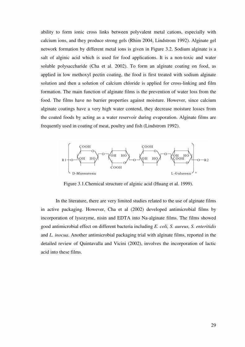
29
ability to form ionic cross links between polyvalent metal cations, especially with
calcium ions, and they produce strong gels (Rhim 2004, Lindstrom 1992). Alginate gel
network formation by different metal ions is given in Figure 3.2. Sodium alginate is a
salt of alginic acid which is used for food applications. It is a non-toxic and water
soluble polysaccharide (Cha et al. 2002). To form an alginate coating on food, as
applied in low methoxyl pectin coating, the food is first treated with sodium alginate
solution and then a solution of calcium chloride is applied for cross-linking and film
formation. The main function of alginate films is the prevention of water loss from the
food. The films have no barrier properties against moisture. However, since calcium
alginate coatings have a very high water contend, they decrease moisture losses from
the coated foods by acting as a water reservoir during evaporation. Alginate films are
frequently used in coating of meat, poultry and fish (Lindstrom 1992).
Figure 3.1.Chemical structure of alginic acid (Huang et al. 1999).
In the literature, there are very limited studies related to the use of alginate films
in active packaging. However, Cha et al (2002) developed antimicrobial films by
incorporation of lysozyme, nisin and EDTA into Na-alginate films. The films showed
good antimicrobial effect on different bacteria including E. coli, S. aureus, S. enteritidis
and L. inocua. Another antimicrobial packaging trial with alginate films, reported in the
detailed review of Quintavalla and Vicini (2002), involves the incorporation of lactic
acid into these films.

30
Figure 3.2. Alginate gel network formed by metal ions (Huang et al. 1999).
3.3.2.5. Carragenan Films
Carragenans are water soluble galactose polymers extracted from red seaweed
(Cha et al. 2002). Carragenan is a mixture of different polymers called: , , �, �, and
carragenan. , and �-carragenan are used in food applications (Gennadios et al. 1997).
-carragenan is highly sulfated and it forms gels with divalent cations like calcium.
carragenan , a less sulfated molecule, forms thermo reversible gels.
Similar to alginate films, carragenan films also act as a reservoir of moisture and
prevent moisture loss from the food (Lindstrom et al.1992). Frozen precooked meat,
poultry, and fish have been coated with calcium carragenan coatings. In the literature,
studies related to use of carragenan films in active packaging are scarce. Cha et al
(2002) developed lysozyme incorporated carragenan films. However, it was reported
that these films showed lower antimicrobial activity than lysozyme incorporated
alginate films tested under the same conditions.

31
3.3.2.6. Chitosan Films
Chitin is the structural polysaccharide found in the exoskeletons of insects,
crustaceans, fungi and algae. It is a linear polymer of �-(1�4) linked N-acetylglucose
amine. However, since it is water insoluble it should be modified to chitosan, a water
soluble form, by deacetylation before used as a packaging film material (Lindstrom et
al. 1992). Due to its cationic properties chitosan films are inherently antimicrobial. The
antimicrobial effect of chitosan is attributed to its interaction with the negatively
charged residues of bacterial surface (Martiral-Gros et al. 2002).
Dip or spray coating with chitosan is applied in the coating of fresh fruits
(Lindstrom et al. 1992). Such coatings reduce the maturation rate and they may also
provide an antimicrobial effect. Recently, Park et al. (2004) developed some lysozyme
incorporated chitosan composite films. These workers found that the inherent
antimicrobial effect of chitosan films can be improved by the incorporation of
lysozyme. Thus, chitosan films may be the most potential candidate for use in
combination with biopreservatives.
3.3.3. Lipid Films
Lipid compounds prevent the transport of moisture. Thus, films made from
lipids have good water vapor barrier properties. However, they are quite fragile,
unstable and relatively inflexible. Also, lipids usually form opaque films (Ryu et al.
2002, Guilbert 1996).Wax and acetylated monoglycerides are given below as examples
of lipid films.
3.3.3.1. Wax Coatings
Waxes consist of esters of long chain alcohols and fatty acids. They are widely
used for coating of fruits and vegetables to prevent moisture loss since they prevent
moisture transport better than most other lipid and non-lipid films. For example, the
moisture loss can be reduced by 50% with waxes that are coated on apples, grapes,
pears, prunes, tomatoes, peppers, and cucumbers, but wax coatings do not reduce decay

32
(Lindstrom et al. 1992). Some of the examples for waxes suitable for food coating
include beeswax, candelilla wax and carnauba wax (Baldwin et al. 1997).
3.3.3.2. Acetylated Monoglycerides
Acetylated glycerides can be obtained by either through the reaction of
glycerides with acetic anyhydride or through the catalyzed interesterification of a fat or
oil with triacetin (Gennadios et al. 1997). Acetylated monoglycerides are waxy and
relatively impermeable to moisture migration (Lindstrom et al. 1992, Kester and
Fennema 1986). They have been applied to meat cuts and poultry to protect against
dehydration during storage (Kester and Fennema 1986). To prevent moisture loss,
dehydrated apricot pieces were also coated by dipping into molten acetylated
monoglyceride (Lindstrom et al. 1992).
3.3.4. Composite Films
Different functional characteristics of several film elements can also be
combined to form a single film or coating. Different kinds of polysaccharides, proteins,
and lipids have been used, either alone or in mixtures, to obtain composite films. For
example, Schultz et al. (1949) incorporated lipids into low-methoxy pectinate films to
improve resistance to water vapor permeation (Kester and Fennema 1986). To maintain
high concentration of sorbic acid at the surface of an intermediate-moisture cheese
analog, Torres et al. (1985) also used a composite film composed of zein and acetylated
monoglycerides and plasticized with glycerol (Kester and Fennema 1986). Emulsion
coatings containing methyl cellulose, vegetable oil, beewax, and glycerol monooleate
are also good examples of composite films (Lindstrom et al. 1992).

33
CHAPTER 4
BIOPRESERVATION AND BIOPRESERVATIVES
4.1. Biopreservation
In food technology, different chemicals such as organic or inorganic acids,
metals, alcohols, ammonium compounds or amines can be used as antimicrobials
(Suppakul et al. 2003, Appendini and Hotchkiss 2002). However, due to the health
concerns of the consumers, producers are now highly interested in the use of
biopreservatives to maintain food safety and quality. For food technologists and
engineers the “biopreservation” is a new term. As reported by Hugas (1998) the term
biopreservation refers to “extended storage life and enhanced safety of foods using their
natural or controlled microflora and (or) their antibacterial products”. The
biopreservation may also be defined as “the use of active substances extracted from
food or obtained by food grade microorganism fermentation to enhance food safety and
quality” (Franchi et al. 2003).
4.2. Biopreservatives
As given in the definitions above, biopreservation is conducted by using some
protective microorganisms and their antibacterial products or by some active
compounds extracted from food. Different biopreservatives were discussed below.
4.2.1. Lactic Acid Bacteria
The use of lactic acid bacteria in food preservation have been practiced for a very
long time. In fact, the fermentation is one of the oldest biopreservation methods used by
humans and it involves the oxidation of carbohydrates by lactic acid bacteria to produce
a range of antimicrobial products including organic acids such as lactic and acetic acid,
hydrogen peroxide, bacteriocins etc. However, since the fermentation changes the
sensory properties of food almost completely, the current trend of using lactic acid

34
bacteria in biopreservation involves the addition of viable bacteriocin-producing pure
cultures to food for inhibition of pathogens and/or extension of the shelf-life while
changing the sensory properties as little as possible (Vermeiren et al. 2004, Hugas
1998). Bacteriocins are ribosomally-produced antimicrobial polypeptides or proteins
that may inhibit some pathogenic bacteria (Ross et al. 2002, Hugas 1998). Thus, the
growth and bacteriocin production of added lactic acid bacteria may increase the safety
of food and/or prolong its shelf life. However, the success of this application depends
on the ability of the culture to grow and produce bacteriocin in the food under the
environmental or technological conditions. For example, it was reported that the
psychotropic bacteriocin-producing lactic acid bacteria have a good potential for use in
biopreservation of meat and meat products (Hugas 1998). Recently, there have also
been studies to characterize lactic acid bacteria capable to produce hydrogen peroxide
under refrigeration temperatures (Villegas and Gilliland 1998, Jaroni and Brashears
2000). The hydrogen peroxide is a strong oxidant that is effective on both gram-positive
and gram-negative bacteria. Thus, inoculation of pure cultures of such bacteria may be a
novel approach for the use of lactic acid bacteria in biopreservation.
Besides addition of pure protective cultures, the use of original micro flora of
food may also be employed in biopreservation. For example, chilled-storage of red meat
under modified atmosphere triggers the change of natural micro flora from putrefactive
gram negative bacilli to fermentative lactic acid bacteria. In such a case, due to the low
carbohydrate content and strong buffering capacity of meat, the lactic acid bacteria
conduct a mild fermentation without any significant change in the sensory
characteristics of food and the shelf-life of product increases considerably (Hugas
1998).
4.2.2. Bacteriocins
As indicated above some lactic acid bacteria produce antimicrobial polypeptides
or proteins called bacteriocins (Vermeiren et al. 2004, Ross et al. 2002, Hugas 1998).
Thus, crude or pure bacteriocin preparations obtained from fermentation mediums of
bacteriocin-producing lactic acid bacteria are potential antimicrobials for
biopreservation of food. Some of the well known bacteriocins include nisin, pediocin
and lacticin. However, nisin is the only bacteriocin that found a widespread commercial
35
use (Ross et al. 2002). Nisin composed of 34 amino acids is a polypeptide that shows
antimicrobial activity mainly against gram positive bacteria. In diary products it shows
strong antibotulinical, antilisterial and antistaphylococcal activity when used at suitable
concentrations (Hugas 1998). However, the use of nisin in fresh meat products is not
very successful since this agent forms an inactive complex with glutathione (Rose et al.
1999). It was reported that the complex formation reaction is catalyzed by Glutathione
S-transerase enzyme in raw meat (Rose et al. 2002). Thus, it is suggested that the nisin
should be used in cooked meat products which do not contain active enzyme.
4.2.3. Antimicrobial Enzymes
4.2.3.1. Lysozyme
Lysozyme (EC3.2.1.17) is a single peptide enzyme found in different sources
including plants, animals and microorganisms (Ibrahim et al. 1996). It contains 129
amino acid residues and it has a molecular weight of 14400 (Takahashi et al. 2000,
Ibrahim et al 1991). The enzyme shows lytic activity on bacteria by hydrolyzing
glycosidic β-linkages between N-acetylhexosamines of pepdidoglycan (PG) in their cell
walls. Therefore, different studies have been conducted to use of lysozyme for the
biopreservation of food. Commercially the enzyme is obtained from hen egg white
(Chang et al. 2000). Thus, most of the studies related to lysozyme have been focused on
hen egg white lysozyme. The enzyme shows antibacterial activity mainly against gram-
positive bacteria, but it is ineffective against gram-negative bacteria which PG layer is
surrounded by a protective lipopolysaccharide (LPS) membrane (Nakamura et al. 1991,
Ibrahim et al. 1991). Thus, native lysozyme alone is currently employed in food
technology for some specific purposes such as prevention of the late blowing and off-
flavor formation in cheeses by Clostridium tyrobutiricum (de Roos et al. 1998). Because
of the absorption of lysozyme by casein micelles and retention of enzyme activity after
absorption, the enzyme may be very suitable for the biopresevation of dairy products
(de Roos et al. 1998). Also, there are some studies related to the control of malolactic
fermentation in wines (Marchal et al. 2000) and reduction of specific pathogenic
bacteria on meat surfaces by lysozyme (Korhonen et al. 1998). The sweet taste of the
enzyme in solutions also encouraged some scientists to study the potential applications

36
of enzyme in beverage industry as both a sweetener and antimicrobial (Masuda et al.
2001). Moreover, the studies related to the incorporation of enzyme on plastic or edible
films or film materials have also been conducted successfully (Padgett et al. 1998, Cha
et al. 2002, Appendini and Hotchkiss 1997). The extensive studies to increase the antimicrobial spectrum of lysozyme and
make it a general purpose biopreservative are also quite promising. For example, it was
found that combination of lysozyme with EDTA makes lysozyme highly effective on
gram-negative bacteria. The increased antimicrobial spectrum of lysozyme in presence
of EDTA is attributed to destabilizing effect of EDTA on the LPS layer which
surrounds PG layer of gram-negative bacteria. The mechanism of the destabilization of
LPS layer is not fully characterized. However, it was thought that it occurs by the
chelating of the divalent cations such as Ca+2 and Mg+2 that are necessary for binding of
LPS to PG (Branen and Davidson 2004). Nakamura et al. (1991, 1992) showed that
conjugates of lysozyme with dextran, galactomannan or xyloglucan have good
antimicrobial activity against both gram-positive and gram-negative bacteria when
applied in combination with mild heating at 50 oC. These conjugates obtained naturally
by maillard reaction also showed excellent emulsifying properties superior to those of
commercial emulsifiers (Nakamura et al. 1992). The conjugates of lysozyme with
glycolipids such as glucose stearic monoester can also be obtained naturally through
maillard reaction to produce perfect emulsifiers (Takahashi et al. 2000). However, such
conjugates show very little antimicrobial activity. The same problem was also observed
in a genetically modified double glycosylated lysozyme which showed excellent
emulsifying properties but no lytic action (Shu et al. 1998). By lipophilization of
lysozyme with different fatty acids, Ibrahim et al. (1991, 1993) and Liu et al. (2000)
produced some modified lysozymes which possess antimicrobial activity against gram-
negative bacteria at room temperature. The main disadvantage of lipophilization is the
low recovery of enzyme activity after this process. However, this may be overcome by
stabilization of enzyme activity with glycosylation applied before lypophilization
process (Liu et al. 2000). Also, Ibrahim et al (1996a, 1996b) found that when lysozyme
is partially denatured by heating in pH 6.0 buffer at 80 oC for 20min, it gained a strong
bactericidal activity against gram-negative bacteria such as E. coli, S. enteritidis and P.
auriginosa. It was reported that the partially denatured enzyme also maintained its
antimicrobial activity against gram-positive bacteria even when it was heated to lose its
37
catalytic activity towards PG completely. Thus, antimicrobial activity of heat treated
lysozyme may not be determined only by its catalytic activity.
Although, the most probable candidate for food preservation is hen egg white
lysozyme, there are some other lysozymes which naturally have broad antimicrobial
activity. For example, Human lysozyme (HL) and bovine stomach lysozyme (BSL) are
reported to have antimicrobial activity towards both gram-positive and gram-negative
bacteria as well as fungi. Wilcox et al. (1997) showed that BSL genetically introduced
to tobacco plant may be recovered and purified to 93 % homogeneity from the
transgenic plants by an easily scalable process. This study showed the possibility of
commercial BSL production from transgenic plants and increased the importance of this
enzyme as hen egg white lysozyme alternatives.
4.2.3.2. Lactoperoxidase
The enzyme lactoperoxidase is a basic glycoprotein, containing a heme group
with Fe+3. It consists of a single polypeptide chain of 612 residues with a molecular
mass of ~ 78.5 kDa and a carbohydrate content of about 10%. It is an oxidoreductase
found in mammalian milk, saliva, and tear (Wolf et al. 2000, Pakkanen and Aalto. 1997,
Jacob et al. 2000, Monzani et al. 1997) and it plays an important role in protecting the
lactating mammary gland and the intestinal tract of new-born infants against pathogenic
microorganisms. The mechanism of the antimicrobial action of LPS is based on
conversion of thiocynate (SCN-) to antimicrobial products such as hipothiocyanite
(OSCN-) ion, hypothiocyanous acid (HOSCN) and some other highly reactive and short
lived oxidation products in presence of H2O2 (Pruitt et al. 1982). This system owing its
antimicrobial effect to oxidation of sulphydryl (-SH) groups in microbial enzymes
shows bactericidal effect on gram-negative bacteria and bacteriostatic effect on gram-
positive bacteria (Seifu et al. 2005). Also, it has antifungal (Min and Krochta 2005,
Jacob et al. 2000) and antiviral (Pakhanen and Aalto 1997, Seifu et al. 2005) activities.
The synergetic effect of LPS with nisin has also been demonstrated (Zapico et al. 1998,
Dufour et al. 2003, Boussouel et al. 2000). Thus, the use of LPS alone or in combination
with nisin may give unique biopreservation systems that are effective on most
pathogens.

38
Due to its broad antimicrobial spectrum and generally recognized as safe
(GRAS) status (Elliot et al. 2003) different studies have recently been conducted related
to use of lactoperoxidase (LPS)-thiocyanate-H2O2 antimicrobial system in
biopreservation. Since LPS system is part of the natural preservation system exists in
milk, the addition of thiocyanate and /or H2O2 to milk to activate naturally occurring
LPS is used to improve microbial quality of milk and cheese (Seifu et al. 2004, Seifu et
al. 2005). Commercially, the enzyme is produced from bovine whey (Ye et al. 2000,
Hahn et al. 1998). Thus, the addition of LPS and other components of this antimicrobial
system to thermally processed skim milk (Zapico et al. 1998) and meat products
(Kennedy et al. 2000, Elliot et al. 2004) and prevention of the development of
pathogenic bacteria have also been studied. The studies related to use of LPS in
antimicrobial packaging, on the other hand, are very limited. In their detailed review
Suppakul et al. (2003) suggested the use LPS in antimicrobial packaging. Recently,
Min and Krochta studied the inhibition of Penicillium commune by LPS impregnated
whey protein films and showed the good potential of this enzyme for use in
antimicrobial packaging.
4.2.3.3. Glucose Oxidase
Although the lysozyme and lactoperoxidase are the most suitable enzymes to use
in biopreservation, there are some other enzymes that may be used to form an
antimicrobial mechanism. The enzyme glucose oxidase, gained a GRAS status in US in
1958 (Labuza and Breene 1989), has long been used to reduce glucose content of
powdered egg (Petruccioli et al. 1999). The enzyme is an oxidoreductase and its
antimicrobial activity is related mainly with its limitation of oxygen needed for the
aerobic microorganisms and production of hydrogen peroxide. The mechanism of the
enzyme depends on its removal of two hydrogen from the –CHOH groups of glucose
with the formation of glucono-delta-lacton and hydrogen peroxide (Labuza and Breene
1989). By use of glucose oxidase-glucose system, it is possible to remove one mole of
oxygen per mole of oxidized glucose. However, in presence of catalase, the
effectiveness of this enzyme system is reduced since the enzymatically formed
hydrogen peroxide is decomposed to water and oxygen. The enzyme is commercially
produced as a by-product of the gluconic acid fermentation from Aspergillus niger or

39
from fermentation of Penicillium amagasakiense (Karmali et al. 2004). The catalase is a
natural contaminant of the glucose oxidase enzyme preparations and the need of further
purification to separate catalase makes the use of glucose oxidase system very
expensive (Labuza and Breene 1989). Thus, currently the use of this enzyme in food
packaging applications is very limited.
4.2.3.4. Chitinase
Another antimicrobial enzyme is chitinase. Chitinase is an enzyme capable to
catalyze the hydrolysis of chitin to its oligomers and/or monomers. Thus, the
antimicrobial effect of this enzyme is against fungi. The commercial production of
enzyme is conducted by fermentation by using different microorganisms such as
Tricoderma harzainum, Streptomyces sp. and Penicillium sp. (Navani and Kapadnis
2005, Binod et al. 2005). However, the studies to use this enzyme in food industry are
scarce. One of the few studies conducted with this enzyme was that of Fenice et al.
(1999) who used enzyme in combination with high hydrostatic pressure. These workers
reported that the application of chitinase reduces the pressure necessary for inactivation
of food spoiling mold Mucor plumbeus. Further studies are needed to increase the
potential food applications of this enzyme.
4.2.4. Bioactive Proteins and Peptides
4.2.4.1. Lactoferrin
Lactoferrin is a member of transferrin family of non heme iron binding
glycoproteins having a molecular weight of 80 kDa. It is found in colostrum, milk, and
other exocrine secretions such as tears (Satuè-Gracia et al. 2000, Recio and Visser 2000,
Pakkanen and Aalto 1997). The protein has bactericidal and/or bacteriostatic activities
on both gram-positive and gram-negative bacteria, and fungistatic activity on fungi. The
bacteriostatic and fungistatic activities of lactoferrin have been attributed mainly to its
ability to bind and reduce the availability of iron for the microorganisms (Andersson et
al. 2000), while its bactericidal activity is related to its direct interaction with cell
membranes. Lactoferrin also exhibits an antiviral activity against some human intestinal

40
viruses which may be potential food contaminants (Superti et al. 1997, Marchetti et al.
1999). The mechanism of the antiviral activity of lactoferrin has not been fully
understood, but it is attributed to its complex formation ability with virus particles or its
interference with the viral attachment to cell receptors (Marchetti et al. 1999). Besides
its broad antimicrobial spectrum, lactoferrin acts also synergistically with lysozyme
(Ellison et al. 1991). Thus, combination of these two proteins may be used when higher
antimicrobial activity is required in food systems. Pakkanen and Aalto (1997), on the
other hand, reported that lactoperoxidase and lactoferrin show additive antibacterial
effect against Streptococcus mutans. The hydrolysis of lactoferrin with pepsin generates
a considerably more potent bactericidal peptide called lactoferricin. Lactoferricin
corresponds to the 24 (17-41) or 25 (17-42) amino acid residues near the N terminus of
lactoferrin (Recio and Visser 1999). This location is reported to be a region distinct
from lactoferrin’s iron-binding sites (Lucca and Walsh 1999). The peptide is also
reported to have a strong antifungal activity (Lucca and Walsh 1999). Liceaga-Gesualdo
et al. (2001) reported that lactoferricin effectively inhibits both spore germination and
mycelial growth of Penicillium spp and suggested the use of peptide as an antifungal in
bottled water. In contrast, Min and Krochta (2005) found whey protein films
incorporated with lactoferrin and lactoferricin ineffective on Penicillium commune. Besides its high potential as an antimicrobial, lactoferrin may also be used as an
antioxidant in food systems. The antioxidant activity of the polypeptide is the result of
its strong iron binding capacity. Lactoferrin has two globular lobes that bind one iron
atom each. This protein-iron complex is very stable between pH 4 and 7 values (Ward
et al. 1996). Also, the protein maintains its iron binding ability and solubility after mild
pasteurization and UHT treatments (Mata et al. 1998). There are only limited studies
about the antioxidant effect of lactoferrin in food systems. Huang et al (1999) showed
the decreased prooxidant effect of iron in corn oil emulsions. Sauté-Garcia et al (2000)
reported the concentration-dependent inhibition of oxidation by lactoferrin in whey-
based and casein predominant infant formulas. Thus, lactoferrin may be used in a wide
range of food systems as a bifunctional food additive.
Bovine milk contains approximately 0.1 mg/mL lactoferrin that is 22% saturated
with iron (Satuè-Gracia et al. 2000). Thus, commercially lactoferrin is obtained form
bovine whey, a by-product of dairy industry, by using standard column
chromatographic procedures or modern membrane techniques (Ulber et al. 2001). Also,
there is an increasing demand for the production of recombinant human lactoferrin (Hlf)

41
in microorganisms or in transgenic animals and plants (Spik and Theisen 2000, Mata et
al. 1998). Mata et al (1998) found that Hlf and recombinanat Hlf from Aspergillus
awamori can be survived from mild pasteurization (72 oC-20 sec) and UHT treatments
(135 oC-8 sec). Thus, they proposed that these Hlfs would have been more suitable for
food which will be subjected to heat treatment.
4.2.4.2. Phosvitin
Phosvitin is a phosphoglycoprotein found in egg yolk (Nakamura et al. 1998,
Khan et al. 1998). Nearly 50 % of its amino acids are serine (Castellani et al. 2003a ,
Castellani et al. 2003b) and it is a highly phosphorylated protein containing
approximately 10 % phosphorus (Lee et al. 2002, Khan et al. 1998). Phosvitin has been
fractionated into � and � components that contain different phosphorus contents and
molecular weights. The main components have molecular mass of 45000 for �-
phosvitin, and 37500, 42000, and 45000 for �-phosvitin (Khan et al. 1998; Castellani et
al. 2003b).
Phosvitin is used as a natural antimicrobial due to its ability to bind and limit
iron necessary for bacterial growth. When combined with mild heating, due to its
emulsifying properties, the protein showed high affinity particularly to gram-negative
bacteria cell wall that consists of lipopolysaccharides (Khan et al. 2000). Phosvitin acts
also as an antioxidant and inhibits metal catalyzed lipid-oxidation (Maheswari et al.
1997). Strong affinity of phosvitin to metal ions is due to its high phosphorus content
(Guérin-Dubiard et al. 2002). Castellani et al. (2003a) investigated the influence of
different factors on iron binding capacity of purified phosvitin found that pH 6.5 was
the best pH for iron fixation of phosvitin. The maximum iron binding capacity of the
protein was 115 µg of iron/mg of phosvitin and this corresponds to one iron per two
phosphate groups (Castellani et al. 2003a). The antioxidant activity of phosvitin may be
increased by modification. For example, Nakamura et al. (1998) modified the phosvitin
by natural maillard reaction with galactomannan and successfully increased its
antioxidant activity. Recently, Lee et al. (2002) showed the antioxidant activity of
phosvitin in muscle food. However, according to these researchers, in muscle food
phosvitin is not a very effective antioxidant and it should be combined with other

42
antioxidants. Further studies are needed to evaluate the real antimicrobial and
antioxidant potential of phosvitin in food systems.

43
CHAPTER 5
MATERIALS AND METHODS
5.1. Materials
Whole milk used in LPS production was obtained in different time intervals
from the same Holstein cow of a local farm in Urla, Izmir (Turkey). Fresh hen eggs
used to produce LSZ were obtained from a supermarket in �zmir (Turkey). Toyopearl
sulphopropyl (SP) cation exchanger (SP-550C, fast flow, size: 100 micron) was
obtained from Supelco (Bellefonte, PA, USA). Corn zein, Micrococcus lysodeikticus,
dialysis tubes (cut off: 12000 MW), dextran (from Leuconostoc mesenteroides, 73.200
MW), ABTS (2,2-Azino-bis-(3-Ethylbenz-Thiazoline-6-Sulfonic acid)) and sodium salt
of alginic acid (from Macrocystis pyrifera , viscosity of 2 % solution at 25 oC is 3500
cpc) were obtained from Sigma (St. louis, Mo., USA). Rennet was obtained from ICN
Biomedicals Inc. (Aurora, Ohio, USA). Glycerol and ascorbic acid were obtained from
Merck (Darmstadt, Germany). Sericin (Silk Biochemical Co. Ltd) was kindly donated
by Assistant Professor Dr. O�uz Bayraktar from Izmir Institute of Technology. The
lyophilized preparations of crude thermally processed chick pea proteins (TP-CP) were
provided by Research Assistant �skender Arcan from �zmir Institute of Technology.
These crude preparations containing almost 51% protein were produced by modification
of the method described by Geneuese and Lajolo (1998) according to Arcan (2005).
5.2. Methods
5.2.1. Production of Biopreservatives
5.2.1.1. Production of Lactoperoxidase (LPS)
The LPS was produced from bovine whey by the method given in Ye et al. (2000)
with slight modifications. The skim milk was obtained by centrifugation of 1L of whole
milk at 5000 x g for 20 min at 30 ºC. The skim milk was then filtrated from cheese-

44
cloth, warmed up to 37 ºC in an incubator and 90 mg rennet was added to 900 mL of the
skim milk. After 1 h at 37 ºC, the precipitates in whey were separated by filtration from
a cheese-cloth and centrifuged at 10000 x g for 25 min at 4 ºC. 400 mL of the clarified
whey was then loaded to a SP Toyopearl column (height: 11.5 cm, diameter: 2.8 cm)
previously equilibrated with 0.05 M Na-phosphate buffer at pH 6.5. The column was
first washed with 500 mL of the equilibration buffer and then it was eluted with a linear
gradient of 600 mL of 0-0.55 M NaCl prepared in the same buffer. 10 mL fractions
collected from the column were assayed for their absorbance at 280 nm and LPS
activity (see section 5.2.3). This study was conducted by using LPS from three different
purification studies. In the first purification study, the activity profile of LPS eluted was
monitored quantitatively whereas in the other studies it was monitored qualitatively by
using the same reaction mixture. The enzyme containing fractions collected from the
column were pooled and dialyzed 24 h at 4 oC. The dialyzed enzyme was then
lyophilized in a Labconco freeze-dryer (FreeZone 6 liter, Kansas City, MO, USA),
working approximately between -44 and -47 oC collector temperature and 50 x 10-3 and
100 x 10-3 mBar vacuum, after dissolving 250-300 mg dextran in the dialyzed enzyme
extract as a supporting material. The sample container volume was two to three times
the sample volume. The lyophilized enzymes used in film making were stored at – 18 oC and their activities were determined as U/mg before each film preparation.
5.2.1.2. Production of Lysozyme (LSZ)
The LSZ was produced by slightly modifying the partial purification step given in
Jiang et al. (2001). Briefly, the egg whites separated carefully without disturbing the
egg yolks were first diluted 3-fold with 0.05 M NaCl solution. To precipitate the egg
white proteins other than the LSZ, the pH of this mixture was set to 4.0 by carefully
adding several drops of 1 N acetic acid and it was diluted with equal volume of 60 %
(v/v) ethanol. After 6 hours incubation at room temperature in presence of 30 %
ethanol, the mixture was centrifuged at 15 000 x g for 15 min at 4 oC and the precipitate
was discarded. The supernatant containing LSZ was first dialyzed for 21 h at 4 oC by
three changes of 2000 mL distilled water and then lyophilized as described above
without using any supporting material. The lyophilized enzymes used in film making

45
were stored at – 18 oC and their activities were determined as U/mg before each film
preparation.
5.2.2. Stability of the Produced LPS and LSZ in Lyophilized Form
The stability of the lyophilized LPS and LSZ was determined by monitoring of
enzymes’ activity during frozen storage at – 18 oC. The preparations were dissolved in
distilled water and their activities were determined as described in sections 5.2.3 and
5.2.4.
5.2.3. Determination of LPS Activity
The activity of LPS was determined spectrophotometrically by using a Shimadzu
(Model 2450, Tokyo, Japan) spectrophotometer equipped with a constant temperature
cell holder working at 30 oC and using ABTS as substrate (Chávarri et al. 1998). The
reaction mixture was formed by mixing 2.3 mL, 0.65 mM ABTS prepared in 0.1 M Na-
phosphate buffer at pH 6.0, 0.1 mL enzyme solution and 0.1 mL, 0.2 mM H2O2 solution.
All components of reaction mixture were brought to 30 oC before mixing. The increase
in absorbance was monitored at 412 nm for 5 min and enzyme activity calculated from
the slope of the initial linear portion of absorbance vs. time curve was expressed as Unit
(0.001 absorbance change in one minute).
5.2.4. Determination of LSZ Activity
The activity of LSZ was determined spectrophotometrically at 660 nm and 30 oC
by modification of the method given in Chang and Chang (2000). The reaction mixture
was prepared by mixing 2.3 mL Micrococcus lysodeikticus cell suspension (at 30 oC)
prepared 0.26 mg/mL in 0.05 M, pH 7.0 Na-phosphate buffer and 0.2 mL enzyme
solution (incubated at 30 oC for 5 min). The reduction in absorbance was monitored for
5 min and enzyme activity was calculated from the slope of the initial linear portion of
absorbance vs. time curve. The enzyme activity was determined as Unit (0.001 change
in absorbance in one minute) or percent initial activity.

46
5.2.5. Preparation of Films
5.2.5.1. Preparation of Alginate Films
To prepare the films, 0.62-5.2 mg LPS preparation and/or 4 or 8 mg antioxidant
protein (TP-CP proteins, sericin or bovine serum albumin) was dissolved per g of 2 %
(w/v) alginic acid solution by mixing slowly with a magnetic stirrer (final
concentrations of LPS in dried films: 80-690 µg/cm2 [516-4325 U/cm2]; final
concentrations of antioxidant proteins in dried film: 0.53 or 1.06 mg/cm2). After
complete solubilization of the ingredients 10 g portions of film preparation solution
were spread onto glass petri dishes (9.8 cm in diameter). The petri dishes were dried at
room temperature for three days and 0.8 mL, 0.3 M CaCl2 was pipetted into them to
cross-link the dried films. The average thickness of dried control films prepared by this
method is determined by scanning electron microscope (Philips XL 30S FEG, FEI
Company, Eindhoven, Netherlands) as 12.8 µm.
5.2.5.2. Preparation of Zein Films
The zein films were produced as described in Padgett et al. (1998). Briefly, 1.4 g
corn zein was dissolved with 8.1 mL of ethanol (97 %) by mixing slowly with a
magnetic stirrer for 25 min. 0.39 mL glycerol was then added to medium and the
temperature of the mixture was increased until it started to boil. The mixing was then
ceased and the film solution was boiled for 5 min. After cooling to room temperature,
1.1-16.2 mg LSZ and/or 9-18 mg antioxidant protein (TP-CP proteins or bovine serum
albumin) was added per g of film preparation solution (final concentrations of LSZ in
dried films: 63-957 µg/cm2 [187-2800 U/ cm2]; final concentrations of antioxidant
proteins in dried films: 0.53 or 1.06 mg/cm2). The mixture was further stirred for 25
minute and 4.3 g of it was spread evenly onto a 8.5 x 8.5 cm glass plate, previously
cleaned with ethanol. The plates were placed in an incubator (Memmert, 108 L, Model
BE 500, Germany) and films were dried at room temperature for 24 h by keeping the
incubator door half open. After drying, the films were peeled from the glass plates
carefully and 6 x 6 cm pieces cut from the middle of films were used in all tests. The
average thicknesses of films determined by a micrometer changed between 0.14 and

47
0.15 mm for control and LSZ incorporated zein films and 0.16 and 0.18 mm for LSZ
and antioxidant protein incorporated films.
5.2.6. Studies Related to Alginate Films Incorporated with LPS
5.2.6.1. Monitoring of LPS Activity Release from Alginate Films
The release tests were conducted in a refrigerated incubator (Sanyo Model MIR-
153, Japan). The films (containing 0.15 mg/cm2 [941 U/cm2 LPS] area: 75.4 cm2) were
placed in glass petri dishes containing 50 mL distilled water (4 oC). The petri dishes
were then covered with nylon stretch films and their glass lids and incubated for 1800
min at 4 oC with continuous stirring at 200 rpm with a magnetic stirrer (2 cm long
Teflon coated rod). The LPS activity in release test solution was monitored by taking
0.6 mL aliquots from the release test solution at different time intervals and conducting
activity measurements for three times by using 0.2 mL of the taken aliquot in a single
measurement. In release tests, the standard reaction mixture was changed as; 1.5 mL,
0.65 mM ABTS prepared in 0.1 M Na-phosphate buffer at pH 6.0, 0.2 mL release test
solution and 0.5 mL 0.1 mM H2O2. This reaction mixture measures enzyme activity
almost 4.3 fold higher than the standard reaction mixture given in section 5.2.3. Thus, it
is quite suitable to measure residual amounts of LPS released from the films.
5.2.6.2. Determination of LPS Affinity to Alginate Films
The affinity of LPS to alginate films was determined by modifying the method
used by Appendini and Hotchkiss (1997) who determined the affinity of lysozyme to
different plastic films. This method was based on considering the amount of enzyme
activity retained in films after repeated activity measurements. In this study, after each
activity measurement, the films were additionally washed with cold distilled water. The
films used in this study contained 3252 U/cm2 (0.52 mg/cm2) LPS. The activity
determination was conducted for 7 times and after each activity determination, to
remove the free enzyme (if existed) and oxidized substrate absorbed, the films were
washed by placing firstly for 3 min into a beaker containing 100 mL of cold distilled
water and then secondly for 15 min into another beaker containing 250 mL of cold

48
distilled water stirred magnetically. In these tests only, the activity measurements were
conducted by a two point assay, by reading standard reaction mixture absorbance (at
412 nm) at 0th and 30th minutes of reaction (Activity = Abs30min-Abs0min). The activity
was expressed as percent initial activity.
5.2.6.3. Determination of Immobilized LPS Activity in Alginate Films
Before assayed for LPS activity, the crosslinked films were peeled from the petri
dishes and immersed into a beaker containing 50 mL distilled water for 30 seconds, to
remove excessive CaCl2 that causes some precipitations in reaction mixture. The films
were then halved carefully with a clean razor and one of these halves (area: 37.7 cm2)
was placed into a glass petri dish containing 23 mL, 0.65 mM ABTS solution prepared
in 0.1 M Na-phosphate buffer at pH 6.0 and 2 mL, 0.1-0.3 mM H2O2 solution brought to
30 oC before placing films. The petri dishes were kept in an incubator at 30 oC and
stirred at 200 rpm with a magnetic stirrer. The activity monitored by measuring the
reaction mixture absorbance at 412 nm at different time intervals was determined from
the slope of the initial linear portion of absorbance vs. time curve. The measurements
were repeated for the remaining halves of the films and average of two measurements
was considered in all measurements. The activity was expressed as U (0.001 absorbance
change in one minute) per cm2 of the films.
5.2.6.4. Characterization of Immobilized LPS Activity in Alginate
Films
In all characterization studies, the activities were determined with the standard
method given in section 5.2.6.3 at 30 oC unless otherwise was indicated. All
measurements were applied for two film halves seperately and averages were used in
determination of an effect on LPS activity, expressed as U/cm2 or percent initial
activity.

49
Effect of LPS preparation concentration on LPS activity of alginate films
The effect of LPS preparation concentration on activity of this enzyme in films
was determined by measuring activity of films containing LPS between 516 and 4325
U/cm2 (0.08 and 0.69 µg/cm2).
Effect of H2O2 concentration on LPS activity of alginate films
The effect of H2O2 concentration on LPS activity of films was determined by
measuring activity of films containing 1200 U/cm2 (0.24-0.28 mg/cm2) LPS at different
H2O2 concentrations (final concentrations in reaction mixture was between 2 and 24
�M).
Effect of temperature on LPS activity of alginate films
The effect of temperature on LPS activity of films was determined by measuring
activity of films containing 998 U/cm2 (0.23 mg/cm2) LPS between 4 ° and 30 °C. The
activation energy (Ea) for the activity was calculated by using Arrhenius plot formed by
plotting natural logarithm of activity (U/cm2) vs. reciprocal of temperature (1/T(oK)).
The temperature quotient (Q10) of the enzyme activity was calculated from the equation
of k2/k1= (Q10) T2-T1, where k is reaction rate (activity) and T is temperature (oC).
Effect of pH on LPS activity of alginate films
The effect of pH on LPS activity of films was determined by measuring enzyme
activity of films containing 1274 U/cm2 (0.19 mg/cm2) LPS in the reaction mixture of
23 mL, 0.65 mM ABTS solution prepared in 0.1 M acetate (at pH 3.1, 4.0 or 5.0) or Na-
phosphate (at pH 6.0 or 7.0) buffers and 2 mL, 0.3 mM H2O2.
pH stability of LPS activity of alginate films
The pH stabilities of LPS activity of films were determined by incubating films
containing 1114 U/cm2 (0.19 mg/cm2) LPS in 25 mL, 0.1 M acetate (at pH 3.1, 4.0 or
5.0) or 0.1 M Na-phosphate (at pH 6.0) buffers at 4 °C for 24 h. The remaining activity

50
of the films were determined at pH 6.0 by using the following reaction mixture, 23 mL
0.65 mM ABTS prepared in 0.1 M Na-phosphate buffer at pH 6.0 and 2 mL, 0.3 mM
H2O2. The remaining LPS activity of films were given as percent of initial activity
determined at pH 6.0 by using films which were not incubated for 24h.
5.2.7. Studies Related to Alginate Films Incorporated with LPS and
Antioxidant Proteins
5.2.7.1. Determination of LPS and Antioxidant Activity in Alginate
Films
In these experiments one half of a dried and cross-linked film was used for the
determination of antioxidant activity whereas the other half was used for the
determination of LPS activity as described in section 5.2.6.3. Before used in activity or
antioxidant activity determination, the films were immersed into a beaker containing 50
mL distilled water for 30 seconds, to remove excessive CaCl2 that causes precipitations
in reaction mixtures. The water used in washing was not discarded and assayed for
antioxidant activity as described in section 5.2.8.1. For the determination of antioxidant
activity of films, a washed film half was placed in 25 mL, 7 mM ABTS radical cation
(ABTS.+) solution (at 30 °C), produced by using potassium persulfate and diluted with 5
mM PBS (pH 7.4) to absorbance of ~0.75 at 734 nm (Re et al. 1999). The inhibition of
ABTS radical was monitored at 734 nm and 30 oC and the percent inhibition at the end
3 or 15 minute was calculated from the following equation;
% inhibition ( )
( )10011
min0
min)153(
min)0(
min153 ��
�
�
��
�
�
���
�
�−−
���
�
�−=
control
controlor
sample
sampleor
Abs
Abs
Abs
Abs
Distilled water was used as control. The obtained inhibitions were also compared with
those inhibitions of the standard antioxidant vitamin C tested under the same conditions
and given as nmol vitamin C/cm2 (see Appendix A).

51
5.2.8. Studies Related to Zein Films Incorporated with LSZ and/or
Antioxidant Proteins
5.2.8.1. Monitoring of LSZ and Antioxidant Activity Release from Zein
Films
The release tests were conducted in a refrigerated incubator at 4 oC. The films (6
x 6 cm) were placed in glass petri dishes containing 50 mL distilled water (4 oC). The
petri dishes were then covered with nylon stretch films and their glass lids and
incubated for 1800 min with continuous stirring at 200 rpm with a magnetic stirrer (2
cm long teflon coated rod).
The LSZ activity released from films was monitored by taking 0.6 mL aliquots
from the release test solution at different time intervals and conducting activity
measurements (see section 5.2.4.) for three times by using 0.2 mL of the taken aliquot in
a single measurement. The activity was expressed as total units released per cm2 of the
films (U/cm2) for a given time period. All calculations were corrected by considering
the total activity removed during sampling. The monitoring of enzyme release was
continued until the activity increase in release test solutions ended and a slight reduction
in activity initiated. The release rates were calculated from the slopes of the best fit
curves of initial portions of activity released vs. time curves as U/cm2/min. The total
activity released from a film was determined by considering the highest activity points
in activity released vs. time curves. The percentage of activity recovered from a film in
a release test was determined from the ratio of total activity released and incorporated
activity, calculated by considering the activity and amount of partially purified LSZ
added to film making solutions.
The antioxidant activity released from films was monitored by taking 0.6
mL aliquots from the release test solution at different time intervals and conducting
antioxidant activity measurements for three times by using 0.2 mL of the taken aliquot
in a single measurement. The antioxidant activity was determined by using the ABTS
radical cation (ABTS.+) prepared and diluted as described in section 5.2.7.1. The
reaction mixture was formed by mixing 0.2 mL sample and 1.9 mL ABTS radical
solution. The inhibition of ABTS radical was monitored at 734 nm and 30 oC and the
percent inhibition at the end of 3 or 15 minute was calculated from the equation given

52
in section 5.2.7.1. Distilled water was used as control. The total antioxidant activity
released from the films at the end of release test was calculated as nmol vitamin C/cm2
by considering the highest activities released from the films (see standard curve for
vitamin C in Appendix B). During calculation of total antioxidant activity released, the
antioxidant activity removed during sampling was also considered.
5.2.8.2. Determination of Immobilized LSZ Activity in Zein Films
Remained after Release Tests
In release tests, the LSZ release from all films ended at 1380 min of the release
test. Also, no measurable activity was determined in selected films exposed to a second
release test following the first release test last for 1800 min. Thus, the enzyme retained
in zein films after the first release test was designated as immobilized enzyme. To
determine the immobilized enzyme activity, the films (6 x 6 cm) obtained from release
tests (for 1800 min) were placed into glass petri dishes containing 25 mL Micrococcus
lysodeikticus solution (at 30 oC) prepared 0.26 mg/mL in 0.05 M, pH 7.0 Na-phosphate
buffer. The petri dishes were kept in an incubator at 30 oC and their contents’
absorbance at 660 nm was monitored periodically. The LSZ activity of the films were
determined from the slopes of the initial linear portions of absorbance vs. time curve
and given as U/cm2 after multiplication of the obtained result by the factor of ten. The
factor was used to obtain comparable results with the measurements of standard reaction
mixture of soluble enzyme that has 10 fold lower reaction volume (see section 5.2.4).
5.2.8.3. Determination of Retained Antioxidant Activity in Zein Films
after Release Test
For the determination of retained antioxidant activity in films, following 1800
min release test, the films were transferred to glass petri dishes containing 25 mL, 7 mM
ABTS radical solution (see section 5.2.7.1 for preparation) at 30 °C. The inhibition of
ABTS radical was monitored at 734 nm and 30 oC and the percent inhibition was
calculated from the equation given in 5.2.7.1. The obtained inhibitions were also
compared with those inhibitions obtained for the standard antioxidant vitamin C tested

53
under the same conditions and given as nmol vitamin C/cm2 (see standard curve for
Vitamin C in Appendix A).
5.2.8.4. Stability of LSZ in Pre-cast Zein Films
For the determination of the stability of enzyme in pre-cast films, 374 U/cm2 (112
µg/cm2) LSZ and 180 µg/cm2 disodium EDTA incorporated films were cold stored at 4 oC. In certain time periods, the films (6 x 6 cm) were placed into glass petri dishes
containing 50 mL distilled water at 4 oC and stirred at 200 rpm as described in the
release tests (see section 5.2.8.1). At the end of 1800 min, a period that enzyme release
ended and a slight decline was observed in activity, the films were removed from
distilled water and the total enzyme activity in release test solutions were determined.
The activity was expressed as total units released per cm2 of the films.
5.2.9. Protein Content
Protein content was determined according to Lowry procedure by using bovine
serum albumin as standard (see Appendix C) (Harris 1987).

54
CHAPTER 6
RESULTS AND DISCUSSIONS
In this study, the biopreservatives produced by simple fractionation, dialysis or
column chromotagraphy were incorporated into edible films. From the edible films,
alginate films were used for incorporation of lactoperoxidase (LPS) and/or antioxidant
proteins, whereas zein films were used for incorporation of lysozyme (LSZ) and/or
antioxidant proteins. The antioxidant proteins, bovine serum albumin (BSA), thermally
processed chick pea proteins (TP-CP) and sericin, were obtained from different
chemical companies or provided by researchers specified in the materials section.
6.1. Production of Biopreservatives
6.1.1. Production of LPS
In different partial purification studies conducted, the LPS elution from SP-
Toyopearl column initiated when salt concentration is between 0.21 and 0.26 M, and
ended when salt concentration reached between 0.42 and 0.51 M. In all elutions the
most active LPS fractions came between 0.3 and 0.4 M salt concentrations. In batch-1, a
large protein peak containing two fractions and lacking LPS activity was detected
before LPS elution (Fig. 6.1). The elution profile of LPS activity suggested the presence
of two major LPS enzyme form in bovine whey (F1 and F2). In batch-2, conducted with
another milk sample after several months, the LPS was not separated from the major
protein peak eluted before LPS active fractions and several protein peaks were observed
in the enzyme active region (Fig. 6.2). This increased the protein content of the
collected LPS active fractions. However, because of the higher recovery of LPS activity
following column chromatography of batch-2, the specific activity of enzyme and
purification fold of separation were not significantly different from those of the same
parameters in batch-1 (Table 6.1). In batch-3, as occurred in batch-1, the major portion
of LPS active fractions eluted separated from the large protein peak coming before
enzyme. The bovine milk used in batch-3 contained the highest LPS activity and its

55
specific activity and recovery after column chromatography was also highest. The
results of these chromatographic separations are quite different from those of Ye et al.
(2000) that used the same chromatographic procedure for the partial purification of LPS
from bovine whey. These workers eluted LPS from the column between 0.16 and 0.19
M salt concentrations as a single peak. Also, they eluted a large protein fraction
containing two protein peaks, reported as lactoferrin-a and lactoferrin-b by these
workers, eluted from the column between 0.42 and 0.55 M salt concentrations. Özdemir
and Floros (2001), on the other hand, determined two LPS fractions after cation
exchange chromatography on CM-Sephadex C-50. However, the LPS gave a single
band in SDS- PAGE electrophoresis.
Figure 6.1. Protein and LPS activity profiles during Toyopearl-SP cation exchange
chromatography of bovine whey (Batch no: 1)
Watanabe et al. (2000) explained the heterogenic profiles of LPS in disk-
electrophoresis and ion-exhange chromatography by differences in the N-terminal
amino acid residues of different LPS forms. However, as LPS is a glycoprotein that
contains almost 10 % carbohydrate, such a heterogeneity may also be related with the
type of carbohydrates attached to enzyme by glycosilation. In fact, Wolf et al. (2000)
0
0.2
0.4
0.6
0.8
1
1.2
1.4
1.6
1.8
2
50 60 70 80 90 100 110 120 130 140 150 160 170
Fraction number
Pro
tein
(ab
s at
280
nm
) o
r N
aCl c
on
cen
trat
ion
(M
)
0
20000
40000
60000
80000
100000
120000
140000
160000
180000
LP
S a
ctiv
ity
(U/m
l)
Protein conc NaCl conc LPS
------------Washing-----------lFF2

56
reported that the glycosilation sites of LPS are fully occupied with different high
mannose and complex structures and this may change molecular weight of LPS between
74 850 and 79 188 Da.
0
0.2
0.4
0.6
0.8
1
1.2
1.4
70 80 90 100 110 120 130 140 150 160 170 180
Fraction number
Pro
tein
(abs
at 2
80 n
m) o
r N
aCl c
once
ntra
tion
(M)
Protein (Batch no: 2) Protein (Batch no: 3) NaCl conc
l LPS (Batch no: 3) l
l LPS (Batch no:2) I l
--- Washing--- l
Figure 6.2. Protein profiles and active fraction regions during Toyopearl-SP cation
exchange chromatography of bovine whey (Batch no: 2 and 3)
6.1.2. Production of LSZ
The partial purification of LSZ was based on the removal of egg white proteins
other than the LSZ by ethanol precipitation (Jiang et al. 2001). During precipitation, the
pH of the medium was set close to the isoelectric point (pI) of the major egg white
protein ovalbumin (pI= 4.5). The purification fold and the recovery of LSZ from
different batches of hen egg whites were given in Table 6.2 for the different stages of
the partial purification process. The pH adjustment and the addition of ethanol caused
the activation of the enzyme. Thus, the recovery of enzyme after 6h incubation period
changed between 123-137 %. Jiang et al. (2001) applied this method with 8h ethanol
incubation period and without dialysis as a partial purification procedure before column
chromatography. However, although these workers attributed the high recovery of LSZ
to the protective effect of ethanol they did not observe the activation of enzyme with the

57
ethanol treatment. Our preliminary studies conducted by measuring the enzyme activity
at pH 7.0 following bringing extract pH to 4.0 showed that the pH adjustment applied
during extraction did not cause any activation. Thus, it is clear that the activation is
related to the ethanol treatment.
Table 6.1. Summary of partial purification studies of LPS from bovine whey
Batch
�
Volume
(mL)
T. activity
(U)
T. protein
(mg)
S. activity
(U/mg)
Purity
(fold)
Recovery
(%)
1. Treatment of skim milk with rennet and centrifugation
1 400 13708000 4374 3134 1 100
2 400 18824000 5032 3741 1 100
3 400 26614000 3632 7328 1 100
2. Toyopearl- SP cation exchange chromatography
1 150 3651750 76 48049 11 27
2 330 9698700 182 53290 14 52
3 248 21588400 95 227246 31 81
3. 24 h dialysis at 4 oC
1 nda nd nd nd nd nd
2 300 9447000 153 61745 17 50
3 267 16950495 74 229061 31 64 a not determined
The effect of ethanol on LSZ depends on its concentration (Liu et al. 2004,
Bonincontro et al. 1997). For example, Bonincontro et al. (1997) reported that at very
low ethanol concentrations at which mole fraction of ethanol in LSZ solution is 0.06,
the �-helix content of the enzyme increases. At higher mole fractions of ethanol
(between 0.06-0.25) the �-helix content approaches to that of its native form but the
enzyme becomes more tightly folded. Liu et al. (2004) reported that ethanol
concentrations between 0 and 20 % (v/v) enhance the LSZ solution stability. It was
hypothesized that the stabilization occurs as a result of the binding of ethanol to the LSZ
surface via hydrophobic interactions. This reduces the contacts among the hydrophobic
patches and increases the local hydrophilicity due to the exposed OH groups of the
bound ethanol (Liu et al. 2004). In fact, this is the basis of the partial purification of
LSZ in the presence of 30 % ethanol used in this study. Following dialysis, on the other

58
hand, the loss of the activity gained by activation and the 28 to 55 % of initial activity
suggested that the activation occurred could be reversible and part of the enzyme was
destabilized in the presence of 30 % ethanol.
Table 6.2. Summary of partial purification studies of LSZ from hen egg white
Batch
#
Volume
(mL)
Total
activity
(U)
Total protein
(mg)
Specific activity
(U/mg)
Purity
(fold)
Recovery
(%)
Dilution of egg white 3-fold with 0.05 M NaCl
1 147 3049000 7218 422 1 100
2 147 3256000 7696 423 1 100
3 147 4010000 7999 501 1 100
pH adjustment + 6 hours incubation in presence of 30 % ethanol
1 250 4167500 925 4505 10.7 137
2 267 3999660 720 5555 13.1 123
3 274 5123800 785 6527 13.0 128
21h dialysis at 4 ºC
1 321 2208480 820 2693 6.4 72
2 375 1477500 680 2173 5.1 45
3 394 2458560 713 3448 6.9 61
In this study, the average recovery (59 %) of the partially purified and dialyzed
LSZ is lower than the recovery (78 %) of the undialyzed partially purified LSZ obtained
by heat denaturation of non LSZ proteins in hen egg white in the presence of ascorbic
acid used as reductant (Chang et al. 2000). However, the average specific activity (2771
U/mg) of the dialyzed enzyme obtained in this study was almost equal to that of (2996
U/mg) the undialyzed enzyme reported by Chang et al. (2000). Since they found the
destabilizing effect of ascorbic acid on LSZ during cold storage, Chang et al. (2000)
also applied dialysis after the partial purification. However, the recovery and the
specific activity of enzyme following dialysis were not reported by these workers. On
the other hand, the average recovery of LSZ obtained in this study after dialysis was
slightly higher than that of 50 % for the undialyzed partially purified LSZ obtained by

59
the selective precipitation with di-(2-ethylhexyl) sodium sulfosuccinate (AOT), an
anionic surfactant, used by Shin et al. (2003).
6.1.3. Stability of the Produced LPS and LSZ in Lyophilized Form
6.1.3.1. Stability of LPS
In the literature, the general stabilizing effect of dextran on enzymes was reported
by different workers (Wasserman 1984, Sasahara et al. 2003, de la Casa et al. 2002). As
given in Table 6.3, different enzymes lyophilized by using dextran maintained almost
70-75 % of their initial activity after 1-2 months at -18 oC. Further storage of one of the
enzymes for almost 5.4 months increased the loss in enzyme activity only slightly (16
%). Thus, it appears that the lyophilized LPS used in film making showed sufficient
stability.
6.1.3.2. Stability of LSZ
The lysozyme is generally known with its high structural stability. However,
Chang et al. (2000) found that the partially purified lyophilized lysozyme is not stable
during cold storage without dialysis. These workers attributed the instability of
undialyzed partially purified lysozyme to the inhibitory effects of reductants such as
ascorbic acid, cysteine or cystine used during enzyme purification. The activities of
different batches of lyophilized lysozymes used in this study varied between 2900 and
3351 U/mg and the enzymes showed a very high stability in lyophilized form at -18 oC
(Table 6.4). For example, the initial activity of enzyme obtained in batch # 2 unchanged
at the end of 8 months storage. These results clearly showed that the partially purified
lysozyme obtained in this study may be a suitable source for food industry.

60
Table 6.3. Storage stability of partially purified and lyophilized LPS at -18 oC
Activity Batch
#
Storage time
(days) U/mg % initial activity
1 0 6271 ±210 100
28 4542 ±73 72
48 4767 ±84 76
161 3768 ±284 60
2 0 4363 ±107 100
32 4424 ±134 101
70 3005 ±178 69
3 0 6576 ±479 100
20 5750 ±132 87
33 4621 ±152 70
Table 6.4. Storage stability of partially purified and lyophilized LSZ at -18 ºC
Activity Batch
#
Storage time
(days) U/mg % initial activity
1 0 3345 ±166 100
10 3658 ±70 109
80 3521 ±128 105
2 0 2900 ±325 100
24 2979 ±234 103
137 3179 ±236 110
240 2941 ±71 101
3 0 3351 ±501 100
45 3374 ±300 101

61
6.2. Studies Related to Alginate Films Incorporated with LPS
6.2.1. Release of LPS from Alginate Films
In release tests conducted with films containing 0.15 mg/cm2 (941 U/cm2) LPS
we did not observe the release of any enzyme activity from the alginate films. This
result clearly showed the strong binding of the added enzyme to films at this
concentration. However, to determine the affinity of enzyme to alginate films in more
details we also conducted some repeated activity measurements with high LPS
containing films.
6.2.2. Affinity of LPS to Alginate Films
As seen in Figure 6.3, after two activity measurements conducted by 30 min
reaction at 30 oC and two washings, the films containing 0.52 mg/cm2 maintained most
of their LPS activity. In characterization studies, the LPS content of films was equal
almost half of the LPS content of films used in repeated activity measurements. Thus,
very little or no LPS leakage is expected during characterization studies. The alginate
films are composed of linear copolymers of D-mannuronic acid and L-guluronic acid
that are cross-linked by the CaCl2 (Lindstrom 1992). Because of its high isoelectric
point (pI=9.6), the LPS is mainly positively charged in the pH values studied. Thus, the
enzyme may bind to alginate films by some ion exchange reactions mediated by the
negatively charged carboxylic acid groups on polymeric chains of alginate. In this study
the LPS was prepared with dextran. Thus, the H-bonding of dextran to enzyme and
alginate films can also make a contribution to the immobilization of enzyme. After 3rd
and 4th measurements and washings, on the other hand, a moderate drop (almost 30 %)
in enzyme activity was observed, whereas significant drops in activity occurred in the
following measurements and washings. The significant reduction in enzyme activity
after 5th measurement and washing might be related with the inhibitory effect of the
substrate’s oxidation products since washing was not very effective in removal of blue
colored oxidized substrate absorbed by the alginate films.

62
Figure 6.3. Stability of LPS activity of alginate films during repeated activity
measurements and washings
6.2.3. Characterization of Immobilized LPS Activity in Alginate Films
6.2.3.1. Effect of LPS Preparation Concentration on LPS Activity of
Alginate Films
Incorporation of LPS between 0.08-0.33 mg/cm2 (516-2046 U/cm2) did not
change the LPS activity of alginate films considerably (Fig. 6.4). However,
incorporation of LPS at greater concentrations increased the LPS activity of films
significantly. The unexpected increase in activity at high LPS concentration may be due
to LPS leakage into reaction mixture. In this study the characterization studies were
conducted with films containing less than 0.30 mg/cm2 LPS. Thus, it is once more
confirmed that the characteristics of LPS in this study reflect mainly the characteristics
of immobilized form of enzyme. In such films, the antimicrobial effect of LPS system is
controlled by the diffusion of antimicrobial compounds enzymatically formed from the
thiocyanate incorporated into films or transformation of thiocyanate added to food or
naturally occurring in food.
0
20
40
60
80
100
1 2 3 4 5 6 7 8
Number of measurements
Rem
aini
ng a
ctiv
ity (%
)

63
Figure 6.4. Effect of LPS preparation concentration on enzyme activity of alginate films
6.2.3.2. Effect of H2O2 Concentration on LPS Activity of Alginate Films
The effect of H2O2 on LPS activity immobilized in alginate films was determined
between 2-24 µ� H2O2 concentrations. As seen in Figure 6.5, increase of H2O2
concentration increased the LPS activity of alginate films linearly. In the H2O2
concentration range studied, the activities of films changed between 0.044 and 1.5
U/cm2 and an almost two fold increase in LPS activity was observed when H2O2
concentration was increased two folds. Thus, these results showed that by regulation of
H2O2 concentration, it is possible to form and control the rate of an antimicrobial
mechanism in alginate films containing small amounts of immobilized LPS.
6.2.3.3. Effect of Temperature on LPS Activity of Alginate Films
The effect of temperature on LPS activity immobilized in alginate films was
determined between 4 o and 30 oC, a temperature range that packaged food generally is
exposed.
0
0.2
0.4
0.6
0.8
1
1.2
1.4
1.6
0 1000 2000 3000 4000 5000Activity of soluble enzyme added to films
(U/cm2)
Act
ivity
of i
mm
obili
zed
enzy
me
dete
rmin
ed in
film
s (U
/cm
2)

64
0
0,2
0,4
0,6
0,8
1
1,2
1,4
1,6
1,8
0 4 8 12 16 20 24 28
H2O2 concentration (�M)
Act
ivity
of i
mm
obili
zed
enzy
me
dete
rmin
ed in
film
s (U
/cm
2 )
Figure 6.5. Effect of H2O2 concentration on LPS activity of alginate films
0
20
40
60
80
100
120
0 5 10 15 20 25 30 35
Temperature (oC)
Rel
ativ
e ac
tivity
(%)
Figure 6.6. Effect of temperature on LPS activity of alginate films
As seen in Figure 6.6, between 4 o and 16 oC the increase of temperature increased the
enzyme activity of films. In this temperature range a 10 oC change in temperature
caused only a 0.26 fold (Q10) change in enzyme activity. The activation energy of
enzyme between 4 o and 16 oC was also calculated as 40.6 kJ/mol. These Ea and Q10
values indicate the low dependency of LPS activity in alginate films on temperature
change. Between 16 o and 30 oC, on the other hand, the temperature had almost no

65
effect on LPS activity of films. The low temperature dependency of the immobilized
LPS in alginate films is an advantage for the food applications of the enzyme when a
certain amount of enzyme activity is desired in the product under variable process
and/or storage temperatures.
6.2.3.4. Effect of pH on LPS Activity of Alginate Films
The effect of pH on LPS activity was determined between pH 3.0 and 7.0. The
pH profile of LPS activity gave an interesting pattern (Figure 6.7). At pH 4.0 the
activity of enzyme was minimum. However, the enzyme showed almost 30 % of its
activity at this pH value. The optimum activity of enzyme was at pH 6.0, but the
enzyme is also very active at pH 7.0. However, at pH 7.0 the alginate films were
decomposed in reaction mixture during activity measurements. It is likely that the
decomposition of films occurred because of the loosening of cross-linking interactions
at this pH by the effect of oxidative changes catalyzed by LPS in reaction mixture. The
enzyme, on the other hand, maintained minimum 50 % of its activity at pH 3.0 and 5.0.
The broad pH spectrum of LPS suggests the suitability of using alginate films
containing immobilized LPS as an antimicrobial coating system for different foods. The
pH optimum and profile of immobilized LPS in alginate films is not significantly
different from that of soluble LPS in bovine milk. For example, Blel et al (2001)
showed that the LPS in milk maintained minimum 50 % of its activity between pH 6.0
and 8.0 and it showed optimum activity at pH 6.7. In their detailed review Seifu et al
(2005) also reported the optimum pH of soluble bovine LPS between 5.0 and 6.0,
depending on the concentrations of the substrates (ABTS and H2O2) used in the assay.
6.2.3.5. pH Stability of LPS Activity of Alginate Films
The stability tests showed that the LPS lost almost 90 % of its activity in 24 h at
pH 3.0 (Fig. 6.8). However, the enzyme activity is very stable between pH 4.0 and 6.0,
a range where most food fall in this category. The retention of most of the enzyme
activity in alginate films in this pH range after 24 h once more showed the strong
binding of LPS by the alginate films.

66
0
20
40
60
80
100
120
2 3 4 5 6 7 8pH
Rel
ativ
e ac
tivity
(%)
Figure 6.7. Effect of pH on LPS activity of alginate films
Figure 6.8. pH stability of LPS activity of alginate films incubated for 24h at 4 oC
0
10
20
30
40
50
60
70
80
90
100
3 4 5 6
Incubation pH
Rem
aini
ng a
ctiv
ity (%
)

67
6.3. Studies Related to Alginate Films Incorporated with LPS and
Antioxidant Proteins
6.3.1. Antioxidant Activity of Alginate Films Incorporated with LPS
and Antioxidant Proteins
To produce alginate films with both LPS and antioxidant activity, in addition to
enzyme, different antioxidant proteins were also incorporated into the films. As
discussed above, when LPS was incorporated into alginate films alone, it bound and
showed a high affinity to alginate films. Our results suggested that particularly at low
LPS concentrations (0.08-0.33 mg/cm2) the enzyme binds to films very tightly.
However, compared to LPS, the antioxidant proteins were incorporated into alginate
films at very high concentrations (0.53 and 1.06 mg/cm2). Thus, we determined
significant releases of antioxidant activities from the films during their washing
following cross-linking (Table 6.5). Thus, it appears that after a food coating application
the cross-linking of the alginate films containing high concentrations of proteins should
not be conducted by dipping of food to CaCl2 solutions. This can cause the significant
leaching of the antioxidant proteins incorporated into the films. Thus, it is essential to
spray CaCl2 solutions instead of dipping into the solution of this cross-linking agent.
Also, it is clear that the major portion of antioxidant activity in such alginate coatings
will release at the initial stages of a packaging application.
On the other hand, the antioxidant activity released from the films by washing
was significantly greater when sericin protein was incorporated instead of TP-CP
proteins. The antioxidant activity of washed films incorporated with sericin was also
greater than those of films incorporated with TP-CP proteins (Fig. 6.9A and B). For
example, 0.53 mg/cm2 sericin incorporated washed films exhibited the same antioxidant
activity with 1.06 mg/cm2 TP-CP protein incorporated washed films in two fold shorter
time period. In 1.06 mg/cm2 sericin and 1200 U/cm2 LPS incorporated washed films, on
the other hand, the antioxidant activity of 1.06 mg/cm2 TP-CP protein and 1200 U/cm2
LPS incorporated washed films was achieved in almost four fold shorter time period.
Thus, it should be concluded that compared to TP-CP protein incorporated films, sericin
incorporated films are more effective against active radicals.

68
Table 6.5. LPS and/or antioxidant activity of alginate films incorporated with LPS
and/or antioxidant proteins
Incorporated
concentrations in films
Antioxidant
protein
(mg/cm2)
LPS
(U/cm2)
LPS
activity
(U/cm2)a
Antioxidant
activity of washed
films
(nmol Vit C/cm2)
Antioxidant activity
released in washing
water
(nmol Vit C/cm2)c
- - 0 6.3 (60)b -
- 1200 1.00 8.7 (70) -
0.53 (TP-CP) - - 18.9 (25) -
0.53 (TP-CP) 1200 0.96 18.8 (40) -
1.06 (TP-CP) - - 18.5 (20) 24.5
1.06 (TP-CP) 1200 1.64 18.3 (15) 58.6
0.53 (BSA) - - 13.9 (75) -
0.53 (BSA) 1200 0.78 13.3 (70) -
0.53 (Sericin) - - 18.3 (10) 268
0.53 (Sericin) 1200 1.60 17.9 (15) 206
1.06 (Sericin) - - 18.0 (7.5) 318
1.06 (Sericin) 1200 1.15 18.1 (3.5) 353 a the final concentration of H2O2 in reaction mixture was 16 µM; b the time period (min) necessary to
reach the indicated antioxidant activity; c before antioxidant activity determination, the films were washed
in 50mL distilled water for 30 seconds to remove the cross-linking agent CaCl2 which causes
precipitations in reaction mixtures.

69
Figure 6.9. Antioxidant activities of different alginate films incorporated with LPS
and/or antioxidant proteins (A: low protein concentrations; B: high
protein concentrations)
0
10
20
30
40
50
60
70
80
90
100
0 2 4 6 8 10 12 14 16 18 20 22
Time (minute)
% In
hibi
tion
of A
BTS
rad
ical
1,06 mg/cm2 TP-CP protein
1,06 mg/cm2 TP-CP protein + 1200 U/cm2 LPS
1,06 mg/cm2 Sericin
1,06 mg/cm2 Sericin + 1200 U/cm2 LPS
B
0
10
20
30
40
50
60
70
80
90
100
0 5 10 15 20 25 30 35 40 45 50 55 60 65 70 75
Time (minute)
% In
hibi
tion
of A
BTS
rad
ical
Control 1200 U/cm2 LPS
0,53 mg/cm2 BSA 0,53 mg/cm2 BSA+1200 U/cm2 LPS
0,53 mg/cm2 TP-CP protein 0,53 mg/cm2 TP-CP protein + 1200 U/cm2 LPS0,53 mg/cm2 Sericin 0,53 mg/cm2 Sericin +1200 U/cm2 LPS
A

70
6.3.2. LPS Activity of Alginate Films Incorporated with LPS and
Antioxidant Proteins
The effect of incorporation of different antioxidant proteins on LPS activity of
alginate films was also investigated (Figure 6.10). The incorporation of 1200 U/cm2
LPS or 1200 U/cm2 LPS and 0.53 mg/cm2 TP-CP protein into alginate films did not
cause any change in LPS activity of films (Table 6.5). However, a two fold increase in
TP-CP protein concentration in alginate films caused the increase of the measured LPS
activity in films almost 1.6 fold. The sericin and LPS containing films also showed
higher LPS activity than films containing only LPS. Since the soluble enzymes are
much more active than the bound enzymes, the increase in LPS activity in TP-CP
protein and sericin containing films may be related with the solubilization of part of the
LPS in these films during activity determination. In contrast, a slight reduction observed
in LPS activity of BSA containing films suggests the release of a portion of enzyme
activity in these films during washing step applied before activity determination.
6.3.3. Effect of Hydrogen Peroxide Concentration on LPS Activity of
Alginate Films Incorporated with LPS and Antioxidant Proteins
The LPS activity curves of alginate films incorporated with LPS and different
antioxidant proteins and assayed at different H2O2 concentrations were given in Figure
6.11. In alginate films incorporated with only 1221 U/cm2 LPS, the activity of films
increased almost 3 fold by the same fold increase in the concentration of H2O2 (from 8
to 24 µ�) used in activity measurement (Table 6.6.). In TP-CP and sericin containing
films, the increase of H2O2 concentration from 8 to 24 µ� increased the activity of
enzyme 3.6 and 2.1 fold, respectively. At 8 and 24 µ� H2O2 concentrations, the films
incorporated with TP-CP proteins gave 2 to 3 fold higher LPS activity than those films
incorporated with only LPS or LPS and sericin. At 16 µ� H2O2 concentration, 0.53
mg/cm2 sericin and 1200 U/cm2 LPS containing films showed higher activity (1.6
U/cm2) (see Table 6.5). Thus, it seems that the increase in H2O2 concentration did not
always cause an increase in LPS activity of antioxidant containing alginate films.

71
0
0.1
0.2
0.3
0.4
0.5
0.6
0.7
0.8
0.9
0 5 10 15 20 25 30 35 40 45 50 55
Time(min)
LPS
act
ivity
(abs
at 4
12 n
m)
Control
1200 U/cm2 LPS
0,53 mg/cm2 BSA+1200 U/cm2 LPS
0,53 mg/cm2 TP-CP protein + 1200 U/cm2 LPS
1,06 mg/cm2 TP-CP protein +1200 U/cm2 LPS
0,53 mg/cm2 Sericin + 1200 U/cm2 LPS
1,06 mg/cm2 Sericin + 1200 U/cm2 LPS
Figure 6.10. LPS activities of different alginate films incorporated with LPS and
antioxidant proteins
0
0.2
0.4
0.6
0.8
1
1.2
1.4
0 5 10 15 20 25 30 35 40 45 50 55
Time(min)
LPS
act
ivity
(abs
at 4
12nm
)
1221 U/cm2 LPOX (0.1 mM H2O2) 1221 U/cm2 LPOX (0.3 mM H2O2)
1221 U/cm2 LPOX+0.53 mg/cm2 TP-CP protein (0.1 mM H2O2) 1221 U/cm2 LPOX+0.53 mg/cm2 TP-CP protein (0.3 mM H2O2)
1221 U/cm2 LPOX+0,53 mg/cm2 Sericin (0,1 mM H2O2) 1221 U/cm2 LPOX+0.53 mg/cm2 Sericin (0,3 mM H2O2)
Figure 6.11. Effect of H2O2 concentration on LPS activity of alginate films incorporated
with LPS and antioxidant protein
This result suggests that the differences in the LPS activity of antioxidant protein
containing films may also be related with changes in the kinetics of enzyme or

72
properties of film reactive surface. Further studies are needed to determine the actual
reasons of the varying reactivities of LPS in alginate films incorporated with different
antioxidant proteins.
Table 6.6. Effect of type of antioxidant protein and H2O2 concentration on LPS activity
of alginate films
Incorporated concentrations in films
Antioxidant
protein (mg/cm2)
LPS
(U/cm2)
Final concentration
of H2O2 in reaction
mixture (µ�)
Activity
(U/cm2)
Ratioa
- 1221 8 0.40 2.9
- 1221 24 1.16
0.53 TP-CP 1221 8 0.95 3.6
0.53 TP-CP 1221 24 3.45
0.53 Sericin 1221 8 0.44 2.1
0.53 Sericin 1221 24 0.91 a the ratio of activity at 24 µ� H2O2 and 8 µ� H2O2
6.4. Studies Related to Zein Films Incorporated with LSZ
6.4.1. Release of LSZ from Zein Films
Unlike to LPS that bind strongly to alginate films, LSZ did not bind zein films
strongly. Thus, significant amount of LSZ release was observed during release tests of
zein films. For films incorporated with 187, 374 and 541 U/cm2 LSZ, the initial release
rates of enzyme changed between 7 and 9 U/cm2/min (Table 6.7). In films incorporated
with 374 U/cm2 LSZ, the total activity released was close to the incorporated activity.
However, in films incorporated with 187 and 541 U/cm2 LSZ, the total activities
released were 32 and 35 % greater than the incorporated activities in films, respectively.
This showed the activation of the LSZ incorporated into zein films. As observed during
partial purification studies, the activation may occur by the effect of ethanol used as
film preparation solvent. However, the substantial increases (over 300 % of
incorporated activity) of the activities recovered from films incorporated with 708 and
1318 U/cm2 LSZ suggest that the enzyme concentration may also be effective on the

73
activation (Fig. 6.12). Also, it appears that the 97 % ethanol used in the film preparation
had a stronger activating effect on LSZ than the 30 % ethanol used in the partial
purification medium. At high concentrations, the ethanol is more effective on the
protein conformation. In fact, the high concentrations of ethanol may denature or
destabilize proteins (Liu et al. 2004). However, it seems that the conformational
changes in LSZ induced by high ethanol concentration pronounced the activation that
occurred less intensively at low ethanol concentration. Also, at high ethanol
concentrations, the activity gained by activation was stable and not destabilized as
occurred in dialysis applied during the partial purification. For example, during release
tests of films incorporated with 708 and 1318 U/cm2 LSZ, the activity loss of LSZ in the
following 24h of maximal release in distilled water at 4 oC (between 360th and 1800th
minutes of release test) were only 25 and 14 %, respectively.
For films incorporated with 708 and 1318 U/cm2 LSZ, the enzyme release rates
increased 3-4 folds. Particularly in films incorporated with 1318 U/cm2 enzyme, almost
18 % of the total activity released freed immediately at the first 2.5th min of the release
test. The rapid release occurred due to the structural changes in films at this high LSZ
concentration and it caused an initial burst in the release curve. At high concentrations,
the partially purified enzyme preparation showed very limited solubility in zein film
solutions prepared with ethanol. Thus, after casting and drying, the excessive enzyme
and the other proteins in enzyme preparation aggregated in the films. The aggregated
proteins, formed some semi-transparent light yellow spots on the film surfaces (Fig.
6.13), are hydrophilic and their greater interaction with water might have caused the
increases in enzyme release rates. The aggregates also existed in films incorporated with
541 U/cm2 or less LSZ, but in these films they were small and rare. Following release
tests (for 1800 min) the aggregates in the films were hardly perceptible. However, the
surfaces of LSZ incorporated rehydrated films, particularly those incorporated with
1318 U/cm2 LSZ, become rough. Moreover, following release tests, both control and
LSZ incorporated rehydrated films were elastic and they maintained their integrity.
However, when films were stretched, the eroded locations of aggregates in LSZ
incorporated films appeared and films torn at these locations if further stretching was
applied.

74
Table 6.7. Some kinetic parameters related to LSZ activity release from zein films at 4 oC
Incorporated
LSZ activity
in films
(U /cm2)
LSZ release
rate
(U/ cm2/min)
Total LSZ
activity released
(U/cm2)
LSZ activity
recovered from
films (%)d
Immobilized
LSZ activity
retained in
films (U/cm2)
187 (63)a 9 (0-10)b 246 ±60 (1380)c 132 1.1
374 (126) 8 (0-30) 323 ±25 (360) 86 4.6
541 (148) 7 (0-60) 731 ±85 (1380) 135 11
708 (244) 27 (0-60) 2229 ±71 (360) 315 5
1318 (455) 29 (0-120) 4034 ±37 (360) 306 5 aLSZ incorporated into films as µg/cm2; bTime periods of data used in best fit to calculate initial rates; cRelease test periods (min) to achieve highest activity released; d (total activity released / incorporated
activity) x 100
Incorporated activities
0
500
1000
1500
2000
2500
3000
3500
4000
0 100 200 300 400 500 600 700 800 900 1000 1100 1200 1300 1400
Time (minute)
Act
ivity
rel
ease
d (U
nit/c
m2 )
187 U/cm2 374 U/cm2 541 U/cm2 708 U/cm2 1318 U/cm2
Figure 6.12. Release of LSZ from different zein films in distilled water at 4 oC

75
Figure 6.13. Photographs of zein films incorporated with LSZ (2678 U/mg)
(incorporated activities: A: Control; B: 187 U/cm2; C: 708 U/cm2; D: 1318
U/cm2; The background was dark to make the spots of light yellow
semitransparent protein aggregates visible)
6.4.2. Immobilized LSZ Activity Retained in Zein Films
In packaging of foods, the retention of part of the LSZ in films may be
beneficial to maintain the aseptic nature of films and to obtain greater antimicrobial
effect at the food contact surfaces. In this study, following 1800 min release test in
distilled water at 4 oC, the immobilized activity retained in films initially containing 187
U/cm2 LSZ was insignificant (Table 6.7). However, the immobilized activity of films
initially containing 374 and 541 U/cm2 LSZ increased 4 and 10 fold, respectively
(Figure 6.14). In films initially containing 708 and 1318 U/cm2 LSZ, on the other hand,
the activity of films dropped. It seems that the deformations occurred because of protein
aggregation caused the release of most LSZ retained by these films. In the literature,
reports related to affinity of different edible films to LSZ are scarce. However,
Appendini and Hotchkiss (1997), investigated the immobilization of LSZ on different
polymers, determined the high retention of enzyme on cellulose triacetate (CTA) films.
These workers incorporated 10-300 mg LSZ (3x crystallized, Sigma Chem. Co.) per g
dry weight of CTA used in film preparation and determined 0.6-5.5 U/cm2 activity in 10
mL Micrococcus lysodeikticus cell suspension at pH 6.2 phosphate buffer. Although the
LSZ incorporated CTA films were assayed for immobilized enzyme activity directly,
the activities of these films are not significantly greater than those determined in this
study for zein films exposed to 1800 min release test before activity measurements.
A B D C
23mm

76
Thus, as these workers also specified, the measured activity of films does not reflect
their LSZ content but it reflects only the activity of enzyme immobilized at the film
surface. Due to its ability to bind on hydrophobic surfaces (Wertz and Santore 2002),
LSZ in zein films that are also mainly hydrophobic may be retained by hydrophobic
interactions. However, it was reported that in hydrophobic CTA membranes, the LSZ is
absorbed mainly because of the cation-exchange properties of these membranes (Murata
and Tonioka 1997). Further studies are needed to reveal the binding mechanism of LSZ
on zein films and to show the contribution of immobilized LSZ activity to their
antimicrobial effect.
6.4.3. Stability of Partially Purified LSZ in Pre-cast Zein Films
Since the combination of disodium EDTA with LSZ was suggested (Padgett et
al. 1998, Mecito�lu et al. 2005) to enhance the antimicrobial activity of enzyme against
gram-negative microorganisms, the stability tests were conducted in presence of this
additive. In zein films incorporated with 374 U/cm2 LSZ and 180 µg/cm2 disodium
EDTA and cold stored at 4 oC for 15 days, the enzyme lost almost 15 % of its total
activity determined in initial release tests. However, in the following months of storage
the total activity released increased almost to the level of activity initially released from
the films. The importance of some hydrophobic groups such as tryptophan-108 in
maintaining LSZ activity and conformational stability has been shown recently (Inoue
et al. 1992). Thus, the stability of the enzyme in zein films may be related with the
protective effect of hydrophobic environment in these films.
Table 6.8. Stability of partially purified LSZ in pre-cast zein filmsa cold stored at 4 oC
LSZ activity Storage time
(days) U/cm2 % initial activity
0 382 ±87 100
15 321 ±56 84
45 378 ±81 99
120 417 ±87 109 a Incorporated with 374 U/cm2 lysozyme and 180 µg/cm2 disodium EDTA. In antimicrobial tests this
combination was found effective on E.coli (Mecito�lu et al. 2005)

77
Incorporated activities
0
0.1
0.2
0.3
0.4
0.5
0.6
0.7
0.8
0.9
1
0 5 10 15 20 25 30 35 40 45 50 55 60
Time(minute)
Abs
orba
nce
at 6
60nm
187 U/cm2 374 U/cm2 541 U/cm2 708 U/cm2 1318 U/cm2
Figure 6.14. Immobilized LSZ activity retained in different zein films determined in
Micrococcus lysodeikticus solutions after 1800 min release test in distilled
water at 4 oC
The results of this study showed also the lack of any inhibitory effect of
ingredients used in zein film preparation. Thus, it is clear that the ready to use pre-cast
zein films incorporated with LSZ and disodium EDTA may be supplied commercially
for packaging applications.
6.5. Studies Related to Zein Films Incorporated with LSZ and
Antioxidant Proteins
6.5.1. Homogeneity of Zein Films Incorporated with LSZ and
Antioxidant Proteins
As discussed in section 6.4.1, the incorporation of LSZ caused the formation of
hydrophilic protein aggregates in zein films (compare Fig. 6.15A and B). The
incorporation of LSZ into zein films with water soluble TP-CP proteins further
increased the hydrophilic nature of the films. As seen in Figure 6.15C, the incorporation
of 1400 U/cm2 LSZ and 0.53 mg/cm2 TP-CP proteins caused the increase of the tiny
hydrophilic spots in films significantly. Since the LSZ preparation used in this study

78
was highly hydrophilic this caused a better distribution of LSZ on film surface. The
increase of the LSZ content of films to 2800 U/cm2 while keeping TP –CP protein
concentration at 0.53 mg/cm2, on the other hand, caused significant changes in film
structure (Fig. 6.15D). The films became more transparent and more homogenous.
Since the emulsifying activity is a well known function of legume proteins (Vliet et al.
2002, Horax et al. 2004), it is possible that an emulsion formed among LSZ, TP-CP
proteins and zein. The further increase of TP-CP protein content to 1.06 mg/cm2 and
reduction of LSZ content to 1400 U/cm2 gave similar films with those of 2800 U/cm2
LSZ and 0.53 mg/cm2 TP-CP protein containing films.
This study did not intent the characterization of the mechanical properties of the
obtained films. However, it is observed that the incorporation of TP-CP proteins
increased also the film elasticity. When control and LSZ containing films were kept
several months at room temperature they became quite brittle. However, TP-CP protein
containing films kept several months under the same conditions maintained their
elasticity and did not brake up when folded. The incorporation of BSA, on the other
hand, made similar effect with incorporation of LSZ alone. The BSA formed large and
transparent spots on film surfaces and did not make contribution in distribution of
hydrophilic aggregates in films. This may be due to the low emulsifying activity of BSA
(Kang et al. 2003). Further studies are needed to characterize the mechanical properties
of films incorporated with different proteins.
Figure 6.15. Photographs of zein films incorporated with LSZ and TP-CP proteins
(incorporated activities: A: Control; B: 1400 U/cm2LSZ; C: 1400 U/cm2
LSZ + 0.53 mg/cm2 TP-CP proteins; D: 2800 U/cm2 LSZ + 0.53 mg/cm2
TP-CP proteins; E: 1400 U/cm2 LSZ + 1.06 mg/cm2 TP-CP proteins. The
background was dark to make the spots of light yellow semitransparent
protein aggregates visible.
A B C D E

79
6.5.2. Release of Antioxidant Activity from Zein Films Incorporated
with LSZ and Antioxidant Proteins
The results of the release tests conducted in distilled water at 4 oC were given in
Figure 6.16. The incorporation of antioxidant TP-CP proteins or BSA did not change
the initial release rate of antioxidant activity significantly (Table 6.9). The release tests
also clearly showed the inherent antioxidant activity of zein films. In protein assays
conducted in release test solutions significant amount of protein release was determined
from the control films. Thus, it seems that the antioxidant activity of controls is due to
the released soluble protein fractions in the zein films. The incorporation of LSZ and
BSA did not affect the total released antioxidant activity from the films. However, the
incorporation 0.53 or 1.06 mg/cm2 TP-CP proteins increased the antioxidant activity
released from the films almost 17 and 25 %, respectively. In this study due to the
limitation of extensive sampling, the antioxidant activities during release tests were
based on a 3 min inhibition test. Thus, it should be considered that the actual antioxidant
activity of the films may be slightly greater than the reported values if test periods were
extended over 3 min.
6.5.3. Retained Antioxidant Activity of Zein Films Incorporated with
LSZ and Antioxidant Proteins
Following 1800 min release tests, the antioxidant activities retained in films
were also determined (Table 6.9). As seen in Figure 6.17, the antioxidant activity
retained in films incorporated initially with 0.53 mg/cm2 BSA was slightly higher than
that retained in control films. The retained antioxidant activity of 0.53 mg/cm2 TP-CP
incorporated films, on the other hand, was almost 1.5-2 fold higher than the retained
antioxidant activity of control and BSA incorporated films. Thus, the results obtained in
this study clearly showed the good potential of TP-CP proteins to increase both the
homogeneity and the radical scavenging activity of zein films.

80
0
50
100
150
200
250
300
350
400
450
500
0 200 400 600 800 1000 1200 1400 1600 1800
Time(minute)
Ant
ioxi
dant
act
ivity
(nm
ol V
it C
/cm
2)
Control
2019 U/cm2 LSZ
0,53 mg/cm2 TP-CP protein + 2019 U/cm2 LSZ
1,06 mg/cm2 TP-CP protein + 2019 U/cm2 LSZ
0,53 mg/cm2 BSA
0,53 mg/cm2 BSA+2019 U/cm2 LSZ
Figure 6.16. Antioxidant activity released from zein films incorporated with LSZ
and/or different antioxidant proteins at 4 oC
0
10
20
30
40
50
60
70
80
90
100
0 5 10 15 20 25 30 35 40 45 50 55 60 65
Time (minute)
Ant
ioxi
dant
act
ivity
(%
inhi
bitio
n of
AB
TS r
adic
al)
Control
0,53 mg/cm2 BSA+2019 U/cm2 LSZ
0,53 mg/cm2 TP-CP protein+2019 U/cm2 LSZ
Figure 6.17. Antioxidant activity remained in films following 1800 min release test.

81
Table 6.9.Kinetic parameters related to antioxidant activity release from zein films at
4oC
Incorporated
concentrations in films
Antioxidant
protein
(mg/cm2)
LSZ
(U /cm2)
Release rate
of antioxidant
activity
(nmol Vit. C/
cm2/min)
Total antioxidant
Activity released
From films
(nmol Vit. C/cm2)
Antioxidant
activity retained
in films
(nmol Vit.C/cm2)
- -
4.4b 357 ±13 (1800)c 10.7 (25)d
15.0 (50)
- 2019
(546)a
4.1 349 ±4.4 (1380) -
0.53 (TP-CP) 2019 (770) 5.7 416 ±8.6 (1380) 20.4 (25)
1.06 (TP-CP) 2019 (680) 3.2 444 ±21 (1800) -
0.53 (BSA) - 4.2 373 ±4.0 (1800) -
0.53 (BSA) 2019 (670) 4.8 349 ±6.3 (360) 14.3 (25)
20.1 (50) aLSZ incorporated into films as µg/cm2; bThe data in the first 60 min were used in best fit to calculate
initial rates; cRelease test periods (min) to achieve highest antioxidant activity released; dthe time period
necessary to reach the indicated antioxidant activity
6.5.4. Release of LSZ from Zein Films Incorporated with LSZ and
Antioxidant Proteins
The release of LSZ from different films incorporated with LSZ and antioxidant
proteins was seen in Figure 6.18. Similar to our previous release tests, a significant
activity release was observed from films incorporated only with 2019 U/cm2 LSZ. The
activation of the enzyme in these films was minimum 227 % (Table 6.10). The
activation was observed also in BSA containing films (234 %). In 2019 U/cm2 LSZ and
0.53 mg/cm2 TP-CP containing films, on the other hand, the released activity was

82
almost equal to incorporated activity. However, in all other TP-CP containing films, the
released activity was lower than the incorporated activity. Thus, it seems that the
incorporation of TP-CP protein prevented the activation of LSZ. Also, it is possible that
the incorporation of TP-CP proteins increased the fraction of LSZ molecules bound to
zein films.
0
500
1000
1500
2000
2500
3000
3500
4000
4500
5000
5500
0 50 100 150 200 250 300 350 400 450 500 550 600
Time (min)
Act
ivity
rel
ease
d (U
nit/c
m2 )
0,53 mg/cm2 TP-CP proteins+2800 U/cm2 LSZ 0,53 mg/cm2 BSA+2019 U/cm2 LSZ
0,53 mg/cm2 TP-CP proteins+2019 U/cm2 LSZ 2019 U/cm2 LSZ0,53 mg/cm2 TP-CP proteins+1400 U/cm2 LSZ 1,06 mg/cm2 TP-CP proteins+1400 U/cm2 LSZ
Figure 6.18. LSZ activity released from zein films incorporated with LSZ and/or
antioxidant proteins at 4 oC
6.5.5. Immobilized LSZ Activity of Zein Films Incorporated with LSZ
and Antioxidant Proteins
In 0.53 mg/cm2 TP-CP containing films, the incorporation of 1400 or 2019 U/cm2
LSZ did not change the initial release rate of LSZ. However, the immobilized LSZ
activity of zein films incorporated with 0.53 mg/cm2 TP-CP and 2019 U/cm2 LSZ was
almost two fold higher (Table 6.10). The increase of incorporated LSZ to 2800 U/cm2
while keeping the concentration of incorporated antioxidant protein at 0.53 mg/cm2, on

83
the other hand, increased the LSZ release rate and immobilized activity of LSZ 2-3 fold.
Thus, it seems that the increase in LSZ concentration increased both the soluble and
bound LSZ fractions. In this study, the lowest LSZ release rate was obtained for 1400
U/cm2 LSZ and 1.06 mg/cm2 TP-CP protein containing films. The low release rate and
low enzyme activity recovered (53 %) from these films suggest the retention of most of
the LSZ in these films. However, interestingly, the immobilized activity retained in
these films was almost equal to that of 1400 U/cm2 LSZ and 0.53 mg/cm2 TP-CP protein
containing films (Fig. 6.19).
Table 6.10. Kinetic parameters related to LSZ release from zein films at 4 oC
Incorporated
concentrations in films
Antioxidant
protein
(mg/cm2)
LSZ
(U /cm2)
LSZ
release
rate
(U/ cm2/min)
Total LSZ
activity
released
from films
(U/cm2)
LSZ
activity
recovered
from films
(%)d
Immobilized
LSZ activity
retained in
films
(U/cm2)
- 2019
(670)a
23
(0-120)b
4577 ±400
(360)c
227 9.1
0.53
(TP-CP)
1400
(479)
16
(0-60)
1084 ±286
(360)
77 34
0.53
(TP-CP)
2019
(770)
15
(0-120)
2158 ± 101
(360)
107 62
0.53
(TP-CP)
2800
(958)
40
(0-60)
2344 ±186
(360)
84 141
1.06
(TP-CP)
1400
(479)
8.2
(0-60)
740 ±18
(360)
53 33
0.53
(BSA)
2019
(670)
95
(0-60)
4729 ±302
(60)
234 61
aLysozyme incorporated into films as µg/cm2; bTime periods of data used in best fit to calculate initial
rates; cRelease test periods (min) to achieve highest activity released; d (total activity released /
incorporated activity) x 100
The immobilized LSZ activities retained in 2019 U/cm2 LSZ and 0.53 mg/cm2
BSA containing films which released significant amount of LSZ activity and 2019
U/cm2 LSZ and 0.53 mg/cm2 TP-CP protein containing films that released two fold

84
lower LSZ activity were also same. These results once more confirmed that the
immobilized enzyme activity of films does not reflect their retained enzyme content but
it reflects the amount of enzyme immobilized at film surface and capable to show
activity.
0
0.1
0.2
0.3
0.4
0.5
0.6
0.7
0.8
0.9
0 2 4 6 8 10 12 14 16 18 20 22 24
Time (minute)
LSZ
activ
ity (a
bsor
banc
e at
660
nm)
2019 U/cm2 LSZ
0,53 mg/cm2 TP-CP protein+2019 U/cm2 LSZ
0,53 mg/cm2 TP-CP protein+1400 U/cm2 LSZ
0,53 mg/cm2 TP-CP protein+2800 U/cm2 LSZ
1,06 mg/cm2 TP-CP protein+1400 U/cm2 LSZ
0,53 mg/cm2 BSA+2019 U/cm2 LSZ
Figure 6.19. Immobilized LSZ activity retained in zein films incorporated with LSZ
and/or antioxidant proteins and tested in Micrococcus lysodeikticus
solutions after 1800 min release test in distilled water at 4 oC

85
CHAPTER 7
CONCLUSIONS
Production of Biopreservatives
• Partially purified LSZ obtained by alcohol and isoelectric pH precipitation of
nonlysozyme proteins and dialysis showed very high storage stability in
lyophilized form. The application of this method is very simple and practical.
Also, it may easily be applied in industrial scale for the mass production of
enzyme. Thus, the enzyme produced by this method may be a commercial
source for the food applications of LSZ.
• Partially purified LPS may be obtained by direct application of bovine whey
onto a SP-Toyopearl column and dialysis. Depending on the changes in enzyme
molecular properties, the elution profiles of enzyme from the column may show
variations. However, the enzyme elutes from the column in almost the same salt
concentration range. The LPS showed a moderate stability in lyophilized form
prepared with dextran as supporting material. Thus, further studies are needed to
improve the long term storage stability of the enzyme.
Production of Alginate Films Incorporated with LPS
• The incorporation of LPS into alginat films causes the immobilization of
enzyme. Thus, the potential antimicrobial effects of these films will be
controlled by the diffusion of converted antimicrobial products of thiocyanate
incorporated into films with LPS enzyme.
• The activity of immobilized enzyme showed a very low response to temperature
changes. It has a broad pH spectrum and good pH stability between pH 4 and 6.
Also, the activity of the immobilized enzyme may easily be controlled by
regulation of H2O2 concentration. Thus, the antimicrobial mechanism of these
films may be very suitable for various food systems having different pH and
storage temperatures.

86
Production of Alginate Films Incorporated with LPS and Antioxidant
Proteins
• The antioxidant activity of alginate films may be increased by incorporation of
TP-CP and sericin protein into films.
• The antioxidant proteins did not show affinity to alginate films and released
easily from the films in case of contact with water. Thus, it is essential to apply
the cross-linking of food treated by these films with spraying of CaCl2, instead
of dipping into the solution of this cross-linking agent.
• The incorporation of TP-CP and sericin protein into alginate films enhanced the
LPS activity.
Production of Zein Films Incorporated with LSZ
• The LSZ showed very low affinity to zein films and it released rapidly from the
films.
• The ethanol used in the production of zein films activates LSZ significantly and
this brings the advantage of obtaining a high enzyme activity by use of
minimum amount of enzyme.
• The LSZ showed a very high stability in zein films during cold storage. Thus,
pre-cast LSZ containing zein films may be supplied for commercial food
applications.
Production of Zein Films Incorporated with LSZ and Antioxidant
Proteins
• Incorporation of TP-CP proteins causes a better distribution of LSZ in films.
• The zein films have inherent antioxidant activity, since zein used in film
production already contains antioxidant protein fractions. However,
incorporation of TP-CP improves the antioxidant activity of films significantly.

87
• Incorporation of TP-CP proteins reduces the release rate and total activity of
LSZ recovered from the films. However, the films incorporated with antioxidant
proteins showed higher immobilized enzyme activity.
Current Status of the Project
• The antimicrobial effects of zein films incorporated with LSZ or LSZ and
antioxidant proteins were fully characterized by using E. coli, B. subtilis and L.
plantarum.
• The tests for determination of antimicrobial effects of LPS incorporated alginate
films are still continuing.
• The test of obtained zein films in real food systems is still continuing. The
applications is concentrated more on meat products.
Future Plans
• Test of obtained films on different foods.
• Characterization and improvement of mechanical properties of film.

88
REFERENCES Andersson, Y., Lindquist, S., Lagerqvist, C., Hernell, O. 2000. “Lactoferrin is
Responsible for the Fungistatic Effect of Human Milk”, Early Human Development. Vol. 59, pp.95-105.
Appendini, P. & Hotchkiss, J.H. 1997. “Immobilization of Lysozyme on Food Contact
Polymers as Potential Antimicrobial Films”, Packaging Technology and Science. Vol.10, pp. 271-279.
Appendini, P. and Hotchkiss, H. J. 2002. “Review of Antimicrobial Food Packaging”,
Innovative Food Science & Emerging Technologies. Vol. 3, pp.113-126. Arcan, �. and Yemenicioglu A. 2005. “Characterization and modification of antioxidant
proteins from plant materials”, �zmir Institute of Technology, Department of Food Engineering (Unpublished data from master thesis)
Arts, M.J.T.J., Haenen, G.R.M.M., Wilms, L.C., Beetstra, S.A.J.N., Heunen, C.G.M.,
Voos, H.P., Bast, A. 2002. “Interactions between Flavonoids and Proteins: Effect on the Total Antioxidant Capacity”, J.Agric.Food Chem. Vol. 50, pp. 1184-1187.
Baldwin, E.A., Nisperos, M.O., Hagenmaier, R.D., Baker, R.A., 1997. “Use of Lipids in
Coatings for Food Products”, Food Technology. Vol. 51, No. 6, pp. 56-64. Binod, P., Pusztahelyi, T., Nagy, V., Sandhya, C., Szakács, G., Pócsi, I., Pandey, A.
2005. “Production and Purification of Extracellular Chitinases from Penicillium aculeatum NRRRL 2129 under Solid-State Fermentation”, Enzyme and Microbial Technology. Vol.36, pp. 880-887.
Blel, M., Guingamp, M.F., Gaillard, J.L., Humbert, G. 2001. “Improvement of a
Method for the Measurement of Lactoperoxidase Activity in Milk”, International Dairy Journal. pp. 795-799.
Bonincontro, A., De Francesco, A., Matzeu, M., Onori, G., Santucci, A. 1997.
“Conformational Changes of Lysozyme in Water-Ethanol Mixtures”, Colloids and Surfaces B: Biointerfaces. Vol. 10, pp. 105-111.
Boussouel, N., Mathieu, F., Revol-Junells, A.M., Milliere, J.B. 2000. “Effects of
Combinations of Lactoperoxidase System and Nisin on the Behaviour of Listeria monocytogenes ATCC 15313 in Skim Milk”, Int. J. Food Microbiol. Vol. 61, pp. 169-175.
Branen, J.K. & Davidson, P.M. 2004. “Enhancement of Nisin, Lysozyme, and
Monolaurin Antimicrobial Activities by Ethylenediaminetetraacetic Acid and Lactoferrin”, International Journal of Food Microbiology. Vol. 90, pp. 63-74.
Cagri, A., Ustunol, Z., Ryser, E.T. 2002. “Inhibition of Three Pathogens on Bologna
and Summer Sausage Using Antimicrobial Edible Films”, Journal of Food Science. Vol. 67, No. 6, pp. 2317-2324.

89
Castellani O., Dubiard-Guérin C., Briand-David E., Anton M. 2003a. “Influence of
Physicochemical Conditions and Technological Treatments on the Iron Binding Capacity of Egg Yolk Phosvitin”, Food Chemistry. Vol. 46, pp. 1-9.
Castellani, O., Martinet, V., Briand-David, E., Dubiard-Guérin, C., Anton, M. 2003b.
“Egg Yolk Phosvitin: Preparation of Metal-Free Purified Protein by Fast Protein Liquid Chromatography Using Aqueous Solvents”, Journal of Chromatography B. Vol. 791, pp. 273-284.
Cemero�lu, B. and Acar, J., 1986. “Konserve Üretim teknolojisi”, in Meyve ve Sebze
��leme Teknolojisi, edited by B. Cemero�lu, pp. 158-328. Cha, D.S., Choi, J.H., Chinnan, M.S., Park, H.J. 2002. “Antimicrobial Films Based on
Na-Alginate and -Carrageenan”, Lebensm.-Wiss. U.-Technol. Vol. 35, pp. 715-719.
Chang, H. Y. C. & Chang, Y. 2000. “Rapid Separation of Lysozyme from Chicken Egg White by Reductants and Thermal Treatment”, Journal of Agricultural and Food Chemistry. Vol. 48, No. 2. pp.161-164.
Chávarri, F., Santisbean, A., Virto, M., de Renobales, M. “Alkaline Phosphatase. Acid
Phosphates, Lactoperoxidase, and Lipoprotein Lipase Activities in Industrial Ewe’s Milk and Cheese”, J. Agric. Food Chem. Vol. 46, pp. 2926-2932.
Chen, H., Muramoto, K.,Yamauchi, F. 1995. “Structural Analysis of Antioxidative
Peptides from Soybean �-Conglycinin”, J.Agric.Food Chem. Vol. 43, pp. 574-578.
Choi, S.G., Kim, K.M., Hanna, C.L., Weller, C.L. Kruw, W.L. 2003. “Molecular
Dynamics of Soy-Protein Isolate Films Plasticized by Water and Glycerol”, Journal of Food Science. Vol. 68, No. 8, pp. 516-2522.
Cisneros-Zevallos, L. and Krochta, J.M. 2003. “Whey Protein Coatings for Fresh Fruits
and Relative Humidity Effects”, Journal of Food Science. Vol. 68, No.1, pp. 176-181.
Cooksey, K. 2001. “Antimicrobial Food Packaging Materials”, Additives for Polymers.
pp. 6-10. Dawson, P. Hoffman, K. & Han, I. 2000. “Biocide-impregnated food films to inhibit
food pathogens”, Second NSF International Conference on Food Safety, Savannah,GA, October, pp. 11-13.
de la Casa, R. M., Guisán, J.M., Sánchez-Montero, J.M., Sinisterra, J.V. 2002.
“Modification of the Activities of Two Different Lipases from Candida rugosa with Dextrans”, Enzyme and Microbial Technology. Vol. 30, pp. 30-40.
De Roever, C. 1998. “Microbiological Safety Evaluations and Recommendations on
Fresh Produce”, Food Control. Vol. 9, pp. 321-347.

90
de Roos, A.L., Walstra, P., Geurts, T.J. 1998. “The Association of Lysozyme with Casein”, Int. Dairy Journal. Vol. 8, pp. 319-324.
de Schoos, S.S., Oliver, G., Fernandez, F.M. 1999. “Relationships Between
Lactoperoxidase System Components in Creole Goat Milk”, Small Ruminant Research. Vol. 32, pp. 69-75.
Del Nobile, M.A., Buonocore, G.G., Fava, P., Piergiovanni, L. 2002. “Modelling of
Hexalnal Sorption Kinetic in an Aldehydes Scavenger Film Intended for Food Packaging Applications”, Journal of Food Science. Vol. 67, No. 7, pp. 2687-2691.
Del Nobile, M.A., Piergiovanni, L., Buonocore, G.G., Fava, P., Puglisi, M.L., Nicolais,
L. 2003. “Naringinase Immobilization in Polymeric Films Intended for Food Packaging Applications”, Journal of Food Science. Vol. 68, No. 6, pp.2046-2049.
Dufour, M., Simmonds, R.S., Bremer, P.J. 2003. “Development of a Method to
Quantify in Vitro the Synergistic activity of “natural” antimicrobials”, International Journal of Food Microbiology, Vol. 85, pp. 249-258.
Elliot, R.M., McLay, J.C., Kennedy, M.J., Simmons, R.S. 2003. “Inhibition of
Foodborne Bacteria by the Lactoperoxidase System in a Beef Cube System”, International Journal of Food Microbiology. Vol. 91, pp. 73-81.
Fenice, M., Giambattisa, R.D., Leuba, J., Federici, F. 1999. “Inactivation of Mucor
plumbeus by the Combined Actions of Chitinase and High Hydrostatic Pressure”, International Journal of Food Microbiology. Vol. 52, pp. 109-113.
Fishman, M.L. and Coffin, D.R. 1998. “Mechanical, Microstructural and Solubility
Properties of Pectin/poly (vinyl alcohol) Blends”, Carbohydrate Polymers. Vol. 35, pp.195-203.
Forssel, P., Lahtinen, R., Lahelin, M., Mylläärinen, P. 2002. “Oxygen Permeability of
Amylose and Amylopectin Films”, Carbohydrate Polymers. Vol. 47, pp. 125-129.
Franchi, M.A., Serra, G.E., Cristianini, M. 2003. “The Use of Biopreservatives in the
Control of Bacterial Contaminants of Sugarcane alcohol Fermentation”, Journal of Food Science. Vol. 68, No. 7, pp. 2310-2315.
Gennadios, A., Hanna, M.A., Kurth, L.B. 1997. “Application of Edible Coatings on
Meats, Poultry and Seafoods: A review”, Lebensm.-Wiss.u.-Technol. Vol. 30, pp. 337-350.
Genovase, M.I. and Lajolo, F.M. 1998. “Influence of Naturally Acid-Soluble Proteins
from Beans (Phaseolus vulgaris L.) on in vitro Digestibility Determination”, Food Chemistry. Vol. 62, No. 3, pp. 315-323.

91
Gilbert, R.D. and Kadla, J.F., 1998. “Polysaccharides-Cellulose”, in Biopolymers from Renewable Resources, edited by D.L. Kaplan (Springer-Verlag Berlin Heidelberg New York), pp. 47-95.
Guérin-Dubiard, C., Anton, M., Garcia-Dhene, A., Martinet, V., Brulé, G. 2002. “Hen
Egg and Fish Egg Phosvitins: Composition and Iron Binding Properties”, Eur Food Res Technol. Vol. 214, pp. 460-464.
Guilbert, S., Gontard, N., Gorris, L.G.M. 1996. “Prolongation of the Shelf-Life of
Perishable Food Products Using Biodegradable Films and Coatings”, Lebensm.-Wiss.u.-Technol. Vol. 29, pp.10-17.
Guilbert, S. and Gontard, N. 1995. “Edible and biodegradable food packaging”, Foods
and Packaging Materials-Chemical Interactions, pp.159-167. Hahn , R., Schulz, P.M., Schaupp, C., Jungbauer, A. 1998. “Bovine Whey Fractionation
Based on Cation-Exchange Chromatography”, Journal of Chromotography A, Vol. 795, pp. 227-287.
Han, J.H. 2000. “Antimicrobial Food Packaging”, Food Technology. Vol. 54, No.3,
pp.56-65. Harris, D. A., 1987. “Spectrophotometric assays”, in Spectrophotometry and
Spectrofluorometry, edited by D. A. Harris, C. L. Bashford, (Eds. ; IRL Press: Oxford, U.K.), pp 59_60.
Haugard, V.K., Udsen, A.M., Mortensen, G., Hoegh, L., Petersen, K., Monahan, F.
2001. “Potential Food Applications of Biobased Materials. An EU-concerted action project”, Starch/Stärke. Vol. 53, pp. 189-200.
Herald, T.J., Hachmeister, K.A., Huang, S., Bowers, J.R. 1996. “Corn Zein Packaging
Materials for Cooked Turkey”, Journal of Food Science. Vol. 61, No. 2, pp. 415-418.
Hoffman, KL., Han, I.Y., Dawson, P.L. 2001. “Antimicrobial Effects of Corn Zein
Films Impregnated with Nisin, Lauric Acid, and EDTA”, Journal of Food Protection. Vol. 64, No. 6, pp. 885-889.
Hong, S.I. and Krochta, J.M. 2003. “Oxygen Barrier Properties of Whey Protein Isolate
Coatings on Poly Propylene Films”, Journal of Food Science. Vol. 68, No. 1, pp. 224-228.
Horax, R., Hettiarachchy, N.S., Chen, P., Jalaluddin, M., 2004. “Preparation and
Characterization of Protein Isolate from Cowpea (Vigna unguiculata L. Walp.)”, Journal of Food Science. Vol. 69, No. 2, pp. 114-118.
Hu, M., McClements, D. J., Decker, E.A. 2003. “ Lipid Oxidation �n Corn Oil-�n-Water
Emulsions Stabilized By Casein, Whey Protein Isolate, and Soy Protein Isolate”, J.Agric.Food Chem. Vol. 51. pp. 1696-1700.

92
Huang, R.Y.M., Pal, R., Moon, G.Y. 1999. “Characteristics of Sodium Alginate Membranes for the Pervaporation Dehydration of Ethanol-Water and Isopropanol-Water Mixtures”, Journal of Membrane Science. Vol.160, pp.101-113.
Huang, S.W., Gracia-Satué, T.M., Frankel, E.N., German, J.B. 1999. “Effect of
Lactoferrin on Oxidative Stability of Corn Oil Emulsions and Liposomes”, J.Agric.Food Chem. Vol. 47, pp. 1356-1361.
Hugas, M. 1998. “Bacteriocinogenic Lactic Acid Bacteria for the Biopreservation of
Meat and Meat Products”, Meat Science. Vol. 49, No. 1, pp. 39-50. Ibrahim, H.R., Kato, A., Kobayashi, K. 1991. “Antimicrobial Effects of Lysozyme
Against Gram-Negative Bacteria due to Covalent Binding of Palmitic Acid”, J. Agric. Food Chem. Vol. 39, pp. 2077-2082.
Ibrahim, H.R., Kobayashi, K., Kato, A. 1993. “Length of Hydrocarbon Chain and
Antimicrobial Action to Gram-Negative Bacteria of Fatty Acylated Lysozyme”, J. Agric. Food Chem. Vol. 41, pp. 1164-1168.
Ibrahim, H.R., Higashiguchi, S., Junega, L.R., Kim, M., Yamamoto, T. 1996a. “A
Structural Phase of Heat-Denaturated Lysozyme with Novel Antimicrobial Action”, J. Agric. Food Chem. Vol. 44, pp. 1416-1423.
Ibrahim, H.R., Higashiguchi, S., Mamoru, K., Junega, L.R., Kim, M., Yamamoto, T.
Sugimato, Y., Aoki, T. 1996b. “Partially Unfolded Lysozyme at Neutral ph Agglutinates and Kills Gram-Negative And Gram-Positive Bacteria through Membrane Damage Mechanism”, J. Agric. Food Chem. Vol. 44, pp. 3799-3806.
Inoue, M., Yamada, H., Yasukochi, T., Kuroki, R., Miki, T., Horiuchi, T., Imoto, T.
1992. “Multiple Role of Hydrophobicity of Tryptophan-108 in Chicken Lysozyme: Structural Stability, Saccharide Binding Ability, and Abnormal pka of Glutamic Acid-35”, Biochemistry. Vol. 31, No. 24, pp. 5545-5553.
Jacob, B.M., Antony, E.K., Sreekumar, B., Haridas, M. 2000. “Thiocyanate Mediated
Antifungal and Antibacterial Property of Goat Milk Lactoperoxidase”, Life Sciences. Vol. 66, No. 25, pp. 2433-2439.
Janes, M.E., Kooshesh, S., Johnson, M.G. 2002. “Control of Listeria Monocytogenes on
the Surface of Refrigerated, Ready-To-Eat Chicken Coated With Edible Zein Film Coatings Containing Nisin and/or Calcium Propionate”, Journal of Food Science. Vol. 67, No. 7, pp. 2754-2757.
Jaroni, D and Brashears, M.M. 2000. “Production of Hydrogen Peroxide by
Lactobacillus Delbrueckii Subsp. Lactis as Influenced by Media Used for Propagation of Cells”, Journal of Food Science. Vol. 65, No. 6, pp. 1033-1036.

93
Jiang, C.M., Wang, M.C., Chang, W.H., Chang, H.M. 2001. “Isolation of Lysozyme from Hen Egg Albumen by Alcohol-�nsoluble Cross-Linked Pea Pod Solid �on-Exchange Chromatography”, Journal of Food Science. Vol. 66, No. 8, pp. 1089-1092.
Judge, R., Forsythe, E.L., Pusey, M.L. 1998. “The Effect of Protein Impurities on
Lysozyme Crystal Growth”, Biotechnology and Bioengineering. Vol. 59, No.6, pp. 776-785.
Kang, Y.N., Kim, H., Shin, W.S., Woo, G., Moon, T.W. 2003. “Effect of Disulfide
Bond Reduction on Bovine Serum Albumin-Stabilized Emulsion Gel Formed by Microbial Transglutaminase”, Journal of Food Science. Vol. 68, No. 7, pp. 2215-2219.
Kanner, J., Harel, S., Granit, R., 2001. “Betalains-A New Class of Dietary Cationized
Antioxidants”, J. Agric. Food Chem. Vol. 49, pp. 5178-5185. Karmali, K., Karmali, A., Teixeira, A., Curto, M.J.M. 2004. “Assay for Glucose
Oxidase From Aspergillus Niger And Penicillium Amagasakiense by Fourier Transform Infrared Spectroscopy”, Analytical Biochemistry. Vol. 333, Pp. 320-327.
Kennedy, M., O’Rourke, A.L., McLay, J., Simmonds, R. 2000. “Use of Ground Beef
Model to Assess the Effect of The Lactoperoxidase System on The Growth of Escherichia Coli O157:H7, Listeria Monocytogenes and Staphylococcus Aureus in Red Meat”, Int. J. Food Microbiol. Vol. 57, pp. 147-158.
Kester, J.J. and Fennema, O.R. 1986. “Edible Films and Coatings: A Review”, Food
Technology. pp.47-58. Khan Sattar, M.A., Babiker, E.E., Azakami, H., Kato, A. 1998. “Effect of Protease
Digestion and Dephosphorylation on High Emulsifying Properties of Hen Egg Yolk Phosvitin”, J. Agric. Food Chem. Vol. 46, pp. 4977-4981.
Khan Sattar, M.A., Nakamura, S., Ogawa, M., Akita, E., Azakami, H., Kato, A. 2000.
“Bactericidal Action of Egg Yolk Phosvitin Against Ecsherichia Coli Under Thermal Stress”, J. Agric. Food Chem. Vol. 48, pp. 1503-1506.
Kim, S.J. and Ustünol, Z. 2001. “Thermal Properties, Heat Sealability and Seal
Attributes of Whey Protein Isolate/ Lipid Emulsion Edible Films”, Journal of Food Science.Vol. 66, No. 7, pp. 985-990.
Kim, S., Sessa, D.J., Lawton, J.W. 2004. “Characterization of Zein Modified With A
Mild Cross-Linking Agent”, Industrial Crops and Products. pp.1-9. Korhonen, H., Leppälä, A.P., Rantamäki, P., Tupasela, T., 1998. “Impact of Processing
on Bioactive Proteins and Peptides”, Trends in Food Science and Technology. Vol. 9, pp. 307-319.

94
Kouoh, F., Gressier, B., Luyckx, M., Brunet, C., Thierry, D., Cazin, M., Cazin, J.C. 1999. “Antioxidant Properties of Albumin: Effect on Oxidative Metabolism of Human Neutrophil Granulocytes”, Il Farmaco. Vol. 54, pp. 695-699.
Krochta, K.M. and Johnston-De Mulder, C. 1997. “Edible and Biodegradable Polymer
Films: Challanges and Opportunities”, Food Technology. Vol. 51, No. 2, pp. 60-74.
Labuza, T.P. & Breene, W.M. 1989. “Applications of “Active Packaging” for
Improvement of Shelf-Life and Nutritional Quality of Fresh and Extended Shelf-Life Foods”, Journal of Food Processing and Preservation. Vol. 13, pp. 1-69.
Lai, H.M. and Padua, G.W. 1997. “Properties And Microstructure of Plasticized Zein
Films”, Cereal Chem. Vol. 74, No. 6, pp. 771-775. Le Tien, C., Vacho, C., Mateescu, A., Lacroix, M. 2001. “Milk Protein Coatings
Prevent Oxidative Browning of Apples and Potatoes”, Journal of Food Science. Vol. 66, No. 4, pp. 512-516.
Lee, S.K., Han, J.H., Decker, E.A. 2002. “Antioxidant Activity of Phosvitin in
Phosphatidylcholine Liposomes and Meat Model Systems”, Journal of Food Science. Vol. 67, No. 1, pp. 37-41.
Liceaga-Gesualdo, A., Li-Chan, E.C.Y., Skura, B.J. 2001. “Antimicrobial Effect of
Lactoferrin Digest on Spores of A Penicillium Sp. Isolated from Bottled Water”, Food Research International. Vol. 34, pp. 501-506.
Lin, S.Y. and Krochta, J.M. 2003. “Plasticizer Effect on Grease Barrier and Color
Properties of Whey-Protein Coatings on Paperboard”, Journal of Food Science. Vol. 68, No. 1, pp. 229-233.
Lindstrom, T.R., Morimoto, K., Cante, C.J., 1992. “Edible Films and Coatings”, in
Encyclopedia of Food Science and Technology, edited by Y.H. Hui (John Wiley and Sons. Inc., New York) Vol. 2, pp. 659-663.
Liu, S., Azakami, H., Kato, A. 2000. “Improvement in the Yield of Lipophilized
Lysozyme by the Combination with Maillard-Type Glycosylation”, Nahrung. Vol. 44, No. 6, pp. 407-410.
Liu, S., Sugimato, T., Azakami, H., Kato, A. 2000. “Lipophilization of Lysozyme by
Short and Middle Chain Fatty Acids”, J. Agric. Food Chem. Vol. 48, pp. 265-269.
Liu, W., Bratko, D., Prausnitz, J.M., Blanch, H.W. 2004 “Effect of Alcohols on
Aqueous Lysozyme-Lysozyme Interactions from Static Light-Scattering Measurements”, Biophysical Chemistry. Vol. 107, pp. 289-298.
Lucca, A.J. and Walsh, T.J. 1999. “Antifungal Peptides: Novel Therapeutic Compounds
Against Emerging Pathogens”, Antimicrobial Agents and Chemotherapy. Vol. 43, No.21, pp. 1-11.

95
Madhavi, D.L., Deshpande, S.S., Salunkhe, D.K., 1996. “Toxicological Aspects of
Food Antioxidants”, in Food Antioxidants, edited by D.L. Madhavi, S.S. Deshpande, D.K. Salunke (Marcel Dekker, Inc., New York), pp. 267-359.
Madhavi, D.L. and Salunkhe, D.K., 1996. “Summary, Conclusions, and Future
Research Needs”, in Food Antioxidants, edited by D.L. Madhavi, S.S. Deshpande, D.K. Salunke (Marcel Dekker, Inc., New York), pp. 471-477.
Madhavi, D.L., Singhal, R.S., Kulkarni, P.R., 1996 “Tecnological Aspects of Food
Antioxidants”, in Food Antioxidants, edited by D.L. Madhavi, S.S. Deshpande, D.K. Salunke (Marcel Dekker, Inc., New York), pp. 159-265.
Maftoonazad, N. and Ramaswamy, H.S. 2005. “Postharvest Shelf-Life Extension of
Avocados Using Methyl Cellulose-Based Coating”, LWT. Vol.38, pp. 617-624. Maheswari, S.U., Ramadoss, C.S., Krishnaswamy, P.R. 1997. “Inhibition of Fe(II)
Catalyzed Linoleic Acid Oxidation and DNA Damage By Phosvitin”, Molecular and Cellular Biochemistry. Vol. 177, pp. 47-51.
Marchal, R., Chaboche, D., Marchal-Delahaut, L., Gerland, C., Gandon, J.P., Jeandet, P.
2000. “Detection and Qualification of Lysozyme in Champagne Wines”, J. Agric. Food Chem. Vol. 48, pp. 3225-3231.
Marchetti, M., Superti, F., Ammendolia, M.G., Rossi, P., Valenti, P., Seganti, L. 1999.
“Inhibition of Polivirus Type 1 Infection by Iron-, Manganase- and Zinc-Saturated Lactoferrin”, Med Microbiol Immunol. Vol. 187, pp. 199-204.
Masuda, T., Ueno, Y., Kitabatake, N. 2001. “Sweetness and Enzymatic Activity of
Lysozyme”, J. Agric. Food Chem. Vol. 49, No. 10, pp. 4937-4941. Mata, L., Sánchez, L., Headon, D.R., Calvo, M. 1998. “Thermal Denaturation of
Human Lactoferrin and Its Effect on the Ability to Bind Iron”, J. Agric. Food Chem. Vol. 46, pp. 3964-3970.
Mecito�lu, Ç., Yemenicio�lu, A., Arslano�lu, S., Elmacı, S.Z., Korel, F., Çetin, A.E,
2005. “Incorporation of Partially Purified Hen Egg White Lysozyme into Zein Films for Antimicrobial Food Packaging”, Food Research International. , in press.
Miller, K.S. and Krochta, J.M. 1997. “Oxygen and Aroma Barrier Properties of Edible
Film: A Review”, Trends in Food Science & Technology. Vol. 8, pp. 228-237. Min, S. and Krochta, J.M. 2005. “Inhibiton of Penicillium Commune by Edible Whey
Protein Films Incorporating Lactoferrin, Lactoferrin Hydrosylate, and Lactoperoxidase Systems”, Journal of Food Science. Vol. 70, No. 2, pp. 87-94.

96
Monzani, E., Gatti, A.L., Profumo, A., Casella, L., Gullotti, M. 1997. “Oxidation of Phenolic Compounds by Lactoperoxidase. Evidence for the Presence of A Low-Potential Compound II During Catalytic Turnover”, Biochemistry. Vol. 36, pp. 1918-1926.
Moore, M.E., Han, I.Y., Action, J.C., Ogale, A.A., Barmore, C.R., Dawson, P.L. 2003.
“Effects of Antioxidants in Polyethylene Film on Fresh Beef Color”, Journal of Food Science. Vol. 68, No. 1, pp. 99-104.
Murata, T. & Tonioka, A. 1997. “Interfacial State Change of Cellulose Triacetate
Membrane by Adsorption of Polyelectrolyte”, Journal of Colloid and Interface Science. Vol. 192, pp. 26-36.
Nakamura, S., Kato, A., Kobayashi, K. 1991. “New Antimicrobial Characteristics of
Lysozyme-Dextran Conjugate”, J. Agric. Food Chem. Vol. 39, pp. 647-650 Nakamura, S., Kato, A., Kobayashi, K. 1992. “Bifunctional Lysozyme-Galactomannan
Conjugate Having Excellent Emulsifying Properties and Bactericidal Effect”, J. Agric. Food Chem. Vol. 40, pp. 735-739.
Nakamura, S., Ogawa, M., Nakai, S., Kato, A., Kitts, D.D. 1998. “Antioxidant Activity
of A Maillard-Type Phosvitin-Galactomannan Conjugate with Emulsifying Properties and Heat Stability”, J.Agric. Food. Chem. Vol. 46, pp. 3958-3963.
Nattress, F.M. and Baker, L.P. 2002. “Effects Of Treatment With Lysozyme and Nisin
on the Microflora and Sensory Properties of Commercial Pork”, International Journal of Food Technology.Vol. 2672, pp. 1-9.
Navani, N.N. and Kapadnis, B.P. 2005. “Optimization of Chitinase Production Using
Statistics Based Experimental Designs”, Process Biochemistry. Vol. 40, pp. 651-660.
Okada, Y. and Okada, M. 1998. “Scavenging Effect of Water Soluble Proteins in Broad
Beans on Free Radicals and Active Oxygen Spices”, J. Agric. Food Chem. Vol. 46, pp. 401-406.
Ou, S., Wang, Y., Tang, S., Huang, C., Jackson, M.G. 2005. “Role of Ferulic Acid in
Preparing Edible Films from Soy Protein Isolate”, Journal of Food Engineering. Vol. 70, pp. 205-210.
Ozdemir, M. and Floros, J.D. 2001. “Analysis And Modeling of Potassium Sorbate
Diffusion through Edible Whey Protein Films”, Journal of Food Engineering. Vol. 47, pp.149-155.
Padgett, T., Han, I.Y., Dawson, P.L. 1998. “Incorporation of Food-Grade Antimicrobial
Compounds into Biodegradable Packaging Films”, Journal of Food Protection. Vol. 61, No. 10, pp. 1330-1335.

97
Padgett, T., Han, I.Y., Dawson, P.L. 2000. “Effect of Lauric Acid Addition on the Antimicrobial Efficacy and Water Permeability of Corn Zein Films Containing Nisin”, Journal of Food Processing and Preservation. Vol. 24, pp. 423-432.
Pakkanen, R. and Aalto, J. 1997. “Growth Factors and Antimicrobial Factors of Bovine
Colostrum”, Int. Dairy Journal. Vol. 7, pp. 285-297. Park, H.J. 1999. “Development of Advanced Edible Coating for Fruits”, Trends in Food
Science & Technology. Vol. 10, pp. 254-260. Park, S.I., Daeschel, M.A., Zhao, Y. 2004. “Functional Properties of Antimicrobial
Lysozyme-Chitosan Composite Films”, Journal of Food Science. Vol. 69, No. 8, pp.215-221.
Peterson, K., Nielsen, P.V., Bertelsen, G., Lawther, M., Olsen, M.B., Nilsson, N.H.,
Mortensen, G. 1999. “Potential of Biobased Materials for Food Packaging”, Trends in Food Science & Technology. Vol. 10, pp. 52-68.
Petruccioli, M., Federici, F., Bucke, C., Keshavarz, T. 1999. “Enhancement of Glucose
Oxidase Production by Penicillium Variabile P16”, Enzyme and Microbial Technology Vol. 24, pp. 397-401.
Pruitt, K.M., Tenovuo, J., Andrews, R.W., McKane, T. 1982. “Lactoperoxidase-
Catalyzed Oxidation of Thiocyanate: Polarographic Study of the Oxidation Products”, Biochemistry. Vol. 21, pp. 562-567.
Quintavalla, S. and Vicini, L. 2002. “Antimicrobial Food Packaging in Meat Industry”,
Meat Science. Vol. 62, pp. 373-380. Østdal, H., Bjerrum, M.J., Pedersen, J.A., Andersen, H.J. 2000. “Lactoperoxidase-
Induced Protein Oxidation in Milk”, J. Agric. Food Chem. Vol. 48, pp. 3939-3944.
Rajalakshmi, D. and Narasimhan, S., 1996. “Food Antioxidants: Sources and Methods
of Evaluation”, in Food Antioxidants, edited by D.L. Madhavi, S.S. Deshpande, D.K. Salunke (Marcel Dekker, Inc., New York), pp. 65-157.
Rakotonirainy, A.M., Wang, Q., Padua, G.W. 2001. “Evaluation of Zein Films as
Modified Atmosphere Packaging for Fresh Broccoli”, Journal of Food Science. Vol. 66, No. 8, pp. 1108-1111.
Re R. Pellegrini N., Proteggente A., Pannala A., Yang M., Evans-Rice C. 1999.
“Antioxidant Activity Applying an Improved ABTS Radical Cation Decolorization Assay”, Free radical Biology & Medicine. Vol. 26, No. 9/10, pp. 1231-1237.
Recio, I. and Visser, S. 1999. “Two Ion-Exchange Chromatographic Methods for the
Isolation of Antibacterial Peptides from Lactoferrin in Situ Enzymatic Hyrolysis on An Ion-Exchange Membrane”, Journal of Chromatography A. Vol. 831, pp. 191-201

98
Recio, I. and Visser, S. 2000. “Antibacterial and Binding Characteristics of Bovine,
Ovine and Caprine Lactoferrins: A Comparative Study”, International Dairy Journal. Vol. 10, pp. 597-605.
Rhim, J.W. 2004. “Physical and Mechanical Properties of Water Resistant Sodium
Alginate Films”, Lebensm.-Wiss. u.-Technol. Vol. 37, pp. 323-330. Rimac- Brn�i�, S., Lelas, V., Rade, D., Šimundi�, B. 2004. “Decreasing of Oil
Absorbtion in Potato Strips During Deep Fat Frying”, Journal of Food Engineering. Vol. 64, pp.237-241.
Rose, N.L., Sporns, P., Stiles, M.E., McMullen, L.M. 1999. “Inactivation of Nisin by
Glutathione in Fresh Meat”, Journal of Food Science. Vol. 64, No. 5, pp. 759-762.
Rose, N.L., Palcic, M.M., Sporns, P., McMullen, L.M. 2002. “Nisin: A Novel Sustrate
for Glutathione S-Transferase Isolated from Fresh Beef”, Journal of Food Science. Vol. 67, No. 6, pp. 2288-2293.
Ross, R.P., Morgan, S., Hill, C. 2002. “Preservation and Fermentation: Past, Present
And Future”, International Journal of Food Microbiology. Vol. 79, pp. 3-16. Ryu, S.Y., Rhim, J.W., Roh, J.H., Kim, S.S. 2002. “Preparation and Physical Properties
cf Zein-Coated High-Amylose Corn Starch Film”, Lebensm.-Wiss.u.Technol. Vol. 35, pp. 680-686.
Sasahara, K., McPhie, P., Minton, A.P. 2003. “Effect Of Dextran on Protein Stability
and Conformation Attributed to Macromolecular Crowding”, J. Mol. Biol. Vol. 326, pp. 1227-1237.
Satué-Gracia, M.T., Frankel, E.N., Rangavajhyala, N., German, J.B. 2000. “Lactoferrin
in Infant Formulas: Effect On Oxidation”, J.Agric. Food Chem. Vol. 48, pp. 4984-4990.
Scannel, A.G.M., Hill, C., Ross, R.P., Marx, S., Hartmeier, W., Arendt, E.K. 2000.
“Development of Bioactive Food Packaging Materials Using Immobilized Bacteriocins Lacticin 3147 and Nisaplin”, International Journal of Food Microbiology. Vol. 60, pp. 241-249.
Seifu, E., Buys, E.M., Donkin, E.F., Petzer I.M. 2003. “Antibacterial Activity of the
Lactoperoxidase System Against Food-Borne Pathogens in Saanen and South African Indigenous Goat Milk”, Food Control. pp.1-6.
Seifu, E., Buys, E.M., Donkin, E.F. 2004. “Quality Aspects of Gouda Cheese Made
from Goat Milk Preserved by the Lactoperoxidase System” International Dairy Journal, Vol. 14, pp. 581-589.

99
Seifu, E., Buys, E.M., Donkin, E.F. 2005. “Significance of the Lactoperoxidase System in the Dairy �ndustry and its Potential Applications: A Review”, Trends in Food Science&Technology. pp. 1-18.
Shin, Y.O., Rodil, E., Vera, J.H. 2003. “Selective Preparation of Lysozyme from Egg
White Using AOT”, Journal of Food Science. Vol. 68, No. 2, pp. 595-599. Shogrun, R.L., 1998. “Starch: Properties and Materials Applications”, in Biopolymers
from Renewable Resources, edited by D.L. Kaplan (Springer-Verlag Berlin Heidelberg New York), pp. 30-46.
Shu, Y.W., Sahara, S., Nakamura, S., Kato, A. 1996. “Effects of the Length of
Polysaccharide Chains on the Functional Properties of The Maillard- Type Lysozyme-Polysaccharide Conjugate”, J. Agric. Food Chem. Vol. 44, pp. 2544-2548.
Shukla, R. and Cheryan, M. 2001. “Zein: The Industrial Protein from Corn”, Industrial
Crops and Products. Vol. 13, pp. 171-192. Specchio, J.J., 1992. “Antioxidants”, in Encyclopedia of Food Science and Technology,
edited by Y.H. Hui (John Wiley and Sons. Inc., New York) Vol. 1, pp. 73-79. Spik, G. and Theisen, M. 2000. “Characterization of the Post-Translational Biochemical
Processing of Human Lactoferrin Expressed in Transgenic Tobacco”, Bundesgesundheitsbl-Gesundheitsforsch-Gesundheitsschutz 2. Vol. 43, pp. 104-109.
Suppakul, P., Miltz, J., Sonneveld, K., Bigger, S.W. 2003. “Active Packaging
Technologies with an Emphasis on Antimicrobial Packaging and Its Applications”, Journal of Food Science. Vol. 68, No. 2, pp. 408-420.
Superti, F., Ammendolia, M.G., Valenti, P., Seganti, L. 1997. “Antirotaviral Activity of
Milk Proteins:Lactoferrin Prevents Rotavirus Infection in the Enterocyte-Like Cell Line HT-29”, Vol.186, pp.83-91.
Takahashi, K., Lou, X.F., Ishii, Y., Hattori, M. 2000. “Lysozyme-Glucose Stearic Acid
Monoester Conjugate Formed through the Maillard Reaction as an Antibacterial Emulsifier”, J. Agric. Food Chem. Vol. 48, pp. 2044-2049.
Teerakarn, A., Hirt, D.E., Acton, J.C., Rieck, J.R., Dawson, P.L. 2002. “Nisin
Diffusion in Protein Films: Effects of Film Type and Temperature”, Journal of Food Science. Vol. 67, No. 8, pp. 3019-3025.
Tong, L.M., Sasaki, S., McClements, D.J., Decker, E.A., 2000. “Mechanisms of the
Antioxidant Activity of a High Molecular Weight Fraction of Whey”, J. Agric. Food Chem. Vol. 48, pp. 1473-1478.
Ulber, R., Plate, K., Weiss, T., Demmer, W., Buchholz, H., Scheper, T. 2001.
“Downstream Processing of Bovine Lactoferrin from Sweet Whey”, Acta Biotechnol. Vol. 21, No.1, pp. 27-34.

100
Vachon, C., D’aprano, G., Lacroix, M., Letendre, M. 2003. “Effect of Edible Coating
Process and Irradiation Treatment of Strawberry Fragaria Spp. on Storage-Keeping Quality”, Journal of Food Science. Vol. 68, No. 2, pp. 608-612.
Vermeiren, L., Devlieghere, F., Debevere, J. 2004. “Evaluation of Meat Born Lactic
Acid Bacteria as Protective Cultures for the Biopreservation of Cooked Meat Products”, International Journal of Food Microbiology. Vol. 96, pp. 149-164.
Villegas, E. and Gilliland, S.E. 1998. “Hydrogen Peroxide Production by Lactobacillus
Delbrueckii Subsp. Lactis I At 5 ºC”, Journal of Food Science. Vol. 63, No. 6, pp. 1070-1074.
Vliet, T.V., Anneke, A.H., Bos, M.A. 2002. “Gelation and Interfacial Behaviour of
Vegetable Proteins”, Current opinion in colloid and interface science. Vo. 7. pp. 462-468.
Ward, P.P., Zhou, X., Conneely, O.M. 1996. “Cooperative Interactions Between the
Amino- and Carboxyl-Terminal Lobes Contribute to the Unique Iron-Binding Stability of Lactoferrin”, The Journal of Biological Chemistry. Vol. 271, No. 22, pp. 12790-12794
Wasserman, B.P. 1984. “Thermostable Enzyme Production”, Food Technology. pp. 78-
89. Watanabe, S., Murata, S., Kumura, H., Nakamura, S., Bollen, A., Moguilevsky, N.,
Shimazaki, K. 2000. “Bovine Lactoperoxidase and Its Recombinant: Comparison of Structure and Some Biochemical Properties”, Biochemical and Biophysical Research Communications. Vol. 274, pp. 756-761.
WEB_1, 1999. CyberFoodSci, 28/10/2003. http://www.dcn.davis.ca.us/~han/
CyberFoodsci/volume1999.html WEB_2, 2000. CyberFoodSci, 28/10/2003. http://www.dcn.davis.ca.us/~han/
CyberFoodsci/volume2000.html
WEB_3, 2001. CyberFoodSci, 28/10/2003. http://www.dcn.davis.ca.us/~han/ CyberFoodsci/volume2000.html
Wertz, C.F. & Santore, M.M. 2002. “Adsorption and Reorientation Kinetics of
Lysozyme on Hydrophobic Surfaces”, Langmuir. Vol. 18, No. 4, pp. 1190-1199.
Wilcox, C.P., Weissinger, A.K., Long, R.C., Fitzmaurice, L.C., Mirkov, T.E.,
Swaisgood, H.E. 1997. “Production And Purification of An Active Bovine Lysozyme in Tobacco (Nicotiana Tabacum): Utilization of Value Added Crop Plants Traditionally Grown Under Intensive Agriculture”, J. Agric. Food Chem. Vol. 45, pp. 2793-2797.

101
Wolf, S.M., Ferrari, R.P., Traversa, S., Biemann, K. 2000. “Determination of the Carbohydrate Composition and the Disulfide Bond Linkages of Bovine Lactoperoxidase by Mass Spectrometry”, Journal of Mass Spectrometry. Vol. 35, pp. 210-217.
Yamada, F.H. Fuwa, N.F. Nomura, M.T. 2000. “Use of Sericin as Antioxidants and
Tyrosinase Inhibitors”, United States Patent Documents. Patent number. 6,165,982, pp. 1-12
Ye, X., Yoshida, S., Ng, T.B. 2000. “Isolation of Lactoperoxidase, Lactoferrin, �-
Lactalbumin, �-Lactoglobulin B and �-Lactoglobulin A From Bovine Rennet Whey Using Ion Exchange Chromatography”, The international Journal of Biochemistry & Cell Biology. Vol. 32, pp. 1143-1150.
Yemenicio�lu, A. and Cemero�lu, B. 1996. “Gıdaların Modifiye Atmosferde
Paketlenme Tekni�i”, Gıda Teknolojisi. Vol. 1, No. 3, pp. 34-40 Yemenicio�lu, A. 2002. “Control of Polyphenols Oxidase in Whole Potatoes by Low
Temperature Blanching”, Eur Food Res Technol. Vol. 214, pp. 313-319. Yıldırım, A., Mavi, A., Oktay, M., Kara, A.A., Algur, Ö.F., Bilalo�lu, V. 2000.
“Comparison of Antioxidant and Antimicrobial Activities of Tilia (Tilia Argenta Desf Ex DC), Sage (Salvia Triloba L.) and Black Tea (Camelia Sinensis) Extracts”, J. Agric. Food Chem. Vol. 48, pp. 5030-5034.
Zapico, P., Medina, M., Gaya, P., Nunez, M. 1998. “Synergistic Affect of Nisin and the
Lactoperoxidase System on Listeria Monocytogenes in Skim Milk”, Int. J. Food Microbiol. Vol. 40, pp. 35-42.

102
APPENDIX A
Vitamin C Standard (1) for ABTS Radical Cation
Discoloration Method
Figure A.1. Standard curve (1) for vitamin C
Reaction Mixture = 1 mL Vitamin C + 24 mL ABTS.+ solution (prepared as described in section 5.2.7.1.)
y = 0.1222xR2 = 0.994
0
10
20
30
40
50
60
70
80
90
100
0 100 200 300 400 500 600 700 800
Ascorbic acid (nmol/reaction mixture)
% In
hibi
ton

103
APPENDIX B
Vitamin C Standard (2) for ABTS Radical Cation
Discoloration Method
y = 0,9816xR2 = 0,9596
0
10
20
30
40
50
60
70
80
90
100
0 20 40 60 80 100
Vitamin C (nmol/reaction mixture)
% In
hibi
tion
Figure B.1. Standard curve (2) for vitamin C
Reaction mixture = 0.2 mL Vitamin C+1.9 mL ABTS.+ solution (prepared as described in section 5.2.7.1.)

104
APPENDIX C
Protein Standard for Lowry Method
Figure C.1. Protein standard curve for Lowry method
y = 0.0075xR2 = 0.9958
0
0.05
0.1
0.15
0.2
0.25
0.3
0.35
0 10 20 30 40 50
Protein concentration(microgram/0.2 ml)
Abs
orba
nce
(at 7
50 n
m)