profiling physical characteristics of the swimmer’s
TRANSCRIPT

Profiling Physical Characteristics of the Swimmer’s Shoulder: Comparison to Baseball
Pitchers and Non-Overhead Athletes
BS, Oregon State University, 2004
Sakiko Oyama
by
of the requirements for the degree of Master in Science
School of Health and Rehabilitation Science in partial fulfillment
Submitted to the Graduate Faculty of
2006
University of Pittsburgh

School of Health and Rehabilitation Science
UNIVERSITY OF PITTSBURGH
Sakiko Oyama
by
This thesis was presented
Thesis Director: Joseph B. Myers, PhD, ATC, Director of Graduate Sports Medicine /
Assistant Professor
Craig A. Wassinger, MS, PT, Graduate Student
Kevin M. Conley, PhD, ATC, Undergraduate Athletic Training Program Director
Joseph B. Myers, PhD, ATC, Director of Graduate Sports Medicine / Assistant Professor
and approved by
August 2nd, 2006
It was defended on
ii

2006
Copyright © by Saki Oyama
iii

PROFILING PHYSICAL CHARACTERISTICS OF THE SWIMMER’S SHOULDER:
COMPARISON TO BASEBALL PITCHERS AND NON-OVERHEAD ATHLETES
Sakiko Oyama, BS, ATC
University of Pittsburgh, 2006
Introduction: Despite being classified together as “overhead athletes,” the shoulders of
swimmers and baseball pitchers were expected to differ in physical characteristics due to the
distinctive demands placed upon their shoulders. The purpose of this study was to compare
shoulder characteristics between male swimmers, pitchers, and non-overhead athletes (controls).
It was hypothesized that swimmers’ bilateral shoulders and pitchers’ dominant shoulders would
present adaptive changes from participation in their respective sport.
Methods: Glenohumeral range of motion (ROM), posterior shoulder tightness (PST),
scapular kinematics, forward shoulder posture (FSP), and shoulder strength were compared
between 15 male intercollegiate swimmers, 15 intercollegiate pitchers, and 15 controls. All
subjects were free of shoulder pain. ROM and PST were measured using standard
goniometer/carpenters square, and FSP was assessed using a double-square device. Strength was
assessed using an isokinetic dynamometer, and scapular kinematics were assessed using an
electromagnetic tracking device.
Results: Pitchers dominant shoulder exhibited greater external rotation ROM, compared
to their non-dominant shoulder (p= 0.049) and the control’s dominant shoulder (p= 0.049). No
between-group differences in internal rotation ROM and total ROM were found. Glenohumeral
internal rotation deficit was greater in pitchers than in swimmers (p< 0.001) and controls (p<
iv

0.001). External rotation gain was also greater in pitchers compared to swimmers (p=0.025).
Swimmers (p= 0.002~0.004) and pitchers (p= 0.015~0.047) exhibited greater bilateral flexion
ROM than controls. There were no significant between-group differences in abduction and
extension ROM. PST was greater in pitchers compared to controls in supine method. No
between-group or between-limb differences were found in strength variables. No between-group
differences in scapular kinematic variables were found. Dominant shoulders were positioned
anteriorly compared to the non-dominant shoulder (p= 0.012).
Conclusions: The results of the study demonstrated differences in shoulder
characteristics among swimmers, pitchers, and controls. These differences may be due to the
unique demands of each sport. The ROM characteristics (GIRD, ERG, and PST) were observed
only in pitchers due to their dominant use of a unilateral limb. Between-group difference in
strength, scapular kinematics, and FSP were not observed in this study. Further research and
advancement in assessment techniques may reveal differences in these variables.
v

TABLE OF CONTENTS
TABLE OF CONTENTS ...........................................................................................................VI
1.0 INTRODUCTION........................................................................................................ 1
2.0 MATERIALS AND METHODS .............................................................................. 16
2.1 SUBJECTS ......................................................................................................... 16
2.2 INSTRUMENTATIONS................................................................................... 17
2.2.1 Motion Monitor electromagnetic tracking device.................................... 17
2.2.2 Biodex System 3 Isokinetic Dynamometer ............................................... 18
2.3 PROCEDURES.................................................................................................. 18
2.4 DATA REDUCTION AND ANALYSIS.......................................................... 34
3.0 RESULTS ................................................................................................................... 37
3.1 EXTERNAL ROTATION RANGE OF MOTION ........................................ 37
3.2 INTERNAL ROTATION RANGE OF MOTION ......................................... 39
3.3 TOTAL RANGE OF MOTION ....................................................................... 39
3.4 GLENOHUMERAL INTERNAL ROTATION DEFECIT/ EXTERNAL
ROTATION GAIN............................................................................................................. 39
3.5 FLEXION/ ABDUCTION/ EXTENSION ROTATION RANGE OF
MOTION............................................................................................................................. 42
3.6 POSTERIOR SHOULDER TIGHTNESS ...................................................... 43
3.7 EXTERNAL/ INTERNAL ROTATION STRENGTH.................................. 44
3.8 PROTRACTION/ RETRACTION STRENGTH........................................... 45
3.9 SCAPULAR KINEMATICS ............................................................................ 46
3.10 FORWARD SHOULDER POSTURE............................................................. 51
4.0 DISCUSSION ............................................................................................................. 52
vi
4.1 EXTERNAL/ INTERNAL ROTATION RANGE OF MOTION................. 52
4.2 FLEXION/ ABDUCTION/ EXTENSION RANGE OF MOTION............... 54
4.3 POSTERIOR SHOULDER TIGHTNESS ...................................................... 55
4.4 EXTERNAL/ INTERNAL ROTATION STRENGTH.................................. 57
4.5 PROTRACTION/ RETRACTION STRENGTH........................................... 58
4.6 SCAPULAR KINEMATICS ............................................................................ 59
4.7 FORWARD SHOULDER POSTURE............................................................. 60
4.8 CLINICAL RELEVANCE ............................................................................... 62
4.9 LIMITATIONS OF THE STUDY ................................................................... 63
4.10 FUTURE DIRECTIONS................................................................................... 63
5.0 CONCLUSIONS ........................................................................................................ 65
BIBLIOGRAPHY....................................................................................................................... 66
vii

LIST OF TABLES
Table 1: Shoulder ROM in swimmers in previous studies ............................................................. 9
Table 2: Shoulder ROM in Non-pathological baseball players reported in previous studies....... 10
Table 3: Dependent Variables....................................................................................................... 15
Table 4: Subject Demographics .................................................................................................... 17
Table 5: Descriptions of Anatomical Landmarks ......................................................................... 26
Table 6: Definitions of Local Coordinate Systems....................................................................... 27
Table 7: Internal/ External Rotation Range of Motion ................................................................. 37
Table 8: Glenohumeral Internal Rotation Deficit/ External Rotation Gain.................................. 40
Table 9: Flexion/ Extension/ Abduction Range of Motion........................................................... 42
Table 10: Posterior Shoulder Tightness Side-lying Method......................................................... 43
Table 11: Posterior Shoulder Tightness Supine Method .............................................................. 43
Table 12: Internal/ External Rotation Strength at 60 o/sec............................................................ 45
Table 13: Internal/ External Rotation Strength at 300 o/sec.......................................................... 45
Table 14: Protraction/ Retraction Strength at 12.2cm/sec ............................................................ 46
Table 15: Protraction/ Retraction Strength at 36.6/sec ................................................................. 46
Table 16: Scapular Kinematics Data Dominant Shoulder ............................................................ 48
Table 17: Scapular Kinematics Non-dominant Shoulder ............................................................. 49
Table 18: Forward Shoulder Posture ............................................................................................ 51
Table 19: Forward Shoulder Posture (Main Effect) ..................................................................... 51
viii

LIST OF FIGURES
Figure 1: Forward Shoulder Posture Assessment ......................................................................... 20
Figure 2: Posterior Shoulder Tightness Assessment Side-lying Method...................................... 23
Figure 3: Local Coordinate System .............................................................................................. 28
Figure 4: Scapular Kinematic Assessment ................................................................................... 29
Figure 5: Biodex patient setup for scapular protraction/ retraction strength testing..................... 31
Figure 6: Biodex patient setup for humeral internal/ external rotation strength testing ............... 33
Figure 7: Scapular positions and orientations assessed in the current study ................................ 35
Figure 8: External Rotation ROM................................................................................................. 38
Figure 9: Glenohumeral Internal Rotation Deficit........................................................................ 40
Figure 10: External Rotation Gain................................................................................................ 41
Figure 11: Posterior Shoulder Tightness (Supine Method) .......................................................... 44
Figure 12: Scapular Kinematics in Baseball Pitchers ................................................................... 50
ix
1.0 INTRODUCTION
Intercollegiate swimmers train 6 days/week, up to 20 hours/week, as regulated by
National Collegiate Athletic Association (NCAA) regulation.1 The total distance swam per day
by any individual swimmer may reach as high as 20,000 yards.1 Swimming 10,000 yards during
a typical practice (60% free style/back stroke/butterfly, 40% kicking/breast stroke) would
amount to over 10,000 strokes a week.1
Chronic shoulder pain is extremely common among competitive swimmers.1-7 Past
studies have reported that 15 to 80% of competitive swimmers experience pain that interferes
with their training at some point in their career.6, 8, 9 One study 8 reported that 66% of swimmers
have shoulder injuries, compared to 57% of professional baseball players, 44% of college
volleyball players, 29% of college javelin throwers, and 7% of professional golfers. Despite this
high prevalence of shoulder pain in swimmers, research devoted to the swimming population is
relatively scarce compared to studies of other overhead athletes, particularly baseball players.
Biomechanics, pathomechanics, and treatment/rehabilitation guidelines have been investigated
extensively for shoulder injuries that occur in baseball, but little information has focused on
shoulder injuries that occur due to swimming. Both swimming and baseball pitching are
categorized as overhead activities. However the overhead motion performed by swimmers and
throwers differs in terms of kinematics, muscle action, and number of repetitions .1-3, 8-12 The
specific adaptation to imposed demand (SAID) principle states that the body adapts specifically
1

to the demands that are placed upon it. Based on this principle, it was speculated that the
shoulders of swimmers and baseball pitchers, despite being commonly grouped together in
clinical literature, would differ significantly in physical characteristics.
When qualitatively comparing the action of the swimming stroke with that of the baseball
pitch it is obvious that there are characteristic differences. A swim stroke consists of pulling the
arm through the water like an “oar” in order to propel the body forward. In contrast, the arm is
used like a “whip” to accelerate the ball during a baseball pitch. These differences in motion
arise from the need to accomplish different goals. Simply stated, the goal of a free-style stroke is
to maximize the volume of water displaced by a single stroke, thereby increasing the velocity of
the swimmer. Whereas the goal of a baseball pitch is to maximize ball velocity by increasing the
angular velocity of the arm at the time of ball release.
When examing the demands and goals of each respective sport, there is an obvious trade
off between force production and repetitions performed. The baseball pitch is an explosive
action with emphasis on maximizing the velocity of each pitched ball. Therefore, baseball
pitchers perform relatively low repetitions (<1000 pitches/week8) of overhead motions with high
angular velocities (~7,000 deg/sec10). This allows them to optimize their performance without
risking injury. Inversely, swimming requires the shoulder to perform work over a greater period
of time and thus a greater number of repetitions. Swimmers typically perform very high
repetitions of strokes (16,000 revolution/week8) at a relatively lower angular velocity (80
deg/sec) 2. In comparison, professional tennis players typically perform 1000 revolutions of
overhead motion per week, and collegiate javelin throwers perform 300 revolutions/week.8
Clearly the number of repetitions of overhead motion performed by the other overhead athletes
does not compare with the repetitions performed by swimmers.
2
The range of motion required to perform the swimming stroke and the baseball pitch is
also different. During each stroke, a swimmer reaches out forward, achieving full humeral
abduction as the arm enters the water so that he/she can “catch the water.”12 This position creates
large torques at the shoulder joint, due to the hand receiving water resistance and creating a long
moment arm.12 This force causes the shoulder to abduct beyond the maximum active ROM.
Achieving full humeral abduction for every stroke requires well-coordinated, energy-efficient
glenohumeral/ scapular movement. Conversely, during a baseball pitch humeral abduction is
maintained at 90-110 degrees throughout the action.10, 13 Kinematic analysis of the mechanics of
the swimming stroke demonstrated that approximately 110 degrees of maximum external
rotation is achieved, and the humerus is positioned anterior to the frontal plane for a majority of
the stroke time..12 A kinematic assessment of baseball pitching showed that external rotation at
the glenohumeral joint can reach as high as 178 degrees10 during the cocking phase, with the
humerus positioned 14-30 degrees posterior to the frontal plane of the trunk (horizontal
abduction).10 The combined excessive external rotation and horizontal abduction seen in baseball
pitching places high sheer force on the anterior joint capsule.14
With both the baseball pitch and swim stroke, force generated at the upper-extremity is
produced primarily by a means of humeral adduction and internal rotation.1, 9, 10, 15-21 In
swimming, the propulsive force is produced during the pull-through phase when the arm is
adducted and internally rotated under the water.1, 15, 21 The baseball pitch consists of the throwing
arm accelerating to an extremely high velocity during the acceleration phase and then is
decelerated to 0 degrees/sec during the deceleration and follow-through phases. 6, 10, 17, 20, 22, 23
Humeral external rotator muscles along with the other glenohumeral and scapular stabilizing
muscles are responsible for eccentrically slowing the arm during this phase.13, 17, 19, 23-26 In
3
swimming, water resistance reduces internal rotation velocities to approximately 80
degrees/sec.2, 12 Therefore, the demand on the humeral external rotator muscle group and
scapular stabilizers to decelerate the arm during the swim stroke is not as high. This absence of
the deceleration phase maybe one of the most critical differences between the swimming and
pitching actions. This needs to be noted when treating these athletes because the deceleration
phase in pitching is thought to be associated with an alteration in ROM and posterior shoulder
tightness in baseball players.17, 20, 23, 25-28 The specifics of this topic will be elaborated on later.
It would be interesting to see how the forces experienced at the shoulder joint during
swimming compares to the forces experienced at the shoulder during the baseball pitch.
Unfortunately, kinetics of the shoulder during swimming have not been studied due to
methodological barriers. However, it has been reported that the shoulder distraction force during
the acceleration phase of pitching equals one to one and a half times the individuals body weight,
and the anterior sheer force at the shoulder reaches 310 N*m in the late cocking phase.29
Muscle strength imbalance between shoulder internal and external rotators has been
investigated in overhead athletes.15, 16, 18-21, 23, 27-31 Repetitive strokes that emphasize shoulder
adduction and internal rotation lead to muscle imbalances between shoulder adductors/abductors
and internal/external rotators.9, 15, 16, 21, 32 The propulsive force in swimming is produced mainly
from shoulder adduction and internal rotation in freestyle, backstroke, and butterfly.1 McMaster
et al21 found swimmers to have significant increases in internal rotation strength with a smaller
degree of increase in external rotation strength, which resulted in lower external rotation: internal
rotation isokinetic strength ratio (0.53~0.65 in swimmers, 0.74~0.78 in control subjects). This
finding agrees with data presented by others.15, 16 On the contrary, Layton et al11 recently
reported a decrease in isometric strength of the internal rotators, external rotators, middle
4

trapezius, and lower trapezius muscles in Division I collegiate swimmers when compared to
college-age control subjects. This data, which suggests that swimmers have weaker
glenohumeral internal rotation strength than the non-athletes, conflicts with the data presented by
the other authors.21 The disparity amongst the data may be due to the differences in testing
procedures.
A majority of studies investigating these strength profiles of the baseball player’s have
consistently reported lower external rotation to internal rotation concentric strength ratios of their
throwing arm compared to their non-throwing arm when tested isokinetically at 300
degrees/sec.19, 27, 33-35 The reported external rotation : internal rotation strength ratio for the
throwing arms ranges from 0.61-0.7019, 27, 33, 35, whereas the reported ranges for the non-
dominant arm in baseball pitchers was 0.74-0.80.35 These studies consistently show increased
internal rotation strength in the throwing arm.27, 33-35 Some studies have reported greater external
rotation strength in the dominant arm, while others have reported less or equal external rotation
strength in the dominant arm when compared to the non-dominant limb.19, 27, 33-35 The external
rotation: internal rotation strength ratio reported by Noffel et al35 examined only non-throwers in
their study and found an external rotation: internal rotation strength ratio of 0.75 in the dominant
and 0.80 in the non-dominant shoulder. Therefore, baseball pitchers intrinsically have lower
external rotation: internal rotation strength in their throwing shoulder when compared to their
non-throwing arm and extrinsically have a lower ratio than both the dominant and non-dominant
arms of non-throwers. When assessing the strength of overhead athletes it is important to note
not only the strength characteristics and ratios of the rotator cuff muscles, but also those of the
scapular stabilizers. Cools et al 36 reported a high reliability for measuring protraction/retraction
strength using an isokinetic dynamometer (ICC= 0.88~0.96). The authors subsequently noted
5

lower protraction strength and protraction/retraction strength ratios in overhead athletes'
shoulders with impingement symptoms. As of present, no study has evaluated a
protraction/retraction isokinetic strength profile in specific groups of overhead athletes.37
Along with specific and observable changes in both ROM and strength comes changes in
the actual structures that surround and support the shoulder. Of interest in particular, is that of
the posterior shoulder. The posterior shoulder structures consist of the posterior band of the
inferior glenohumeral ligament complex, the posterior glenohumeral joint capsule, and the
posterior shoulder musculature.17 The relationship between posterior shoulder tightness (PST)
and shoulder dysfunction is now recognized both clinically and experimentally.17, 25, 26, 38 This
tightness of the posterior shoulder is quantified by the amount of horizontal abduction that can be
produced with the scapula stabilized.25, 26 Baseball players are reported to have significantly
greater PST on their dominant arm when compared to both their non-dominant arm and non-
baseball players.26 The posterior band of the inferior glenohumeral ligament complex, (IGHLC),
is the primary restraint to the humeral inferior translation when humerus is abducted to 90
degrees and has been found to be thicker in pathological subjects.17 Unfortunately, current
measurement techniques are unable to distinguish which structures are responsible for the
reduction in horizontal abduction ROM.26
Burkhart et al17 has proposed the mechanism by which PST leads to shoulder problems.
They proposed that when the humerus is abducted and externally rotated to 90 degrees, the
posterior band of the IGHLC shifts underneath the humeral head.17 In this position, the tight
posterior band of the IGHLC pushes the glenohumeral joint contact point posterior-superiorly
and creates a slack in the anterior band of the IGHLC.17 This combined effect of posterior-
superior migration of the joint contact point and the pseudo-laxity in the anterior band of the
6
IGHLC permits hyper-external rotation of the humerus by allowing the humerus to externally
rotate beyond normal ROM without the greater tuberosity of the humerus abutting the posterior-
superior glenoid rim. Burkhart et al17 went on to claim that PST is the ultimate culprit in the
development of shoulder injuries such as SLAP lesions, internal impingement, anterior joint
capsule failure, and/or rotator cuff tears.
The possible cause of PST is speculated to be thickening and contracture of the
posterior-inferior glenohumeral joint capsule occurring in response to repetitive loading of the
posterior shoulder structures during the follow-through phase of throwing.13, 17, 20, 22, 23 As
described previously, the shoulder incurs high distraction forces during this phase, and the forces
not completely resisted by eccentric contraction of the posterior shoulder musculature will stress
the passive posterior shoulder structures.17
As mentioned previously, the swimming stroke does not have a deceleration phase where
a high eccentric load is imposed on the posterior shoulder. This suggests that PST may not
develop in swimmers' shoulders. To date PST and its implications have not been examined in a
population of swimmers.
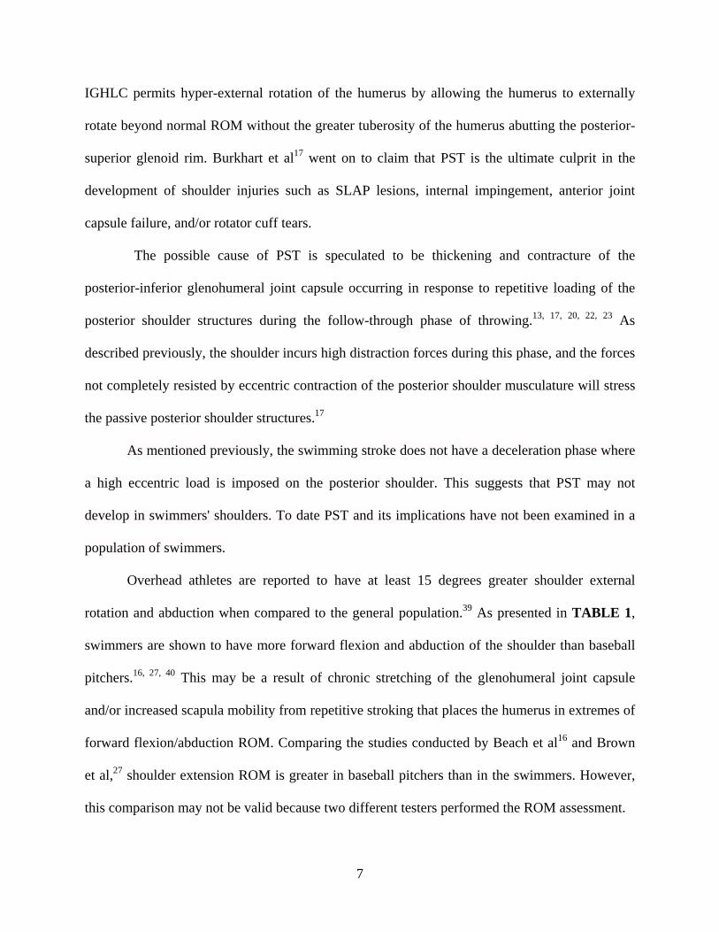
Overhead athletes are reported to have at least 15 degrees greater shoulder external
rotation and abduction when compared to the general population.39 As presented in TABLE 1,
swimmers are shown to have more forward flexion and abduction of the shoulder than baseball
pitchers.16, 27, 40 This may be a result of chronic stretching of the glenohumeral joint capsule
and/or increased scapula mobility from repetitive stroking that places the humerus in extremes of
forward flexion/abduction ROM. Comparing the studies conducted by Beach et al16 and Brown
et al,27 shoulder extension ROM is greater in baseball pitchers than in the swimmers. However,
this comparison may not be valid because two different testers performed the ROM assessment.
7

Bak et al15 reported greater than normal external rotation ROM and normal internal
rotation ROM in a group of 8 non-impaired elite swimmers (TABLE 1). On the other hand, less
than normal internal rotation ROM has been reported in swimmers evaluated in a study by Beach
et al16 (TABLE 1). This discrepancy may be attributed to the fact that 69% of the subjects who
participated in the study were experiencing some degree of shoulder pain.16
There have been many studies that have evaluated shoulder internal/external rotation
ROM in baseball players (TABLE 2).10, 19, 26, 27, 40-42 Baseball players are reported to have a
combination of decreased internal rotation ROM, or glenohumeral internal rotation deficit
(GIRD), and increased external rotation ROM, or external rotation gain (ERG), in the throwing
arm.20, 23, 27, 30 Because the loss of internal rotation ROM in baseball players is accompanied by
the increase in external rotation ROM, the arc of the total internal-external rotation ROM is
preserved.20, 23, 27, 30
Theories have been proposed to explain why alterations in ROM occur in these overhead
athletes.3, 17, 20, 40, 42-45 Tightness of the posterior shoulder structures has been suggested as one of
the key factors for the shift in ROM in baseball players.17, 25 It has been reported that the surgical
release of the posterior glenohumeral joint capsule in people with decreased internal rotation
ROM resulted in restoration of normal ROM.46 Additionally, a stretching technique that
specifically isolates the posterior-inferior shoulder structures is reported to improve internal
rotation ROM at the glenohumeral joint.38 The Professional Baseball Athletic Training Society
reported that baseball players with an internal rotation deficit of more than 30 degrees have
increased their internal rotation ROM after performing the “Sleeper’s stretch” for 3-12 weeks.38
These reports support the theory that PST may be one of the factors leading to GIRD.
8
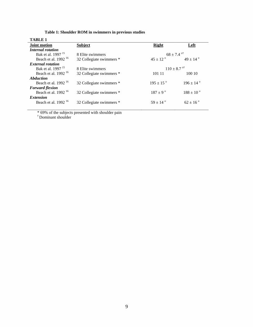
Table 1: Shoulder ROM in swimmers in previous studies
TABLE 1 Joint motion Subject Right LeftInternal rotation Bak et al. 1997 15 8 Elite swimmers 68 ± 7.4 o†
Beach et al. 1992 16 32 Collegiate swimmers * 45 ± 12 o 49 ± 14 oExternal rotation Bak et al. 1997 15 8 Elite swimmers 110 ± 8.7 o†
Beach et al. 1992 16 32 Collegiate swimmers * 101 11 100 10 Abduction Beach et al. 1992 16 32 Collegiate swimmers * 195 ± 15 o 196 ± 14 oForward flexion Beach et al. 1992 16 32 Collegiate swimmers * 187 ± 9 o 188 ± 10 o
Extension Beach et al. 1992 16 32 Collegiate swimmers * 59 ± 14 o 62 ± 16 o
* 69% of the subjects presented with shoulder pain † Dominant shoulder
9

Table 2: Shoulder ROM in Non-pathological baseball players reported in previous studies
TABLE 2 Joint motion Subject Throwing Non-throwingInternal rotation Borsa et al. 2004 42 43 Professional baseball players 68.6 ± 9.2 o 78.3 ± 10.6 o Reagan et al. 2002 40 54 College baseball players 43.0 ± 7.4 o 51.2 ± 7.3 o Tyler et al. 1999 26 23 College baseball pitchers 50.0 ± 2.0 o 69.5 ± 2.5 o Brown et al. 1988 27 18 Professional Baseball pitchers 83 ± 13.9 o 98 ± 13.2 oExternal rotation Borsa et al.2004 42 43 Professional baseball players 134.8 ± 10.2 o 125.8 ± 8.7 o
Reagan et al. 2002 40 54 College baseball players 116.3 ± 11.4 o 106.6±11.2 o Tyler et al. 1999 26 23 College baseball pitchers 109.7 ± 2.4 o 98.9 ± 1.6 o Bigliani et al. 1997 72 Professional baseball pitchers 118.0 o 102.8 o Brown et al. 1988 27 18 Professional Baseball pitchers 141 ± 14.7 o 132 ±14.6 oAbduction Brown et al. 1988 27 18 Professional Baseball pitchers 168 ± 8.4 o 172 ± 11.6 o
98 ± 10.8 o* 105 ± 10.3 o* Forward flexion Reagan et al. 2002 40 54 College baseball players 175.1 ± 7.0 o 175.6 ± 5.5 o Bigliani et al. 1997 41 72 Professional baseball pitchers 174.9 o 177.3 o Brown et al. 1988 27 18 Professional Baseball pitchers 163 ± 7.9 o 168 ± 6.3 oExtension Brown et al. 1988 27 18 Professional Baseball pitchers 72 ± 15.5 o 78 ± 13.3 o
* Isolated glenohumeral range of motion
10

The scapula needs to move in coordination with the humerus to keep the humeral head
centered in the glenoid fossa to maintain joint stability throughout full ROM.13 Appropriate
positioning of the scapula and its alignment with the humerus is said to lead to optimal shoulder
function, both physiologically and biomechanically.13 The scapula moves in three dimensions
with six degrees of freedom; upward-downward rotation, internal-external rotation, anterior-
posterior tilting, elevation-depression, and protraction-retraction.47-52 Most 3-dimentional
scapular kinematics studies on healthy subjects have shown that the scapula upwardly rotates,
externally rotates, and posteriorly tilts with humeral elevation.48-52 Pathological shoulders are
reported to have altered scapular kinematics when compared to non-pathological shoulders.24, 53,
54 Lukasiewicz et al 54 reported that subjects with shoulder impingement demonstrated a
significantly lower posterior tilting of the scapula during humeral elevation.54 Fatigued shoulders
have been shown to have altered scapular kinematics as well.55-57 Su et al56 measured the amount
of scapular upward rotation in swimmers before and after practice. They found that swimmers
have significantly less upward rotation with humeral elevation after practice.56 Tsai et al57
assessed scapular kinematics before and after a fatigue protocol for the external rotator muscles
and found significantly decreased posterior tilting, external rotation, and upward rotation in early
to middle phases of humeral elevation. These studies may suggest that the overhead athletes
become more prone to sustaining shoulder injury when they are fatigued due to the alteration in
scapula kinematics.
A recent study investigating 3-dimentional scapular kinematics targeting throwing
athletes showed that throwing athletes have significantly increased upward rotation, internal
rotation, and retraction of the scapula during humeral elevation compared to non-throwers.52 It is
speculated that these changes in scapular kinematics are due to chronic adaptations in order to
11

perform a throwing motion more efficiently.52 to date, there has been no published research
examining the 3-dimentional scapular kinematics in swimmers. Considering that a swimmer’s
shoulder abducts beyond its active ROM during each stroke, swimmers may display chronic
adaptations in the form of increased scapular mobility to efficiently achieve humeral abduction.
Swimmers are anecdotally notorious for “poor posture,” which is commonly
characterized by forward neck, increased thoracic kyphosis, and rounded shoulders.1, 2, 8, 9, 11
Postural malalignment can change resting scapular posture, alter scapular kinematics, decrease
strength in the surrounding muscle, and restrict shoulder ROM.13, 58-60 Kebaetse et al58 compared
active shoulder ROM, isometric abduction strength, and 3-dimensional scapular kinematics
between erect and slouched posture. They reported that slouched posture resulted in greater
scapular elevation, internal rotation, and less upward rotation and posterior tilting.58 The authors
also reported that the subjects had decreased scapular abduction ROM and muscle force when
they were in slouched posture. 58
Recently, increased forward neck inclination and rounded shoulder posture with reduced
posterior shoulder girdle muscle strength has been reported in swimmers.11 Forward shoulder is
described as protraction and elevation of the scapula and a forward position of the shoulders.61, 62
A protracted scapula has been shown with MRI to decrease subacromial space.63 This can be
problematic in swimmers, since there are phases in the swimming stroke where the shoulder is
placed in positions prone to evoking subacromial impingement pain.2, 12, 64 A protracted scapula
also places the scapula in an unfavorable position to cause thoracic outlet syndrome, a condition
where vascular and/or neural structures get impinged under tight anterior neck/ chest
musculature or costoclavicular structures.65 Performing thousands of strokes with poor posture
will increase the risk of developing shoulder problems, and therefore addressing poor posture in
12

swimmers should help improve shoulder function and possibly reduce the risk of developing
shoulder pathologies. No study to date has quantitatively compared forward shoulder posture
between swimmers, baseball players and how they differ from non-overhead athletes.
The purpose of this study was to compare physical characteristics of the shoulder among
groups of overhead athletes participating in two different sports (swimming and baseball) and
non-overhead athletes. Although many studies in the past have evaluated the shoulder
characteristics in baseball players and swimmers, there were no comparative studies examining
the two groups of overhead athletes using the same testing procedure. Data obtained from non-
overhead athletes served as a control for comparison. Any deviation from this value found in
baseball players and swimmers was considered to be associated with the participation in their
respective sports. Only non-pathological male subjects participated in this study, since
pathological changes in shoulder characteristics may confound the inter-sports differences. The
data obtained from this study may serve to provide normative values for shoulder characteristics
in healthy male intercollegiate baseball pitchers and swimmers. Clinicians treating these
populations can use the information to identify alterations in physical characteristics in
pathological athletes, and set appropriate treatment/rehabilitation goals. The results from this
study emphasize the importance of treating overhead athletes accordingly based on their sports.
Shoulder ROM, isokinetic strength, PST, scapular kinematics during humeral elevation,
and FST were assessed in 15 healthy male intercollegiate swimmers, 15 intercollegiate baseball
pitchers, and 15 control (non-overhead) athletes. Specific dependent variables of interest are
summarized in TABLE 3.
It was hypothesized that: 1) swimmers would have greater external rotation ROM, and an
equal amount of internal rotation ROM, resulting in a greater total ROM arc compared to the
13

control subjects, 2) baseball pitchers would have greater external rotation ROM , but less internal
rotation ROM, resulting in an equal amount of total ROM arc compared to the control subjects,
3) swimmers would have greater forward flexion/abduction ROM than both the baseball pitchers
and the control subjects, 4) baseball pitchers would have greater extension ROM than both the
swimmers and the control subjects, 5) swimmers and control subjects would both have less PST
than baseball pitchers, 6) swimmers and baseball pitchers would both have greater internal
rotation strength and lower external to internal rotation strength ratios than control subjects, 7)
swimmers and baseball pitchers would both have greater scapular upward rotation, internal
rotation, protraction, and posterior tilting during humeral elevation than control subjects, and 8)
swimmers and baseball pitchers would both have greater FSP compared to control subjects.
These differences were expected to be present bilaterally in swimmers, but exhibited only in the
dominant arms of baseball pitchers. The unilateral alteration in ROMs in baseball pitchers are
expected to lead to GIRD, ERG, and PST.
14
Table 3: Dependent Variables
TABLE 3 Type of tests Dependent VariablesROM Internal rotation ROM (deg.)
External rotation ROM (deg.) GIRD (IR non-dominant – IR dominant) (deg.) ERG (ER non-dominant – ER dominant) (deg.) Total ROM arc (IR ROM + ER ROM) (deg.) Forward flexion ROM (deg.) Extension ROM (deg.)
Abduction ROM (deg.) PST Side lying cross body horizontal abduction test
PST dominant – PST non-dominant Supine cross body horizontal abduction test
PST non-dominant - PST dominant Strength ER peak torque normalized to body weight @ 60deg/sec (Nm/kg)
IR peak torque normalized to body weight @ 60deg/sec (Nm/kg) ER: IR strength ratio @ 60deg/sec* IR peak torque normalized to body weight @ 300deg/sec (Nm/kg) ER peak torque normalized to body weight @ 300deg/sec (Nm/kg) ER: IR strength ratio @ 300deg/sec* Protraction peak torque normalized to body weight @12.2cm/sec (Nm/kg) Retraction peak torque normalized to body weight @ 12.2cm/sec (Nm/kg) Protraction: retraction strength ratio @ 12.2cm/sec* Protraction peak torque normalized to body weight @36.6cm/sec (Nm/kg) Retraction peak torque normalized to body weight @ 36.6cm/sec (Nm/kg) Protraction: retraction strength ratio @ 36.6cm/sec*
Scapular kinematics Scapula internal/external rotation (deg.)
Scapula upward/downward rotation (deg.) @ 0, 30, 60, 90, and 120 Scapula anterior/posterior tilt (deg.) degrees of humeral Scapula protraction/retraction (deg.) elevation Scapula elevation/depression (deg.)
Forward shoulder Posture Forward shoulder posture (cm) FSP dominant – FSP non-dominant
* The strength ratios are ratios between peak torques normalized to body weight
15
2.0 MATERIALS AND METHODS
2.1 SUBJECTS
15 intercollegiate swimmers, 15 intercollegiate baseball pitchers and 15 control subjects
participated in this study. Only male subjects were recruited and participated in this study to
control for possible gender differences. Due to differences in the stroke mechanics, swimmers
who solely compete in breaststroke were excluded from this study. Swimmers and baseball
pitchers were required to have at least 5 years of participation in their respective sport.
Intercollegiate non-overhead athletes from track, cross country, and soccer teams served as the
control subjects. Athletes who had participated in formal overhead sport activities for over a year
within the past 4 years (swimming, baseball, tennis, volleyball, water polo etc.) did not qualify as
control subjects. Subjects with a previous history of shoulder surgery, traumatic injury
(dislocation/ subluxation/ AC joint sprain) were excluded from this study. Subjects who had
experienced shoulder pain that interfered with the training in the past 6 months were also
excluded from this study. The demographic information of the subjects is presented in TABLE
5.
16
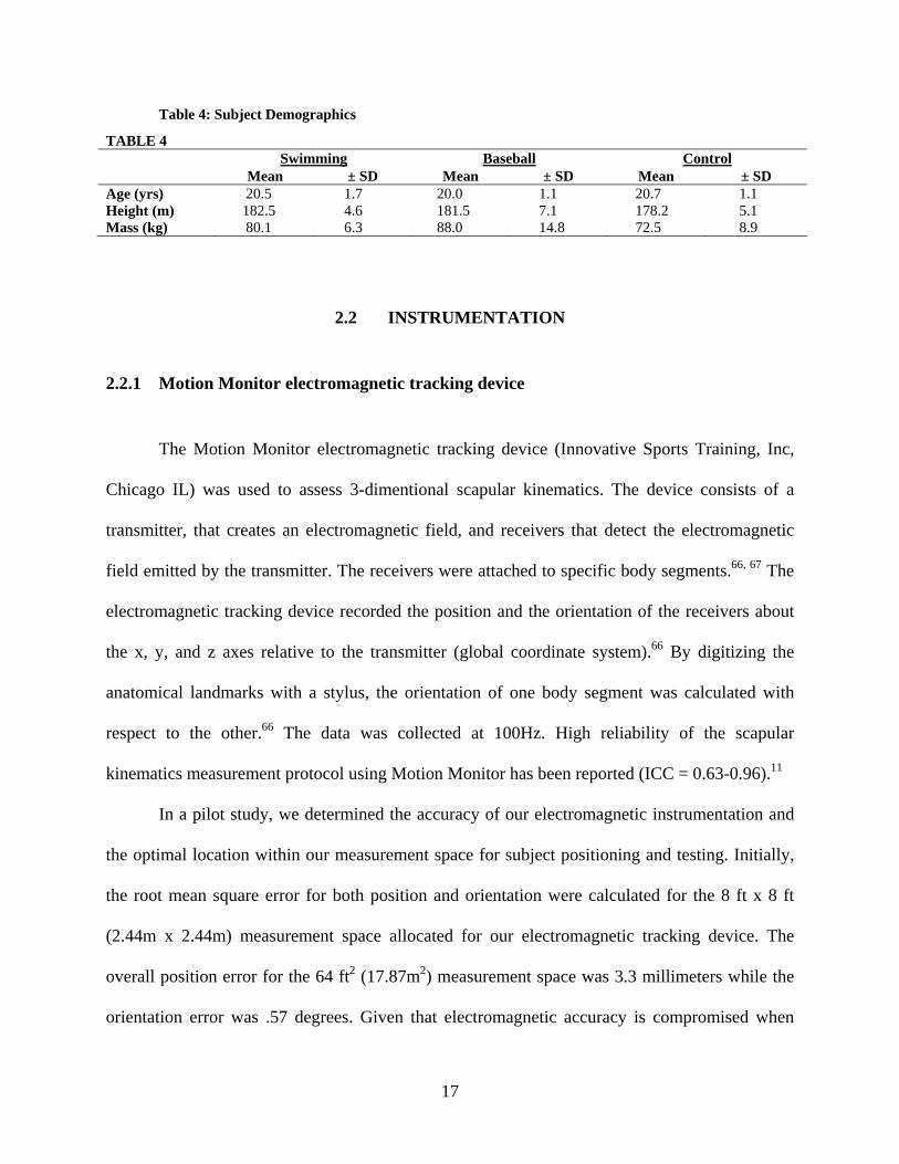
Table 4: Subject Demographics
TABLE 4 Swimming Baseball Control
Mean ± SD Mean ± SD Mean ± SD Age (yrs) 20.5 1.7 20.0 1.1 20.7 1.1 Height (m) 182.5 4.6 181.5 7.1 178.2 5.1 Mass (kg) 80.1 6.3 88.0 14.8 72.5 8.9
2.2 INSTRUMENTATION
2.2.1 Motion Monitor electromagnetic tracking device
The Motion Monitor electromagnetic tracking device (Innovative Sports Training, Inc,
Chicago IL) was used to assess 3-dimentional scapular kinematics. The device consists of a
transmitter, that creates an electromagnetic field, and receivers that detect the electromagnetic
field emitted by the transmitter. The receivers were attached to specific body segments.66, 67 The
electromagnetic tracking device recorded the position and the orientation of the receivers about
the x, y, and z axes relative to the transmitter (global coordinate system).66 By digitizing the
anatomical landmarks with a stylus, the orientation of one body segment was calculated with
respect to the other.66 The data was collected at 100Hz. High reliability of the scapular
kinematics measurement protocol using Motion Monitor has been reported (ICC = 0.63-0.96).11
In a pilot study, we determined the accuracy of our electromagnetic instrumentation and
the optimal location within our measurement space for subject positioning and testing. Initially,
the root mean square error for both position and orientation were calculated for the 8 ft x 8 ft
(2.44m x 2.44m) measurement space allocated for our electromagnetic tracking device. The
overall position error for the 64 ft2 (17.87m2) measurement space was 3.3 millimeters while the
orientation error was .57 degrees. Given that electromagnetic accuracy is compromised when
17
measurements are taken too close to or too far from the transmitter, we determined where within
that measurement space yielded the lowest amount of error. It was determined that the region of
the measurement space that is between 3 ft (.91m) and 4 ft (1.2m) directly in front of the
transmitter demonstrated the least amount of position (.7 mm) and orientation (.27 degrees) error.
Thus all kinematic assessments in the current study were performed with the subjects standing
with their heels 3 feet away from the transmitter.
2.2.2 Biodex System 3 Isokinetic Dynamometer
Biodex System 3 isokinetic dynamometer (Biodex Medical, Shirley, NY) was used to
assess shoulder strength. The dynamometer contains strain gauges and potentiometers, which
measures the force exerted by the body segments to the arm moving at a constant speed.52, 68
Reliability and the validity of the Biodex System 3 isokinetic dynamometer in assessing strength
in rotational movement has been demonstrated to be very high (ICC=0.99~1.00) through a wide
range of velocities.68 The reliability and the validity of the instrument in assessing scapular
protraction/retraction peak torque have been reported to be high (ICC 0.94-0.96 on non-dominant
side, ICC 0.88-0.92 on dominant side).36 Shoulder internal/external rotation strength at 60
degrees/sec and 300 degrees/sec, and scapular protraction/retraction strength at 12.2cm/sec and
36.6cm/sec were assessed using this device in a seated position.
2.3 PROCEDURES
Prior to testing, each subject provided informed consent as required by the University of
Pittsburgh Institutional Review Board. After signing the consent form, subjects proceeded to
18

forward shoulder posture assessment, described by Peterson et al.69 Subjects were asked to stand
in front of the wall, march 10 times in place, roll their shoulders forward and backward three
times70 and then nod their head back and forth 5 times.71 This sequence of motion is performed
to produce a natural standing posture.70, 71 The subjects were then asked to move backwards to
the wall until their buttocks touched the wall, and remain in this position until testing was
completed. The tester measured the distance (cm) between the wall and the anterior tip of the
acromion process using the Double Square device (FIGURE 1). Measurements were performed
three times on each shoulder by the same investigator for all subjects. Peterson et al69
investigated the validity and the reliability of the four different methods of postural assessment,
and reported the method using Double Square had moderate correlation with the radiographic
measurement (r=0.65) and high reliability (ICC=0.89). In our laboratory, high intrasession
(ICC=0.98, SEM=.32cm) and intersession (ICC=.992, SEM=.16cm) reliability was obtained
from the pilot data. The average of the distances between the wall and the anterior tip of the
acromion process was recorded bilaterally, and the ratio between the FSP on the dominant versus
non-dominant shoulder was calculated (TABLE 3).
19

Figure 1: Forward Shoulder Posture Assessment
After the postural assessment, passive humeral internal/external rotation, forward flexion,
extension, and abduction ROM were assessed using procedures described by Norkin and
White.72 The subject laid supine on the treatment table with the testing shoulder placed in 90
degrees of abduction, and the elbow slightly off the edge of the table. A rolled towel was placed
under the arm to align the humerus level with the acromion process.72 The first tester stabilized
the shoulder against the table with one hand to prevent any accessory motion, while using the
other hand to passively move the humerus into maximal internal/external rotation. The second
tester measured the ROM using a goniometer. The angle of the forearm, with respect to the plane
parallel to the floor, was recorded as glenohumeral internal/external rotation ROM. The
difference between the dominant and the non-dominant shoulder were recorded as GIRD and
ERG, and the internal/external rotation total ROM arc was calculated (TABLE 3). A level was
attached to the stationary arm of the goniometer to ensure that the stationary arm was kept
20
parallel to the ground. Intrasession reliability and precision for the goniometric measurements
obtained from the pilot study were high for both internal rotation (ICC= .985, SEM= 1.51) and
external rotation ROM (ICC= .942, SEM= 1.75).
Each subject remained supine for forward flexion and abduction ROM assessments. The
subject’s shoulder was moved passively into forward flexion until the end range while the
scapula was stabilized against the treatment table by the tester’s hand. Each subject’s elbow was
kept straight to ensure that the long head of the triceps brachii muscle would not restrict the
ROM.72 The angle between the midaxillary line and the midline of the humerus was recorded as
the arm was forward flexed.72 The same manner was used to obtain abduction ROM, except the
angle between a line passing through the acromion process that is parallel to the midline of the
sternum relative to the midline of the humerus was recorded as the abduction angle.72
Each subject was asked to lie prone for the shoulder extension ROM assessment. The
humerus was passively extended with slight elbow flexion so that the long head of the biceps
brachii muscle would not restrict ROM. The angle between the midaxillary line and the midline
of the humerus was recorded as the extension ROM.72
All goniometric measurements were performed bilaterally by the same testers for all
subjects. Internal/external rotation, forward flexion, extension, and abduction ROM were
reported (TABLE 3).
Each subject remained on the table for the PST assessments. PST was first assessed with
subject side-lying in a side-lying position, followed by a supine assessment. The side-lying cross
body humeral abduction test is the standard PST testing procedure described by Tyler et al
(FIGURE 2).25, 26 The supine procedure was performed in addition to the side-lying PST
assessment in this study because unpublished data collected at our lab suggests that although
21

both side-lying and supine testing procedures can be performed reliably, the supine method can
be performed with higher precision.73 The subject was asked to lay on his side for the side-lying
cross body humeral adduction test. The subject’s thorax was aligned perpendicular to the
treatment table with the spine in neutral flexion, extension, and rotation. With the tester facing
the subject, excessive scapular movement was restricted by stabilizing the lateral border of the
scapula in a retracted position. Starting from a position of 90 degrees humeral abduction and
neutral humeral rotation, the tester passively lowered the arm into horizontal adduction by
griping the subject’s forearm just distal to the humeral epicondyles. The arm was lowered until
the humeral horizontal adduction motion has ceased or until the humerus started to internally
rotate.25, 26 At the end of the ROM, the second tester recorded the distance, in centimeters,
between the medial epicondyle and the surface of the treatment table using a carpenter’s square.
This distance quantified the amount of horizontal adduction, which reflected the degree of
tightness in the posterior shoulder structures. High reliability has been reported in the literature
for this testing procedure (ICC dominant=0.92, ICC non-dominant=0.95). Intrasession ICC
(SEM) and interssession ICC (SEM) obtained in our laboratory were .87 (.37cm) and .23 (.74
cm), respectively. The tests were performed three times bilaterally on each shoulder by the same
testers for consistency. The distance between the table and the subject’s medial epicondyle were
recorded, and the ratio between the PST of dominant versus non-dominant shoulders was
calculated (TABLE 3).
22

Figure 2: Posterior Shoulder Tightness Assessment Side-lying Method
23

For the supine PST assessment, the subject lied supine on the treatment table. One tester
was positioned beside the table of the shoulder being tested and asked the subject to lift their
shoulder off the table. The tester placed one hand under the scapula, pressing their thenar
eminence against the lateral border of the scapula, stabilizing the scapular in a retracted position.
The tester then used the other hand to passively move the subject’s arm into horizontal
adduction. At the end ROM, the second tester recorded the angle formed between the humerus
and the horizontal plane from the superior aspect of the shoulder. The fulcrum of the goniometer
was placed over the estimated glenohumeral joint center, and the movement arm was aligned
with the humerus. The stationary arm was kept parallel to the floor, and confirmed using the
attached level. Intrasession ICC (SEM) and interssession ICC (SEM) obtained in our laboratory
for the supine method were .93(1.1o) and .64 (2.2 o), respectively. The tests were performed three
times on each shoulder by the same testers for consistency. The angle between the humerus and
the horizontal plane, which represents the horizontal adduction ROM, was recorded. The
difference between the horizontal adduction ROM of the dominant versus non-dominant
shoulder was calculated.
Following the PST assessment, the subject was prepared for bilateral scapular kinematics
assessment using the Motion Monitor. A total of six receivers were used in this study. The first
receiver was attached to the spinous process of the seventh cervical vertebrae (C7). Two
receivers were attached bilaterally on the flat portion of the bilateral acromion processes, and
another two receivers were attached bilaterally to the mid-shaft of the posterior humerus56. All
receivers were secured on the skin using double sided adhesive disks (3M Health Care, St. Paul,
Minn), pre-wrap, athletic tape, and a velcro strap to minimize skin movement. The sixth receiver
was attached to the stylus that was used to palpate and digitize the anatomical landmarks on the
24
upper arm, scapula, and thorax. The anatomical landmarks digitized included the eighth thoracic
vertebrae (T8), processus xiphoideus (PX), seventh cervical vertebrae (C7), incisura jugularis
(IJ), acromion-clavicular joint (AC), trigonum spinae (TS), angulus inferior (AI), medial
epicondyle (ME), lateral epicondyle (LE), and glenohumeral joint center. The digitized
landmarks appear in TABLE 5.52 Because the glenohumeral joint center cannot be palpated, it
was estimated as the point that moves least with respect to the scapula when the humerus is
passively moved through several short arcs.74 Digitization of these anatomical landmarks on each
segment allowed construction of the local coordinate system for each body segments; thorax,
scapula, and humerus (FIGURE 3). The definition of the local coordinate system appears on
TABLE 6.52 Using local coordinate systems, orientation of the humerus with respect to the
thorax (humeral elevation angle) and the position and the orientation of scapula with respect to
the thorax were calculated. Each subject performed 10 repetitions of bilateral full shoulder
elevation in the scapular plane (30 degrees anterior to the frontal plane) (FIGURE 4). The
subject elevated the arm in two seconds, and lowerd the arm in two seconds. PVC pipe guided
the motion, and the speed was moderated by the metronome. After the testing, all sensors were
removed. Scapular kinematics variables recorded are summarized in TABLE 3.
25
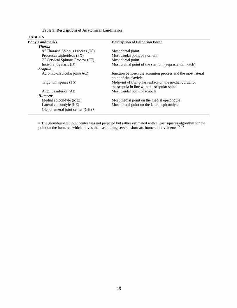
Table 5: Descriptions of Anatomical Landmarks
TABLE 5 Bony Landmarks Description of Palpation Point
Thorax 8th Thoracic Spinous Process (T8) Most dorsal point Processus xiphoideus (PX) Most caudal point of sternum 7th Cervical Spinous Process (C7) Most dorsal point Incisura jugularis (IJ) Most cranial point of the sternum (suprasternal notch) Scapula
Acromio-clavicular joint(AC) Junction between the acromion process and the most lateral point of the clavicle
Trigonum spinae (TS) Midpoint of triangular surface on the medial border of the scapula in line with the scapular spine Angulus inferior (AI) Most caudal point of scapula Humerus Medial epicondyle (ME) Most medial point on the medial epicondyle Lateral epicondyle (LE) Most lateral point on the lateral epicondyle Glenohumeral joint center (GH) ∗
∗ The glenohumeral joint center was not palpated but rather estimated with a least squares algorithm for the point on the humerus which moves the least during several short arc humeral movements.74, 75
26
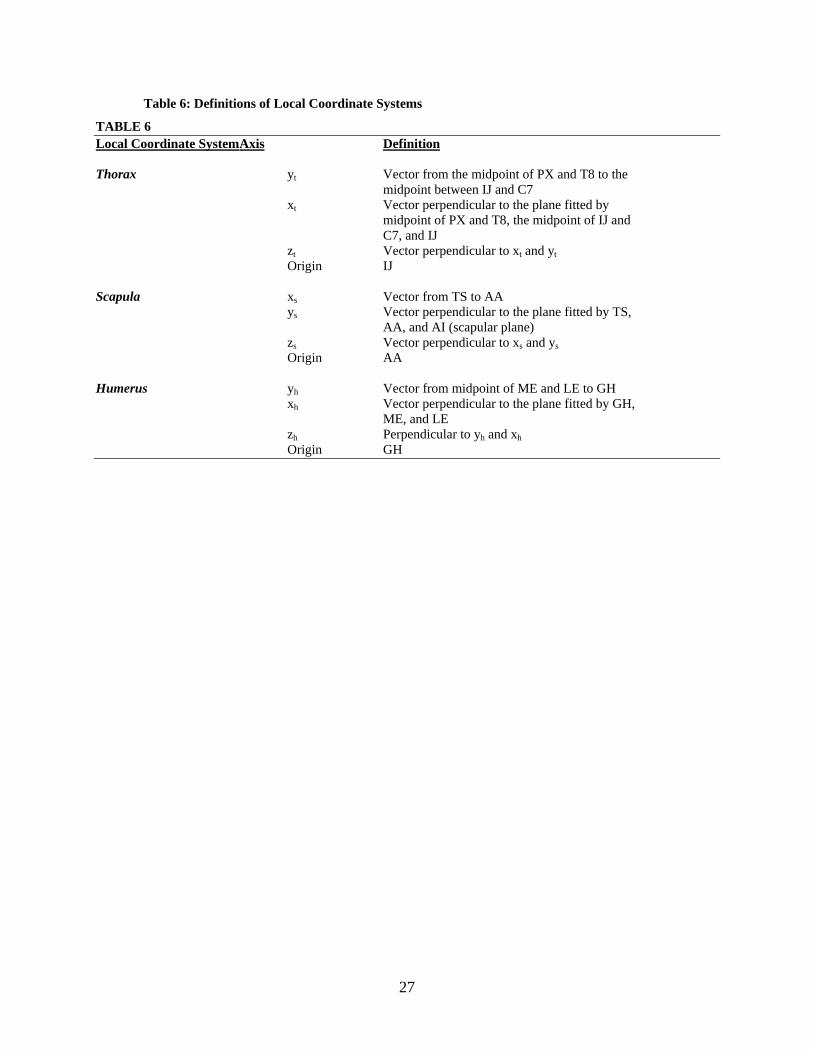
Table 6: Definitions of Local Coordinate Systems
TABLE 6 Local Coordinate System Axis Definition Thorax yt Vector from the midpoint of PX and T8 to the
midpoint between IJ and C7 xt Vector perpendicular to the plane fitted by midpoint of PX and T8, the midpoint of IJ and C7, and IJ zt Vector perpendicular to xt and yt
Origin IJ Scapula xs Vector from TS to AA ys Vector perpendicular to the plane fitted by TS, AA, and AI (scapular plane) zs Vector perpendicular to xs and ys Origin AA Humerus yh Vector from midpoint of ME and LE to GH xh Vector perpendicular to the plane fitted by GH, ME, and LE zh Perpendicular to yh and xh Origin GH
27

Figure 3: Local Coordinate System
28
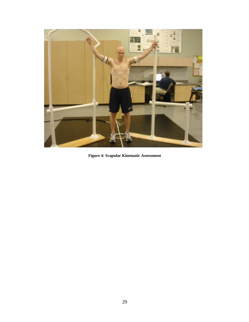
Figure 4: Scapular Kinematic Assessment
29

Following the scapular kinematic assessment, shoulder strength (protraction/retraction
and internal/external rotation) was assessed using the Biodex System 3 isokinetic dynamometer.
Prior to strength testing, each subject was weighed, and the body weight was entered into the
Biodex subject profile in order for the recorded peak torques to be normalized to the subject’s
body weight. This allowed for accurate comparison of shoulder strength for subjects with
different body sizes. Shoulder protraction and retraction strength was assessed first. The closed
kinetic chain features of the Biodex setup were used as described by Cools et al.37, 76 The subject
was seated on the Biodex chair and stabilized to the chair with two diagonal straps over the trunk
to prevent any accessory motion (FIGURE 5). The closed chain attachment was connected to
the dynamometer parallel to the floor at the subject’s shoulder height. The chair and the
dynamometer were rotated so that the subject’s arm was positioned 30 degrees in front of the
body. With the elbow extended, the subject held the handgrip on the attachment and performed
shoulder protraction/retraction against the handgrip. The subject was instructed to hold the
dynamometer handgrip move their scapula forward and backward keeping the elbow straight.
The subject performed 5 repetitions at the slower speed (12.2 cm/s) and 10 repetitions at the
higher speed (36.6 cm/s) with 1 minute rest in between. Data were collected bilaterally. Average
protraction and retraction forces normalized to body weight as well as protraction: retraction
strength ratios at two testing speeds were recorded (TABLE 3).
30

Figure 5: Biodex patient setup for scapular protraction/ retraction strength testing
31

After a 5 minutes rest, an internal/external rotation strength assessment was performed.
Each subject was seated in the Biodex chair as before. The tilt angle and position of the
dynamometer, as well as the seat height were adjusted so that the humeral internal/external
rotation could be performed comfortably in the scapular plane: subjects’ shoulder was placed in
neutral position with their humerus abducted to approximately 10 degrees in scapular plane (30
deg anterior to the frontal plane) (FIGURE 6). Each subject practiced the movement until they
felt comfortable to perform the task. Subjects then performed 5 repetitions of concentric
external/internal rotation isokinetic strength tests at 60 degrees/sec on both shoulders. After the
strength testing at 60 degrees/sec, the subject rested for a minute, and then practiced testing
movement at 300 degrees/sec until they felt comfortable to perform the task. The subjects
performed 10 repetitions of internal/external rotation at 300 degrees/sec. The peak internal and
external rotation torques normalized to the body weight as well as external: internal rotation
strength ratios at 2 testing speeds were recorded (TABLE 3).
32
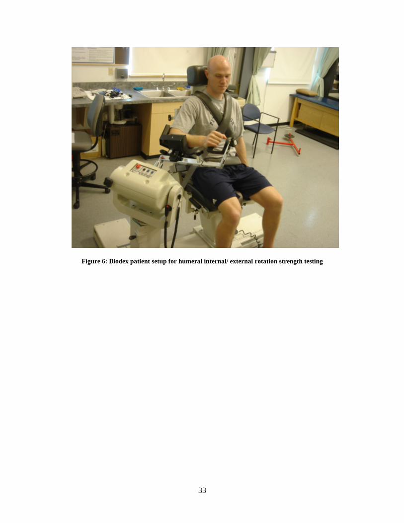
Figure 6: Biodex patient setup for humeral internal/ external rotation strength testing
33

2.4 DATA REDUCTION AND ANALYSIS
Raw scapular kinematic data was filtered with a low-pass 10Hz Butterworth filter. The
position and orientation data of the receivers and the digitized anatomical landmarks were used
to construct local coordinate systems for the thorax, scapula, and humerus. The coordinate
system used were in accordance with recommendations from the International Shoulder Group of
the International Society of Biomechanics.77 When the subject stood in an anatomical position,
the coordinate system for each segment was vertical (y-axis), horizontal to the right (x-axis), and
posterior (z-axis). Orientation of the scapula was determined as rotation about the y-axis of the
scapular (internal/external rotation), rotation about the z-axis of the scapula (upward/downward
rotation), and rotation about the x-axis of the scapula (anterior/posterior tipping) (FIGURE 7).
Euler angle decompositions were used to determine the scapular and humeral orientation with
respect to the thorax. The rotation sequence of the Euler angle was chosen based on the
recommendation of the International Shoulder Group.77 The scapula was attached to the thorax
via the clavicle; a rigid body with a fixed length, therefore the position of the scapula could be
described as the orientation of the vector extending from incisura jugularis (IJ) to acromion-
clavicular joint (AC) with respect to the local coordinate system of the thorax. Anatomically, the
vector extending from IJ to AC closely represents the orientation of the clavicle. The scapular
protraction/retraction angle was calculated as the angle formed between the vector extending
from IJ to AC joint points and the frontal plane of the thorax, and the scapular
elevation/depression angle was calculated as an angle formed between the vector and the
transverse plane of the thorax.
34

ANTERIOR/POSTERIOR TIPPING
UPWARD/DOWNWARD ROTATION
INTERNAL/EXTERNAL ROTATION
PROTRACTION/RETRACTIONELEVATION/DEPRESSION
Figure 7: Scapular positions and orientations assessed in the current study
35

The position and the orientation of the scapula when the humerus was at the side (0), 30,
60, 90, and 120 degrees of humeral elevation were recorded. Due to the reported inaccuracy in
the data above 120 degrees of humeral elevation, no data was collected beyond 120 degrees.48
Variables were calculated and processed using Matlab 12 (The MathWorks inc., Natick,
Massachusetts).
One-within, one-between analysis of variance (ANOVA) was used to determine any
inter-group and inter-limb differences for internal and external ROM, total ROM arc (IR ROM +
ER ROM), forward flexion ROM, extension ROM, abduction ROM, ER/IR strength at 60o/sec
and 300o/sec, protraction/ retraction strength at 12.2cm/sec and 36.6cm/sec, and forward
shoulder posture. Amount of GIRD (IR non-dominant – IR dominant), ERG (ER non-dominant –
ER dominant), and the dominant to non-dominant shoulder PST ratio (PST non-dominant / PST
dominant) were determined by a one-way ANOVA. A two-within, one-between AVOVA was
used to analyze scapular kinematics variables. Scapular kinematic variables
(protraction/retraction, elevation/depression, upward/downward rotation, internal/external
rotation, anterior/posterior tilt angles) were compared between groups, limbs, and humeral
positions (0, 30, 60, 90, and 120 degrees humeral elevation). A tukey post-hoc test was
performed following any significant differences that arose. SPSS 12, statistical analysis software
(SPSS Inc, Chicago IL) was used to run statistical analysis for all the variables. The level of
significance was set at an alpha level of .05 prior to the study.
36

3.0 RESULTS
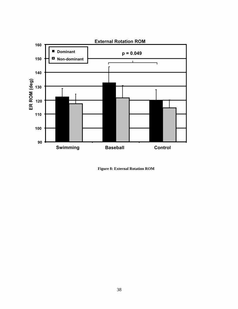
3.1 EXTERNAL ROTATION RANGE OF MOTION
The external rotation ROM data are presented in TABLE 7 and Figure 8. A significant
group by limb interaction (p = 0.004) was found in external rotation ROM. The dominant
shoulders of baseball pitchers exhibited greater external rotation ROM compared to the dominant
shoulders of control subjects (p = 0.049, HSD = 12.25). There was no between-group difference
in the non-dominant shoulder external rotation ROM. The baseball pitchers displayed
significantly greater external rotation ROM in their dominant shoulders when compared to their
non-dominant shoulder (p = 0.049, HSD = 12.25). No side-to-side differences in external
rotation ROM were found in swimmers or control subjects.
Table 7: Internal/ External Rotation Range of Motion
TABLE 7 Swimming Baseball Control
Mean ± SD Mean ± SD Mean ± SD Dominant Internal rotation (deg) 46.7 13.0 41.7 5.9 43.7 8.3 External rotation (deg) 122.4 5.9 132.0 10.4 119.9 8.0 Total range of motion (deg) 169.1 10.6 173.7 10.3 163.6 11.0 Non-dominant Internal rotation (deg) 48.8 12.4 54.3 8.3 45.0 8.8 External rotation (deg) 117.4 7.0 119.7 9.5 113.8 5.0 Total range of motion (deg) 166.1 11.2 174.0 13.9 158.8 9.6
37
External Rotation ROM
Swimming Baseball Control
ER R
OM
(deg
)
Dominant
Non-dominantp = 0.049
130
120
90
100
150
110
140
160
Figure 8: External Rotation ROM
38
3.2 INTERNAL ROTATION RANGE OF MOTION
The internal rotation ROM data are presented in TABLE 7. A significant group by limb
interaction (p < 0.001) was found in internal rotation ROM. However, the post-hoc analysis
revealed no difference between groups or limbs.
3.3 TOTAL RANGE OF MOTION
The total ROM data are presented in TABLE 7. There was no significant group by limb
interaction for the total ROM (p = 0.337).
3.4 GLENOHUMERAL INTERNAL ROTATION DEFECIT/ EXTERNAL
ROTATION GAIN
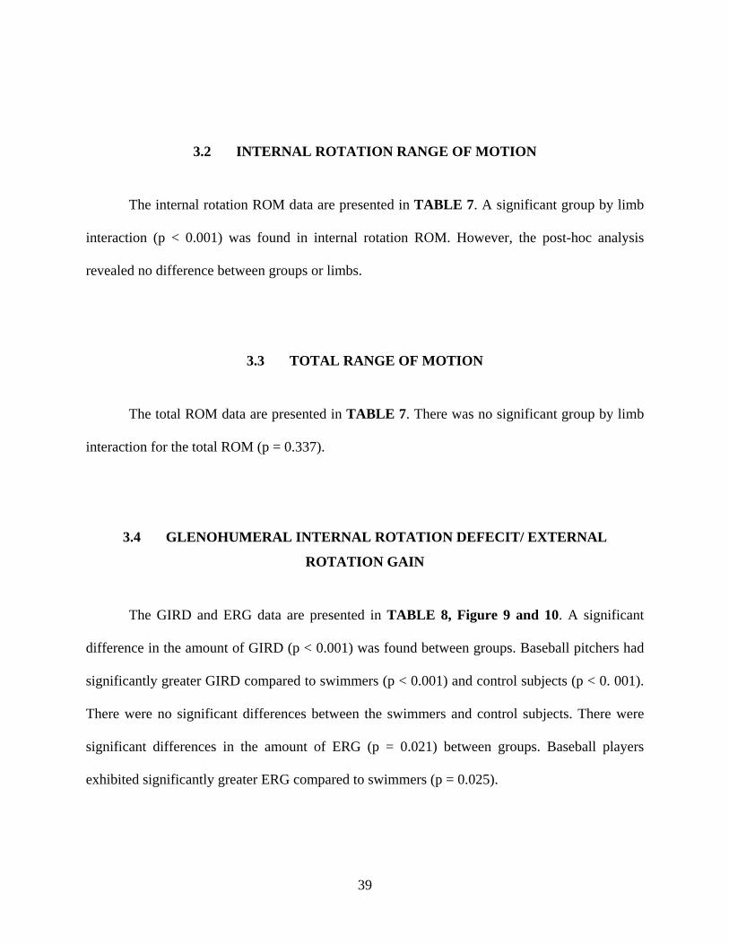
The GIRD and ERG data are presented in TABLE 8, Figure 9 and 10. A significant
difference in the amount of GIRD (p < 0.001) was found between groups. Baseball pitchers had
significantly greater GIRD compared to swimmers (p < 0.001) and control subjects (p < 0. 001).
There were no significant differences between the swimmers and control subjects. There were
significant differences in the amount of ERG (p = 0.021) between groups. Baseball players
exhibited significantly greater ERG compared to swimmers (p = 0.025).
39
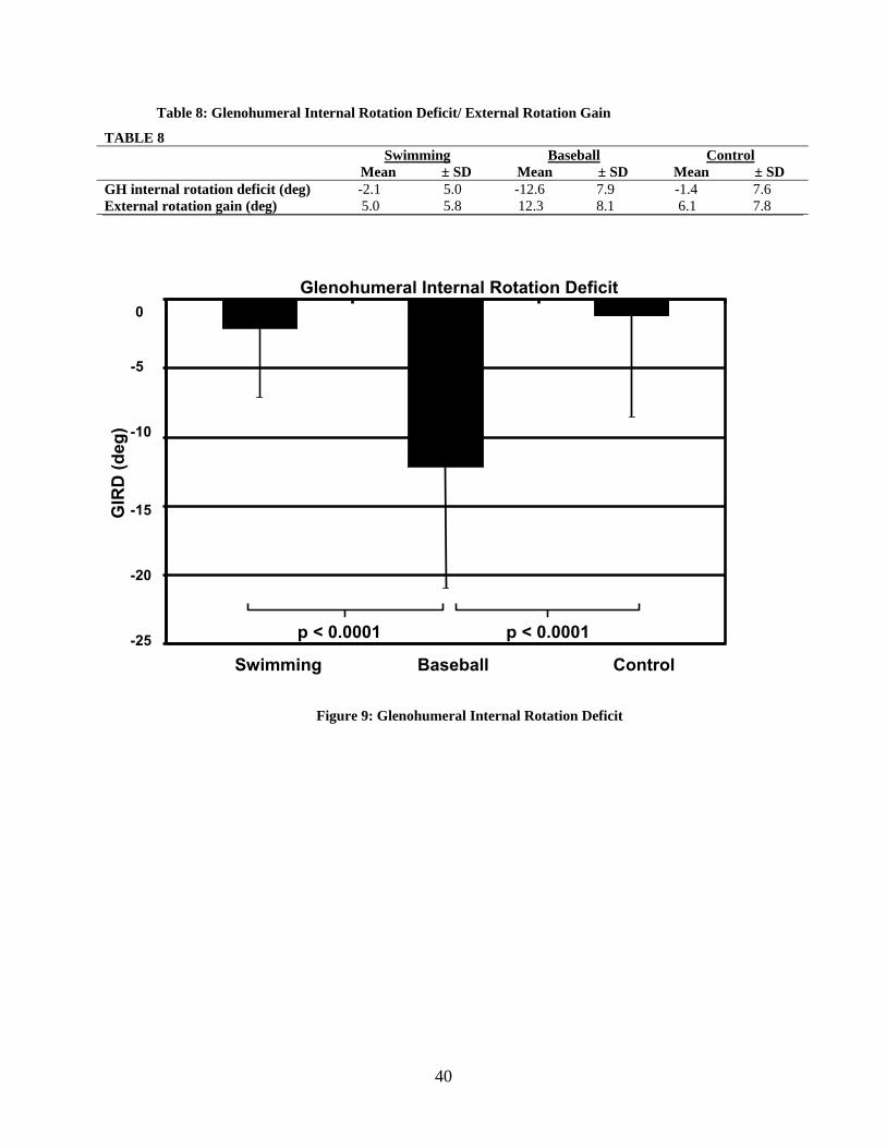
Table 8: Glenohumeral Internal Rotation Deficit/ External Rotation Gain
TABLE 8 Swimming Baseball Control
Mean ± SD Mean ± SD Mean ± SD GH internal rotation deficit (deg) -2.1 5.0 -12.6 7.9 -1.4 7.6 External rotation gain (deg) 5.0 5.8 12.3 8.1 6.1 7.8
p < 0.0001
Glenohumeral Internal Rotation Deficit
Swimming Baseball Control
GIR
D (d
eg)
p < 0.0001
-20
-25
-10
-15
-5
0
Figure 9: Glenohumeral Internal Rotation Deficit
40
External Rotation Gain
12
6
Swimming Baseball Control
p = 0.025
10
14
16
18
0
22
20
8
4
2
Exte
rnal
Rot
atio
n G
ain
(deg
)
Figure 10: External Rotation Gain
41

3.5 FLEXION/ ABDUCTION/ EXTENSION ROTATION RANGE OF MOTION
The flexion, abduction, and extension ROM data are presented in TABLE 9. There was a
significant group by limb interaction for flexion ROM (p = 0.005). Control subjects had
significantly less flexion ROM in their dominant shoulders compared to swimmers (p = 0.004,
HSD = 10.43) and baseball players (dominant: p = 0.047, HSD = 10.43)). Control subjects had
significantly less flexion ROM in their non-dominant shoulders compared to swimmers (p =
0.002, HSD = 10.43) and baseball players (p = 0.015, HSD = 10.43). There were no differences
in flexion ROM between the dominant and non-dominant shoulders for all groups. No group by
sports interaction for the abduction (p = 0.814) and extension (p = 0.224) ROM was found.
Table 9: Flexion/ Extension/ Abduction Range of Motion
TABLE 9 Swimming Baseball Control Mean ± SD Mean ± SD Mean ± SD
Dominant Flexion (deg) 198.6 9.2 189.7 13.3 178.6 9.5 Extension (deg) 78.9 10.4 79.5 12.0 72.2 5.7 Abduction (deg) 168.9 12.5 158.2 9.0 157.8 11.1 Non-dominant Flexion (deg) 197.3 9.6 193.7 11.8 176.0 9.7 Extension (deg) 79.0 13.8 82.2 10.2 68.8 8.8 Abduction (deg) 166.3 9.6 157.1 6.2 155.9 10.4
42

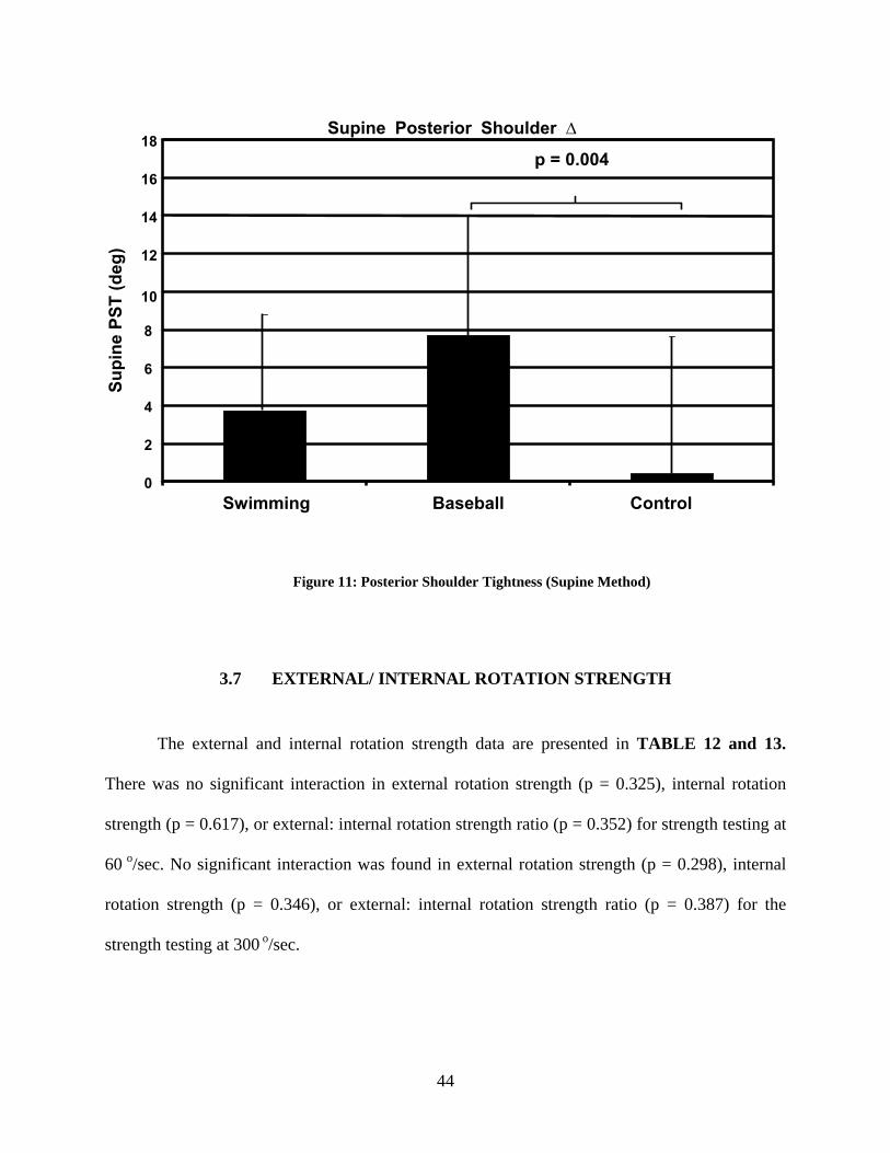
3.6 POSTERIOR SHOULDER TIGHTNESS
The PST data are presented in TABLE 10, 11, and Figure 11. PST was not significantly
different between groups for the side-lying assessment (p = 0.178). However, PST was
significantly different between groups for the supine assessment (p = 0.006). Baseball players
exhibited significantly greater PST compared to the control subjects (p= 0.004).
Table 10: Posterior Shoulder Tightness Side-lying Method
TABLE 10 Swimming Baseball Control Mean ± SD Mean ± SD Mean ± SD
Dominant (cm) 31.47 3.89 31.94 4.31 30.71 2.86 Non-dominant (cm) 31.15 2.97 29.67 3.66 30.27 3.76 Dominant - non-dominant (cm) 0.32 2.18 2.27 4.49 0.44 0.22
Table 11: Posterior Shoulder Tightness Supine Method
TABLE 11 Swimming Baseball Control Mean ± SD Mean ± SD Mean ± SD
Dominant (deg) 105.0 11.4 105.9 5.8 106.0 6.3 Non-dominant (deg) 108.7 10.0 114.0 9.3 106.0 8.9 Non-dominant – dominant (deg)
3.73 5.03 8.02* 7.08 0.190* 7.07
∗ Significant difference between the groups
43
Supine Posterior Shoulder ∆
Swimming Baseball Control
Supi
ne P
ST (d
eg)
p = 0.004
6
4
2
0
8
12
10
18
16
14
Figure 11: Posterior Shoulder Tightness (Supine Method)
3.7 EXTERNAL/ INTERNAL ROTATION STRENGTH
The external and internal rotation strength data are presented in TABLE 12 and 13.
There was no significant interaction in external rotation strength (p = 0.325), internal rotation
strength (p = 0.617), or external: internal rotation strength ratio (p = 0.352) for strength testing at
60 o/sec. No significant interaction was found in external rotation strength (p = 0.298), internal
rotation strength (p = 0.346), or external: internal rotation strength ratio (p = 0.387) for the
strength testing at 300 o/sec.
44
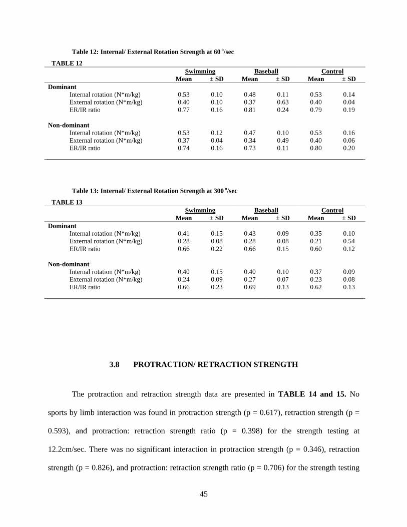
Table 12: Internal/ External Rotation Strength at 60 o/sec
TABLE 12 Swimming Baseball Control Mean ± SD Mean ± SD Mean ± SD
Dominant Internal rotation (N*m/kg) 0.53 0.10 0.48 0.11 0.53 0.14 External rotation (N*m/kg) 0.40 0.10 0.37 0.63 0.40 0.04 ER/IR ratio 0.77 0.16 0.81 0.24 0.79 0.19 Non-dominant Internal rotation (N*m/kg) 0.53 0.12 0.47 0.10 0.53 0.16 External rotation (N*m/kg) 0.37 0.04 0.34 0.49 0.40 0.06 ER/IR ratio 0.74 0.16 0.73 0.11 0.80 0.20
Table 13: Internal/ External Rotation Strength at 300 o/sec
TABLE 13 Swimming Baseball Control Mean ± SD Mean ± SD Mean ± SD
Dominant Internal rotation (N*m/kg) 0.41 0.15 0.43 0.09 0.35 0.10 External rotation (N*m/kg) 0.28 0.08 0.28 0.08 0.21 0.54 ER/IR ratio 0.66 0.22 0.66 0.15 0.60 0.12 Non-dominant Internal rotation (N*m/kg) 0.40 0.15 0.40 0.10 0.37 0.09 External rotation (N*m/kg) 0.24 0.09 0.27 0.07 0.23 0.08 ER/IR ratio 0.66 0.23 0.69 0.13 0.62 0.13
3.8 PROTRACTION/ RETRACTION STRENGTH
The protraction and retraction strength data are presented in TABLE 14 and 15. No
sports by limb interaction was found in protraction strength (p = 0.617), retraction strength (p =
0.593), and protraction: retraction strength ratio (p = 0.398) for the strength testing at
12.2cm/sec. There was no significant interaction in protraction strength (p = 0.346), retraction
strength (p = 0.826), and protraction: retraction strength ratio (p = 0.706) for the strength testing
45

at 36.6/sec.
Table 14: Protraction/ Retraction Strength at 12.2cm/sec
TABLE 14 Swimming Baseball Control Mean ± SD Mean ± SD Mean ± SD
Dominant Protraction (N/kg) 2.03 0.50 2.24 1.07 2.37 1.01 Retraction (N/kg) 2.19 0.42 2.07 0.80 2.44 1.06 Pro/Ret ratio 0.93 0.17 1.10 0.30 1.03 0.35 Non-dominant Protraction (N/kg) 2.35 0.52 2.41 1.02 2.39 0.99 Retraction (N/kg) 2.54 0.85 2.10 0.98 2.55 1.07 Pro/Ret ratio 0.90 0.32 1.18 0.16 0.97 0.22
Table 15: Protraction/ Retraction Strength at 36.6/sec
TABLE 15 Swimming Baseball Control Mean ± SD Mean ± SD Mean ± SD
Dominant Protraction (N/kg) 1.69 0.47 1.87 0.71 1.74 0.85 Retraction (N/kg) 1.95 0.54 1.98 0.87 1.93 0.88 Pro/Ret ratio 0.81 0.29 1.02 0.35 0.93 0.40 Non-dominant Protraction (N/kg) 1.81 0.54 1.92 0.83 1.74 0.59 Retraction (N/kg) 2.06 0.80 1.90 0.87 2.02 0.86 Pro/Ret ratio 0.87 0.36 1.03 0.19 0.90 0.17
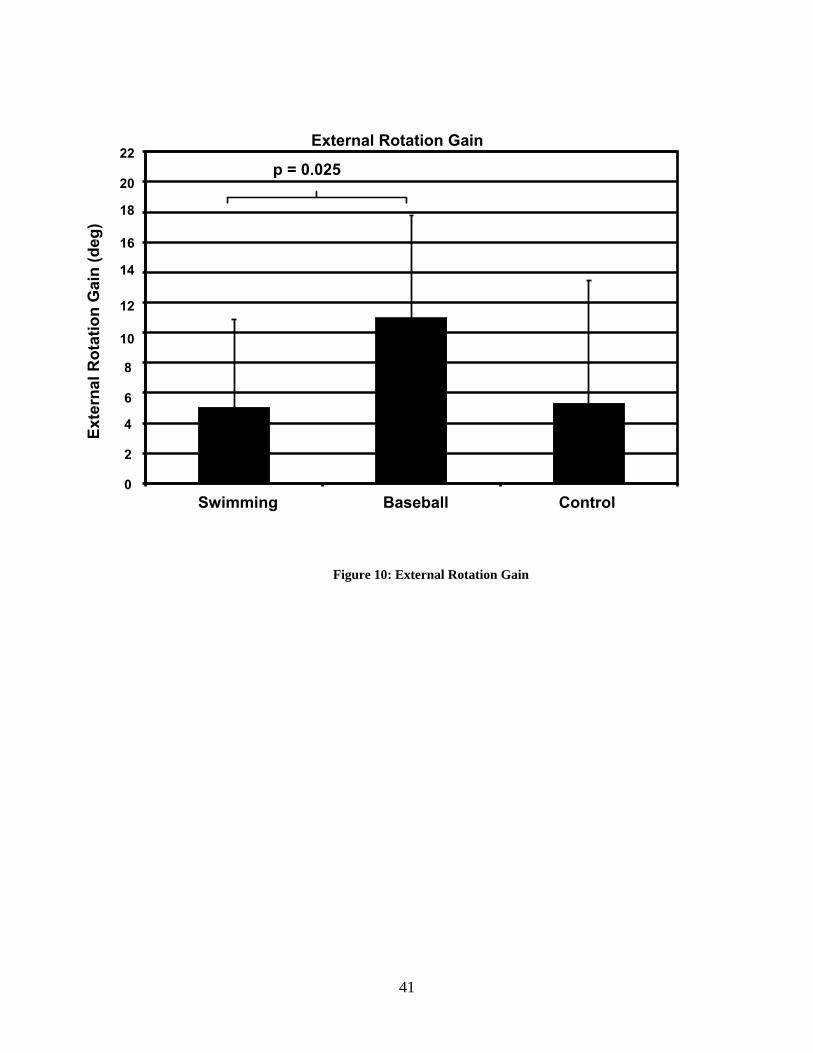
3.9 SCAPULAR KINEMATICS
The raw scapular kinematic data are presented in TABLE 16, 17, and Figure 12. The
five scapular kinematic variables (upward rotation, external rotation, posterior tipping,
protraction, and elevation) were analyzed using a two-within one-between ANOVA to compare
each variable between the three groups, limbs, and five humeral elevation angles. There was a
46
significant interaction with scapular upward rotation (p < 0.001). Baseball pitchers had greater
scapular upward rotation on their dominant side compared to their non-dominant side (p =
0.045). No statistically significant interactions were found for scapula external rotation (p =
0.292), posterior tipping (p = 0.679), protraction (p = 0.469), and elevation (p = 0.064).
47

Table 16: Scapular Kinematics Data Dominant Shoulder
TABLE 16 Swimming Baseball Control Mean ± SD Mean ± SD Mean ± SD Scapular upward/downward rotation 0o humeral elevation 2.86 6.04 1.91 6.74 1.04 7.13 30o humeral elevation 9.80 7.57 7.92 5.68 6.75 6.01 60o humeral elevation 20.52 8.57 19.80 6.44 18.12 5.98 90o humeral elevation 29.96 9.09 31.32 8.69 28.13 6.15 120o humeral elevation 33.11 9.82 40.45 13.88 36.53 8.11 Scapular external /internal rotation 0o humeral elevation 22.40 5.80 29.92 7.89 28.29 9.25 30o humeral elevation 20.89 4.47 26.84 8.09 25.45 8.31 60o humeral elevation 22.20 4.82 25.06 8.63 24.23 7.74 90o humeral elevation 25.47 6.40 24.97 11.49 27.12 6.98 120o humeral elevation 34.59 7.47 27.80 15.24 36.25 8.28 Scapular posterior/anterior tilt 0o humeral elevation -13.73 12.58 -16.13 3.94 -15.66 5.30 30o humeral elevation -9.38 13.22 -12.76 4.37 -12.49 5.61 60o humeral elevation -7.27 13.49 -12.00 4.92 -10.19 6.92 90o humeral elevation -5.94 13.76 -11.07 6.67 -8.44 8.14 120o humeral elevation -2.90 15.02 -4.92 9.34 -3.55 9.05 Scapular protraction/retraction 0o humeral elevation -18.39 5.21 -16.06 5.19 -17.51 5.88 30o humeral elevation -21.84 5.25 -18.87 4.77 -20.73 5.64 60o humeral elevation -24.85 5.44 -22.12 4.83 -24.80 5.81 90o humeral elevation -27.96 5.69 -26.49 5.64 -28.63 5.48 120o humeral elevation -33.63 5.51 -34.55 6.39 -35.40 5.08 Scapular elevation 0o humeral elevation 8.55 3.43 7.26 4.80 7.19 4.29 30o humeral elevation 11.69 3.94 9.33 4.26 9.31 4.18 60o humeral elevation 17.84 4.35 15.88 4.12 15.45 4.33 90o humeral elevation 23.60 4.59 22.99 4.88 21.19 4.57 120o humeral elevation 29.20 5.69 30.36 5.60 28.08 4.34
48

Table 17: Scapular Kinematics Non-dominant Shoulder
TABLE 17 Swimming Baseball Control Mean ± SD Mean ± SD Mean ± SD Scapular upward/downward rotation
0o humeral elevation 2.41 8.22 1.01 7.17 1.00 8.40 30o humeral elevation 8.88 7.50 6.33 6.70 7.41 8.30 60o humeral elevation 19.41 7.04 15.64 6.77 17.75 9.13 90o humeral elevation 28.42 7.28 22.42 8.06 26.75 10.19 120o humeral elevation 33.80 8.49 26.81 7.48 35.23 10.18 Scapular external /internal rotation 0o humeral elevation 20.41 5.94 28.72 8.75 24.38 6.77 30o humeral elevation 18.40 4.57 25.37 7.76 20.46 5.96 60o humeral elevation 19.21 5.33 23.48 7.00 20.21 6.67 90o humeral elevation 22.16 7.26 24.46 7.23 23.97 8.69 120o humeral elevation 29.68 11.87 30.87 11.18 34.98 13.90 Scapular posterior/anterior tilt 0o humeral elevation -13.74 6.03 -13.33 5.67 -15.12 9.16 30o humeral elevation -10.01 5.37 -10.55 5.71 -12.68 8.47 60o humeral elevation -7.94 5.84 -8.92 6.29 -10.92 7.28 90o humeral elevation -7.00 7.82 -6.80 7.72 -9.54 7.43 120o humeral elevation -3.52 13.05 -1.70 9.14 -6.39 9.41 Scapular protraction/retraction 0o humeral elevation -22.80 4.19 -19.57 6.45 -19.99 4.68 30o humeral elevation -26.78 4.30 -22.69 6.03 -24.16 4.20 60o humeral elevation -30.21 4.63 -26.14 6.23 -27.53 4.30 90o humeral elevation -33.60 4.68 -30.51 6.67 -30.50 5.01 120o humeral elevation -38.54 4.54 -38.54 6.87 -36.43 5.69 Scapular elevation 0o humeral elevation 9.12 4.88 7.13 4.48 6.71 3.63 30o humeral elevation 11.58 4.65 8.90 4.37 9.35 4.22 60o humeral elevation 17.10 4.38 14.29 3.97 15.14 4.78 90o humeral elevation 22.63 3.90 19.69 4.06 20.84 5.44 120o humeral elevation 28.14 4.21 25.69 5.43 28.61 6.09
49

Scapula Upward Rotation in Baseball Pitchers
0 30 60 90 120
Humeral Elevation Angle (deg)
Scap
ula
Upw
ard
Rot
atio
n (d
eg) Dominant
Non-dominant
p = 0.045
60
70
50
40
30
20
10
0
Figure 12: Scapular Kinematics in Baseball Pitchers
50
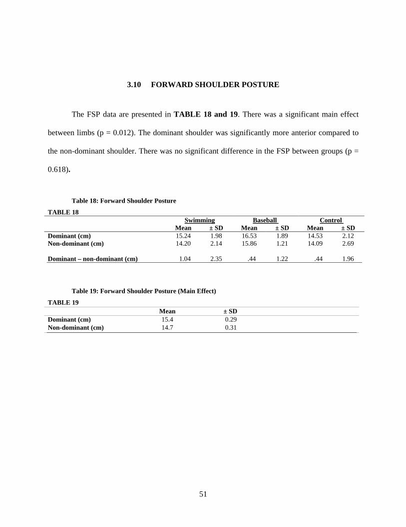
3.10 FORWARD SHOULDER POSTURE
The FSP data are presented in TABLE 18 and 19. There was a significant main effect
between limbs (p = 0.012). The dominant shoulder was significantly more anterior compared to
the non-dominant shoulder. There was no significant difference in the FSP between groups (p =
0.618).
Table 18: Forward Shoulder Posture
TABLE 18 Swimming Baseball Control Mean ± SD Mean ± SD Mean ± SD
Dominant (cm) 15.24 1.98 16.53 1.89 14.53 2.12 Non-dominant (cm) 14.20 2.14
15.86 1.21 14.09 2.69
Dominant – non-dominant (cm) 1.04 2.35 .44 1.22 .44 1.96
Table 19: Forward Shoulder Posture (Main Effect)
TABLE 19 Mean ± SD
Dominant (cm) 15.4 0.29 Non-dominant (cm) 14.7 0.31
51
4.0 DISCUSSION
The purpose of this study was to compare the physical characteristics of the shoulder
between swimmers, baseball pitchers, and control subjects. Swimmers’ and baseball pitchers’
shoulders are subjected to distinctively different demands in terms of kinematics, muscle action,
and repetition of overhead motion performed. Therefore, based on the SAID principle, swimmers
and baseball pitchers were expected to have different physical characteristics.
4.1 EXTERNAL/ INTERNAL ROTATION RANGE OF MOTION
As hypothesized, baseball pitchers exhibited significantly greater external rotation ROM
of the dominant shoulder compared to control subjects. This greater amount of external rotation
ROM found in the dominant shoulder of baseball pitchers has been extensively reported and is
comparable to that of previous studies.19, 25, 27, 40-42 The increase in external ROM is often
attributed to the repetitive ballistic motion at the end ROM in the late cocking phase of pitching
Although swimmers are often stereotyped as having a “hyper-mobile” shoulder joint, this
study found their external rotation ROM to be similar to the amount found in control subjects.
120 o The external rotation ROM of swimmers in this study was higher (~20 degrees) compared
to the results reported in a previous study by Beach et al 16 This difference could be due to the
use of pathological subjects in the previous study.16
52

Internal rotation ROM of the dominant shoulder was not significantly different between
groups, thus, our hypothesis was rejected. Although not statistically significant, the mean internal
rotation ROM of baseball pitchers was less in the dominant shoulder compared to the non-
dominant shoulder by more than 10 degrees. The high within-group variability in ROM may
account for this finding not being statistically significant. The internal rotation ROM found in
this study for swimmers and baseball pitchers is similar to the results from previous studies. 19, 25,
40-42, 78
As hypothesized, there were no bilateral differences in glenohumeral external rotation
and internal rotation ROM in swimmers. This should be due to the bilateral nature of the
swimming stroke. Small amount of ERG and GIRD was found in the dominant shoulders of
swimmers and control subjects. This suggests that a small degree of GIRD and ERG exist in
non-throwers, presumably from the hand dominance. Non-overhead athletes examined in a few
other studies similarly exhibited small amount of GIRD and ERG in their dominant shoulders.25,
26, 79
Baseball pitchers exhibited significantly greater GIRD compared to the other groups. The
amount of GIRD exhibited by baseball pitchers in this study is similar to those reported by
Myers et al 78 and Borsa et al 42. The baseball pitchers also exhibited greater ERG compared to
swimmers. This ERG is due to the increased external rotation ROM of the dominant shoulder in
baseball pitchers, because the external rotation ROM of the non-dominant shoulders was not
different between groups. As mentioned earlier, this increase in the external rotation ROM of the
dominant shoulder is a common observation in baseball pitchers 19, 25, 40-42, 78.
Despite ERG and GIRD, the total ROM in baseball pitchers was similar bilaterally. This
result is in agreement with the unpublished data of Wilk and Arrigo 20. The 372 professional
53

baseball pitchers they tested exhibited 7o greater external rotation ROM and 7o less internal
rotation ROM on the dominant shoulder compared to the non-dominant shoulder, resulting in
equal (within 5 o) total ROM with bilateral comparison 20. Wilk 20 referred to this preservation in
the total ROM as a “total motion concept”. Excessive GIRD that far exceeds the ERG, results in
decreases in the total ROM. This has been observed in symptomatic throwing shoulders. 20, 80
Decreases in the total ROM are considered an important physical sign associated with shoulder
pathologies 20, 80. Ruotolo et al 80 have recently reported that the total arc of shoulder ROM in the
painful dominant shoulder was decreased by 9.2o compared to the non-dominant shoulder in
collegiate baseball players. All the baseball pitchers in this study were non-pathological for at
least 3 months, and had no history of acute trauma or shoulder surgery. Symmetric total ROM on
the in our subjects supports this idea that the loss of total range of motion arc is associated with
shoulder pathology.
4.2 FLEXION/ ABDUCTION/ EXTENSION RANGE OF MOTION
Swimmers and baseball pitchers had a greater flexion ROM compared to control subjects.
The flexion ROM of swimmers and baseball pitchers was comparable to previous studies of
similar populations 15, 16, 27, 40, 41. The small difference between the studies possibly arose from
the inter-tester variability. Greater flexion ROM in swimmers compared to control subjects can
be explained by the repetitive use of their shoulders through the full ROM during swimming 27.
In a study by Brown et al 27, baseball pitchers had less flexion ROM of the dominant shoulder
compared to the non-dominant shoulder. Brown et al 27 explained this decreased flexion ROM as
a result of not using the throwing arm through a full ROM during pitching, and the unilateral
54
tightness of the pectoralis major and latissimus dorsi muscles of the dominant side. The pitchers
in our study exhibited statistically equal flexion ROM. This may be explained by the fact that the
baseball pitchers in the study by Brown et al 27 were professionals with an average age of 27
years, whereas the pitchers in this study were collegiate, with an average age of 20 years. This
bilateral imbalance may become more accentuated the longer pitchers throw.
In the current study, abduction ROM was measured with the humerus in neutral rotation,
as the limb was passively abducted to a point where the greater tuberosity abutted the acromion
process. This was done instead of fully externally rotating the humerus and passively moving the
arm through full ROM until ligamentous endfeel from the postero-inferior joint capsule. The
examiner felt that the testing procedure would be a better assessment for isolating the
glenohumeral ROM. This may explain why the abduction data from our study are considerably
lower than previously reported data.16
4.3 POSTERIOR SHOULDER TIGHTNESS
The results of the PST assessment showed a significant between-group difference in the
supine-method. This difference was not present for the side-lying method. Recent data from our
laboratory indicate that supine and side-lying methods of PST assessment have similar accuracy
and precision, but the supine method has higher reliability and sensitivity 73. During the testing
sessions, subjects subjectively report more difficulty relaxing the shoulder girdle muscles during
the side-lying method compared to the supine method. The results from this study reflect the
higher sensitivity of the supine method, which could be a result of better patient comfort.
The findings of this study showed that baseball pitchers exhibited significantly greater
55

PST compared to swimmers. This was in agreement with our hypothesis. PST in baseball players
has been documented in previous literature 17, 20, 25, 26, 75, 78, yet PST in swimmers has never
previously been evaluated. Due to the bilateral nature of the swim stroke, it was expected that the
swimmers would have no bilateral difference in PST.
Several studies have implicated PST as a contributing factor to shoulder pathologies in
overhead athletes. 17, 20, 25, 26, 73, 78, 81, 82 It has been documented that tightness in the posterior
shoulder structures tended to shift the glenohumeral joint center postero-superiorly during
maximal external rotation.75 This change in position increases the risk of developing labral
pathologies in overhead athletes, especially pitchers who repetitively position their shoulder in a
maximal external rotation position. 75
As expected, swimmers in this study presented with very small bilateral difference in
PST. The bilateral difference between PST was less than 4o, the dominant shoulder being slightly
tighter than the non-dominant shoulder. From this data alone, it is inconclusive whether the
swimmers have PST on both shoulders, or they do not have bilateral PST bilaterally. However,
an inference can be made from as to the strong relationship that exists between PST and the loss
of internal rotation ROM.17, 20, 25, 26, 73, 78, 81, 82 Tyler et al 25 stated that for every 4o of internal
rotation ROM loss, 1cm of PST can be expected to exist. Although statistically insignificant,
mean internal rotation ROM of the dominant shoulders of swimmers was greater than control
subjects’. This could be an indication that swimmers do not have PST. Further analysis of ROM
and PST is needed to further understand the swimmer’s shoulder.
56

4.4 EXTERNAL/ INTERNAL ROTATION STRENGTH
The hypothesis of this study was that swimmers and baseball pitchers would have higher
internal rotation strength, and a lower external: internal rotation strength ratio compared to
control subjects. This was expected because forces generated in swimming and baseball pitching
are produced primarily from humeral adduction and internal rotation at the shoulder1, 9, 10, 13, 15-21
Contrary to our expectation, the results of this study showed no between-group differences in
internal rotation strength, external rotation strength, or external: internal rotation strength ratio
tested at 60o/sec and 300 o /sec. Additionally, no bilateral strength differences were detected in
baseball players.
In this study, the external: internal rotation strength ratio tested at 60 o /sec for swimmers
was similar to that found by Beach et al 16, but higher than the results reported by McMaster et
al. 21 The external: internal rotation strength ratio tested at 300 o /sec in baseball pitchers was
similar to that of previous studies 10, 19, 27, 33. The lack of differences in the internal and external
rotation strength profile between the groups were likely due to the control subjects having very
low external: internal rotation strength ratios. This could be attributed to the fact that every
control subject in this study was an intercollegiate athlete who participated in regular weight
training activities.
Using a hand-held dynamometer, Layton et al11 evaluated shoulder strength in
intercollegiate swimmers and found decreased muscle strength among the glenohumeral internal
rotators, glenohumeral external rotator, trapezius, and serratus anterior muscles compared to age-
matched non-swimmers. The swimmers who participated in our study did not exhibit weaker
shoulder rotational strength compared to non-overhead athletes. This discrepancy is likely
attributed to differences in testing procedures between the two studies.
57

The majority of studies that have evaluated isokinetic strength in baseball players have
found higher internal strength and lower external: internal rotation strength ratio in the dominant
shoulder compared to the non-dominant shoulder. The majority of the baseball players in this
study were tested during the early pre-season period, when the bilateral difference in strength
might be minimal. This factor along with high variability in external rotation strength, marked by
the high standard deviation relative to the mean, may be the reason for the lack of significant
differences.
4.5 PROTRACTION/ RETRACTION STRENGTH
Few studies have evaluated scapular protraction and retraction strength using an
isokinetic dynamometer 36, 37, 76. The protraction and retraction strength values obtained in this
study were low compared to the values previously reported by Cools et al. 36, 37, 76 The values in
this study were decreased because we reported strength as an average peak torque normalized to
body weight. We felt average peak torque was more representative of strength compared to the
peak torque, because the value is less likely affected by a single repetition that is remarkably
higher than the rest of the repetitions. The protraction: retraction strength ratios found in this
study were comparable to those of other studies examining overhead. 36, 37, 76 However, the
current study was the first of our knowledge to specifically evaluate protraction and retraction
strength in baseball pitchers and swimmers. Our hypothesis was that swimmers and baseball
pitchers would exhibit greater protraction and retraction strength compared to the control
subjects due to the extensive use of their shoulders. However, no difference between the baseball
pitchers, swimmers, and control subjects was found in protraction strength, retraction strength, or
58

the strength ratio in our study. In a study by Cools et al 76, no bilateral difference in protraction
and retraction strength was found in healthy overhead athletes, but the protraction: retraction
strength ratio was significantly different at 12.2cm/sec. Perhaps isokinetic testing is not sensitive
enough to discern the bilateral differences in a healthy population.
4.6 SCAPULAR KINEMATICS
This study was the first to evaluate 3-dimentional scapular kinematics in swimmers. Our
hypothesis that swimmers would have enhanced scapular mobility due to the repetitive use of
their shoulder through a large ROM was not supported by our data. The swimmers who
participated in this study had scapular kinematics similar to that of control subjects. The possible
reason no difference in scapular kinematic between the control subjects and the swimmers was
found is because the scapular kinematics were assessed in a single plane of motion. Because
swimming stroke is a multi-planer motion, scapular kinematics assessment in a single plane may
not be appropriate to evaluate the sports specific adaptation in the scapular kinematics. In
addition, the swimming stroke is unique from a baseball pitch, tennis serve, or volleyball spike in
that the shoulders are repeatedly placed in maximal flexion and abduction. Thus, unique scapular
kinematics characteristics in swimmers may be present towards the end-range of humeral
elevation.
Myers et al 52 have demonstrated that baseball players possess greater scapular upward
rotation, internal rotation, and retraction of the scapula of the dominant shoulder during humeral
elevation. However, in this study there were no between group differences in scapular
59
kinematics. The only notable difference found was an increased scapular upward rotation of the
baseball pitchers’ dominant shoulders compared to the non-dominant side at high humeral
elevation angles. This finding has been reported by other studies.82-84 Myers et al 52 reported that
scapular external rotation linearly increased as humeral elevation increased, whereas our data
demonstrated a decrease in scapular external rotation at the lower humeral elevation angles (0o to
60 o) , and an increase at higher elevation angles (90o to 120o). In addition, the range of external
rotation in our study was significantly lower (~10 o) compared to that reported by Myers et al 52,
and the elevation angle found in this study (range: 6.5o-30.5o) was considerably higher compared
to the data by Myers et al 52 (range: 0.33o-12.23o). The difference between the findings of these
studies could be from the difference in the anatomical landmarks used to create the scapular local
coordinates. Our study used trigonum spinae (the intersection of the medial scapula border and
the scapula spine) and acromioclavicular joint to define the medial-lateral axis of the scapula,
while Myers et al 52 used trigonum spinae and angulus acromialis (the most lateral-dorsal point
of scapula). Differences could also be attributed to the unknown measurement error or the low
subject size in our study.
4.7 FORWARD SHOULDER POSTURE
There were no differences in FSP between groups contradicted our hypothesis that the
dominant shoulder of baseball players would be positioned more anteriority compared to the
other groups due to the unilateral nature of their sports activity.
Swimmers have been shown to have increased forward head angle and scapular
abduction compared to non-swimmers.11 Our FSP measurement using a double-square method
60

was unable to show differences in posture between swimmers and non-overhead athletes. The
double square device was chosen for our study because it can be easily performed to assess
shoulder posture in a clinical setting. A limitation of this method is that the measurement can be
affected by postural sway, as well as rotation of the lower extremity and torso. This could have
masked potential group differences in the FSP between groups.
When all groups were combined, the FSP data showed that the dominant shoulder was
positioned significantly anterior compared to the non-dominant shoulder when the average of
each side was compared. Significantly “dropped” dominant shoulder has been reported in
individuals with shoulder pathology.81 This “dropped” appearance of the shoulder is attributed to
the upper scapula rotating antero-inferiorly as a result of increased scapular protraction and
internal rotation.81 The protraction and internal rotation of the scapula positions an acromion
process anteriorly. Burkhart et al 81 use the acronym S.I.C.K., Scapular malposition, Inferior
medial scapula border prominence, Coracoid pain and malposition, and dysKinesis of scapular
movement, as a condition characterized by an apparent “dropping” of the dominant pathological
shoulder, anterior shoulder pain, and altered scapular kinematics.81 The S.I.C.K. scapula is
clinically shown to be associated with labral pathologies, impingement syndrome, and rotator
cuff lesions.81 All the subjects in this study were asymptomatic, yet exhibited significant
asymmetry in the forward shoulder posture. This suggests that presence of asymmetry alone may
not be an indication of shoulder pathologies. Clinically observed postural asymmetry in patients
with shoulder pathology may be present in greater degree compared to the asymmetry found in
non-pathologic subjects. Comparison of FSP between pathologic and non-pathologic individuals
needs to be done in the future.
61
4.8 CLINICAL RELEVANCE
Some of the variables evaluated in this study (PST, protraction/ retraction strength, and
three-dimensional scapular kinematics) have never been assessed in intercollegiate swimmers,
and therefore can serve as the normative data for this population.
No previous literature has examined PST in swimmers. Our data suggest that PST may
not exist in swimmers. The swimmers who participated in this study were all free of shoulder
pain, thus PST in swimmers with shoulder pathology is still unknown. PST has been reported in
individuals with shoulder subacromial impingement 25 and internal impingement.78 Considering
subacromial impingement is the most common shoulder injury in swimmers, assessment of PST
should be included as a part of the clinical evaluation of shoulder pain in swimmers.1, 2, 12, 64
Posterior shoulder stretching is widely performed by baseball players to correct PST and such
stretches may be indicated for swimmers with impingement syndrome.
The protraction: retraction strength ratios of swimmers in our study were 0.90-0.93 at
12.2 cm/sec, and 0.81-0.87 at 36.6 cm/sec. These can be used as normative values for healthy
intercollegiate swimmers, and can be used for comparison in future studies.
Analysis of the three dimensional scapular kinematics during the humeral elevation task
showed symmetric scapular kinematics in swimmers, whereas baseball players exhibited
asymmetry in scapular upward rotation at higher humeral elevation angles. This suggests that
asymmetry in the scapular kinematics of swimmers may indicate pathology, while some degree
of asymmetry may be expected in the baseball pitchers.
This study showed differences in shoulder ROM characteristics and PST between
swimmers, baseball players, and control subjects. Understanding differences in the physical
62
characteristics that exist between the groups may help clinicians perform sports-specific shoulder
evaluations, and design rehabilitation/ treatment programs.
4.9 LIMITATIONS OF THE STUDY
One of the limitations of this study was that subjects were considered healthy based on
the absence of symptoms 6 months prior to participation. No x-rays or magnetic resonance
imaging (MRI) were performed to rule out pathology. There is a possibility that some subjects
may have had underlying pathology that had yet to become symptomatic.
Another limitation of this study was that data collection was conducted over 5 months,
meaning some athletes were tested during the early preseason, while others were tested during
the competitive season. A ROM change that may occur in baseball pitchers during the season has
been documented in the literature. Lack of control over the timing of the testing during the
season may have confounded the results of the study.
4.10 FUTURE DIRECTIONS
Further research examining the shoulder characteristics of pathological swimmers is
needed. Comparison of the data from this study to pathological subjects will help identify the
possible risk factors for shoulder pathology in swimmers. Furthermore, prospective cohort
studies that track shoulder physical characteristics and the development of shoulder pathology in
a group of swimmers may be able to identify the factors that predict future shoulder injury. Once
key factors of injury are identified, the development of specific intervention programs that focus
63
on reducing the risk factors associated with injury will help prevent the development of shoulder
pathologies.
Currently, assessment of the 3-dimensional scapular kinematics is limited to angles below
120 o of humeral elevation due to the reduced validity in the measurements above 120 o.48
Scapular kinematics assessment using bone pins or multi-planar x-ray will be required to
evaluate the scapular kinematics above 120 o in swimmers. The shoulder is internally rotated and
elevated greater than 180 o at hand entry during the swim stroke. This position closely resembles
that of the Neer impingement sign test, the special test one of the most commonly used to assess
subacromial impingement. In future research assessment of scapular kinematics at humeral
elevation angles above 120 o may reveal unique scapular characteristics in swimmers, and may
also help identify the scapular kinematics differences between the healthy and pathological
swimmers.
64
5.0 CONCLUSIONS
The results of this current study demonstrated differences in the physical characteristics
of the shoulders among swimmers, baseball pitchers, and controls due to the sports specific
demands placed on their shoulders. Although swimmers are referred to as a population with
hyper-mobile shoulder joints, increased ROM was observed only in flexion. The shoulder
extension and rotational ROM of swimmers did not exhibit hyper-mobility compared to baseball
pitchers and controls. The ROM characteristics that arise from the unilateral use of the dominant
limb (GIRD, ERG, and PST) were observed in baseball players but not in swimmers. Differences
in strength, scapular kinematics, and FSP between groups were not identified in this study.
Further research and advancement in assessment techniques may reveal differences in these
variables in the future.
65

BIBLIOGRAPHY
1. Richardson AB, Jobe FW, Collins HR. The shoulder in competitive swimming. Am J
Sports Med. May-Jun 1980;8(3):159-163. 2. Bak K, Faunl P. Clinical findings in competitive swimmers with shoulder pain. Am J
Sports Med. Mar-Apr 1997;25(2):254-260. 3. Borsa PA, Scibek JS, Jacobson JA, Meister K. Sonographic stress measurement of
glenohumeral joint laxity in collegiate swimmers and age-matched controls. Am J Sports Med. Jul 2005;33(7):1077-1084.
4. McMaster WC. Anterior glenoid labrum damage: a painful lesion in swimmers. Am J
Sports Med. Sep-Oct 1986;14(5):383-387. 5. McMaster WC, Roberts A, Stoddard T. A correlation between shoulder laxity and
interfering pain in competitive swimmers. Am J Sports Med. Jan-Feb 1998;26(1):83-86. 6. McMaster WC, Troup J. A survey of interfering shoulder pain in United States
competitive swimmers. Am J Sports Med. Jan-Feb 1993;21(1):67-70. 7. Scovazzo ML, Browne A, Pink M, Jobe FW, Kerrigan J. The painful shoulder during
freestyle swimming. An electromyographic cinematographic analysis of twelve muscles. Am J Sports Med. Nov-Dec 1991;19(6):577-582.
8. Johnson D. In Swimming, shoulder the burden. Sportcare and fitness. 1988(May-
June):24-30. 9. Kennedy JC, Hawkins R, Krissoff WB. Orthopaedic manifestations of swimming. Am J
Sports Med. Nov-Dec 1978;6(6):309-322. 10. Dillman CJ, Fleisig GS, Andrews JR. Biomechanics of pitching with emphasis upon
shoulder kinematics. J Orthop Sports Phys Ther. Sep 1993;18(2):402-408. 11. Layton JA, Thigpen,C.A., Pauda,D.A., Prentice,W.E., Karas,S.G. A Comparison
Between Swimmers and Non-swimmers on Posture, Range of Motion, Strength, and Scapular Motion. Paper presented at: National Athletic Training Association Annual Meeting, 2005; Indianapolis.
66

12. Yanai T, Hay JG. Shoulder impingement in front-crawl swimming: II. Analysis of stroking technique. Med Sci Sports Exerc. Feb 2000;32(1):30-40.
13. Kibler WB. The role of the scapula in athletic shoulder function. Am J Sports Med. Mar-
Apr 1998;26(2):325-337. 14. Fleisig GS, Barrentine SW, Escamilla RF, Andrews JR. Biomechanics of overhand
throwing with implications for injuries. Sports Med. Jun 1996;21(6):421-437. 15. Bak K, Magnusson SP. Shoulder strength and range of motion in symptomatic and pain-
free elite swimmers. Am J Sports Med. Jul-Aug 1997;25(4):454-459. 16. Beach ML, Whitney SL, Dickoff-Hoffman SA. Relationship of Shoulder Flexibility,
Strength, and Endurance to Shoulder Pain in Competitive Swimmers. J Orthop Sports Phys Ther. 1992;16(6):262-268.
17. Burkhart SS, Morgan CD, Kibler WB. The disabled throwing shoulder: spectrum of
pathology. Part I: pathoanatomy and biomechanics. Arthroscopy. Apr 2003;19(4):404-420.
18. Wilk KE, Andrews JR, Arrigo CA. The abductor and adductor strength characteristics of
professional baseball pitchers. Am J Sports Med. Nov-Dec 1995;23(6):778. 19. Wilk KE, Andrews JR, Arrigo CA, Keirns MA, Erber DJ. The strength characteristics of
internal and external rotator muscles in professional baseball pitchers. Am J Sports Med. Jan-Feb 1993;21(1):61-66.
20. Wilk KE, Meister K, Andrews JR. Current concepts in the rehabilitation of the overhead
throwing athlete. Am J Sports Med. Jan-Feb 2002;30(1):136-151. 21. McMaster WC, Long SC, Caiozzo VJ. Shoulder torque changes in the swimming athlete.
Am J Sports Med. May-Jun 1992;20(3):323-327. 22. Kibler WB. Role of the Scapula in the Overhead Throwing Motion. Contemporary
Orthopaedics. 1991;22(5). 23. Meister K. Injuries to the shoulder in the throwing athlete. Part one:
Biomechanics/pathophysiology/classification of injury. Am J Sports Med. Mar-Apr 2000;28(2):265-275.
24. Kibler WB, McMullen J. Scapular dyskinesis and its relation to shoulder pain. J Am Acad
Orthop Surg. Mar-Apr 2003;11(2):142-151. 25. Tyler TF, Nicholas SJ, Roy T, Gleim GW. Quantification of posterior capsule tightness
and motion loss in patients with shoulder impingement. Am J Sports Med. Sep-Oct 2000;28(5):668-673.
67
26. Tyler TF, Roy T, Nicholas SJ, Gleim GW. Reliability and validity of a new method of measuring posterior shoulder tightness. J Orthop Sports Phys Ther. May 1999;29(5):262-269; discussion 270-264.
27. Brown LP, Niehues SL, Harrah A, Yavorsky P, Hirshman HP. Upper extremity range of
motion and isokinetic strength of the internal and external shoulder rotators in major league baseball players. Am J Sports Med. Nov-Dec 1988;16(6):577-585.
28. Donatelli R, Ellenbecker TS, Ekedahl SR, Wilkes JS, Kocher K, Adam J. Assessment of
shoulder strength in professional baseball pitchers. J Orthop Sports Phys Ther. Sep 2000;30(9):544-551.
29. McMaster WC, Long SC, Caiozzo VJ. Isokinetic torque imbalances in the rotator cuff of
the elite water polo player. Am J Sports Med. Jan-Feb 1991;19(1):72-75. 30. Meister K. Injuries to the shoulder in the throwing athlete. Part two: evaluation/treatment.
Am J Sports Med. Jul-Aug 2000;28(4):587-601. 31. Wang HK, Cochrane T. Mobility impairment, muscle imbalance, muscle weakness,
scapular asymmetry and shoulder injury in elite volleyball athletes. J Sports Med Phys Fitness. Sep 2001;41(3):403-410.
32. Weldon EJ, 3rd, Richardson AB. Upper extremity overuse injuries in swimming. A
discussion of swimmer's shoulder. Clin Sports Med. Jul 2001;20(3):423-438. 33. Ellenbecker TS, Mattalino AJ. Concentric isokinetic shoulder internal and external
rotation strength in professional baseball pitchers. J Orthop Sports Phys Ther. May 1997;25(5):323-328.
34. Mikesky AE, Edwards JE, Wigglesworth JK, Kunkel S. Eccentric and concentric strength
of the shoulder and arm musculature in collegiate baseball pitchers. Am J Sports Med. Sep-Oct 1995;23(5):638-642.
35. Noffal GJ. Isokinetic eccentric-to-concentric strength ratios of the shoulder rotator
muscles in throwers and nonthrowers. Am J Sports Med. Jul-Aug 2003;31(4):537-541. 36. Cools AM. Test-retest reproducibility of concentric strength values for shoulder girdle
protraction and retraction using the Biodex isokinetic values for dynamometer. Isokinet Exer Sci. 2002;10:129-136.
37. Cools AM, Witvrouw EE, Declercq GA, Vanderstraeten GG, Cambier DC. Evaluation of
isokinetic force production and associated muscle activity in the scapular rotators during a protraction-retraction movement in overhead athletes with impingement symptoms. Br J Sports Med. Feb 2004;38(1):64-68.
68
38. The relationship Between Glenohumeral Internal Rotation and Shoulder and Elbow Pain. PBATS Newsletter. 2002;15(2).
39. Harner CD. The unstable shoulder pathophyisiology and diagnosis Annual Panther
Sports Medicine Symposium. Pittsburgh, PA; 1989. 40. Reagan KM, Meister K, Horodyski MB, Werner DW, Carruthers C, Wilk K. Humeral
retroversion and its relationship to glenohumeral rotation in the shoulder of college baseball players. Am J Sports Med. May-Jun 2002;30(3):354-360.
41. Bigliani LU, Codd TP, Connor PM, Levine WN, Littlefield MA, Hershon SJ. Shoulder
motion and laxity in the professional baseball player. Am J Sports Med. Sep-Oct 1997;25(5):609-613.
42. Borsa PA, Wilk KE, Jacobson JA, et al. Correlation of Range of Motion and
Glenohumeral Translation in Professional Baseball Pitchers. Am J Sports Med. Jul 7 2005.
43. Borsa PA, Jacobson JA, Scibek JS, Dover GC. Comparison of dynamic sonography to
stress radiography for assessing glenohumeral laxity in asymptomatic shoulders. Am J Sports Med. May 2005;33(5):734-741.
44. Crockett HC, Gross LB, Wilk KE, et al. Osseous adaptation and range of motion at the
glenohumeral joint in professional baseball pitchers. Am J Sports Med. Jan-Feb 2002;30(1):20-26.
45. Osbahr DC, Cannon DL, Speer KP. Retroversion of the humerus in the throwing shoulder
of college baseball pitchers. Am J Sports Med. May-Jun 2002;30(3):347-353. 46. Warner JJ, Allen AA, Marks PH, Wong P. Arthroscopic release of postoperative capsular
contracture of the shoulder. J Bone Joint Surg Am. Aug 1997;79(8):1151-1158. 47. Borstad JD, Ludewig PM. Comparison of scapular kinematics between elevation and
lowering of the arm in the scapular plane. Clin Biomech (Bristol, Avon). Nov-Dec 2002;17(9-10):650-659.
48. Karduna AR, McClure PW, Michener LA, Sennett B. Dynamic measurements of three-
dimensional scapular kinematics: a validation study. J Biomech Eng. Apr 2001;123(2):184-190.
49. Ludewig PM, Cook TM, Nawoczenski DA. Three-dimensional scapular orientation and
muscle activity at selected positions of humeral elevation. J Orthop Sports Phys Ther. Aug 1996;24(2):57-65.
50. Ludewig PM, Cook TM, Shields RK. Comparison of Surface Sensor and Bone-fixed
Measurment of Humeral Motion. J Applied Biomech. 2002;18:163-170.
69
51. McClure PW, Michener LA, Sennett BJ, Karduna AR. Direct 3-dimensional measurement of scapular kinematics during dynamic movements in vivo. J Shoulder Elbow Surg. May-Jun 2001;10(3):269-277.
52. Myers JB, Laudner KG, Pasquale MR, Bradley JP, Lephart SM. Scapular position and
orientation in throwing athletes. Am J Sports Med. Feb 2005;33(2):263-271. 53. Ludewig PM, Cook TM. Alterations in shoulder kinematics and associated muscle
activity in people with symptoms of shoulder impingement. Phys Ther. Mar 2000;80(3):276-291.
54. Lukasiewicz AC, McClure P, Michener L, Pratt N, Sennett B. Comparison of 3-
dimensional scapular position and orientation between subjects with and without shoulder impingement. J Orthop Sports Phys Ther. Oct 1999;29(10):574-583; discussion 584-576.
55. Crotty NM, Smith J. Alterations in scapular position with fatigue: a study in swimmers.
Clin J Sport Med. Oct 2000;10(4):251-258. 56. Su KP, Johnson MP, Gracely EJ, Karduna AR. Scapular rotation in swimmers with and
without impingement syndrome: practice effects. Med Sci Sports Exerc. Jul 2004;36(7):1117-1123.
57. Tsai NT, McClure PW, Karduna AR. Effects of muscle fatigue on 3-dimensional
scapular kinematics. Arch Phys Med Rehabil. Jul 2003;84(7):1000-1005. 58. Kebaetse M, McClure P, Pratt NA. Thoracic position effect on shoulder range of motion,
strength, and three-dimensional scapular kinematics. Arch Phys Med Rehabil. Aug 1999;80(8):945-950.
59. Finley MA, Lee RY. Effect of sitting posture on 3-dimensional scapular kinematics
measured by skin-mounted electromagnetic tracking sensors. Arch Phys Med Rehabil. Apr 2003;84(4):563-568.
60. Wang CH, McClure P, Pratt NE, Nobilini R. Stretching and strengthening exercises: their
effect on three-dimensional scapular kinematics. Arch Phys Med Rehabil. Aug 1999;80(8):923-929.
61. Kendall FP, McCreary EK. Muscles: Testing and Function. 3 ed. Baltimore, MC:
Williams and Wilkins; 1983. 62. Kendall HO, Kendall FP, Boynton DA. Posture and Pain. Huntington, NY: Krieger
Publishing Company, Inc.; 1970. 63. Solem-Bertoft E, Thuomas KA, Westerberg CE. The influence of scapular retraction and
protraction on the width of the subacromial space. An MRI study. Clin Orthop Relat Res. Nov 1993(296):99-103.
70

64. Yanai T, Hay JG, Miller GF. Shoulder impingement in front-crawl swimming: I. A method to identify impingement. Med Sci Sports Exerc. Feb 2000;32(1):21-29.
65. Richardson AB. Thoracic outlet syndrome in aquatic athletes. Clin Sports Med. Apr
1999;18(2):361-378. 66. Meskers CG, Vermeulen HM, de Groot JH, van Der Helm FC, Rozing PM. 3D shoulder
position measurements using a six-degree-of-freedom electromagnetic tracking device. Clin Biomech (Bristol, Avon). Jun 1998;13(4-5):280-292.
67. Meskers CG, Fraterman H, van der Helm FC, Vermeulen HM, Rozing PM. Calibration of
the "Flock of Birds" electromagnetic tracking device and its application in shoulder motion studies. J Biomech. Jun 1999;32(6):629-633.
68. Drouin JM, Valovich-mcLeod TC, Shultz SJ, Gansneder BM, Perrin DH. Reliability and
validity of the Biodex system 3 pro isokinetic dynamometer velocity, torque and position measurements. Eur J Appl Physiol. Jan 2004;91(1):22-29.
69. Peterson DE, Blankenship KR, Robb JB, et al. Investigation of the validity and reliability
of four objective techniques for measuring forward shoulder posture. J Orthop Sports Phys Ther. Jan 1997;25(1):34-42.
70. Greenfield BH, Kraus S, Lawrence S, Wolf S. The influence of cephalostatic ear rods on
the positions of head and neck during postural recordings. Am J Dentofac Orthop. 1989;95(4):312-318.
71. Molhave A. En biostaisk undersogelse. Menneshets stuende stilling teorestik og
statometrisk belgst. With an English summary (A biostatic investigation of human erect posture), 1958; Munksgard, Copenhagen.
72. Norkin CC, White DJ. Measurment of Joint Motion: A guide to Goniometry. 2 ed.
Philadelphia: F.A. Davis Company; 1995. 73. Myers JB, Wassinger CA, Oyama S, Ricci RD, Lephart SM. Clinical assessments of
posterior shoulder tightness: Accuracy, reliability, precision, and validity. Paper presented at: American Shoulder and Elbow Society 23rd Open Meeting, 2007; San Diego, CA.
74. Stokdijk M, Nagels J, Rozing PM. The glenohumeral joint rotation centre in vivo. J
Biomech. Dec 2000;33(12):1629-1636. 75. Harryman DT, 2nd, Sidles JA, Clark JM, McQuade KJ, Gibb TD, Matsen FA, 3rd.
Translation of the humeral head on the glenoid with passive glenohumeral motion. J Bone Joint Surg Am. Oct 1990;72(9):1334-1343.
71

76. Cools AM, Witvrouw EE, Mahieu NN, Danneels LA. Isokinetic Scapular Muscle Performance in Overhead Athletes With and Without Impingement Symptoms. J Athl Train. Jun 2005;40(2):104-110.
77. Van der Helm FC. A standardized Protocol for the Description of Shoulder Motions.
University Park, PA: International Shoulder Group of the International Society of Biomechanics; 2002.
78. Myers JB, Laudner KG, Pasquale MR, Bradley JP, Lephart SM. Glenohumeral range of
motion deficits and posterior shoulder tightness in throwers with pathologic internal impingement. Am J Sports Med. Mar 2006;34(3):385-391.
79. Magnusson SP, Gleim GW, Nicholas JA. Shoulder weakness in professional baseball
pitchers. Med Sci Sports Exerc. Jan 1994;26(1):5-9. 80. Ruotolo C, Price E, Panchal A. Loss of total arc of motion in collegiate baseball players.
J Shoulder Elbow Surg. Jan-Feb 2006;15(1):67-71. 81. Burkhart SS, Morgan CD, Kibler WB. The disabled throwing shoulder: spectrum of
pathology. Part III: The SICK scapula, scapular dyskinesis, the kinetic chain, and rehabilitation. Arthroscopy. Jul-Aug 2003;19(6):641-661.
82. Downar JM, Sauers EL. Clinical Measures of Shoulder Mobility in the Professional
Baseball Player. J Athl Train. Mar 2005;40(1):23-29. 83. Downer JM, Sauers EL, Mourrtacos SL. Chronic adaptations in the throwing shoulder of
professiona; baseball players. J Athl Train. 2002;37:S17-S18. 84. Mourtacos SL, Sauers EL, Downar JM. Adolescent baseball players exhibit differences in
shoulder mobility between the throwing and non-throwing shoulder and between divisions of play. J Athl Train. 2003(S72).
72