progressive engineering of a homing endonuclease … 9 dna transfection reagent with manu-facturer...
TRANSCRIPT
Nucleic Acids Research, 2014 1doi: 10.1093/nar/gku224
Progressive engineering of a homing endonucleasegenome editing reagent for the murine X-linkedimmunodeficiency locusYupeng Wang1, Iram F. Khan1, Sandrine Boissel1,2, Jordan Jarjour3, Joseph Pangallo1,Summer Thyme2, David Baker2, Andrew M. Scharenberg1,4,* and David J. Rawlings1,4,*
1Center for Immunity and Immunotherapies, Seattle Children’s Research Institute, Seattle, WA 98101, USA,2Department of Biochemistry, University of Washington, Seattle, WA 98195, USA, 3Pregenen Inc., Seattle, WA98103, USA and 4Departments of Pediatrics and Immunology, University of Washington, Seattle, WA 98195, USA
Received November 13, 2013; Revised March 03, 2014; Accepted March 5, 2014
ABSTRACT
LAGLIDADG homing endonucleases (LHEs) are com-pact endonucleases with 20–22 bp recognition sites,and thus are ideal scaffolds for engineering site-specific DNA cleavage enzymes for genome edit-ing applications. Here, we describe a general ap-proach to LHE engineering that combines ratio-nal design with directed evolution, using a yeastsurface display high-throughput cleavage selection.This approach was employed to alter the bindingand cleavage specificity of the I-Anil LHE to recog-nize a mutation in the mouse Bruton tyrosine kinase(Btk) gene causative for mouse X-linked immunod-eficiency (XID)––a model of human X-linked agam-maglobulinemia (XLA). The required re-targeting ofI-AniI involved progressive resculpting of the DNAcontact interface to accommodate nine base differ-ences from the native cleavage sequence. The en-zyme emerging from the progressive engineeringprocess was specific for the XID mutant allele ver-sus the wild-type (WT) allele, and exhibited activityequivalent to WT I-AniI in vitro and in cellulo reporterassays. Fusion of the enzyme to a site-specific DNAbinding domain of transcription activator-like effec-tor (TALE) resulted in a further enhancement of geneediting efficiency. These results illustrate the poten-tial of LHE enzymes as specific and efficient tools fortherapeutic genome engineering.
INTRODUCTION
Homing endonucleases (HEs) are sequence-specific en-zymes that recognize and cleave DNA at long target sites(typically 20 bp). They are typically encoded within intronsor inteins, and behave as mobile genetic elements that copytheir genetic information into intron- or intein-less allelesof their host gene. This genetic mobility is catalyzed by HEendonuclease-mediated DNA double-strand breaks (DSBs)in intein/intron-less alleles of the host gene. This facilitatesrepair by homologous recombination using the intron- orintein-containing gene, resulting in copying of the intron orintein into the new allele site (1,2).
LAGLIDADG homing endonucleases (LHEs) are a par-ticularly attractive system for the development of gene-specific reagents because they possess 20–22 bp recognitionsites, and cleavage activity is tightly coupled to DNA targetsite recognition (1,3,4). A variety of approaches have beenapplied to generate LHE variants with new cleavage speci-ficities, most of them involving ‘local’ variant library gener-ation through random mutation or structural-based modifi-cation of the LHE protein interface that contacts the DNAtarget site, followed by selection based on DNA cleavage orrecombination activities (5–8). These methods currently areable to generate variants with changes in cleavage specificityin a ‘local’ region of the LHE DNA/protein interface cover-ing a relatively small number of contiguous base pairs. Forphysiologic targets, where multiple base pair mismatchesmust be targeted by variants that possess alterations in adja-cent or overlapping regions of the DNA/protein interface,the engineering of LHE variants with high specificity andactivity requires combinations of local changes that ofteninclude conflicting sets of amino acid (AA) changes in theinterface. The development of methods to overcome limita-tions in large scale LHE re-specification is necessary to ex-
*To whom correspondence should be addressed. Tel: +1 206 987 7319; Fax: +1 206 987 7310. Email: [email protected] may also be addressed to Andrew M. Scharenberg. Tel: +1 206 987 7314; Fax: +1 206 987 7310; Email: [email protected] address:Summer Thyme, Department of Molecular and Cellular Biology, Harvard University, Cambridge, MA 02138, USA.
C© The Author(s) 2014. Published by Oxford University Press on behalf of Nucleic Acids Research.This is an Open Access article distributed under the terms of the Creative Commons Attribution License (http://creativecommons.org/licenses/by/3.0/), whichpermits unrestricted reuse, distribution, and reproduction in any medium, provided the original work is properly cited.
Nucleic Acids Research Advance Access published March 25, 2014 at U
niversity of Washington on A
pril 16, 2014http://nar.oxfordjournals.org/
Dow
nloaded from
2 Nucleic Acids Research, 2014
pand the application of LHEs to such extremely challengingtargets.
Using a yeast surface display high-throughput cleavageselection system (9), here we show the application of ra-tional design with directed evolution in a progressive ap-proach to achieve a large scale re-engineering of the I-Anilhoming endonuclease to specifically recognize a unique se-quence in the mouse Bruton tyrosine kinase (Btk) gene dif-fering by 9 bp from the native I-AniI sequence. Fusion ofthis enzyme to a sequence-specific TALE DNA binding do-main was used to further increase the activity and specificityof the most refined variant for the XID target site. Takentogether, our results provide a roadmap for engineering ofLHEs to create highly specific and active reagents for ther-apeutic genome engineering.
MATERIALS AND METHODS
DNA constructs and substrates for binding and cleavage as-says
The I-Anil scaffold used here is the Y2 variant, containingtwo additional mutations, F13Y and S111Y, which enhanceboth DNA-binding affinity and cleavage efficacy (10). TheTALE repeat variable diresidue (RVD) arrays were assem-bled using Golden Gate TAL effector kit, and the TAL ef-fectors were fused to the N-terminus of XID through a Zn4linker (VGGS) (11,12). The 52 bp HE substrates were gen-erated by PCR using single-strand oligonucleotides as tem-plate with HE recognition sites in the middle flanked by 16bp primer binding site on each end. Biotin and fluorophorelabels were introduced by 5′ biotin-conjugated primer and 3′Alexa Fluor-647-conjugated primer, respectively. HE sub-strates were purified from single-stranded contaminants byExo1 digestion (New England Biolabs) and size exclusionthrough a G-100 column (GE Healthcare), then analyzedfor purity by gel electrophoresis (determined to be >98%)(9).
Yeast growth, transformation, library construction and plas-mid recovery
Saccharomyces cerevisiae strain EBY100 was transformedusing the lithium-acetate (LiAc) method (13). For random-ization library construction, randomization oligos with de-generative code on selected bases were ordered from Sigma.After PCR amplification, oligos were cloned into pET-CON2 vector through homologous recombination in yeast.The distribution of codon frequencies was verified by se-quencing an unselected library and determined to exhibitno major biasing of the type at positions of randomization(9). For random mutagenesis library construction, error-prone PCR was performed over selected region of the I-Anil variant using the GeneMorph-II Random Mutagen-esis kit (Stratagene) according to the manufacturer’s proto-col. Library size was determined by plating serial dilutionson selective plates. Mutation distribution and frequencieswere verified by sequencing an unselected library and de-termined to be in the range of 7–10 mutations per kilo basewith no major biases in the type or position of mutations.Yeast propagation was performed in the presence of 2% raf-finose + 0.1% glucose at 30◦C for at least 12 h prior to in-
duction. Cells were induced in 2% galactose for 2–3 h at30◦C followed by 18–26 h at 20◦C. Plasmids were isolatedfrom yeast populations using the Zymoprep-II kit (ZymoResearch) and transformed into Escherichia coli DH5� byheat shock for amplification and/or sequencing. Sequenc-ing was performed on 40–60 clones for a given selection out-put.
Yeast surface cleavage and sorting
The yeast surface-based cleavage assay has been describedpreviously (9). In brief, ∼30–100 × 106 (at least 3-fold thesize of the input population) induced cells were stained firstwith 1:300 dilution biotinylated anti-HA (Covance), thenwith pre-conjugated streptavidin-PE:biotin-DNA-A647 ina yeast binding buffer containing 180 mM KCl, 10 mMNaCl, 10 mM HEPES, 0.2% bovine serum albumin (BSA),0.1% galactose, pH 7.5. Samples were then washed twice inthe cleavage buffer containing 150 mM KCl, 10 mM NaCl,10 mM HEPES, 10 mM K-glutamine, 0.5 mg/ml BSA, pH8.25, and then transferred to cleavage buffer containing 7.5mM of either CaCl2 or MgCl2, and placed at 37◦C for theindicated time points. The reaction was stopped by transfer-ring cells to three reaction volume cold staining buffer with1:200 dilution FITC-conjugated anti-Myc antibody (LCLlabs) for HE surface expression staining. The catalytic activ-ity of HEs was measured by the decrease in Alex647 fluores-cence intensity associated with dsOligo cleavage and releasefrom yeast surface on a BD ARIAII cytometer, and result-ing data were analyzed using Flowjo software (Tree Star).For XID-Ani libraries, ∼0.3–1% population with the high-est cleavage activity were sorted for enrichment, and eachlibrary was enriched for three times before final analysis.
In vitro cleavage assay and cleavage specificity
3 × 10⁶ displaying yeasts with an estimated concentration of1–10 nM in a 40 �l reaction (assuming 104–105 moleculesper yeast cell surface) (9,14) were incubated with cleavagebuffer + 20 nM Alexa-647-conjugated dsOligo with 5 mMMgCl2, 5 mM dithiothreitol (DTT) and placed at 37◦C for1 h. The reaction was stopped by adding 50 mM ethylene-diaminetetraacetic acid (EDTA) and DNA sample buffer.After spinning down, 20 �l of supernatant was loaded on a10% non-denaturing polyacrylamide gel. HE cleavage sitesare in the middle of oligo substrates, which will generate twoproducts of the same size in cleavage assays. The cleavageproduct detected in in vitro cleavage assay is the 3′ half withAlexa Fluor-647 label. Quantification was performed withan Odyssey infrared imaging system (Li-Cor Biosciences),and cleavage activity was calculated by ratio of cleaved sub-strate to total substrate. The specificity profiles of XID-Aniand WT-Ani were generated by determining the in vitrocleavage of the enzyme to all 60 possible target sequenceswith one of the three other bases at each position. The per-centage of cleavage was normalized to the cleavage of nativetarget sequence.
Yeast surface-based binding assay
The yeast surface-based binding assay and affinity calcula-tion has been described previously (9). Briefly, 1 × 10⁶ dis-
at University of W
ashington on April 16, 2014
http://nar.oxfordjournals.org/D
ownloaded from

Nucleic Acids Research, 2014 3
playing yeasts were incubated in 50 �l staining buffer con-taining biotin-labeled dsOligo ranging from 1 to 500 nMat 4◦C for 2 h. After washing twice with excess stainingbuffer, yeasts were co-stained with streptavidin-PE (BD bio-sciences) and FITC-conjugated anti-Myc antibody for an-other 30 min. The binding affinity of HEs was measuredby the median PE fluorescence value of around 10% se-lected population based on equal Myc epitope yeast surfaceexpression on a BD LSRII cytometer, and resulting datawere analyzed using Flowjo software. The median PE val-ues were plotted versus dsOligo concentrations using theLevenberg–Marquardt (LM) algorithm in the VisualEn-zymics (SoftZymics) module for IGOR Pro 6 (WaveMet-rics).
In cellulo assay in HEK293T cells and primary MEFs
I-Anil (tgaggaggtttctctgtaaa), XID (agtgcctgtttctcttgact),Ani-XID (agtgcctgtttctcttgactctgaggaggtttctctgtaaa) andTALE-XID (tcacctttaaacttcaagaagtgcctgtttctcttgact) tar-get sites were inserted into Traffic Light Reporter (TLR)vector using standard molecular biology techniques, andcorresponding lentivirus was produced as described previ-ously (15,16). Reporter HEK293T cells were generated bytransducing cells with serial-diluted reporter lentiviral vec-tors (LVs) to obtain a population of cells with single copychromosomal integration, and selected by adding 1 �g/mlpuromycin in the culture medium 48 h after transduction.The reporter cells were sorted against mCherry fluorescenceto remove background resulting from integration errors.Open reading frames for WT-Ani, XID-Ani, and TALE-XID enzymes were amplified by PCR and ligated into theCVLlentiviral backbone, which also co-expresses blue flu-orescent protein (BFP) by T2A peptide linker. Open read-ing frame for the 3′ repair exonuclease 2 (TREX2) was am-plified by PCR and ligated into either CVL lentiviral orpEndo backbone, which also co-express iRFP by T2A pep-tide linker. For TLR assay, 1.5 × 10⁵ human embryonickidney (HEK) reporter cells were seeded in a 12-well plate,and transiently co-transfected with 0.8 �g WT-Ani/XID-Ani/TALE-XID and TREX2 expression constructs usingX-tremeGENE 9 DNA transfection reagent with manu-facturer protocols (Roche Applied Science). Seventy-twohours after transfection, the cleavage activity of enzymeswas measured by the percentage of mCherry positive cells,which represents double-strand break-induced mutagenicnon-homologous end-joining (NHEJ) events, within a BFPmarked nuclease-expressing population. Genomic DNAwas isolated from BFP marked cells, and the precise cleav-age rate of integrated target site was determined by sequenc-ing after PCR amplification and cloning (CloneJETTM
PCR Cloning Kit––Thermo Scientific). For homology-directed repair (HDR) assay, the 11RVD-TALE-XID nu-clease was amplified by PCR and ligated into the CVLlentiviral backbone with d14GFP donor template. Seventy-two hours after transfection, HDR and NHEJ events weremeasured by the percentage of GFP and mCherry positivecells within a BFP positive population, respectively, and theprecise gene modification rates within this population weredetermined by genomic sequencing. Mouse embryonic fi-broblasts (MEFs) were isolated from homozygous XID em-
bryos at 12–14 days of gestation. MEFs were cultured inDulbecco’s Modified Eagle’s medium supplemented with 2mM glutamine, 10 mM HEPES and 10% fetal bovine serum(FBS). For XID MEF experiments, 1.2 × 10⁶ cells wereplated over 6 cm dishes. The following day, cells were trans-duced with LVs expressing 6RVD-TALE-XID and TREX2in the presence of 4 �g/ml polybrene. Seventy two hourspost-transduction, BFP and iRFP double positive cells weresorted and re-seeded in a 6 cm dish. Ten days after transduc-tion, cells were harvested for genomic DNA. XID and itshomologous sites were amplified from the harvested DNAand disruption rates were determined by genomic sequenc-ing. All PCR primers used for genomic amplification werelisted in Supplementary Table S3.
RESULTS
Human XLA is a rare X-linked genetic disorder causedby mutations in the human BTK gene, which is expressedat all stages of B-lineage development and is required forpre-B cell expansion and mature B cell survival and ac-tivation (17,18). XLA patients lack mature B cells andimmunoglobulin, and experience recurrent bacterial infec-tions. Current life-long antibody replacement therapy isonly partially effective, is expensive, and is associated withseveral long-term complications. While gene addition ther-apy with recombinant gammaretroviral and lentiviral vec-tors has shown promise (19,20), these approaches have thepotential to cause insertional mutagenesis and gene expres-sion mis-regulation. An ideal method for therapy of XLAwould be to directly repair the BTK mutation in hematopoi-etic stem cells by double-strand break-induced homologousrecombination (3). However, to achieve this efficiently re-quires the identification of a Btk-specific nuclease reagentwith sufficient cleavage specificity for therapeutic use.
Target selection and cluster-based engineering of an XID-specific variant of I-AniI
To explore the potential of using LHEs as gene modificationtools for therapy of XLA, we selected a single base pair mu-tation within Exon 2 of the murine Btk gene as target site(this mutation is found in the XID mouse, a murine modelof human XLA) (19,20). We endeavored to re-program thespecificity of the LHE, I-Anil, to uniquely target the mu-tant allele. This was a significant undertaking, as there are9 bp differences between wild-type (WT) I-Anil target se-quence and XID sequence (Figure 1A), including multipleresidues known to be extremely important for I-AniI activ-ity. Furthermore, targeting this site required engineering ofAAs that comprise the protein–DNA interface contactingboth the −7 to −5 and the +5 to +7 positions of the targetsite, residues that previous combinatorial strategies electedto bypass due to the high number base pair contacts in thesepositions (21).
Recently, a number of I-AniI variants have been isolatedthat cleave target sites with single base pair mismatchesfrom the original I-AniI target (22). As the direct combi-nation of re-designed variants targeted to single base mis-match did not generate active enzymes (−6C−5C−4T and+6T+7G) (data not shown), we selected a ‘cluster’-based
at University of W
ashington on April 16, 2014
http://nar.oxfordjournals.org/D
ownloaded from
4 Nucleic Acids Research, 2014
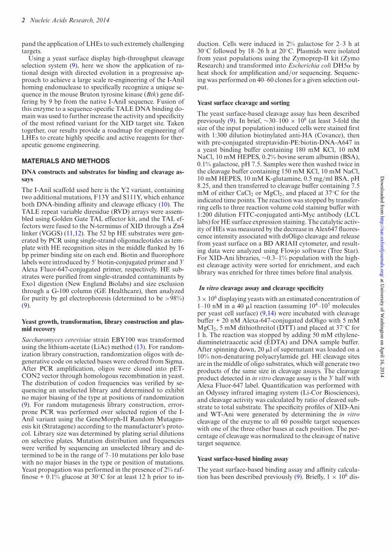
Figure 1. Target selection and cluster-based engineering of an XID-specific variant of I-AniI. (A) Alignment of the native I-Anil LHE recognition sequencewith mouse XID site. Mismatches (red) were divided into four clusters. The single base XID mutation at −8 position (C→T) was shaded in gray. (B) DNAinterface AA residues (red) targeted to DNA sequence mismatches (yellow) were selected for randomization. (C) The workflow for engineering I-Aniltoward XID target site. Based on rational design, AA residues targeted to mismatch clusters were selected for randomization to generate yeast libraries.After cleavage selection, random mutations were introduced into selected active variants by error-prone PCR to generate random mutagenesis library forfurther cleavage selection. Finally, selected designs were characterized and combined to generate XID full site enzyme.
at University of W
ashington on April 16, 2014
http://nar.oxfordjournals.org/D
ownloaded from

Nucleic Acids Research, 2014 5
engineering strategy by dividing the XID sequence into four‘cluster’ of mismatched residues (−10A−8T, −6C−5C−4T,+6T+7G and +9C+10T) based on enzyme structure andAni/XID target sequence mismatch positions (Figure 1Aand B). Based on structural information, AA residues in-teracting with these clusters were selected for randomiza-tion. I-AniI variants bearing alterations in these residueswere incorporated into a yeast surface expression vector,thereby taking advantage of the high homologous recombi-nation efficiency in yeast to generate an LHE yeast surfacedisplay library. Using a previously reported yeast surfacecleavage assay (9), the yeast library was subjected to threerounds of flow cytometry-based selection to enrich highlyactive variants. To further increase enzyme activity, randommutations were introduced by error prone PCR into openreading frames (ORFs) of variants emerging from primaryscreens targeting each cluster. After three rounds of cleav-age selection from the random mutation library, enzyme-expressing DNA vectors were isolated from the final en-riched populations, and individual clones were sequencedand characterized in vitro (Figure 1C; with details of engi-neering experiments performed for each cluster provided inSupplementary Figures S1–S4). As shown in +6T+7G and−6C−5C−4T cluster libraries (Supplementary Figure S2D,S3F, and Supplementary Table S1), variants with the high-est cleavage activity were highly enriched in the final selectedpopulation (up to 80% for a single variant), and these clonestypically exhibited similar patterns of AA variation at thediversified positions.
Combination of cluster designs into an enzyme able to cleavethe XID target site
Using the output of the cluster selections, we generated li-braries in which cluster designs were combined to create en-zymes with active plus (+) and negative (−) half sites. How-ever, we found that directly combining active (+) and (−)half-site designs from these libraries did not generate ac-tive full site variants, suggesting conformational conflict be-tween (+) and (−) half-site designs (data not shown). There-fore, we evaluated partial half-site combinations, and wereable to isolate a −6C−5C−4T+6T+7G+9C+10A-Ani vari-ant with moderate activity on yeast surface. Using this ORFas template for a random mutagenesis library (Supplemen-tary Table S1, Supplementary Figure S4A), we screenedfor variants that exhibited increased activity using our flowcytometry-based cleavage assay. From this screen, we iden-tified a R243W mutation that was highly enriched in themost active population (Supplementary Figure S4B and C).Structural analysis of the R243 residue indicated that it ispositioned a short distance from the native +9 A:T pair, andmutation at that position to C:G pair causes steric clash be-tween this residue and +9 nucleotide pair. When combining(+) and (−) site designs, we speculate that this steric clash af-fects the positioning of catalytic domain, an effect that canbe compensated by the R243W mutation. This conclusion isfurther supported by the ability of the R243W mutation torescue all previously inactive XID variants, following whichtheir cleavage activity could be further increased by randommutagenesis and selection (Supplementary Table S1, Sup-plementary Figure S4D and E).
In vitro characterization of XID-Ani
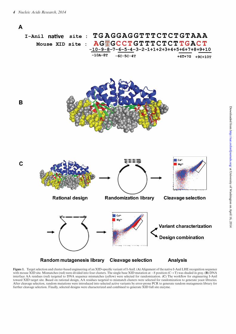
The final selected XID-Ani variant (enriched up to 80%in the final population) includes 31 AA alterations fromWT-Ani, with nearly half of them (14) selected from ran-dom mutagenesis libraries (Supplementary Table S1, Fig-ure 2A). In a yeast surface-based cleavage assay, XID-Anishowed similar cleavage efficacy as WT-Ani without de-tectable activity toward the WT target (Figure 2B). Oncedissociated from the yeast surface, XID-Ani also showedsimilar cleavage kinetics as WT-Ani in an in vitro cleavageassay (Figure 2C). We previously showed that the specificityof re-designed enzymes targeted to partial XID site (−6C,+6T+7G) has significantly improved specificity comparedwith WT-Ani (Supplementary Figures S2D and S3B). Toevaluate the specificity of the engineered XID-Ani enzyme,we compared one-off cleavage specificity profile of XID-Aniwith WT-Ani, and also measured their binding affinity (Fig-ure 2D). ‘One-off’ target site specificity for XID-Ani rangedfrom relatively high at 9 bp positions (where the efficiency ofcleavage of any other three bases was less than 50% of theXID target base), to partial or complete degeneracy at 11positions (where at least one other base was cleaved with anefficiency >50% of the XID target site) (Figure 2F). This isa significant improvement from the WT-Ani, which showedpartial or complete degeneracy at 19 positions (Figure 2E)(9), and is explained at least in part by the fact that the re-designed enzyme has a reduced affinity for the XID targetsite––thus, any single base pair mismatch is more likely tocompromise binding to a sufficient extent that enzymaticactivity is compromised. The improved specificity of XID-Ani using the highly sensitive flow cytometry assay is an im-portant achievement, as it emphasizes the capacity of theI-AniI scaffold to be engineered so as to achieve a high effi-ciency of on-site cleavage while reducing off-target cleavageover nearly the entire DNA/protein interface, an importantconsideration for therapeutic applications where specificityis paramount.
In cellulo performance of XID-Ani
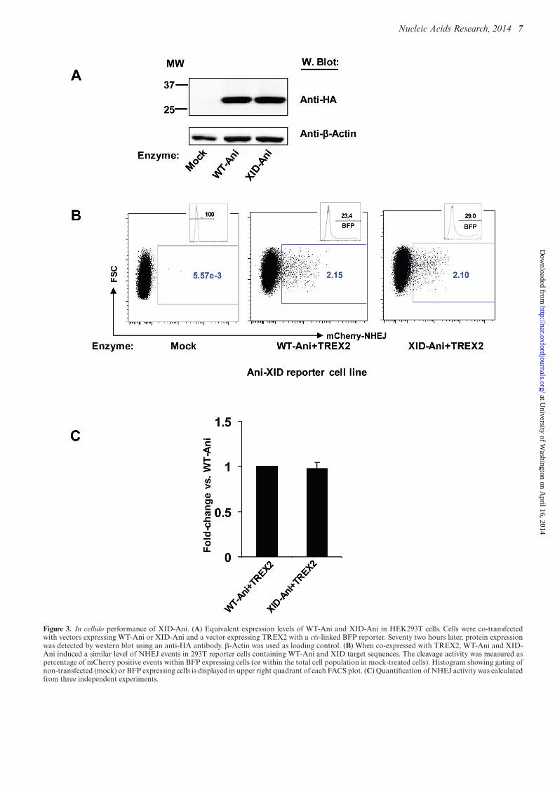
To characterize the activity of the re-designed XID-Ani in acell-based model, we used a previously described TLR sys-tem, that is able to report the capacity of an enzyme to gen-erate both mutagenic NHEJ and targeted HDR (16). Thein cellulo cleavage activity of XID-Ani and WT-Ani weremeasured by monitoring each enzyme’s ability to generatemCherry positive cells when co-expressed with the 3′ exonu-clease TREX2 (TREX2 degrades the 3′ overhang of DSBsgenerated by HEs, and thus its overexpression leads to in-crease rates of end processing, and so an increased prob-ability that a cleavage event will be converted to a muta-genic outcome (23)). To facilitate direct in cellulo cleavageefficacy comparison, a reporter cell line in which both XIDand Ani target sites were included in a single TLR was uti-lized for both enzymes. Reporter cell lines with single tar-get sites were used, in parallel, as controls. Both enzymeswere stably expressed in HEK293T cells with similar levelsof protein detected by western blot (Figure 3A). Similar toWT-Ani, XID-Ani only induced NHEJ events in 293T re-porter cells that possessed the Ani-XID target, but not theWT-Ani target site alone (Figure 3B, Supplementary Figure
at University of W
ashington on April 16, 2014
http://nar.oxfordjournals.org/D
ownloaded from
6 Nucleic Acids Research, 2014
Figure 2. In vitro characterization of XID-Ani enzyme. (A) The final selected XID-Ani variant has 31 AA mutations (red) from WT-Ani. (B) XID-Anishowed similar cleavage efficacy as WT-Ani on yeast toward its target sequence as indicated by allophycocyanin (APC) signal shift in the presence of Mg2+.In contrast, XID-Ani exhibits no activity toward the WT-Ani sequence. (C) XID-Ani demonstrated similar cleavage in vitro kinetics toward its target siteas WT-Ani. Top bands show uncut dsOligo candidate HE substrates. Lower bands represent the 3′ half of HE-cleaved dsOligo substrates detected on thebasis of the Alexa Fluor-647 label. (D) XID-Ani showed lower binding affinity than WT-Ani toward their respective target sites on a yeast surface-basedbinding affinity titration assay. (E and F) One-off in vitro cleavage profiles for WT and engineered XID-Ani. Upper panels show cleavage activity of WT-Aniand XID-Ani toward their respective targets and one-off sites in an in vitro cleavage assay. Lower panels show quantification of relative cleavage efficacywith cleavage toward Ani and XID sites set as 100%, respectively. Quantification and standard error were calculated from three independent experiments.
at University of W
ashington on April 16, 2014
http://nar.oxfordjournals.org/D
ownloaded from
Nucleic Acids Research, 2014 7
Figure 3. In cellulo performance of XID-Ani. (A) Equivalent expression levels of WT-Ani and XID-Ani in HEK293T cells. Cells were co-transfectedwith vectors expressing WT-Ani or XID-Ani and a vector expressing TREX2 with a cis-linked BFP reporter. Seventy two hours later, protein expressionwas detected by western blot using an anti-HA antibody. �-Actin was used as loading control. (B) When co-expressed with TREX2, WT-Ani and XID-Ani induced a similar level of NHEJ events in 293T reporter cells containing WT-Ani and XID target sequences. The cleavage activity was measured aspercentage of mCherry positive events within BFP expressing cells (or within the total cell population in mock-treated cells). Histogram showing gating ofnon-transfected (mock) or BFP expressing cells is displayed in upper right quadrant of each FACS plot. (C) Quantification of NHEJ activity was calculatedfrom three independent experiments.
at University of W
ashington on April 16, 2014
http://nar.oxfordjournals.org/D
ownloaded from
8 Nucleic Acids Research, 2014
S5). The cleavage efficacy of XID-Ani in cellulo was similarto WT-Ani, consistent with their observed relative in vitrocleavage activities (Figure 3B and C).
TALE DNA binding domain fusion with XID-Ani signifi-cantly increases its cleavage efficacy
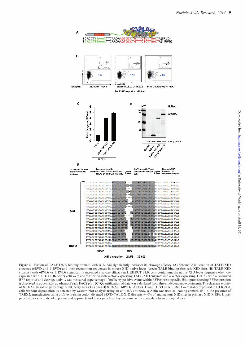
To further increase the efficiency and specificity of the XID-Ani enzyme, we used a recently developed TALE–LHE fu-sion hereafter referred to as megaTAL architecture (12). Wecreated megaTAL fusion enzymes with TALE DNA bind-ing domains targeted to 6 or 11 bp of the native Btk lo-cus sequences located upstream of murine XID site in con-junction with a 7 bp spacer (Figure 4A). The cleavage ef-ficacy of these TALE-XID endonucleases was comparedwith XID-Ani LHE in a HEK293T TLR cell line with inte-grated TALE-XID sequence (Figure 4A). With both TALEarray fusions, the ‘on-site’ cleavage efficacy of TALE-XIDwas substantially increased as reported by the TLR flow cy-tometry readout (Figure 4B and C; with level of protein ex-pression demonstrated in Figure 4D). As cleavage measuredby TLR cell lines has been found to under report true mu-tation rates in some cases (24), we also assessed cleavage atthe XID target in this line via amplicon-based sequencing.Analysis of amplicon sequences demonstrated that nearlycomplete disruption of the XID site was achieved in cells co-expressing 11RVD-TALE-XID and TREX2 (27/28 read-outs, 96.4%). Importantly, the ratio of cleavage efficacy de-termined by genomic sequencing between different enzymeswas consistent with the ratio indicated by the mCherry re-porter readout of the TLR, validating the use of the TLR asa tool for relative comparisons of enzymatic activity (datanot shown).
Because we were not able to compare relative bindingaffinity of 11RVD versus XID-Ani using the yeast surface-based binding assay (due to the difficulty of expressing theTALE domain on the yeast surface), we compared the rela-tive contribution of TALE and HE by combining differentTALE domains and HE variants. Using a 17 RVD TALE(‘L538’––TCATTACACCTGCAGCT) (25), we were able toincrease the cleavage activity of XID-Ani. We also obtainedsimilar cleavage rates using another XID variant with nearlyidentical turnover rate (Kcat) but a significantly lower bind-ing affinity and cleavage activity (∼20% of XID-Ani in theTLR assay). Based on these findings, we argue that the bind-ing affinity of this 17 RVD TALE is sufficient to bring HEswith different binding affinity to their maximum activity.With the 11 RVD TALE (which has lower binding affin-ity than ‘L538’), the cleavage efficacy of this XID variantwas around 70% of XID-Ani (data not shown). Based onthis observation, we estimate the binding affinity of 11 RVDcontributes 70–80% of the total observed activity, which isconsistent with the data showed in Figure 4C.
We next determined the cleavage efficacy of the 6RVD-TALE-XID enzyme at the XID mutant Btk genomic lo-cus. Primary MEFs derived from XID embryos were co-transduced by LVs expressing the 6RVD-TALE-XID andTREX2 (multiplicity of infection (MOI) of 10). Of note, thecodons of the highly repetitive RVD array sequences werediverged to reduce sequence rearrangements occurring dur-ing reverse transcription (26) thereby permitting efficient
LV packaging and expression of this novel nuclease withoutevidence for protein degradation (data not shown). Ten daysafter transduction, native XID target disruption rate wasdetermined by genomic sequencing within cells marked byboth viruses. Although the XID site is thought to be withinthe silent Btk locus in primary MEFs, nearly 40% disrup-tion (21/53 readouts, 39.6%) was detected in XID MEFs,primarily including small deletions within the central fourbases of XID-Ani enzyme recognition site (Figure 4E).
Off-target cleavage of TALE-XID enzyme
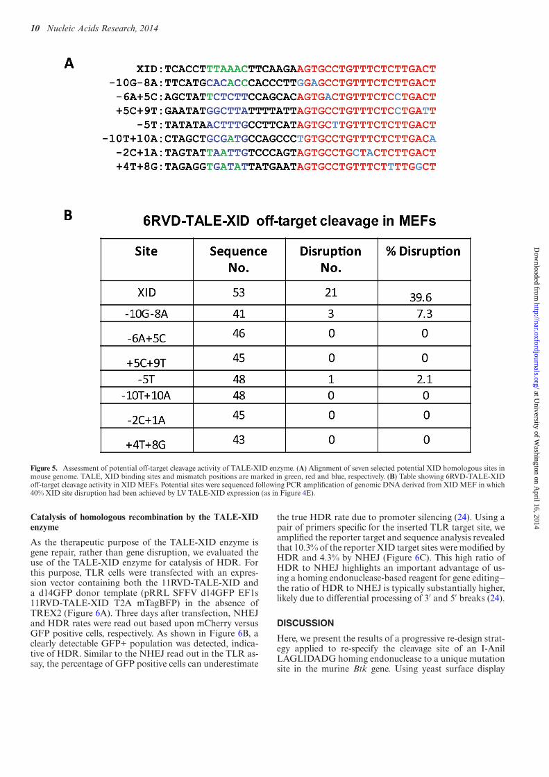
One of the most important considerations for therapeuticapplication of rare-cutting endonucleases is maintaining arate of off-target cleavage that is as low as possible (27). Al-though XID-Ani showed significantly improved specificityfrom WT-Ani, cleavage profiling identified 19 one-off cleav-age sites tolerated by XID-Ani in vitro (based on >50% pre-dicted cleavage activity at candidate target nucleotides; Fig-ure 2F). To examine the specificity of TALE-XID when ex-pressed in vivo, we searched for potential cleavage sites iden-tified in the mouse genome. We identified 23 potential sitesincluding: two sites with a 1 bp mismatch and 21 sites with2 bp mismatches. Sequence and chromosomal positions ofthese sites are provided in Supplementary Table S2. Theone-off cleavage profile predicted that the 1 bp mismatchsites (including the WT btk sequence) would be partiallytolerated. Among the 2 bp mismatches, the cleavage profilepredicted that seven sites would be tolerated (both singlemismatches tolerated by XID-Ani) and 14 sites would notbe tolerated (at least one mismatch is not tolerated).
To directly test the rate of cleavage at predicted off-target sites in vivo, seven potential sites including candi-dates from each of these three categories were sequencedfrom XID MEF genomic DNA isolated from a cell popula-tion in which a 40% XID site disruption had been achievedby LV TALE-XID expression. As shown in Figure 5A,none of these sites possessed upstream homologous TALEbinding sequences. No disruption was found at sites thatwere predicted in vitro to be cleavage resistant (−10T+10A,−2C+1A, +4T+8G). As for sites for which in vitro cleavagewas observed, we detected no disruption at the −6A+5Cand +5C+9T sites, but 7.3% (3/41 readouts) disruption atthe −10G−8A site (Figure 5B). The tolerance of the −10G-8A site is likely due to their positions on nearby residues−10 and −8 within the N terminal loop, where WT I-AniIhas little specificity, while the −6A+5C and +5C+9T aretargeted by distinct domains of XID-Ani and, thus, aremore likely to be incompatible with cleavage when com-bined together. Finally, we observed low-level, 2.1% cleav-age activity at the partially tolerated −5T site. Importantly,the disruption rate for −10G−8A and −5T sites was at least5-fold lower than that of the XID site, consistent with theobserved difference in cleavage efficacy of XID-Ani withand without the fused TALE DNA binding domain in theTLR assay (Figure 4C). Considering the stringency of theassay, which enhances detection of nuclease-mediated dis-ruption via extended lentiviral vector driven nuclease andTREX2 co-expression, these combined results further illus-trate the importance of the TALE domain in specifically en-hancing ‘on site’ activity.
at University of W
ashington on April 16, 2014
http://nar.oxfordjournals.org/D
ownloaded from
Nucleic Acids Research, 2014 9
Figure 4. Fusion of TALE DNA binding domain with XID-Ani significantly increases its cleavage efficacy. (A) Schematic illustration of TALE-XIDenzymes (6RVD and 11RVD) and their recognition sequences in mouse XID native locus (green: TALE binding site; red: XID site). (B) TALE-XIDenzymes with 6RVDs or 11RVDs significantly increased cleavage efficacy in HEK293T TLR cells containing the native XID locus sequence when co-expressed with TREX2. Reporter cells were co-transfected with vectors expressing TALE-XID enzymes and a vector expressing TREX2 with a cis-linkedBFP reporter and cleavage activity was measured as percentage of mCherry positive events within BFP expressing cells. Histogram showing BFP expressionis displayed in upper right quadrant of each FACS plot. (C) Quantification of data was calculated from three independent experiments. The cleavage activityof XID-Ani based on percentage of mCherry was set as one (D) XID-Ani, 6RVD-TALE-XID and 11RVD-TALE-XID were stably expressed in HEK293Tcells without degradation as detected by western blot analysis using an anti-HA antibody. �-Actin was used as loading control. (E) In the presence ofTREX2, transduction using a LV expressing codon diverged 6RVD-TALE-XID disrupts ∼40% of endogenous XID sites in primary XID MEFs. Upperpanel shows schematic of experimental approach and lower panel displays genomic sequencing data from disrupted loci.
at University of W
ashington on April 16, 2014
http://nar.oxfordjournals.org/D
ownloaded from
10 Nucleic Acids Research, 2014
Figure 5. Assessment of potential off-target cleavage activity of TALE-XID enzyme. (A) Alignment of seven selected potential XID homologous sites inmouse genome. TALE, XID binding sites and mismatch positions are marked in green, red and blue, respectively. (B) Table showing 6RVD-TALE-XIDoff-target cleavage activity in XID MEFs. Potential sites were sequenced following PCR amplification of genomic DNA derived from XID MEF in which40% XID site disruption had been achieved by LV TALE-XID expression (as in Figure 4E).
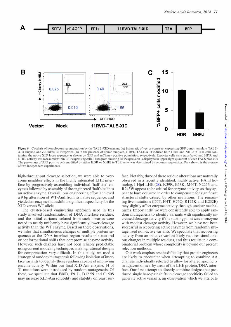
Catalysis of homologous recombination by the TALE-XIDenzyme
As the therapeutic purpose of the TALE-XID enzyme isgene repair, rather than gene disruption, we evaluated theuse of the TALE-XID enzyme for catalysis of HDR. Forthis purpose, TLR cells were transfected with an expres-sion vector containing both the 11RVD-TALE-XID anda d14GFP donor template (pRRL SFFV d14GFP EF1s11RVD-TALE-XID T2A mTagBFP) in the absence ofTREX2 (Figure 6A). Three days after transfection, NHEJand HDR rates were read out based upon mCherry versusGFP positive cells, respectively. As shown in Figure 6B, aclearly detectable GFP+ population was detected, indica-tive of HDR. Similar to the NHEJ read out in the TLR as-say, the percentage of GFP positive cells can underestimate
the true HDR rate due to promoter silencing (24). Using apair of primers specific for the inserted TLR target site, weamplified the reporter target and sequence analysis revealedthat 10.3% of the reporter XID target sites were modified byHDR and 4.3% by NHEJ (Figure 6C). This high ratio ofHDR to NHEJ highlights an important advantage of us-ing a homing endonuclease-based reagent for gene editing––the ratio of HDR to NHEJ is typically substantially higher,likely due to differential processing of 3′ and 5′ breaks (24).
DISCUSSION
Here, we present the results of a progressive re-design strat-egy applied to re-specify the cleavage site of an I-AnilLAGLIDADG homing endonuclease to a unique mutationsite in the murine Btk gene. Using yeast surface display
at University of W
ashington on April 16, 2014
http://nar.oxfordjournals.org/D
ownloaded from
Nucleic Acids Research, 2014 11
Figure 6. Catalysis of homologous recombination by the TALE-XID enzyme. (A) Schematic of vector construct expressing GFP donor template, TALE-XID enzyme, and cis-linked BFP reporter. (B) In the presence of donor template, 11RVD-TALE-XID induced both HDR and NHEJ in TLR cells con-taining the native XID locus sequence as shown by GFP and mCherry positive population, respectively. Reporter cells were transfected and HDR andNHEJ activity was measured within BFP expressing cells. Histogram showing BFP expression is displayed in upper right quadrant of each FACS plot. (C)The percentage of BFP positive cells modified by either HDR or NHEJ in TLR assay was determined by genomic sequencing. Data shown is the averageof two independent experiments.
high-throughput cleavage selection, we were able to over-come neighbor effects in the highly integrated LHE inter-face by progressively assembling individual ‘half site’ en-zymes followed by assembly of the engineered ‘half site’ intoan active enzyme. Overall, our engineering effort achieveda 9 bp alteration of WT-AniI from its native sequence, andyielded an enzyme that exhibits significant specificity for theXID versus WT allele.
The cluster-based engineering approach used in thisstudy involved randomization of DNA interface residues,and the initial variants isolated from such libraries werenoted to nearly uniformly have significantly lower cleavageactivity than the WT enzyme. Based on these observations,we infer that simultaneous changes of multiple protein se-quences at the DNA interface region results in structuralor conformational shifts that compromise enzyme activity.However, such changes have not been reliably predictableusing current modeling techniques, making rational designsfor compensation very difficult. In this study, we used astrategy of random mutagenesis following isolation of inter-face variants to identify those residues capable of improvingenzyme activity. Within our final XID-Ani enzyme, 14 of31 mutations were introduced by random mutagenesis. Ofthese, we speculate that E86D, F91L, D122N and C150Smay increase XID-Ani solubility and stability on yeast sur-
face. Notably, three of these residue alterations are naturallyobserved in a recently identified, highly active, I-Anil ho-molog, I-HjeI LHE (28). K39R, E63K, M66T, N226Y andR243W appear to be critical for enzyme activity, as they ap-pear to have occurred in order to compensate for significantstructural shifts caused by other mutations. The remain-ing five mutations (I55T, I64T, H76Q, R172K and K232E)may slightly affect enzyme activity through unclear mecha-nisms. Importantly, we were consistently able to apply ran-dom mutagenesis to identify variants with significantly in-creased cleavage activity, if the starting point was an enzymewith modest cleavage activity. However, we have not beensuccessful in recovering active enzymes from randomly mu-tagenized non-active variants. We speculate that recoveringactivity from an inactive variant likely requires simultane-ous changes in multiple residues, and thus results in a com-binatorial problem whose complexity is beyond our presentselection methods.
Our work emphasizes the difficulty that protein engineersare likely to encounter when attempting to combine AAchanges individually selected to allow for altered specificityin adjacent or nearby areas of the LHE protein/DNA inter-face. Our first attempt to directly combine designs that pro-duced single base-pair shifts in cleavage specificity failed togenerate active variants, an observation which we attribute
at University of W
ashington on April 16, 2014
http://nar.oxfordjournals.org/D
ownloaded from
12 Nucleic Acids Research, 2014
to neighbor effects between designs due to the highly inte-grated LHE protein/DNA interface. We also observed inseveral cases that active variants selected against a cluster(e.g. +6T+7G) were able to cut combined +6T and +7Gbut not +6T or +7G individually (Supplementary FigureS2D). This is consistent with structure-based predictionsthat these 2 bp are targeted by an overlapping and struc-turally dependent area of the protein/DNA interface, andthus separate designs would likely generate steric clashesthat compromise enzyme activity. Similarly, our attempts tocombine two adjacent regions targeted to −10A−8T and−6C−5C−4T by a direct combination strategy also didnot work. This observation could be explained if the struc-tural shift caused by incorporations of the −6C−5C−4Tdesign had a neighbor effect on the loop region targeted to−10A−8T. Only after that shift was compensated by muta-tions from another region of the interface, was it possible tore-design this loop (Supplementary Figure S3G-I). Finally,the combination of XID (+) and (−) half designs, which re-side in the I-Anil N-term and C-term domains, respectively,and target to distant sequences, unexpectedly did not gen-erate any active variants as a direct combination. However,following random mutagenesis, a single R243W mutationwas recovered that allowed all inactive variants to regaincleavage activity. We hypothesize that the re-designed halfenzyme resulted in a shift in the catalytic residues, which wassubsequently compensated for by the R243W mutation.
While we achieved significant success in resculpting aDNA/protein interface that allowed high specificity and ac-tivity toward the XID target, this interface was not able toachieve a high binding affinity for the XID site. Despite itsreduced binding affinity in vitro, XID-Ani exhibited a levelof activity in in vitro and in cellulo reporter assays that wasequivalent to WT I-AniI. It also exhibited significantly im-proved specificity, critical for reducing the risk of off-targetcleavage in therapeutic applications. We attribute the re-duced binding affinity to the nature of the resculpted in-terface of the (−) half site, which disrupts several hydrogenbonds that are involved in generating the high affinity ofWT I-AniI for its target site. We speculate that to achievethe same level of cleavage activity as WT I-AniI, the reducedbinding affinity was partially compensated by an increasedturnover rate (Kcat).
Importantly, the introduction of a site-specific TALEDNA binding domain using the megaTAL platform wasable to overcome the reduced binding affinity of XID-Anifor the XID site, thereby providing markedly improved invivo activity. Fusion of XID-Ani to TALE DNA bind-ing domains containing as few as six RVDs, resulted inmarkedly enhanced cleavage efficacy in both reporter celllines and primary XID MEFs. The TALE DNA bindingdomain significantly increased XID-Ani cleavage efficacyto its target site but is not anticipated to alter the bindingaffinity of the XID-Ani cleavage head to its homologoussequence. Thus, we do not anticipate that the megaTALfusion changes the intrinsic specificity of the HE cleav-age head. However, by increasing on-site activity, a desiredlevel of on-site modification can be achieved with a reducedprotein expression level and/or a more limited period ofexpression thereby resulting in reduced off site cleavage.Thus, megaTAL fusions result in an increase in the effec-
tive specificity of the enzyme (12). We speculate that futureLHE engineering for therapeutic applications may focus onthe intentional development of low affinity, high specificityLHE’s, with the planned addition of a TALE or other affin-ity enhancing domain to provide for site specific activity toa desired single target site.
Finally, a notable aspect of the performance of the XID-Ani megaTAL was that it was able to achieve a high ratioof HDR to NHEJ. High HDR to NHEJ ratios have typ-ically been observed with homing endonucleases, whereasboth zinc finger nucleases (ZFNs) and TALENs have typi-cally yielded the reverse––rates of NHEJ that are higher thanHDR (24). This is an important observation, as it suggeststhat the DNA ends created by megaTAL reagents are pro-cessed in a manner akin to their homing endonuclease cleav-age heads, as opposed to being processed equivalently toa FOK-I based TALEN. It also supports the concept thatgene editing reagents based on homing endonuclease cleav-age domains may be superior choices for gene editing appli-cations that are dependent on homology-directed repair.
In summary, here we have shown that a yeast sur-face display-based high throughput selection system forHE engineering can be applied in a progressive re-designstrategy to execute an aggressive resculpting of an LHEprotein/DNA interface. By adopting a progressive strategythat incorporated random mutagenesis to boost activity be-tween interface resculpting steps, we were able to achieve a9 bp alteration in cleavage specificity from the native targetsite. Furthermore, we show that compensating XID bindingaffinity through TALE DNA binding domains significantlyimproved the cleavage activity of the final enzyme, yield-ing a highly active and specific gene editing reagent able tocatalyze high rates of homology-directed repair at its targetsite. The success of our progressive strategy in achieving asignificant specificity shift and successful incorporation ofthe re-designed XID-Ani into the megaTAL format offer aroadmap for future LHE engineering projects aimed at cre-ating highly specific and active gene editing reagents.
SUPPLEMENTARY DATA
Supplementary Data are available at NAR Online.
ACKNOWLEDGMENTS
The authors would like to thank all members of the North-west Genome Engineering Consortium (NGEC) (http://ngec-seattle.org) for their insightful input and discussions.Y.W. designed and performed experiments, analyzed dataand wrote the manuscript; I.K., S.B., J.J. and J.P. performedexperiments; S.B.T. and D.B. designed experiments; A.M.S.and D.J.R. designed experiments and wrote the manuscript.
FUNDING
Ruth L. Kirschstein National Research Service Award[HL092555 to Y.W. in part]; National Heart, Lung, andBlood Institute (NHLBI) and National Cancer Insitute(NCI) of the National Institutes of Health [PL1-HL092557to D.J.R., RL1-HL092553 to D.J.R., R01AI071163 toD.J.R., R01-HL075453 to D.J.R., NIH RL1CA133832 to
at University of W
ashington on April 16, 2014
http://nar.oxfordjournals.org/D
ownloaded from
Nucleic Acids Research, 2014 13
A.M.S., UL1DE019582 to A.M.S.]; Seattle Children’s Re-search Institute and the Center for Immunity and Im-munotherapies (Program for Cell and Gene Therapy). Thecontent is solely the responsibility of the authors and doesnot necessarily represent the official views of the NationalInstitutes of Health.Funding for open access charge: SeattleChildren’s Research Institute and the Center for Immunityand Immunotherapies (Program for Cell and Gene Ther-apy).Conflict of interest statement. A.M.S. receives salary andequity compensation from Cellectis, and is a founder andmember of the board of directors of Pregenen. Both of thesecompanies are involved in commercialization of nuclease-related genome engineering technologies.
REFERENCES1. Stoddard, B.L. Stoddard, B.L. (2011) Homing endonucleases: from
microbial genetic invaders to reagents for targeted DNAmodification. Structure, 19, 7–15.
2. Belfort, M. and Roberts, R.J.Belfort, M. and Roberts, R.J. (1997)Homing endonucleases: keeping the house in order. Nucleic AcidsRes., 25, 3379–3388.
3. Hafez, M. and Hausner, G.Hafez, M. and Hausner, G. (2012)Homing endonucleases: DNA scissors on a mission. Genome, 55,553–569.
4. Schiffer, J.T., Aubert, M., Weber, N.D., Mintzer, E., Stone, D., andJerome, K.R.Schiffer, J.T., Aubert, M., Weber, N.D., Mintzer, E.,Stone, D., and Jerome, K.R. (2012) Targeted DNA mutagenesis forthe cure of chronic viral infections. J. Virol., 86, 8920–8936.
5. Ashworth, J., Taylor, G.K., Havranek, J.J., Quadri, S.A., Stoddard,B.L., and Baker, D.Ashworth, J., Taylor, G.K., Havranek, J.J.,Quadri, S.A., Stoddard, B.L., and Baker, D. (2010) Computationalreprogramming of homing endonuclease specificity at multipleadjacent base pairs. Nucleic Acids Res., 38, 5601–5608.
6. Grizot, S., Smith, J., Daboussi, F., Prieto, J., Redondo, P., Merino, N.,Villate, M., Thomas, S., Lemaire, L., and Montoya, G. et al.Grizot,S., Smith, J., Daboussi, F., Prieto, J., Redondo, P., Merino, N., Villate,M., Thomas, S., Lemaire, L., and Montoya, G. (2009) Efficienttargeting of a SCID gene by an engineered single-chain homingendonuclease. Nucleic Acids Res., 37, 5405–5419.
7. Chen, Z., Wen, F., Sun, N., and Zhao, H.Chen, Z., Wen, F., Sun, N.,and Zhao, H. (2009) Directed evolution of homing endonucleaseI-SceI with altered sequence specificity. Protein Eng. Des. Sel., 22,249–256.
8. Baxter, S., Lambert, A.R., Kuhar, R., Jarjour, J., Kulshina, N.,Parmeggiani, F., Danaher, P., Gano, J., Baker, D., and Stoddard, B.L.et al.Baxter, S., Lambert, A.R., Kuhar, R., Jarjour, J., Kulshina, N.,Parmeggiani, F., Danaher, P., Gano, J., Baker, D., and Stoddard, B.L.(2012) Engineering domain fusion chimeras from I-OnuI familyLAGLIDADG homing endonucleases. Nucleic Acids Res., 40,7985–8000.
9. Jarjour, J., West-Foyle, H., Certo, M.T., Hubert, C.G., Doyle, L.,Getz, M.M., Stoddard, B.L., and Scharenberg, A.M.Jarjour, J.,West-Foyle, H., Certo, M.T., Hubert, C.G., Doyle, L., Getz, M.M.,Stoddard, B.L., and Scharenberg, A.M. (2009) High-resolutionprofiling of homing endonuclease binding and catalytic specificityusing yeast surface display. Nucleic Acids Res., 37, 6871–6880.
10. Takeuchi, R., Certo, M., Caprara, M.G., Scharenberg, A.M., andStoddard, B.L. Takeuchi, R., Certo, M., Caprara, M.G.,Scharenberg, A.M., and Stoddard, B.L. (2009) Optimization of invivo activity of a bifunctional homing endonuclease and maturasereverses evolutionary degradation. Nucleic Acids Res., 37, 877–890.
11. Cermak, T., Doyle, E.L., Christian, M., Wang, L., Zhang, Y.,Schmidt, C., Baller, J.A., Somia, N.V., Bogdanove, A.J., and Voytas,D.F.Cermak, T., Doyle, E.L., Christian, M., Wang, L., Zhang, Y.,Schmidt, C., Baller, J.A., Somia, N.V., Bogdanove, A.J., and Voytas,D.F. (2011) Efficient design and assembly of custom TALEN andother TAL effector-based constructs for DNA targeting. NucleicAcids Res., 39, e82.
12. Boissel, S., Jarjour, J., Astrakhan, A., Adey, A., Gouble, A.,Duchateau, P., Shendure, J., Stoddard, B.L., Certo, M.T., and Baker,D. et al.Boissel, S., Jarjour, J., Astrakhan, A., Adey, A., Gouble, A.,Duchateau, P., Shendure, J., Stoddard, B.L., Certo, M.T., and Baker,D. (2013) megaTALs: a rare-cleaving nuclease architecture fortherapeutic genome engineering. Nucleic Acids Res., 2013, 1–11.
13. Gietz, R.D. and Schiestl, R.H.Gietz, R.D. and Schiestl, R.H. (2007)Large-scale high-efficiency yeast transformation using the LiAc/SScarrier DNA/PEG method. Nat. Protoc., 2, 38–41.
14. Boder, E.T. and Wittrup, K.D.Boder, E.T. and Wittrup, K.D. (1997)Yeast surface display for screening combinatorial polypeptidelibraries. Nat. Biotechnol., 15, 553–557.
15. Sather, B.D., Ryu, B.Y., Stirling, B.V., Garibov, M., Kerns, H.M.,Humblet-Baron, S., Astrakhan, A., and Rawlings, D.J.Sather, B.D.,Ryu, B.Y., Stirling, B.V., Garibov, M., Kerns, H.M., Humblet-Baron,S., Astrakhan, A., and Rawlings, D.J. (2011) Development ofB-lineage predominant lentiviral vectors for use in genetic therapiesfor B cell disorders. Mol. Ther., 19, 515–525.
16. Certo, M.T., Ryu, B.Y., Annis, J.E., Garibov, M., Jarjour, J.,Rawlings, D.J., and Scharenberg, A.M.Certo, M.T., Ryu, B.Y., Annis,J.E., Garibov, M., Jarjour, J., Rawlings, D.J., and Scharenberg, A.M.(2011) Tracking genome engineering outcome at individual DNAbreakpoints. Nat. Methods, 8, 671–676.
17. Rawlings, D.J. and Witte, O.N.Rawlings, D.J. and Witte, O.N. (1995)The Btk subfamily of cytoplasmic tyrosine kinases: structure,regulation and function. Semin. Immunol., 7, 237–246.
18. Rawlings, D.J. and Witte, O.N.Rawlings, D.J. and Witte, O.N. (1994)Bruton’s tyrosine kinase is a key regulator in B-cell development.Immunol. Rev., 138, 105–119.
19. Yu, P.W., Tabuchi, R.S., Kato, R.M., Astrakhan, A., Humblet-Baron,S., Kipp, K., Chae, K., Ellmeier, W., Witte, O.N., and Rawlings,D.J.Yu, P.W., Tabuchi, R.S., Kato, R.M., Astrakhan, A.,Humblet-Baron, S., Kipp, K., Chae, K., Ellmeier, W., Witte, O.N.,and Rawlings, D.J. (2004) Sustained correction of B-cell developmentand function in a murine model of X-linked agammaglobulinemia(XLA) using retroviral-mediated gene transfer. Blood, 104,1281–1290.
20. Kerns, H.M., Ryu, B.Y., Stirling, B.V., Sather, B.D., Astrakhan, A.,Humblet-Baron, S., Liggitt, D., and Rawlings, D.J.Kerns, H.M., Ryu,B.Y., Stirling, B.V., Sather, B.D., Astrakhan, A., Humblet-Baron, S.,Liggitt, D., and Rawlings, D.J. (2010) B cell-specific lentiviral genetherapy leads to sustained B-cell functional recovery in a murinemodel of X-linked agammaglobulinemia. Blood, 115, 2146–2155.
21. Smith, J., Grizot, S., Arnould, S., Duclert, A., Epinat, J.C., Chames,P., Prieto, J., Redondo, P., Blanco, F.J., and Bravo, J. et al.Smith, J.,Grizot, S., Arnould, S., Duclert, A., Epinat, J.C., Chames, P., Prieto,J., Redondo, P., Blanco, F.J., and Bravo, J. (2006) A combinatorialapproach to create artificial homing endonucleases cleaving chosensequences. Nucleic Acids Res., 34, e149.
22. Thyme, S.B., Jarjour, J., Takeuchi, R., Havranek, J.J., Ashworth, J.,Scharenberg, A.M., Stoddard, B.L., and Baker, D.Thyme, S.B.,Jarjour, J., Takeuchi, R., Havranek, J.J., Ashworth, J., Scharenberg,A.M., Stoddard, B.L., and Baker, D. (2009) Exploitation of bindingenergy for catalysis and design. Nature, 461, 1300–1304.
23. Certo, M.T., Gwiazda, K.S., Kuhar, R., Sather, B., Curinga, G.,Mandt, T., Brault, M., Lambert, A.R., Baxter, S.K., and Jacoby, K.et al.Certo, M.T., Gwiazda, K.S., Kuhar, R., Sather, B., Curinga, G.,Mandt, T., Brault, M., Lambert, A.R., Baxter, S.K., and Jacoby, K.(2012) Coupling endonucleases with DNA end-processing enzymes todrive gene disruption. Nat. Methods, 9, 973–975.
24. Kuhar, R., Gwiazda, K.S., Humbert, O., Mandt, T., Pangallo, J.,Brault, M., Khan, I., Maizels, N., Rawlings, D.J., and Scharenberg,A.M. et al.Kuhar, R., Gwiazda, K.S., Humbert, O., Mandt, T.,Pangallo, J., Brault, M., Khan, I., Maizels, N., Rawlings, D.J., andScharenberg, A.M. (2013) Novel fluorescent genome editing reportersfor monitoring DNA repair pathway utilization atendonuclease-induced breaks, p. e4.
25. Miller, J.C., Tan, S., Qiao, G., Barlow, K.A., Wang, J., Xia, D.F.,Meng, X., Paschon, D.E., Leung, E., and Hinkley, S.J. et al.Miller,J.C., Tan, S., Qiao, G., Barlow, K.A., Wang, J., Xia, D.F., Meng, X.,Paschon, D.E., Leung, E., and Hinkley, S.J. (2011) A TALE nucleasearchitecture for efficient genome editing. Nat. Biotechnol., 29,143–148.
at University of W
ashington on April 16, 2014
http://nar.oxfordjournals.org/D
ownloaded from
14 Nucleic Acids Research, 2014
26. Holkers, M., Maggio, I., Liu, J., Janssen, J.M., Miselli, F., Mussolino,C., Recchia, A., Cathomen, T., and Goncalves, M.A.Holkers, M.,Maggio, I., Liu, J., Janssen, J.M., Miselli, F., Mussolino, C., Recchia,A., Cathomen, T., and Goncalves, M.A. (2013) Differential integrityof TALE nuclease genes following adenoviral and lentiviral vectorgene transfer into human cells. Nucleic Acids Res., 41, e63.
27. Petek, L.M., Russell, D.W., and Miller, D.G.Petek, L.M., Russell,D.W., and Miller, D.G. (2010) Frequent endonuclease cleavage atoff-target locations in vivo. Mol. Ther., 18, 983–986.
28. Jacoby, K., Metzger, M., Shen, B.W., Certo, M.T., Jarjour, J.,Stoddard, B.L., and Scharenberg, A.M.Jacoby, K., Metzger, M.,Shen, B.W., Certo, M.T., Jarjour, J., Stoddard, B.L., andScharenberg, A.M. (2012) Expanding LAGLIDADG endonucleasescaffold diversity by rapidly surveying evolutionary sequence space.Nucleic Acids Res., 40, 4954–4964.
at University of W
ashington on April 16, 2014
http://nar.oxfordjournals.org/D
ownloaded from