progressive metaplastic and dysplastic changes in mouse ... · progressive metaplastic and...
TRANSCRIPT
Progressive Metaplasticand Dysplastic Changes inMouse Pancreas Induced byCyclooxygenase-2 Overexpression1
Jennifer K.L. Colby*, Russell D. Klein†,2,Mark J. McArthur†, Claudio J. Conti*,Kaoru Kiguchi*, Toru Kawamoto*,Penny K. Riggs*,§, Amy I. Pavone*,Janet Sawicki¶ and Susan M. Fischer*
*University of Texas M.D. Anderson Cancer Center,Science Park – Research Division, Smithville, TX 78957,USA; †Department of Human Nutrition, CancerChemoprevention Program, The Ohio State University,Columbus, OH 43210, USA; ‡Michale E. Keeling Centerfor Comparative Medicine and Research, Department ofVeterinary Sciences, University of Texas M.D. AndersonCancer Center, Bastrop, TX 78602, USA; §Department ofAnimal Science, Texas A&M University, College Station,TX, 77843-2471, USA; ¶The Lankenau Institute for MedicalResearch, Wynnewood, PA, 19016, USA
AbstractCyclooxygenase-2 (COX-2) overexpression is an established factor linking chronic inflammation with metaplastic andneoplastic change in various tissues. We generated transgenic mice (BK5.COX-2) in which elevation of COX-2 andits effectors trigger a metaplasia–dysplasia sequence in exocrine pancreas. Histologic evaluation revealed a chronicpancreatitis-like state characterized by acinar-to-ductal metaplasia and a well-vascularized fibroinflammatory stromathat develops by 3 months. By 6 to 8 months, strongly dysplastic features suggestive of pancreatic ductal adeno-carcinoma emerge in the metaplastic ducts. Increased proliferation, cellular atypia, and loss of normal cell/tissueorganization are typical features in transgenic pancreata. Alterations in biomarkers associated with human inflamma-tory and neoplastic pancreatic disease were detected using immunohistochemistry. The abnormal pancreatic phe-notype can be completely prevented by maintaining mice on a diet containing celecoxib, a well-characterized COX-2inhibitor. Despite the high degree of atypia, only limited evidence of invasion to adjacent tissues was observed, withno evidence of distant metastases. However, cell lines derived from spontaneous lesions are aggressively tumori-genic when injected into syngeneic or nude mice. The progressive nature of the metaplastic/dysplastic changes ob-served in this model make it a valuable tool for examining the transition from chronic inflammation to neoplasia.
Neoplasia (2008) 10, 782–796
Abbreviations: BK5, bovine keratin 5; CFP, cyan fluorescent protein; COX, cyclooxygenase; CP, chronic pancreatitis; EP, PGE2 receptor; H&E, hematoxylin and eosin; IHC,immunohistochemistry; K19, keratin 19; (m)PanIN, (mouse) pancreatic intraepithelial neoplasia; PaSC, pancreatic stellate cell; PDAC, pancreatic ductal adenocarcinoma;PGE2, prostaglandin E2Address all correspondence to: Susan M. Fischer, The University of Texas M.D. Anderson Cancer Center, Science Park – Research Division, P.O. Box 389, 1808 Park Rd 1C,Smithville, TX 78957. E-mail: [email protected] work was supported by National Institutes of Health grants CA105345 and CA122815 (to S.M.F.) and a National Institute of Environmental Health Sciences (NIEHS)training grant T32 ES07247; and by a National Cancer Institute training grant R25CA57730 (J.K.L.C.) and ES07784 from NIEHS.2In memoriam of Dr. Russell D. Klein (1962–2006): Russell D. Klein died December 1, 2006 after a long battle with leukemia. He was a promising young scientist, anexemplary mentor, and a fine human being. His presence will be greatly missed.Received 26 February 2008; Revised 25 April 2008; Accepted 28 April 2008
Copyright © 2008 Neoplasia Press, Inc. All rights reserved 1522-8002/08/$25.00DOI 10.1593/neo.08330
www.neoplasia.com
Volume 10 Number 8 August 2008 pp. 782–796 782
IntroductionInflammatory conditions of the pancreas predispose individuals todeveloping pancreatic ductal adenocarcinoma (PDAC), exemplifiedby individuals with heritable or sporadic forms of chronic pancreatitis(CP) [1–3]. Among the general population, a number of risk factorsfor pancreatitis and pancreatic cancer have been identified, e.g., alco-hol and tobacco use [4,5], high consumption of nitrosamines [6],and diabetes [7,8], which can lead to chronic inflammatory changeseither in specific target tissues [5] or in the body as a whole [7,8].Although many forms of CP have been linked to a higher risk forPDAC, the relationship between the two is ill-defined [9].Metaplasia, the replacement of one mature cell type by another,
frequently occurs in chronic inflammation, and is often considereda preneoplastic condition (e.g., Barrett metaplasia of the esophagus)[10]. Acinar-to-ductal metaplastic changes in the pancreas are oftenfound in CP and in association with the recently defined pancreaticintraepithelial neoplasias (PanINs) and mouse PanIN (mPanIN) pre-cursor lesions and PDAC [11–15]. Recent articles have attempted toclarify the relationship between CP and PDAC [12,14], and onestudy has demonstrated that acinar cells clearly contribute to the de-velopment of PanIN lesions [13]. Metaplastic ductal cells seem tohave diminished sensitivity to apoptotic stimuli and, therefore, havea selective advantage in the diseased pancreas [16]. Although a greatdeal of evidence exists supporting high-grade PanINs as precursorlesions of PDAC, and this is widely accepted, questions remain withregard to the validity of ductal complexes as precursors to PDAC[17]. The concept is supported by evidence from groups studyingboth rodent and human forms of the disease [18–21].Cyclooxygenases (COX-1 and -2) are rate-limiting enzymes in
the production of prostaglandins (PGs), which are short-lived lipid-signaling molecules involved in a number of biologic functions [22].COX-1 expression is generally constitutive, whereas COX-2 is usuallyinduced by stimuli involved in inflammatory responses. ProstaglandinE2 (PGE2), a primary metabolite of COX-2, has been shown topromote cell survival, proliferation, and angiogenesis and prohibitapoptosis, all processes influencing cancer development [23].Prostaglandin production is frequently a factor in the develop-
ment and maintenance of chronic inflammation, and COX-2 is up-regulated in numerous human cancers and precancerous conditions,including pancreatitis and PDAC [24,25]. Cyclooxygenase-2 expres-sion has also been found to be an early event in the N -nitrosobis-(2-oxo-propyl)amine model of pancreatic carcinogenesis in hamster[26]. Forced COX-2 overexpression is associated with the develop-ment of cancer in mouse models of mammary [27] and bladder[28] tumorigenesis. In the skin of NMRI mice, COX-2 overexpres-sion driven by the bovine keratin 5 (BK5) promoter does not leadto spontaneous tumor development but sensitizes the skin to chemicalcarcinogenesis [29]. In this same model, high PGE2 levels also lead tofibrocystic changes and epithelial lesions in the mammary gland and,to some extent, in the pancreas [30,31].In this article, we present a mouse model, FVB-Tg(KRT5-Ptgs2)7Sf
mice, hereafter referred to as BK5.COX-2 mice, in which the over-expression of COX-2 under the control of a BK5 promoter drivespancreatic acinar-to-ductal metaplasia progressing to severe dysplasiasuggestive of PDAC. Histopathologic analyses reveal similarities tohuman CP, as well as pancreatic lesions observed in other genetically[32,33] or chemically induced [34,35] animal models of pancreaticdisease. BK5.COX-2 pancreatic lesions are characterized by acinar-to-ductal metaplasia, development of a fibroinflammatory stroma,
increased proliferation of metaplastic ductal cells, nuclear pleomor-phism, and abnormal cell and tissue architecture. Pancreata frequentlydevelop multilocular cysts. The BK5.COX-2 pancreatic phenotype isinitiated and primarily driven by high levels of COX-2 activity/PGE2signaling. The malignant potential of spontaneous BK5.COX-2 le-sions to progress to invasive adenocarcinoma is suggested by thepresence of strongly dysplastic cell/tissue morphology and by the es-tablishment of cell lines that form aggressive tumors when injected toeither syngeneic or nude mice. We propose that these mice representa valuable new model for the assessment of chemopreventive andchemotherapeutic agents in pancreatic disease, particularly those asso-ciated with pronounced inflammatory conditions such as CP.
Materials and Methods
MiceAll housing and procedures were carried out in an animal facility
accredited by the American Association for the Assessment and Accredi-tation of Laboratory Animal Care, in accordance with Institutional Ani-mal Care and Use Committee guidelines. Mice were maintained onchow ad libitum, unless specified otherwise. FVB-Tg(KRT5-Ptgs2)7Sf(BK5.COX-2) mice were generated on the FVB background by pro-nuclear injection of a bovine keratin 5 (BK5) promoter construct con-taining the coding region of the mouse COX-2 gene, the rabbitβ-globin intron, and an SV40 poly-A tail. The BK5 promoter directsexpression to several tissue types, namely, basal cells of skin, prostate,bladder, forestomach, mammary myoepithelium, kidney papilla, andpancreatic ductal epithelia [36]. Transgenic mice can consistently beidentified by a sparse hair coat but showed no other visible abnormalities.Two founders (Lines 7 and 9) were generated, both of which had a
pancreatic phenotype. Founder 9 produced no offspring; for all sub-sequent studies, we used mice from Line 7. To maintain the line,BK5.COX-2 males were bred to wild type female FVB mice. Hemi-zygous offspring were used for all subsequent analyses. Prostaglandinproduction due to higher COX-2 expression in transgenic embryoswas sufficient to trigger premature parturition; to avoid this, damswere fed low levels of indomethacin (4 ppm of AIN-76A diet; Re-search Diets, St. Paul, MN) for the last few days of pregnancy. Littersborn to these females were full term and otherwise healthy; we no-ticed no long-term effects attributable to indomethacin treatment.
COX-2 Genotyping Polymerase Chain ReactionPrimers used recognize the rabbit β-globin intron within the trans-
gene; sequences are as follows: 5′-TCA-AAG-ACA-CTC-AGG-TAG-AG-3′ (forward); 5′-CTT-GAG-TTT-GAA-GTG-GTA-AC-3′(reverse). For a single 50-μl reaction, the following amounts wereadded: 37.8 μl of double-distilled H2O, 5 μl of 10× polymerase chainreaction (PCR) buffer, 1.0 μl of 10 mM dNTPs, 0.5 μl each of for-ward and reverse primers, 0.2 μl of Taq polymerase, and 5 μl samplefrom HotShot DNA extraction. DNA was amplified using the fol-lowing PCR conditions: an initial denaturation at 95°C (1 minute),30 cycles of denaturation, annealing, and extension at 95°C (30 sec-onds), 50°C (30 seconds), and 72°C (30 seconds), respectively, and afinal extension at 72°C (5 minutes), followed by a 4°C hold.
Histologic Evaluation and ImmunohistochemistryTissues were embedded in paraffin blocks, and 4-μm sections were
cut. Slides were deparaffinized in a xylene substitute (CitraSolv; LLC,
Neoplasia Vol. 10, No. 8, 2008 COX-2-induced Pancreatic Dysplasia Colby et al. 783
Danbury, CT) for 2 × 5 minutes. Tissues were hydrated in a series ofalcohols and water before undergoing antigen retrieval. The antigenretrieval method used was dependent on the specific antibody. Meth-ods used include microwaving in 10-mM citrate buffer, proteasetreatment, and EDTA treatment. After antigen retrieval, endogenousperoxidase activity was quenched with hydrogen peroxide (3% for10 minutes), and sections were blocked with 10% normal serum inphosphate-buffered saline (PBS) for 30 minutes. Primary antibodieswere applied in the concentrations for the lengths of time stated inTable 1 (detailed antibody protocols are available on request). Slideswere washed two times for 5 minutes in PBS before application ofthe secondary antibody. For most antibodies, slides were incubatedwith the secondary antibodies for 30 minutes, and again washed sev-eral times with PBS. Staining was developed by incubating sectionswith diaminobenzidine; sections were counterstained with hematoxy-lin. Slides were dehydrated in a series of alcohols before coverslipping.
Special StainsTissue sections were prepared as previously mentioned for hema-
toxylin and eosin (H&E). Toluidine blue: slides were stained witha 0.1% solution of toluidine blue for 5 minutes and were brieflydestained before dehydration and coverslipping. Alcian blue andperiodic acid Schiff (PAS): slides were stained using kits (KTABP2.5for Alcian blue and KTPAS for PAS) from American Master-TechScientific, Inc. (Lodi, CA).
BK5.CFP Mice/Detection of Cyan Fluorescent ProteinTo construct pK5.CFP, the plasmid pECFP (Clontech, Mountain
View, CA) was digested with BamHI and Af lII. A 1-kb fragment con-taining the CFP sequence was ligated to (BamHI + Af l II)–digestedpIND (Stratagene, La Jolla, CA) to create pIND.ECFP. pIND.ECFPwas digested with BamHI and NheI. The 1-kb fragment was ligatedto (Bgl II + NheI)–digested pMECA [37] to create pMECA-ECFP. A7-kb fragment, released by KpnI digestion of the plasmid p3/4 (giftfrom Deutsches Krebsforchungzentrum, Heidelberg, Germany),was digested with SalI. The resulting 5.2-kb fragment containingthe BK5 promoter sequence was ligated to (KpnI + XhoI)–digestedpMECA-ECFP to create pK5.CFP. pK5.CFP was digested with NheIand KpnI, and the resulting 6.2-kb transgene fragment, containingthe keratin 5 promoter and the cyan fluorescent protein (CFP), waspurified and microinjected into fertilized B6C3F1 mouse oocytes asdescribed [38]. To observe CFP fluorescence in the pancreas of trans-genic mice, pancreata were fixed in 4% paraformaldehyde for 30 min-utes at room temperature, washed three times in PBS, and mountedin OCT for frozen sectioning. Frozen sections were observed using afluorescent microscope (Axioplan; Zeiss, Jena, Germany) equippedwith a CFP filter set and an Axioplan camera.
RNA ExtractionWe used the following protocol to avoid degradation due to the
high levels of RNAses present in pancreas. The protocol is based on
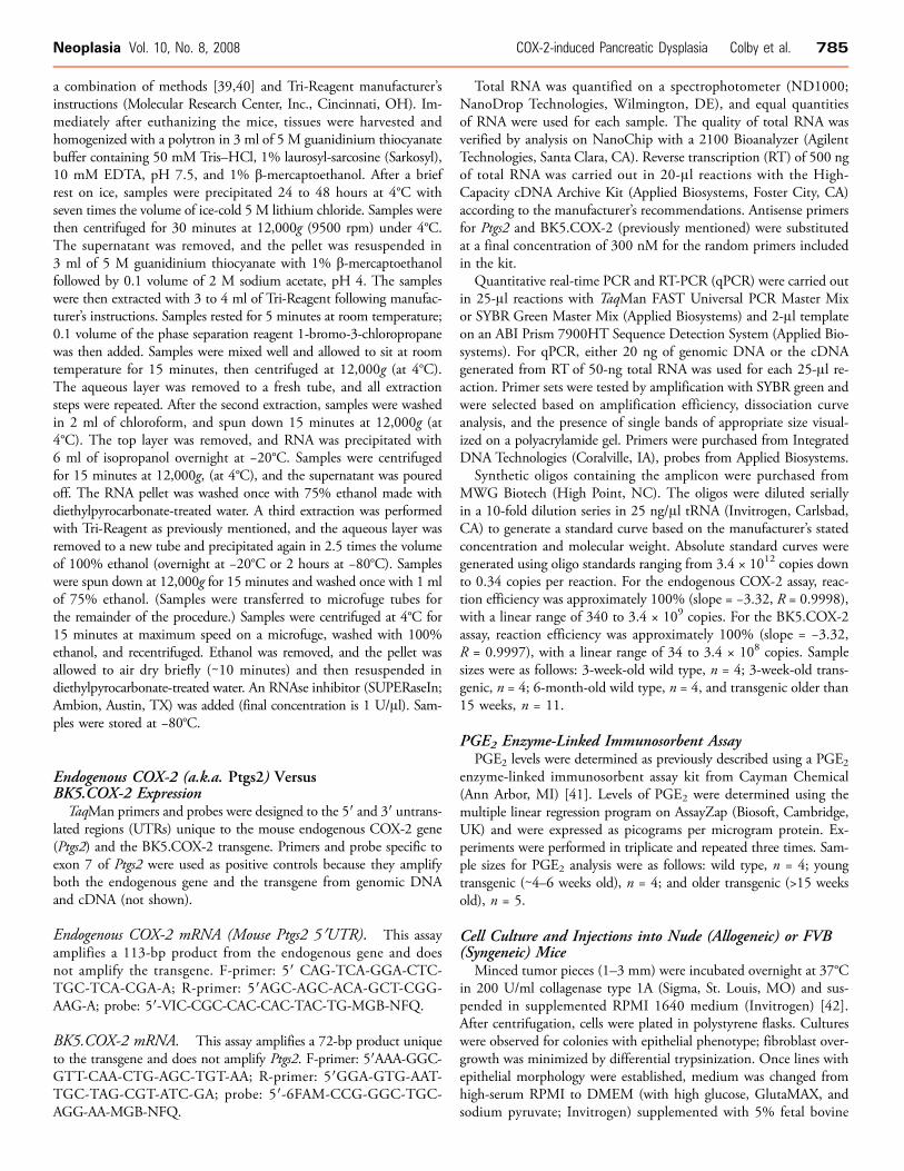
Table 1. List of Antibodies and Conditions Used.
Primary Antibody Application Source Dilution Incubation Time/Temperature Antigen Retrieval Method
α-SMA IHC Dako 1:1000 30 minutes, RT NoneAmylase IHC Sigma 1:5000 30 minutes, RT Citrate*β-Catenin IHC BD Biosciences 1:500 30 minutes, RT CitrateCaspase 3 (active) IHC R&D Systems 1:2000 30 minutes, RT CitrateCD3 IHC Serotec 1:100 2 hours, RT EDTA†
CD31¶ IHC Pharmingen 1:400 ON, 4°C Protease‡
CD45R IHC Serotec 1:2000 1 hour, RT CitrateCOX-1 WB Cayman 1:100 ON, 4°C N/ACOX-2 IHC Cayman 1:500 1 hour, RT CitrateE-Cadherin IHC Santa Cruz Biotechnology 1:50 1 hour, RT CitrateInsulin IHC Zymed 1:500 1 hour, RT NoneK19 IHC AbCam 1:100 1 hour, RT EIER§ (pepsin)Ki-67 IHC Dako 1:200 ON, 4°C CitrateMMP-9 WB Chemicon 1:1000 1 hour, RT N/AMMP-7 IHC LabVision/Neomarkers 1:50 1 hour, RT NonePan-S100 IHC Dako Predilute 1 hour, RT NoneVEGF WB Sigma 1:1000 1 hour, RT N/A
Company Location
Dako Carpinteria, CASigma St. Louis, MOBD Biosciences San Diego, CASerotec Raleigh, NCCayman Ann Arbor, MIAbCam Cambridge, MACell Signaling Danvers, MAInvitrogen/Zymed San Francisco, CAChemicon/Millipore Temecula, CA/Billerica, MACalbiochem (EMD Biosci) San Diego, CA
All immunohistochemical protocols require blocking sections in 10% serum for 30 minutes (to block nonspecific sites) and in 3% H2O2 for 10 minutes (to quench endogenous peroxidase activity).IHC indicates immunohistochemistry; ON, overnight; RT, 25°C; WB, Western blot.*Antigen retrieval method used: 10 mM citrate buffer, pH 6.0, for 10 minutes in microwave and 20 minutes of cool down.†Antigen retrieval method used: 1 mM EDTA, pH 8, for 10 minutes in microwave and 20 minutes of cool down.‡Antigen retrieval method used: 0.06% protease (type 24) in Tris buffer for 10 minutes.§Antigen retrieval method used: enzyme-induced epitope retrieval (EIER) with Digest-all 3 (pepsin; Zymed) for 10 minutes at 37°C. All IHC stains were visualized with diaminobenzidine.¶CD31 IHC uses a tyramide amplification step; contact authors for detailed protocol.
784 COX-2-induced Pancreatic Dysplasia Colby et al. Neoplasia Vol. 10, No. 8, 2008
a combination of methods [39,40] and Tri-Reagent manufacturer’sinstructions (Molecular Research Center, Inc., Cincinnati, OH). Im-mediately after euthanizing the mice, tissues were harvested andhomogenized with a polytron in 3 ml of 5 M guanidinium thiocyanatebuffer containing 50 mM Tris–HCl, 1% laurosyl-sarcosine (Sarkosyl),10 mM EDTA, pH 7.5, and 1% β-mercaptoethanol. After a briefrest on ice, samples were precipitated 24 to 48 hours at 4°C withseven times the volume of ice-cold 5 M lithium chloride. Samples werethen centrifuged for 30 minutes at 12,000g (9500 rpm) under 4°C.The supernatant was removed, and the pellet was resuspended in3 ml of 5 M guanidinium thiocyanate with 1% β-mercaptoethanolfollowed by 0.1 volume of 2 M sodium acetate, pH 4. The sampleswere then extracted with 3 to 4 ml of Tri-Reagent following manufac-turer’s instructions. Samples rested for 5 minutes at room temperature;0.1 volume of the phase separation reagent 1-bromo-3-chloropropanewas then added. Samples were mixed well and allowed to sit at roomtemperature for 15 minutes, then centrifuged at 12,000g (at 4°C).The aqueous layer was removed to a fresh tube, and all extractionsteps were repeated. After the second extraction, samples were washedin 2 ml of chloroform, and spun down 15 minutes at 12,000g (at4°C). The top layer was removed, and RNA was precipitated with6 ml of isopropanol overnight at −20°C. Samples were centrifugedfor 15 minutes at 12,000g, (at 4°C), and the supernatant was pouredoff. The RNA pellet was washed once with 75% ethanol made withdiethylpyrocarbonate-treated water. A third extraction was performedwith Tri-Reagent as previously mentioned, and the aqueous layer wasremoved to a new tube and precipitated again in 2.5 times the volumeof 100% ethanol (overnight at −20°C or 2 hours at −80°C). Sampleswere spun down at 12,000g for 15 minutes and washed once with 1 mlof 75% ethanol. (Samples were transferred to microfuge tubes forthe remainder of the procedure.) Samples were centrifuged at 4°C for15 minutes at maximum speed on a microfuge, washed with 100%ethanol, and recentrifuged. Ethanol was removed, and the pellet wasallowed to air dry briefly (∼10 minutes) and then resuspended indiethylpyrocarbonate-treated water. An RNAse inhibitor (SUPERaseIn;Ambion, Austin, TX) was added (final concentration is 1 U/μl). Sam-ples were stored at −80°C.
Endogenous COX-2 (a.k.a. Ptgs2) VersusBK5.COX-2 ExpressionTaqMan primers and probes were designed to the 5′ and 3′ untrans-
lated regions (UTRs) unique to the mouse endogenous COX-2 gene(Ptgs2) and the BK5.COX-2 transgene. Primers and probe specific toexon 7 of Ptgs2 were used as positive controls because they amplifyboth the endogenous gene and the transgene from genomic DNAand cDNA (not shown).
Endogenous COX-2 mRNA (Mouse Ptgs2 5′UTR). This assayamplifies a 113-bp product from the endogenous gene and doesnot amplify the transgene. F-primer: 5′ CAG-TCA-GGA-CTC-TGC-TCA-CGA-A; R-primer: 5′AGC-AGC-ACA-GCT-CGG-AAG-A; probe: 5′-VIC-CGC-CAC-CAC-TAC-TG-MGB-NFQ.
BK5.COX-2 mRNA. This assay amplifies a 72-bp product uniqueto the transgene and does not amplify Ptgs2. F-primer: 5′AAA-GGC-GTT-CAA-CTG-AGC-TGT-AA; R-primer: 5′GGA-GTG-AAT-TGC-TAG-CGT-ATC-GA; probe: 5′-6FAM-CCG-GGC-TGC-AGG-AA-MGB-NFQ.
Total RNA was quantified on a spectrophotometer (ND1000;NanoDrop Technologies, Wilmington, DE), and equal quantitiesof RNA were used for each sample. The quality of total RNA wasverified by analysis on NanoChip with a 2100 Bioanalyzer (AgilentTechnologies, Santa Clara, CA). Reverse transcription (RT) of 500 ngof total RNA was carried out in 20-μl reactions with the High-Capacity cDNA Archive Kit (Applied Biosystems, Foster City, CA)according to the manufacturer’s recommendations. Antisense primersfor Ptgs2 and BK5.COX-2 (previously mentioned) were substitutedat a final concentration of 300 nM for the random primers includedin the kit.Quantitative real-time PCR and RT-PCR (qPCR) were carried out
in 25-μl reactions with TaqMan FAST Universal PCR Master Mixor SYBR Green Master Mix (Applied Biosystems) and 2-μl templateon an ABI Prism 7900HT Sequence Detection System (Applied Bio-systems). For qPCR, either 20 ng of genomic DNA or the cDNAgenerated from RT of 50-ng total RNA was used for each 25-μl re-action. Primer sets were tested by amplification with SYBR green andwere selected based on amplification efficiency, dissociation curveanalysis, and the presence of single bands of appropriate size visual-ized on a polyacrylamide gel. Primers were purchased from IntegratedDNA Technologies (Coralville, IA), probes from Applied Biosystems.Synthetic oligos containing the amplicon were purchased from
MWG Biotech (High Point, NC). The oligos were diluted seriallyin a 10-fold dilution series in 25 ng/μl tRNA (Invitrogen, Carlsbad,CA) to generate a standard curve based on the manufacturer’s statedconcentration and molecular weight. Absolute standard curves weregenerated using oligo standards ranging from 3.4 × 10¹² copies downto 0.34 copies per reaction. For the endogenous COX-2 assay, reac-tion efficiency was approximately 100% (slope = −3.32, R = 0.9998),with a linear range of 340 to 3.4 × 10⁹ copies. For the BK5.COX-2assay, reaction efficiency was approximately 100% (slope = −3.32,R = 0.9997), with a linear range of 34 to 3.4 × 10⁸ copies. Samplesizes were as follows: 3-week-old wild type, n = 4; 3-week-old trans-genic, n = 4; 6-month-old wild type, n = 4, and transgenic older than15 weeks, n = 11.
PGE2 Enzyme-Linked Immunosorbent AssayPGE2 levels were determined as previously described using a PGE2
enzyme-linked immunosorbent assay kit from Cayman Chemical(Ann Arbor, MI) [41]. Levels of PGE2 were determined using themultiple linear regression program on AssayZap (Biosoft, Cambridge,UK) and were expressed as picograms per microgram protein. Ex-periments were performed in triplicate and repeated three times. Sam-ple sizes for PGE2 analysis were as follows: wild type, n = 4; youngtransgenic (∼4–6 weeks old), n = 4; and older transgenic (>15 weeksold), n = 5.
Cell Culture and Injections into Nude (Allogeneic) or FVB(Syngeneic) MiceMinced tumor pieces (1–3 mm) were incubated overnight at 37°C
in 200 U/ml collagenase type 1A (Sigma, St. Louis, MO) and sus-pended in supplemented RPMI 1640 medium (Invitrogen) [42].After centrifugation, cells were plated in polystyrene flasks. Cultureswere observed for colonies with epithelial phenotype; fibroblast over-growth was minimized by differential trypsinization. Once lines withepithelial morphology were established, medium was changed fromhigh-serum RPMI to DMEM (with high glucose, GlutaMAX, andsodium pyruvate; Invitrogen) supplemented with 5% fetal bovine
Neoplasia Vol. 10, No. 8, 2008 COX-2-induced Pancreatic Dysplasia Colby et al. 785
serum, insulin (10 μg/ml), and penicillin/streptomycin (100 μg/ml).Cells from one of the epithelial lines (JC102) were injected into thespleens of three 7-week-old nude mice (BALB/cAnCr− nu/nu; Na-tional Cancer Institute, Frederick, MD) and were allowed to growfor 7 weeks. Cells were injected intraperitoneally (i.p.; JC101 andJC102) or subcutaneously (JC101) into the FVB mice; mice werekilled and tissues were collected when the tumors were ∼1 cm indiameter or after 30 days.
Chemoprevention StudyGroups of 8 to 12 mice were fed ad libitum diets of AIN-76A diet
(Research Diets, New Brunswick, NJ) containing celecoxib (LKTLaboratories, St. Paul, MN) at concentrations of 500 (4.5 months),1000 (3 months), or 1250 ppm (6–10 months). Age- and sex-matchedcontrols were fed AIN-76A (Research Diets) ad libitum. Mice wereplaced on diets at weaning (3–4 weeks) and killed at the end ofthe feeding periods. Kaplan–Meier survival curves were generatedusing JMP software (SAS Institute, Cary, NC). For survival analysis,11 transgenic mice fed 1250 ppm of celecoxib up to 33 weeks werecompared with 30 mice fed control diet for the same period.
Statistical AnalysisStatistical differences were performed using analysis of variance
(ANOVA) followed by Student’s t test (one-sided, unequal variance).PGE2 and endogenous COX-2 expression data were analyzed usingExcel (Microsoft Corporation, Redmond, WA). Log-rank analysisof Kaplan–Meier survival data was performed using JMP software(SAS Institute).
Results
HistopathologyNecropsies of newborn and perinatal BK5.COX-2 mice did not
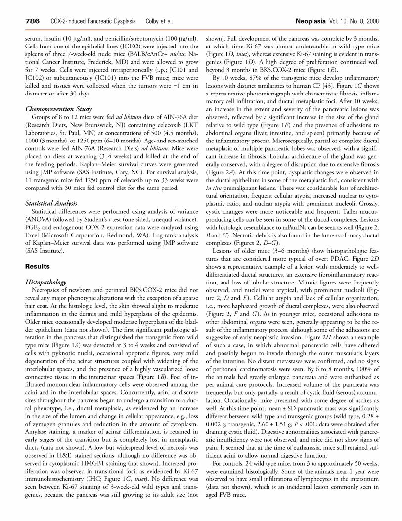
reveal any major phenotypic alterations with the exception of a sparsehair coat. At the histologic level, the skin showed slight to moderateinflammation in the dermis and mild hyperplasia of the epidermis.Older mice occasionally developed moderate hyperplasia of the blad-der epithelium (data not shown). The first significant pathologic al-teration in the pancreas that distinguished the transgenic from wildtype mice (Figure 1A) was detected at 3 to 4 weeks and consisted ofcells with pyknotic nuclei, occasional apoptotic figures, very milddegeneration of the acinar structures coupled with widening of theinterlobular spaces, and the presence of a highly vascularized looseconnective tissue in the interacinar spaces (Figure 1B). Foci of in-filtrated mononuclear inflammatory cells were observed among theacini and in the interlobular spaces. Concurrently, acini at discretesites throughout the pancreas began to undergo a transition to a duc-tal phenotype, i.e., ductal metaplasia, as evidenced by an increasein the size of the lumen and change in cellular appearance, e.g., lossof zymogen granules and reduction in the amount of cytoplasm.Amylase staining, a marker of acinar differentiation, is retained inearly stages of the transition but is completely lost in metaplasticducts (data not shown). A low but widespread level of necrosis wasobserved in H&E–stained sections, although no difference was ob-served in cytoplasmic HMGB1 staining (not shown). Increased pro-liferation was observed in transitional foci, as evidenced by Ki-67immunohistochemistry (IHC; Figure 1C , inset). No difference wasseen between Ki-67 staining of 3-week-old wild types and trans-genics, because the pancreas was still growing to its adult size (not
shown). Full development of the pancreas was complete by 3 months,at which time Ki-67 was almost undetectable in wild type mice(Figure 1D, inset), whereas extensive Ki-67 staining is evident in trans-genics (Figure 1D). A high degree of proliferation continued wellbeyond 3 months in BK5.COX-2 mice (Figure 1E).By 10 weeks, 87% of the transgenic mice develop inflammatory
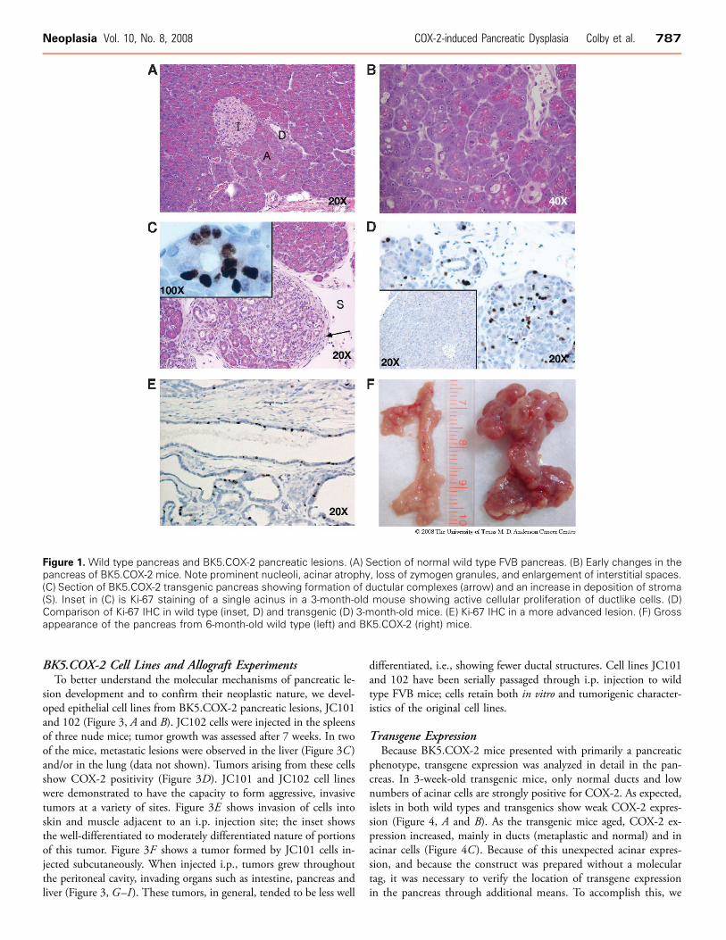
lesions with distinct similarities to human CP [43]. Figure 1C showsa representative photomicrograph with characteristic fibrosis, inflam-matory cell infiltration, and ductal metaplastic foci. After 10 weeks,an increase in the extent and severity of the pancreatic lesions wasobserved, reflected by a significant increase in the size of the glandrelative to wild type (Figure 1F ) and the presence of adhesions toabdominal organs (liver, intestine, and spleen) primarily because ofthe inflammatory process. Microscopically, partial or complete ductalmetaplasia of multiple pancreatic lobes was observed, with a signifi-cant increase in fibrosis. Lobular architecture of the gland was gen-erally conserved, with a degree of disruption due to extensive fibrosis(Figure 2A). At this time point, dysplastic changes were observed inthe ductal epithelium in some of the metaplastic foci, consistent within situ premalignant lesions. There was considerable loss of architec-tural orientation, frequent cellular atypia, increased nuclear to cyto-plasmic ratio, and nuclear atypia with prominent nucleoli. Grossly,cystic changes were more noticeable and frequent. Taller mucus-producing cells can be seen in some of the ductal complexes. Lesionswith histologic resemblance to mPanINs can be seen as well (Figure 2,B and C ). Necrotic debris is also found in the lumens of many ductalcomplexes (Figures 2, D–G ).Lesions of older mice (3–6 months) show histopathologic fea-
tures that are considered more typical of overt PDAC. Figure 2Dshows a representative example of a lesion with moderately to well-differentiated ductal structures, an extensive fibroinflammatory reac-tion, and loss of lobular structure. Mitotic figures were frequentlyobserved, and nuclei were atypical, with prominent nucleoli (Fig-ure 2, D and E ). Cellular atypia and lack of cellular organization,i.e., more haphazard growth of ductal complexes, were also observed(Figure 2, F and G ). As in younger mice, occasional adhesions toother abdominal organs were seen, generally appearing to be the re-sult of the inflammatory process, although some of the adhesions aresuggestive of early neoplastic invasion. Figure 2H shows an exampleof such a case, in which abnormal pancreatic cells have adheredand possibly begun to invade through the outer muscularis layersof the intestine. No distant metastases were confirmed, and no signsof peritoneal carcinomatosis were seen. By 6 to 8 months, 100% ofthe animals had greatly enlarged pancreata and were euthanized asper animal care protocols. Increased volume of the pancreata wasfrequently, but only partially, a result of cystic fluid (serous) accumu-lation. Occasionally, mice presented with some degree of ascites aswell. At this time point, mean ± SD pancreatic mass was significantlydifferent between wild type and transgenic groups (wild type, 0.28 ±0.002 g; transgenic, 2.60 ± 1.51 g; P < .001; data were obtained afterdraining cystic fluid). Digestive abnormalities associated with pancre-atic insufficiency were not observed, and mice did not show signs ofpain. It seemed that at the time of euthanasia, mice still retained suf-ficient acini to allow normal digestive function.For controls, 24 wild type mice, from 3 to approximately 50 weeks,
were examined histologically. Some of the animals near 1 year wereobserved to have small infiltrations of lymphocytes in the interstitium(data not shown), which is an incidental lesion commonly seen inaged FVB mice.
786 COX-2-induced Pancreatic Dysplasia Colby et al. Neoplasia Vol. 10, No. 8, 2008
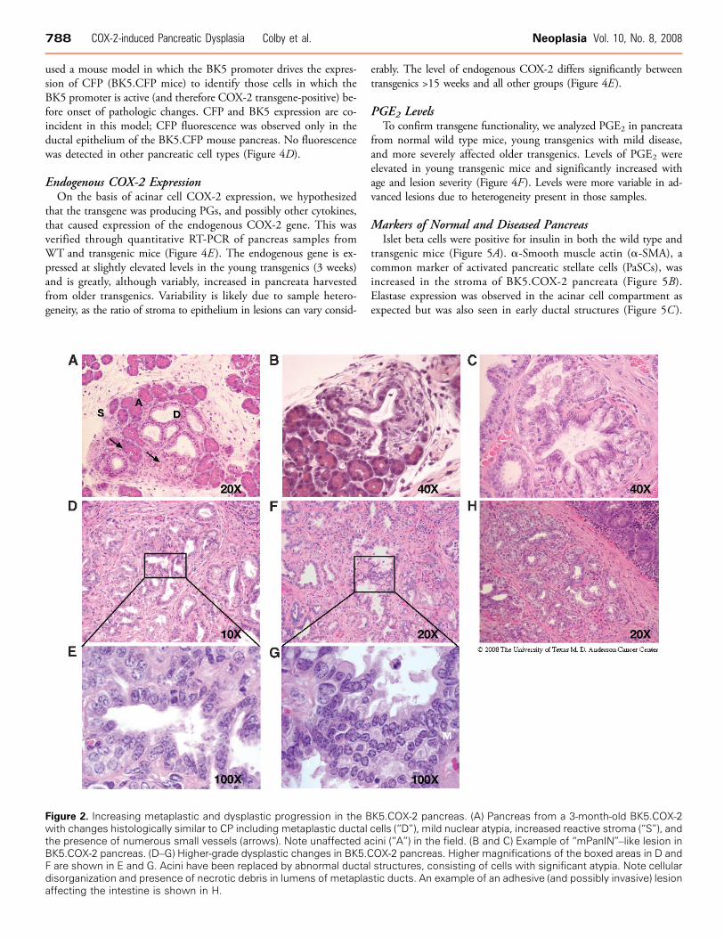
BK5.COX-2 Cell Lines and Allograft ExperimentsTo better understand the molecular mechanisms of pancreatic le-
sion development and to confirm their neoplastic nature, we devel-oped epithelial cell lines from BK5.COX-2 pancreatic lesions, JC101and 102 (Figure 3, A and B). JC102 cells were injected in the spleensof three nude mice; tumor growth was assessed after 7 weeks. In twoof the mice, metastatic lesions were observed in the liver (Figure 3C )and/or in the lung (data not shown). Tumors arising from these cellsshow COX-2 positivity (Figure 3D). JC101 and JC102 cell lineswere demonstrated to have the capacity to form aggressive, invasivetumors at a variety of sites. Figure 3E shows invasion of cells intoskin and muscle adjacent to an i.p. injection site; the inset showsthe well-differentiated to moderately differentiated nature of portionsof this tumor. Figure 3F shows a tumor formed by JC101 cells in-jected subcutaneously. When injected i.p., tumors grew throughoutthe peritoneal cavity, invading organs such as intestine, pancreas andliver (Figure 3, G–I ). These tumors, in general, tended to be less well
differentiated, i.e., showing fewer ductal structures. Cell lines JC101and 102 have been serially passaged through i.p. injection to wildtype FVB mice; cells retain both in vitro and tumorigenic character-istics of the original cell lines.
Transgene ExpressionBecause BK5.COX-2 mice presented with primarily a pancreatic
phenotype, transgene expression was analyzed in detail in the pan-creas. In 3-week-old transgenic mice, only normal ducts and lownumbers of acinar cells are strongly positive for COX-2. As expected,islets in both wild types and transgenics show weak COX-2 expres-sion (Figure 4, A and B). As the transgenic mice aged, COX-2 ex-pression increased, mainly in ducts (metaplastic and normal) and inacinar cells (Figure 4C ). Because of this unexpected acinar expres-sion, and because the construct was prepared without a moleculartag, it was necessary to verify the location of transgene expressionin the pancreas through additional means. To accomplish this, we
Figure 1. Wild type pancreas and BK5.COX-2 pancreatic lesions. (A) Section of normal wild type FVB pancreas. (B) Early changes in thepancreas of BK5.COX-2 mice. Note prominent nucleoli, acinar atrophy, loss of zymogen granules, and enlargement of interstitial spaces.(C) Section of BK5.COX-2 transgenic pancreas showing formation of ductular complexes (arrow) and an increase in deposition of stroma(S). Inset in (C) is Ki-67 staining of a single acinus in a 3-month-old mouse showing active cellular proliferation of ductlike cells. (D)Comparison of Ki-67 IHC in wild type (inset, D) and transgenic (D) 3-month-old mice. (E) Ki-67 IHC in a more advanced lesion. (F) Grossappearance of the pancreas from 6-month-old wild type (left) and BK5.COX-2 (right) mice.
Neoplasia Vol. 10, No. 8, 2008 COX-2-induced Pancreatic Dysplasia Colby et al. 787
used a mouse model in which the BK5 promoter drives the expres-sion of CFP (BK5.CFP mice) to identify those cells in which theBK5 promoter is active (and therefore COX-2 transgene-positive) be-fore onset of pathologic changes. CFP and BK5 expression are co-incident in this model; CFP fluorescence was observed only in theductal epithelium of the BK5.CFP mouse pancreas. No fluorescencewas detected in other pancreatic cell types (Figure 4D).
Endogenous COX-2 ExpressionOn the basis of acinar cell COX-2 expression, we hypothesized
that the transgene was producing PGs, and possibly other cytokines,that caused expression of the endogenous COX-2 gene. This wasverified through quantitative RT-PCR of pancreas samples fromWT and transgenic mice (Figure 4E ). The endogenous gene is ex-pressed at slightly elevated levels in the young transgenics (3 weeks)and is greatly, although variably, increased in pancreata harvestedfrom older transgenics. Variability is likely due to sample hetero-geneity, as the ratio of stroma to epithelium in lesions can vary consid-
erably. The level of endogenous COX-2 differs significantly betweentransgenics >15 weeks and all other groups (Figure 4E).
PGE2 LevelsTo confirm transgene functionality, we analyzed PGE2 in pancreata
from normal wild type mice, young transgenics with mild disease,and more severely affected older transgenics. Levels of PGE2 wereelevated in young transgenic mice and significantly increased withage and lesion severity (Figure 4F ). Levels were more variable in ad-vanced lesions due to heterogeneity present in those samples.
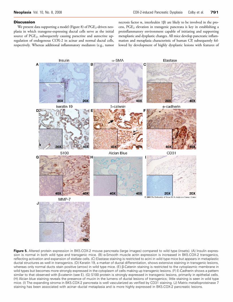
Markers of Normal and Diseased PancreasIslet beta cells were positive for insulin in both the wild type and
transgenic mice (Figure 5A). α-Smooth muscle actin (α-SMA), acommon marker of activated pancreatic stellate cells (PaSCs), wasincreased in the stroma of BK5.COX-2 pancreata (Figure 5B).Elastase expression was observed in the acinar cell compartment asexpected but was also seen in early ductal structures (Figure 5C ).
Figure 2. Increasing metaplastic and dysplastic progression in the BK5.COX-2 pancreas. (A) Pancreas from a 3-month-old BK5.COX-2with changes histologically similar to CP including metaplastic ductal cells (“D”), mild nuclear atypia, increased reactive stroma (“S”), andthe presence of numerous small vessels (arrows). Note unaffected acini (“A”) in the field. (B and C) Example of “mPanIN”–like lesion inBK5.COX-2 pancreas. (D–G) Higher-grade dysplastic changes in BK5.COX-2 pancreas. Higher magnifications of the boxed areas in D andF are shown in E and G. Acini have been replaced by abnormal ductal structures, consisting of cells with significant atypia. Note cellulardisorganization and presence of necrotic debris in lumens of metaplastic ducts. An example of an adhesive (and possibly invasive) lesionaffecting the intestine is shown in H.
788 COX-2-induced Pancreatic Dysplasia Colby et al. Neoplasia Vol. 10, No. 8, 2008
Ductal complexes lose elastase expression as they proliferate. An iden-tical staining pattern is observed with amylase IHC (data not shown).Keratin 19 (K19) is an intermediate filament protein found in pan-creatic ducts and is frequently used to confirm a true ductal pheno-type [44]. Keratin 19 staining is widespread in BK5.COX-2 ductalcomplexes, indicative of a ductal, as opposed to acinar, phenotype(Figure 5D).β-Catenin and E-cadherin have critical roles in adhesion and sig-
naling and are frequently dysregulated and/or expressed in aberrant
patterns in both preneoplastic lesions and neoplasia. In BK5.COX-2mice, cytoplasmic β-catenin levels increase in comparison to wild type,which have a typical membrane-bound pattern of β-catenin staining(Figure 5E ). A similar pattern is observed with E-cadherin staining(Figure 5F). The S100 proteins are a group of calcium-binding pro-teins having numerous effects on cell behavior; they are often over-expressed in cancer and may have prognostic value [45]. Using apan-S100 antibody, we observed a distinct increase in S100 stainingin BK5.COX-2 transgenic pancreas (Figure 5G ). Pancreatic ductaladenocarcinoma frequently shows a dysregulated production of mucins[46]. Alcian blue staining, used for the detection of mucins, is muchmore prominent in BK5.COX-2 lesions versus controls (Figure 5H ).Positive staining for PAS and mucicarmine, additional indicators ofmucins, were also observed in transgenic lesions (data not shown).An increase in vessel number in the stroma of transgenic lesions was
observed in H&E–stained sections and was confirmed by immuno-staining for CD31 (Figure 5I ). Vascular endothelial growth factor(VEGF) is another indicator of an angiogenic response; immunoblotanalysis showed up-regulation of VEGF in transgenic pancreata (notshown). In the pancreas, matrix metalloproteinase 7 (MMP-7) isinvolved in acinar-to-ductal transdifferentiation and is up-regulatedin most human PDAC [47]. Matrix metalloproteinase 9 (MMP-9;gelatinase B) is involved in the remodeling of extracellular matrix innormal and disease processes, and stromal expression of MMP-9 isnecessary for the angiogenesis and progression of tumors in a nudemouse model using orthotopically implanted pancreatic cancer cells[48,49]. In our model, MMP-9 is elevated in transgenic pancreataas determined by immunoblot analysis (not shown). Matrix metallo-proteinase 7 expression was evaluated by IHC; areas of positivity areseen in both stroma and acinar cells undergoing metaplastic change(Figure 5J ).
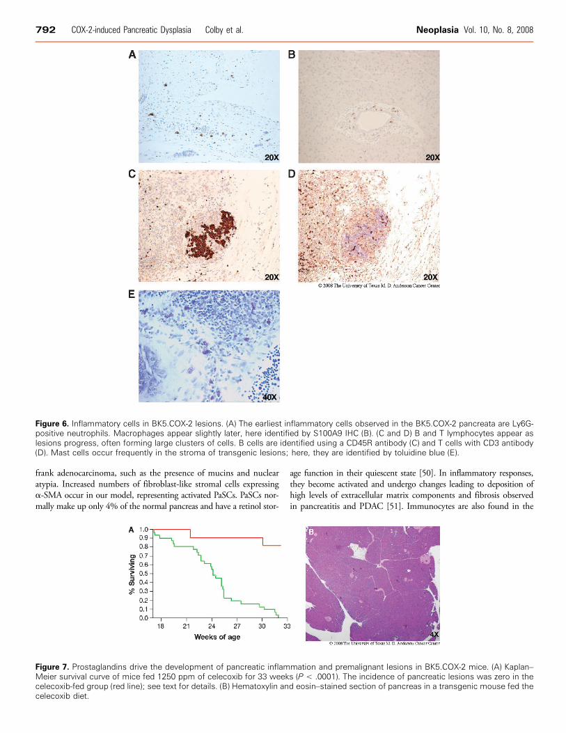
Inflammatory Cell MarkersA range of cells typical of inflammatory responses is observed in
BK5.COX-2 lesions. The earliest abnormal pancreata have an influxof neutrophils, and later macrophages, as verified by Ly6G (Gr-1)and S100A9 IHC (Figure 6, A and B), respectively. More advancedlesions generally possessed numerous B and T lymphocytes, oftenpresent in large clusters (Figure 6, C and D). B- and T-cell identitieswere verified by CD45R (Figure 6C ) and CD3 (Figure 6D) IHC,respectively. Toluidine blue histochemistry demonstrated the pres-ence of mast cells (Figure 6E ).
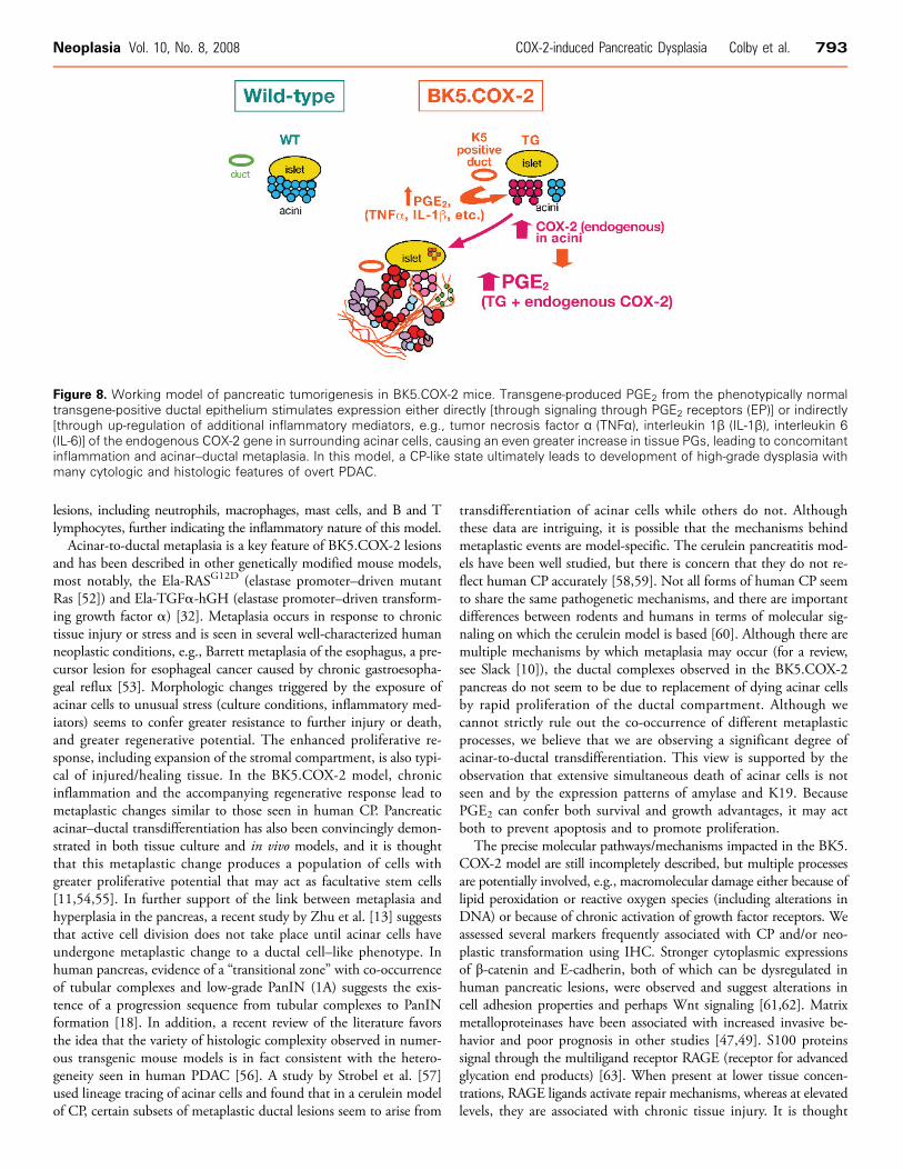
Celecoxib Feeding StudiesTo demonstrate that pancreatic lesions in BK5.COX-2 mice were
due to high levels of PG synthesized by COX-2, transgenic micewere fed diets containing celecoxib (500–1250 ppm), a selectiveCOX-2 inhibitor. Kaplan–Meier analysis revealed significant differ-ences between groups (Figure 7A). None of the mice fed celecoxibshowed any evidence of pancreatic lesions (Figure 7B). Several miceon the highest dose (1250 ppm) died because of gastrointestinaltoxicity, but none had pancreatic abnormalities. The lower doses ofcelecoxib (500 and 1000 ppm) proved equally effective at prevent-ing lesions with significantly less toxicity (data not shown). As notedpreviously, mice on the control diet were euthanized in compliancewith animal care regulations, generally due to the extensive enlarge-ment of the pancreas due to cystic fluid accumulation/fibrosis lead-ing to compression/dysfunction of other organs.
Figure 3. Allograft experiments. H&E–stained sections of JC101(A) and JC102 (B) cells grown on a chamber slide. H&E–stainedsection of the liver metastasis (C) of JC102 cells injected intothe spleen. (D) Immunohistochemistry for COX-2 expression inmetastasis shown in (C). H&E–stained section of tumor arisingnear the injection site of i.p.–injected JC102 cells (E). Note inva-sion to muscle and skin. The inset in E shows a portion of thesame tumor that is well to moderately differentiated. (F) Tumorarising from subcutaneously injected JC101 cells. (G–I) Tumorsarising from i.p.–injected JC101 or 102 cells. Tumors invadingthe small intestine (G), normal pancreas (H), and liver (I) are shown.
Neoplasia Vol. 10, No. 8, 2008 COX-2-induced Pancreatic Dysplasia Colby et al. 789
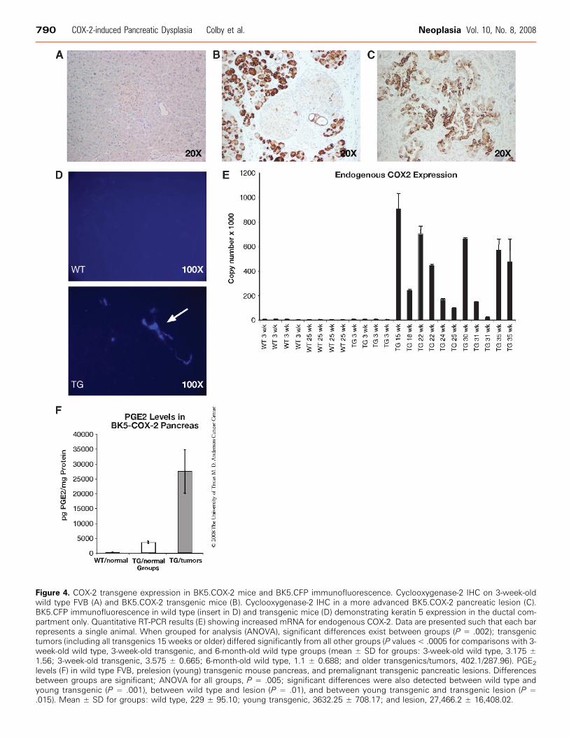
Figure 4. COX-2 transgene expression in BK5.COX-2 mice and BK5.CFP immunofluorescence. Cyclooxygenase-2 IHC on 3-week-oldwild type FVB (A) and BK5.COX-2 transgenic mice (B). Cyclooxygenase-2 IHC in a more advanced BK5.COX-2 pancreatic lesion (C).BK5.CFP immunofluorescence in wild type (insert in D) and transgenic mice (D) demonstrating keratin 5 expression in the ductal com-partment only. Quantitative RT-PCR results (E) showing increased mRNA for endogenous COX-2. Data are presented such that each barrepresents a single animal. When grouped for analysis (ANOVA), significant differences exist between groups (P = .002); transgenictumors (including all transgenics 15 weeks or older) differed significantly from all other groups (P values < .0005 for comparisons with 3-week-old wild type, 3-week-old transgenic, and 6-month-old wild type groups (mean ± SD for groups: 3-week-old wild type, 3.175 ±1.56; 3-week-old transgenic, 3.575 ± 0.665; 6-month-old wild type, 1.1 ± 0.688; and older transgenics/tumors, 402.1/287.96). PGE2
levels (F) in wild type FVB, prelesion (young) transgenic mouse pancreas, and premalignant transgenic pancreatic lesions. Differencesbetween groups are significant; ANOVA for all groups, P = .005; significant differences were also detected between wild type andyoung transgenic (P = .001), between wild type and lesion (P = .01), and between young transgenic and transgenic lesion (P =.015). Mean ± SD for groups: wild type, 229 ± 95.10; young transgenic, 3632.25 ± 708.17; and lesion, 27,466.2 ± 16,408.02.
790 COX-2-induced Pancreatic Dysplasia Colby et al. Neoplasia Vol. 10, No. 8, 2008
DiscussionWe present data supporting a model (Figure 8) of PGE2-driven neo-
plasia in which transgene-expressing ductal cells serve as the initialsource of PGE2, subsequently causing paracrine and autocrine up-regulation of endogenous COX-2 in acinar and normal ductal cells,respectively. Whereas additional inflammatory mediators (e.g., tumor
necrosis factor α, interleukin 1β) are likely to be involved in the pro-cess, PGE2 elevation in transgenic pancreata is key in establishing aproinflammatory environment capable of initiating and supportingmetaplastic and dysplastic changes. All mice develop pancreatic inflam-mation and metaplasia characteristic of human CP, subsequently fol-lowed by development of highly dysplastic lesions with features of
Figure 5. Altered protein expression in BK5.COX-2 mouse pancreata (large images) compared to wild type (insets). (A) Insulin expres-sion is normal in both wild type and transgenic mice. (B) α-Smooth muscle actin expression is increased in BK5.COX-2 transgenics,reflecting activation and expansion of stellate cells. (C) Elastase staining is restricted to acini in wild type mice but appears in metaplasticductal structures as well in transgenics. (D) Keratin 19, a marker of ductal differentiation, shows extensive staining in transgenic lesions,whereas only normal ducts stain positive (arrow) in wild type mice. (E) β-Catenin staining is restricted to the cytoplasmic membrane inwild types but becomes more strongly expressed in the cytoplasm of cells making up transgenic lesions. (F) E-Cadherin shows a patternsimilar to that observed with β-catenin (see E). (G) S100 protein is strongly expressed in transgenic lesions, primarily in epithelial cells.(H) Alcian blue staining reveals the presence of mucin in the lumens of ductal lesions of transgenics; little staining is seen in wild typemice. (I) The expanding stroma in BK5.COX-2 pancreata is well vascularized as verified by CD31 staining. (J) Matrix metalloproteinase 7staining has been associated with acinar–ductal metaplasia and is more highly expressed in BK5.COX-2 pancreatic lesions.
Neoplasia Vol. 10, No. 8, 2008 COX-2-induced Pancreatic Dysplasia Colby et al. 791
frank adenocarcinoma, such as the presence of mucins and nuclearatypia. Increased numbers of fibroblast-like stromal cells expressingα-SMA occur in our model, representing activated PaSCs. PaSCs nor-mally make up only 4% of the normal pancreas and have a retinol stor-
age function in their quiescent state [50]. In inflammatory responses,they become activated and undergo changes leading to deposition ofhigh levels of extracellular matrix components and fibrosis observedin pancreatitis and PDAC [51]. Immunocytes are also found in the
Figure 7. Prostaglandins drive the development of pancreatic inflammation and premalignant lesions in BK5.COX-2 mice. (A) Kaplan–Meier survival curve of mice fed 1250 ppm of celecoxib for 33 weeks (P < .0001). The incidence of pancreatic lesions was zero in thecelecoxib-fed group (red line); see text for details. (B) Hematoxylin and eosin–stained section of pancreas in a transgenic mouse fed thecelecoxib diet.
Figure 6. Inflammatory cells in BK5.COX-2 lesions. (A) The earliest inflammatory cells observed in the BK5.COX-2 pancreata are Ly6G-positive neutrophils. Macrophages appear slightly later, here identified by S100A9 IHC (B). (C and D) B and T lymphocytes appear aslesions progress, often forming large clusters of cells. B cells are identified using a CD45R antibody (C) and T cells with CD3 antibody(D). Mast cells occur frequently in the stroma of transgenic lesions; here, they are identified by toluidine blue (E).
792 COX-2-induced Pancreatic Dysplasia Colby et al. Neoplasia Vol. 10, No. 8, 2008
lesions, including neutrophils, macrophages, mast cells, and B and Tlymphocytes, further indicating the inflammatory nature of this model.Acinar-to-ductal metaplasia is a key feature of BK5.COX-2 lesions
and has been described in other genetically modified mouse models,most notably, the Ela-RASG12D (elastase promoter–driven mutantRas [52]) and Ela-TGFα-hGH (elastase promoter–driven transform-ing growth factor α) [32]. Metaplasia occurs in response to chronictissue injury or stress and is seen in several well-characterized humanneoplastic conditions, e.g., Barrett metaplasia of the esophagus, a pre-cursor lesion for esophageal cancer caused by chronic gastroesopha-geal reflux [53]. Morphologic changes triggered by the exposure ofacinar cells to unusual stress (culture conditions, inflammatory med-iators) seems to confer greater resistance to further injury or death,and greater regenerative potential. The enhanced proliferative re-sponse, including expansion of the stromal compartment, is also typi-cal of injured/healing tissue. In the BK5.COX-2 model, chronicinflammation and the accompanying regenerative response lead tometaplastic changes similar to those seen in human CP. Pancreaticacinar–ductal transdifferentiation has also been convincingly demon-strated in both tissue culture and in vivo models, and it is thoughtthat this metaplastic change produces a population of cells withgreater proliferative potential that may act as facultative stem cells[11,54,55]. In further support of the link between metaplasia andhyperplasia in the pancreas, a recent study by Zhu et al. [13] suggeststhat active cell division does not take place until acinar cells haveundergone metaplastic change to a ductal cell–like phenotype. Inhuman pancreas, evidence of a “transitional zone” with co-occurrenceof tubular complexes and low-grade PanIN (1A) suggests the exis-tence of a progression sequence from tubular complexes to PanINformation [18]. In addition, a recent review of the literature favorsthe idea that the variety of histologic complexity observed in numer-ous transgenic mouse models is in fact consistent with the hetero-geneity seen in human PDAC [56]. A study by Strobel et al. [57]used lineage tracing of acinar cells and found that in a cerulein modelof CP, certain subsets of metaplastic ductal lesions seem to arise from
transdifferentiation of acinar cells while others do not. Althoughthese data are intriguing, it is possible that the mechanisms behindmetaplastic events are model-specific. The cerulein pancreatitis mod-els have been well studied, but there is concern that they do not re-flect human CP accurately [58,59]. Not all forms of human CP seemto share the same pathogenetic mechanisms, and there are importantdifferences between rodents and humans in terms of molecular sig-naling on which the cerulein model is based [60]. Although there aremultiple mechanisms by which metaplasia may occur (for a review,see Slack [10]), the ductal complexes observed in the BK5.COX-2pancreas do not seem to be due to replacement of dying acinar cellsby rapid proliferation of the ductal compartment. Although wecannot strictly rule out the co-occurrence of different metaplasticprocesses, we believe that we are observing a significant degree ofacinar-to-ductal transdifferentiation. This view is supported by theobservation that extensive simultaneous death of acinar cells is notseen and by the expression patterns of amylase and K19. BecausePGE2 can confer both survival and growth advantages, it may actboth to prevent apoptosis and to promote proliferation.The precise molecular pathways/mechanisms impacted in the BK5.
COX-2 model are still incompletely described, but multiple processesare potentially involved, e.g., macromolecular damage either because oflipid peroxidation or reactive oxygen species (including alterations inDNA) or because of chronic activation of growth factor receptors. Weassessed several markers frequently associated with CP and/or neo-plastic transformation using IHC. Stronger cytoplasmic expressionsof β-catenin and E-cadherin, both of which can be dysregulated inhuman pancreatic lesions, were observed and suggest alterations incell adhesion properties and perhaps Wnt signaling [61,62]. Matrixmetalloproteinases have been associated with increased invasive be-havior and poor prognosis in other studies [47,49]. S100 proteinssignal through the multiligand receptor RAGE (receptor for advancedglycation end products) [63]. When present at lower tissue concen-trations, RAGE ligands activate repair mechanisms, whereas at elevatedlevels, they are associated with chronic tissue injury. It is thought
Figure 8. Working model of pancreatic tumorigenesis in BK5.COX-2 mice. Transgene-produced PGE2 from the phenotypically normaltransgene-positive ductal epithelium stimulates expression either directly [through signaling through PGE2 receptors (EP)] or indirectly[through up-regulation of additional inflammatory mediators, e.g., tumor necrosis factor α (TNFα), interleukin 1β (IL-1β), interleukin 6(IL-6)] of the endogenous COX-2 gene in surrounding acinar cells, causing an even greater increase in tissue PGs, leading to concomitantinflammation and acinar–ductal metaplasia. In this model, a CP-like state ultimately leads to development of high-grade dysplasia withmany cytologic and histologic features of overt PDAC.
Neoplasia Vol. 10, No. 8, 2008 COX-2-induced Pancreatic Dysplasia Colby et al. 793
that in stressful environments such as chronically inflamed tissue, li-gands for RAGE may be present as higher-order oligomers, possiblyoverwhelming normal resolution mechanisms [63]. Up-regulation ofS100 proteins also suggests changes in calcium signaling, which canaffect many aspects of cell behavior, including cell cycling and differ-entiation [45].In spite of the presence of molecular markers frequently associated
with more aggressive neoplastic behavior, BK5.COX-2 tumors showedonly occasional invasion and no identifiable metastases. Althoughwe were unable to unequivocally demonstrate metastases originatingfrom spontaneous lesions, cell lines derived from primary lesions wereshown to be tumorigenic, suggesting the presence of additional geneticor epigenetic changes within the cells. Most PDAC cases in humansshow mutations in K-ras [64]; however, mutation-specific sequencingfailed to show any evidence of signature K-ras mutations in BK5.COX-2 tumors (not shown). Because COX-2 up-regulation is down-stream of ras activation, our model may bypass the “need” for K-rasmutation [65]. Activated ras, however, was detected in BK5.COX-2cell lines used for injection experiments (data not shown). Ras activa-tion may reflect a critical change responsible for the more aggressivebehavior of tumors arising from the cell lines, although it is still pos-sible that additional mutations may have been selected for in the pro-cess of deriving the lines from spontaneous tumors. Interestingly, arecent article demonstrated that in adult mice expressing mutant ras(K-RasG12V), inflammatory changes are necessary for the full develop-ment of neoplastic changes [14].An interesting feature seen in most spontaneously occurring BK5.
COX-2 pancreatic lesions is the presence of cystic changes, a featureshared with several other mouse models of pancreatic disease [32,66–68], giving the tumors a gross resemblance to serous cystadenoma/adenocarcinoma [69]. Although the lesions appear grossly like theseneoplasms, microscopically, BK5.COX-2 lesions show much moredramatic dysplasia, with histologic characteristics more in line withductal adenocarcinoma. Newer imaging techniques have lead tomore frequent discovery of pancreatic cysts in humans, and a numberof recent articles have addressed the significance of cystic lesions inpancreatitis and PDAC [70–74]. Although observed less frequently inhumans than solid neoplasms, cystic variants of many lesions occur,some with malignant features [71]. Distinguishing benign cysts fromthose with malignant potential is a diagnostic challenge, particularlyagainst a background of CP [73,75]. Although the full significanceof pancreatic cysts is not known, there are data suggesting an increasedrisk for PDAC in persons with pancreatic cystic changes [72].In a recent publication [31], a COX-2 transgenic mouse generated
on the NMRI background displays some of the characteristics of ourmodel but with critical differences. Similarities exist in terms of thehistopathology of ductal lesions, but a lower percentage of these miceare affected (15% at 6 months; 30% of 12 months), and latency isgreatly increased (lesions take two to three times longer to manifestthemselves). The differences between this model and our own couldbe advantageous in efforts to distinguish important genetic or epi-genetic cofactors influencing the progression of pancreatic disease.Identification of early, premaligant changes is critical for the devel-
opment of improved prevention or intervention approaches. Therehas been mixed success using COX-2 selective inhibitors such ascelecoxib in preclinical/xenograft models of pancreatic cancer, withsome groups reporting inhibition of angiogenesis or growth factor–associated signaling [76–78] and others showing no effect [79].Unfortunately, the efficacy of celecoxib as an adjuvant to standard
chemotherapeutic protocols has been quite limited [79–82], furtherhighlighting the need for the earlier detection of preneoplastic lesionsand a stronger focus on prevention. A number of studies have dem-onstrated causal links between tobacco and/or alcohol use and pan-creatic inflammation and neoplasia [4,5,8]. Cyclooxygenase-2 isfrequently found to be elevated in these studies and can be consid-ered an important biomarker in CP and PDAC. As an example, arecent study by Schuller et al. [83] employed a radiolabeled drugto help identify precancerous lesions that significantly overexpressedCOX-2 in 4-(methylnitrosamino)-1-(3-pyridyl)-1-butanone–treatedhamster tissues (liver, lung, and pancreas). In addition, COX-2 inhi-bition has been shown to limit the progression of mPanINs in theKrasG12D model [84]. We believe that the BK5.COX-2 model hasattributes that will make it a valuable model in furthering our under-standing of how unresolved inflammatory processes can initiate pre-malignant changes linked to pancreatic cancer and how such changescan be prevented. Data obtained from this and other related modelsshould broaden our understanding of the complex relationship be-tween inflammation and pancreatic cancer [12–14,31].
AcknowledgmentsThe authors thank Weidan Peng and Yunhua Bao (Lankenau),Nancy W. Otto, and Kelly Kochan (Science Park) and the personnelat the Science Park Animal Research Facility for expert technical as-sistance. Joi Holcombe (Science Park) provided excellent assistancewith graphics.
References[1] Lowenfels AB, Maisonneuve P, DiMagno EP, Elitsur Y, Gates LK Jr, Perrault J,
and Whitcomb DC (1997). Hereditary pancreatitis and the risk of pancreaticcancer. International Hereditary Pancreatitis Study Group. J Natl Cancer Inst89, 442–446.
[2] Farrow B, Sugiyama Y, Chen A, Uffort E, Nealon W, and Mark Evers B (2004).Inflammatory mechanisms contributing to pancreatic cancer development. AnnSurg 239, 763–769; discussion 769–771.
[3] Hezel AF, Kimmelman AC, Stanger BZ, Bardeesy N, and Depinho RA (2006). Ge-netics and biology of pancreatic ductal adenocarcinoma. Genes Dev 20, 1218–1249.
[4] Maisonneuve P, Lowenfels AB,Mullhaupt B,CavalliniG, Lankisch PG, Andersen JR,Dimagno EP, Andren-Sandberg A, Domellof L, Frulloni L, et al. (2005). Cigarettesmoking accelerates progression of alcoholic chronic pancreatitis. Gut 54, 510–514.
[5] Schuller HM (2002). Mechanisms of smoking-related lung and pancreatic adeno-carcinoma development. Nat Rev Cancer 2, 455–463.
[6] Nkondjock A, Krewski D, Johnson KC, and Ghadirian P (2005). Dietary pat-terns and risk of pancreatic cancer. Int J Cancer 114, 817–823.
[7] Jee SH, Ohrr H, Sull JW, Yun JE, Ji M, and Samet JM (2005). Fasting serumglucose level and cancer risk in Korean men and women. JAMA 293, 194–202.
[8] Stolzenberg-Solomon RZ, Graubard BI, Chari S, Limburg P, Taylor PR, Virtamo J,and Albanes D (2005). Insulin, glucose, insulin resistance, and pancreatic cancerin male smokers. JAMA 294, 2872–2878.
[9] Jura N, Archer H, and Bar-Sagi D (2005). Chronic pancreatitis, pancreatic adeno-carcinoma and the black box in-between. Cell Res 15, 72–77.
[10] Slack JM (2007). Metaplasia and transdifferentiation: from pure biology to theclinic. Nat Rev Mol Cell Biol 8, 369–378.
[11] Means AL, Meszoely IM, Suzuki K, Miyamoto Y, Rustgi AK, Coffey RJ Jr,Wright CV, Stoffers DA, and Leach SD (2005). Pancreatic epithelial plasticitymediated by acinar cell transdifferentiation and generation of nestin-positiveintermediates. Development 132, 3767–3776.
[12] Archer H, Jura N, Keller J, Jacobson M, and Bar-Sagi D (2006). A mouse modelof hereditary pancreatitis generated by transgenic expression of R122H trypsino-gen. Gastroenterology 131, 1844–1855.
[13] Zhu L, Shi G, Schmidt CM, Hruban RH, and Konieczny SF (2007). Acinarcells contribute to the molecular heterogeneity of pancreatic intraepithelial neo-plasia. Am J Pathol 171, 263–273.
[14] Guerra C, Schuhmacher AJ, Canamero M, Grippo PJ, Verdaguer L, Perez-Gallego L, Dubus P, Sandgren EP, and Barbacid M (2007). Chronic pancreatitis
794 COX-2-induced Pancreatic Dysplasia Colby et al. Neoplasia Vol. 10, No. 8, 2008
is essential for induction of pancreatic ductal adenocarcinoma by K-Ras onco-genes in adult mice. Cancer Cell 11, 291–302.
[15] Hruban RH, Adsay NV, Albores-Saavedra J, Anver MR, Biankin AV, Boivin GP,Furth EE, Furukawa T, Klein A, Klimstra DS, et al. (2006). Pathology of geneti-cally engineered mouse models of pancreatic exocrine cancer: consensus reportand recommendations. Cancer Res 66, 95–106.
[16] Schmid RM (2002). Acinar-to-ductal metaplasia in pancreatic cancer develop-ment. J Clin Invest 109, 1403–1404.
[17] Hruban RH, Takaori K, Klimstra DS, Adsay NV, Albores-Saavedra J, BiankinAV, Biankin SA, Compton C, Fukushima N, Furukawa T, et al. (2004). Anillustrated consensus on the classification of pancreatic intraepithelial neoplasiaand intraductal papillary mucinous neoplasms. Am J Surg Pathol 28, 977–987.
[18] Esposito I, Seiler C, Bergmann F, Kleeff J, Friess H, and Schirmacher P (2007).Hypothetical progression model of pancreatic cancer with origin in the centroacinar–acinar compartment. Pancreas 35, 212–217.
[19] Bockman DE, Guo J, Buchler P, Muller MW, Bergmann F, and Friess H (2003).Origin and development of the precursor lesions in experimental pancreaticcancer in rats. Lab Invest 83, 853–859.
[20] Schmid RM, Kloppel G, Adler G, and Wagner M (1999). Acinar–ductal–carcinoma sequence in transforming growth factor-alpha transgenic mice. AnnNY Acad Sci 880, 219–230.
[21] Greten FR, Wagner M, Weber CK, Zechner U, Adler G, and Schmid RM(2001). TGF alpha transgenic mice. A model of pancreatic cancer development.Pancreatology 1, 363–368.
[22] FitzGerald GA (2003). COX-2 and beyond: approaches to prostaglandin inhi-bition in human disease. Nat Rev Drug Discov 2, 879–890.
[23] Dannenberg AJ, Lippman SM, Mann JR, Subbaramaiah K, and DuBois RN(2005). Cyclooxygenase-2 and epidermal growth factor receptor: pharmacologictargets for chemoprevention. J Clin Oncol 23, 254–266.
[24] Yip-Schneider MT, Barnard DS, Billings SD, Cheng L, Heilman DK, Lin A,Marshall SJ, Crowell PL, Marshall MS, and Sweeney CJ (2000). Cyclooxygenase-2 expression in human pancreatic adenocarcinomas. Carcinogenesis 21, 139–146.
[25] Schlosser W, Schlosser S, Ramadani M, Gansauge F, Gansauge S, and Beger HG(2002). Cyclooxygenase-2 is overexpressed in chronic pancreatitis. Pancreas 25,26–30.
[26] Crowell PL, Schmidt CM, Yip-Schneider MT, Savage JJ, Hertzler DA II, andCummings WO (2006). Cyclooxygenase-2 expression in hamster and humanpancreatic neoplasia. Neoplasia 8, 437–445.
[27] Liu CH, Chang SH, Narko K, Trifan OC, Wu MT, Smith E, Haudenschild C,Lane TF, and Hla T (2001). Overexpression of cyclooxygenase-2 is sufficient toinduce tumorigenesis in transgenic mice. J Biol Chem 276, 18563–18569.
[28] Klein RD, Van Pelt CS, Sabichi AL, Dela Cerda J, Fischer SM, Furstenberger G,and Muller-Decker K (2005). Transitional cell hyperplasia and carcinomas in uri-nary bladders of transgenic mice with keratin 5 promoter–driven cyclooxygenase-2 overexpression. Cancer Res 65, 1808–1813.
[29] Muller-Decker K, Neufang G, Berger I, Neumann M, Marks F, and FurstenbergerG (2002). Transgenic cyclooxygenase-2 overexpression sensitizes mouse skin forcarcinogenesis. Proc Natl Acad Sci USA 99, 12483–12488.
[30] Muller-Decker K, Berger I, Ackermann K, Ehemann V, Zoubova S, Aulmann S,Pyerin W, and Furstenberger G (2005). Cystic duct dilatations and proliferativeepithelial lesions in mouse mammary glands upon keratin 5 promoter–drivenoverexpression of cyclooxygenase-2. Am J Pathol 166, 575–584.
[31] Muller-Decker K, Furstenberger G, Annan N, Kucher D, Pohl-Arnold A,Steinbauer B, Esposito I, Chiblak S, Friess H, Schirmacher P, et al. (2006). Pre-invasive duct-derived neoplasms in pancreas of keratin 5–promoter cyclooxygenase-2 transgenic mice. Gastroenterology 130, 2165–2178.
[32] Sandgren EP, Luetteke NC, Palmiter RD, Brinster RL, and Lee DC (1990).Overexpression of TGF alpha in transgenic mice: induction of epithelial hyper-plasia, pancreatic metaplasia, and carcinoma of the breast. Cell 61, 1121–1135.
[33] Hingorani SR, Petricoin EF, Maitra A, Rajapakse V, King C, Jacobetz MA, RossS, Conrads TP, Veenstra TD, Hitt BA, et al. (2003). Preinvasive and invasiveductal pancreatic cancer and its early detection in the mouse. Cancer Cell 4,437–450.
[34] Schmied B, Liu G, Moyer MP, Hernberg IS, Sanger W, Batra S, and Pour PM(1999). Induction of adenocarcinoma from hamster pancreatic islet cells treatedwith N -nitrosobis(2-oxopropyl)amine in vitro. Carcinogenesis 20, 317–324.
[35] Jimenez RE, Z’Graggen K, Hartwig W, Graeme-Cook F, Warshaw AL, andFernandez-del Castillo C (1999). Immunohistochemical characterization ofpancreatic tumors induced by dimethylbenzanthracene in rats. Am J Pathol154, 1223–1229.
[36] Ramirez A, Bravo A, Jorcano JL, and Vidal M (1994). Sequences 5′ of thebovine keratin 5 gene direct tissue- and cell-type–specific expression of a lacZgene in the adult and during development. Differentiation 58, 53–64.
[37] Thomson JM and Parrott WA (1998). pMECA: a cloning plasmid with 44unique restriction sites that allows selection of recombinants based on colonysize. Biotechniques 24922–924, 926, 928.
[38] Hogan BCF and Lacy E (1986). Manipulating the Mouse Embryo. Cold SpringHarbor, NY: Cold Spring Harbor Laboratory Press.
[39] Gomez G, Lee HM, He Q, Englander EW, Uchida T, and Greeley GH Jr(2001). Acute pancreatitis signals activation of apoptosis-associated and survivalgenes in mice. Exp Biol Med (Maywood) 226, 692–700.
[40] Gomez G, Englander EW, Wang G, and Greeley GH Jr (2004). Increasedexpression of hypoxia-inducible factor-1alpha, p48, and the Notch signalingcascade during acute pancreatitis in mice. Pancreas 28, 58–64.
[41] Fischer SM, Lo HH, Gordon GB, Seibert K, Kelloff G, Lubet RA, and ContiCJ (1999). Chemopreventive activity of celecoxib, a specific cyclooxygenase-2inhibitor, and indomethacin against ultraviolet light–induced skin carcino-genesis. Mol Carcinog 25, 231–240.
[42] Jaffee EM, Schutte M, Gossett J, Morsberger LA, Adler AJ, Thomas M, GretenTF, Hruban RH, Yeo CJ, and Griffin CA (1998). Development and character-ization of a cytokine-secreting pancreatic adenocarcinoma vaccine from primarytumors for use in clinical trials. Cancer J Sci Am 4, 194–203.
[43] Bockman DE (1997). Morphology of the exocrine pancreas related to pancrea-titis. Microsc Res Tech 37, 509–519.
[44] Schussler MH, Skoudy A, Ramaekers F, and Real FX (1992). Intermediate fila-ments as differentiation markers of normal pancreas and pancreas cancer. Am JPathol 140, 559–568.
[45] Emberley ED, Murphy LC, and Watson PH (2004). S100 proteins and theirinfluence on pro-survival pathways in cancer. Biochem Cell Biol 82, 508–515.
[46] Moniaux N, Andrianifahanana M, Brand RE, and Batra SK (2004). Multipleroles of mucins in pancreatic cancer, a lethal and challenging malignancy. Br JCancer 91, 1633–1638.
[47] Crawford HC, Scoggins CR, Washington MK, Matrisian LM, and Leach SD(2002). Matrix metalloproteinase-7 is expressed by pancreatic cancer precursorsand regulates acinar-to-ductal metaplasia in exocrine pancreas. J Clin Invest 109,1437–1444.
[48] Vu TH and Werb Z (2000). Matrix metalloproteinases: effectors of develop-ment and normal physiology. Genes Dev 14, 2123–2133.
[49] Nakamura T, Kuwai T, Kim JS, Fan D, Kim SJ, and Fidler IJ (2007). Stro-mal metalloproteinase-9 is essential to angiogenesis and progressive growthof orthotopic human pancreatic cancer in parabiont nude mice. Neoplasia 9,979–986.
[50] Omary MB, Lugea A, Lowe AW, and Pandol SJ (2007). The pancreatic stellatecell: a star on the rise in pancreatic diseases. J Clin Invest 117, 50–59.
[51] Apte MV, Park S, Phillips PA, Santucci N, Goldstein D, Kumar RK, RammGA, Buchler M, Friess H, McCarroll JA, et al. (2004). Desmoplastic reactionin pancreatic cancer: role of pancreatic stellate cells. Pancreas 29, 179–187.
[52] Grippo PJ, Nowlin PS, Demeure MJ, Longnecker DS, and Sandgren EP(2003). Preinvasive pancreatic neoplasia of ductal phenotype induced by acinarcell targeting of mutant Kras in transgenic mice. Cancer Res 63, 2016–2019.
[53] Jankowski JA, Harrison RF, Perry I, Balkwill F, and Tselepis C (2000). Barrett’smetaplasia. Lancet 356, 2079–2085.
[54] Tokoro T, Tezel E, Nagasaka T, Kaneko T, and Nakao A (2003). Differentiationof acinar cells into acinoductular cells in regenerating rat pancreas. Pancreatology3, 487–496.
[55] Sphyris N, Logsdon CD, and Harrison DJ (2005). Improved retention ofzymogen granules in cultured murine pancreatic acinar cells and induction ofacinar–ductal transdifferentiation in vitro. Pancreas 30, 148–157.
[56] Liao JD, Adsay NV, Khannani F, Grignon D, Thakur A, and Sarkar FH (2007).Histological complexities of pancreatic lesions from transgenic mouse modelsare consistent with biological and morphological heterogeneity of human pan-creatic cancer. Histol Histopathol 22, 661–676.
[57] Strobel O, Dor Y, Alsina J, Stirman A, Lauwers G, Trainor A, Castillo CF,Warshaw AL, and Thayer SP (2007). In vivo lineage tracing defines the role ofacinar-to-ductal transdifferentiation in inflammatory ductal metaplasia. Gastro-enterology 133, 1999–2009.
[58] Perides G, Tao X, West N, Sharma A, and Steer ML (2005). A mouse model ofethanol dependent pancreatic fibrosis. Gut 54, 1461–1467.
[59] Schmid RM and Whitcomb DC (2006). Genetically defined models of chronicpancreatitis. Gastroenterology 131, 2012–2015.
Neoplasia Vol. 10, No. 8, 2008 COX-2-induced Pancreatic Dysplasia Colby et al. 795
[60] Saluja AK, Lerch MM, Phillips PA, and Dudeja V (2007). Why does pancreaticoverstimulation cause pancreatitis? Annu Rev Physiol 69, 249–269.
[61] Al-Aynati MM, Radulovich N, Riddell RH, and Tsao MS (2004). Epithelial-cadherin and beta-catenin expression changes in pancreatic intraepithelial neo-plasia. Clin Cancer Res 10, 1235–1240.
[62] Zeng G, Germinaro M, Micsenyi A, Monga NK, Bell A, Sood A, Malhotra V,Sood N, Midda V, Monga DK, et al. (2006). Aberrant Wnt/beta-catenin sig-naling in pancreatic adenocarcinoma. Neoplasia 8, 279–289.
[63] Herold K, Moser B, Chen Y, Zeng S, Yan SF, Ramasamy R, Emond J, Clynes R,and Schmidt AM (2007). Receptor for advanced glycation end products(RAGE) in a dash to the rescue: inflammatory signals gone awry in the primalresponse to stress. J Leukoc Biol 82, 204–212.
[64] Lohr M, Kloppel G, Maisonneuve P, Lowenfels AB, and Luttges J (2005). Fre-quency of K-ras mutations in pancreatic intraductal neoplasias associated withpancreatic ductal adenocarcinoma and chronic pancreatitis: a meta-analysis.Neoplasia 7, 17–23.
[65] Smakman N, Kranenburg O, Vogten JM, Bloemendaal AL, van Diest P, andBorel Rinkes IH (2005). Cyclooxygenase-2 is a target of KRASD12, whichfacilitates the outgrowth of murine C26 colorectal liver metastases. Clin CancerRes 11, 41–48.
[66] Jhappan C, Stahle C, Harkins RN, Fausto N, Smith GH, and Merlino GT(1990). TGF alpha overexpression in transgenic mice induces liver neoplasiaand abnormal development of the mammary gland and pancreas. Cell 61,1137–1146.
[67] Lewis BC, Klimstra DS, and Varmus HE (2003). The c-myc and PyMT onco-genes induce different tumor types in a somatic mouse model for pancreaticcancer. Genes Dev 17, 3127–3138.
[68] Cano DA, Sekine S, and Hebrok M (2006). Primary cilia deletion in pancreaticepithelial cells results in cyst formation and pancreatitis. Gastroenterology 131,1856–1869.
[69] Shintaku M, Arimoto A, and Sakita N (2005). Serous cystadenocarcinoma ofthe pancreas. Pathol Int 55, 436–439.
[70] Strobel O, Z’Graggen K, Schmitz-Winnenthal FH, Friess H, Kappeler A,Zimmermann A, Uhl W, and Buchler MW (2003). Risk of malignancy inserous cystic neoplasms of the pancreas. Digestion 68, 24–33.
[71] Kosmahl M, Pauser U, Anlauf M, and Kloppel G (2005). Pancreatic ductaladenocarcinomas with cystic features: neither rare nor uniform. Mod Pathol18, 1157–1164.
[72] Tada M, Kawabe T, Arizumi M, Togawa O, Matsubara S, Yamamoto N, NakaiY, Sasahira N, Hirano K, Tsujino T, et al. (2006). Pancreatic cancer in patientswith pancreatic cystic lesions: a prospective study in 197 patients. Clin Gastro-enterol Hepatol 4, 1265–1270.
[73] Gomez D, Rahman SH, Won LF, Verbeke CS, McMahon MJ, and Menon KV(2006). Characterization of malignant pancreatic cystic lesions in the back-ground of chronic pancreatitis. JOP 7, 465–472.
[74] Volkan Adsay N (2007). Cystic lesions of the pancreas.Mod Pathol 20 (Suppl 1),S71–S93.
[75] Kloppel G (2007). Chronic pancreatitis, pseudotumors and other tumor-likelesions. Mod Pathol 20 (Suppl 1), S113–S131.
[76] Raut CP, Nawrocki S, Lashinger LM, Davis DW, Khanbolooki S, Xiong H, EllisLM, and McConkey DJ (2004). Celecoxib inhibits angiogenesis by inducingendothelial cell apoptosis in human pancreatic tumor xenografts. Cancer BiolTher 3, 1217–1224.
[77] Wei D, Wang L, He Y, Xiong HQ, Abbruzzese JL, and Xie K (2004). Celecoxibinhibits vascular endothelial growth factor expression in and reduces angiogene-sis and metastasis of human pancreatic cancer via suppression of Sp1 transcrip-tion factor activity. Cancer Res 64, 2030–2038.
[78] Ali S, El-Rayes BF, Sarkar FH, and Philip PA (2005). Simultaneous targeting ofthe epidermal growth factor receptor and cyclooxygenase-2 pathways for pancre-atic cancer therapy. Mol Cancer Ther 4, 1943–1951.
[79] Jimeno A, Amador ML, Kulesza P, Wang X, Rubio-Viqueira B, Zhang X,Chan A, Wheelhouse J, Kuramochi H, Tanaka K, et al. (2006). Assessmentof celecoxib pharmacodynamics in pancreatic cancer. Mol Cancer Ther 5,3240–3247.
[80] El-Rayes BF, Zalupski MM, Shields AF, Ferris AM, Vaishampayan U, HeilbrunLK, Venkatramanamoorthy R, Adsay V, and Philip PA (2005). A phase II studyof celecoxib, gemcitabine, and cisplatin in advanced pancreatic cancer. InvestNew Drugs 23, 583–590.
[81] Xiong HQ, Plunkett W, Wolff R, Du M, Lenzi R, and Abbruzzese JL (2005). Apharmacological study of celecoxib and gemcitabine in patients with advancedpancreatic cancer. Cancer Chemother Pharmacol 55, 559–564.
[82] Cascinu S, Scartozzi M, Carbonari G, Pierantoni C, Verdecchia L, Mariani C,Squadroni M, Antognoli S, Silva RR, Giampieri R, et al. (2007). COX-2 andNF-κB overexpression is common in pancreatic cancer but does not predict forCOX-2 inhibitors activity in combination with gemcitabine and oxaliplatin. AmJ Clin Oncol 30, 526–530.
[83] Schuller HM, Kabalka G, Smith G, Mereddy A, Akula M, and Cekanova M(2006). Detection of overexpressed COX-2 in precancerous lesions of hamsterpancreas and lungs by molecular imaging: implications for early diagnosis andprevention. ChemMedChem 1, 603–610.
[84] Funahashi H, Satake M, Dawson D, Huynh NA, Reber HA, Hines OJ, andEibl G (2007). Delayed progression of pancreatic intraepithelial neoplasia in aconditional Kras(G12D) mouse model by a selective cyclooxygenase-2 inhibitor.Cancer Res 67, 7068–7071.
796 COX-2-induced Pancreatic Dysplasia Colby et al. Neoplasia Vol. 10, No. 8, 2008