progressive multifocal leukoencephalopathy - l.s. neurology diseases/progressive multifocal... ·...
TRANSCRIPT

Progressive MultifocalLeukoencephalopathy
Allen J. Aksamit Jr, MD, FAAN
ABSTRACTPurpose of Review: Progressive multifocal leukoencephalopathy (PML) is an op-portunistic viral infection of the human CNS that has gained new importance be-cause of AIDS and newer immunosuppressive therapies. It destroys oligodendrocytes,leading to neurologic deficits associated with demyelination.Recent Findings: PML most commonly occurs in patients who are HIV infected, butincreasing numbers of patients are being recognized in the context of immunosup-pressive therapies for autoimmune diseases. The precise pathogenesis of infection byJC virus, the etiologic human papovavirus, remains elusive, but much has been learnedsince the original description of the pathologic entity PML in 1958. Detection anddiagnosis of this disorder have become more sophisticated with MRI of the brain andspinal fluid analysis using PCR detection. Immune reconstitution inflammatory syn-drome complicates reversal of immunosuppression when PML has established afoothold in the brain.Summary: No effective therapy exists, but there is hope for better managementof patients by withdrawing exogenous immunosuppression and reconstituting theimmune system, with a projection of better long-term survival.
Continuum Lifelong Learning Neurol 2012;18(6):1374–1391.
INTRODUCTIONProgressive multifocal leukoencephal-opathy (PML) is caused by JC virus in-fection of the oligodendrocytes in thewhite matter of the brain, which leadsto neurologic deficits. JC virusYinducedkilling of oligodendrocytes leads tomyelin loss focally. This is manifest asdysfunction of the cerebral hemispheres,cerebellum, or brainstem. The opticnerves and spinal cord are not clinicallyaffected in PML. This helps to distin-guish it from multiple sclerosis (MS).
JC virusYinduced PML is regarded asan opportunistic infection of the humannervous system. AIDS is the most com-mon associated immunocompromisingillness in recent years, but non-AIDSimmunosuppressive illnesses alsocause PML. In past years, lymphoretic-
ular malignancy, such as chronic lym-phocytic leukemia or non-Hodgkinlymphoma, have caused PML. Theseare considered B-cell neoplasms. Theimportance of B cells in PML patho-genesis is not yet fully understood, butsome researchers have suggested Bcells carry JC virus into the brain. Otherdiseases associated with immunosup-pression, such as those related to organtransplantation, and immunosuppres-sion associated with rheumatoid arthritis,sarcoidosis, systemic lupus erythemato-sus, and dermatomyositis, have allcaused PML. It is unclear whether thesedisorders intrinsically lead to immuno-suppression causing PML or whetherthe treatments are required for PML tooccur. Immunosuppression prolonged for6 months or more is associated with PML.
Address correspondenceto Dr Allen J. Aksamit Jr,Mayo Clinic, Department ofNeurology, 200 First StreetSW, Rochester, MN 55905,[email protected].
Relationship Disclosure:Dr Aksamit reportsno disclosure.
Unlabeled Use ofProducts/InvestigationalUse Disclosure: Since thereare no US Food and DrugAdministration approvedtreatments for progressivemultifocal leukoencephalopathy,all treatments discussed byDr Aksamit are unlabeled.
* 2012, American Academyof Neurology.
1374 www.aan.com/continuum December 2012
Review Article
Copyright @ American Academy of Neurology. Unauthorized reproduction of this article is prohibited.

HISTORYPML, first described as a neuropatho-logic entity in 1958,1 was noted to be amicroscopically multifocal demyelinat-ing disease, with coalescence-formingmacroscopic lesions. Histologically, oli-godendrocyte nuclear enlargementand bizarre astrocyte formation estab-lished the disease as unique. Early on,it was suspected to be a viral illness,based on the pathologic appearanceof the inclusion-bearing oligodendrocytesand its occurrence in immunosuppressedpopulations.2 At the advent of electronmicroscopic investigation, electron mi-crographs revealed polyomavirusYsizeparticles in the nuclei of the infectedoligodendrocytes.3 However, it tookuntil 1971, when brain from a patientwith PML was cultured with human fetalglial cells by Padgett and colleagues,4
to isolate a replicating human DNApolyomavirus. The patient’s name fromwhom this virus was identified was JohnCunningham, leading to the designationas JC virus. Within 2 years, the group atWisconsin did further investigations toshow that much of the population car-ried antibodies to the newly describedJC virus.5 Early on, it was shown thatthe virus exposure occurred as a con-sequence of adolescent or early adult-hood exposure, and persistent serologicreactivity was present in 60% to 70% ofthe population. Other researchers iso-lated another polyomavirus, called SV40virus, from another PML patient,6 butall subsequent PML cases studied bymolecular means have been causedby JC virus.
Part of the unique PML histopathol-ogy was the finding of bizarre astro-cytes. These are cytologically bizarreand have the appearance of tumorlikeastrocytic cells. A case of glioma in ahuman PML patient was reported asearly as 1974.7 Subsequently, however,gliomas have been extraordinarily rare,and controversy about the association
between the JC virus and human gli-oma remains.8Y10
By 1982, PML was reported to beassociated with AIDS.11,12 A review in1984 showed only a few hundred casesof known PML, some of which werefrom the early aspects of the AIDS epi-demic, although most had been asso-ciated with other immunosuppressiveillness.13
By 1984, the complete nucleotidesequence of the double-stranded DNAJC virus was published.14 It was found tobe closely linked to the SV40 virus andanother human polyomavirus, desig-nated BK virus. By 1985, nonradioactivemeans of in situ hybridization providedevidence that oligodendrocytes andbizarre astrocytes in the brain of pa-tients with PML were infected by JCvirus.15 These tools became useful forconfirming the presence of JC virus incases of PML requiring pathologic con-firmation by brain biopsy. Immunohis-tochemical staining for JC virus antigenhas also provided means for pathologicconfirmation of this infection.16
Although the pathogenesis of JCvirus entry into the brain remains some-what unclear, in 1988 B cells were re-ported to contain JC virus in patientswith PML.17 Other authors subsequentlyshowed that JC virus was detected inlymphocytes of PML and HIV-positivepatients by PCR.18
The PCR revolution in virology af-fected the diagnosis of PML. For the firsttime, in 1992, JC virus was detected inthe spinal fluid of patients with PML.19,20
This has become the noninvasive stan-dard for diagnosing PML and has nowbecome accepted as a surrogate markerfor histologic proof of JC virus replica-tion in the brain.21
Despite the severe and usually fatalnature of PML, no proven therapy hasbeen defined. A landmark article test-ing cytarabine in AIDS-related PML waspublished and found to be ineffective
KEY POINT
h Progressive multifocalleukoencephalopathyis a reactivation,opportunistic infectionby JC virus.
1375Continuum Lifelong Learning Neurol 2012;18(6):1374–1391 www.aan.com/continuum
Copyright @ American Academy of Neurology. Unauthorized reproduction of this article is prohibited.

in patients with AIDS.22 A variety ofagents have been tried, but none withany reliable success.
More recently, PML has occurred inpatients with MS treated with natalizu-mab, an immunomodulatory monoclo-nal antibody. Natalizumab was removedfrom the United States drug market fora time because of its association withPML infection23Y25 but later returned tothe market under stricter prescribingguidelines.26 This, however, became aharbinger of PML occurring with othercommonly used immunosuppressiveagents, including rituximab and myco-phenolate mofetil.26,27 It is anticipatedthat additional immunosuppressivetherapies may likewise continue to pre-dispose to PML as an opportunisticinfection.
In opportunistic infections, particu-larly those associated with AIDS, im-mune reconstitution after initiation ofantiretroviral therapy can create a re-bound inflammatory response that isdamaging to the infected organ. Thiscomplication was named the immunereconstitution inflammatory syndrome(IRIS).28,29 IRIS may become manifestin patients with AIDS-associated PMLtreated with antiretroviral therapy andwas recognized in patients with MS inwhom natalizumab was stopped afterrecognition of PML infection, leading toan immune response that is damagingto the brain. Treating PML in thiscomplex context of immunosuppressivedisease has created uncertainty abouttreatment and challenges for the future.
PATHOGENESISThe viral structure of JC virus demon-strates that it is double-stranded DNAhuman polyomavirus. The virus is ac-quired in childhood or young adult-hood.5 It is thought to enter the bodyby a respiratory or oral route. Latencyis established in the body after infec-tion. At least 50% of adults are sero-
positive; recent interest in stratifyingpatients with MS who are candidatesfor natalizumab therapy reconfirmed aseroprevalence in this patient popula-tion of adults as 50% to 60%.30 Sero-positive individuals harbor latent virusin the kidney, lymphoreticular tissue,or brain. This occurs after primaryinfection, but no illness is produced.Periodic asymptomatic systemic reactiva-tion also occurs without consequence,principally detected as asymptomaticshedding of virus in the urine. PML isconsidered a JC virus reactivation infec-tion in which a second event occurs.When reactivation of viral replicationoccurs systemically, immunosuppressionmay cause dissemination to the brain.However, it is also possible that latencyreactivation occurs in the brain andcauses PML (Figure 7-1).31
It is likely that JC virus is carried intothe brain via white blood cells. Clin-ically, this has been suspected becausethe subcortical gray-white junction local-ization is typical of PML. Clinically andpathologically this represents an endarteriole location for hematogenouslydisseminated cells, as occurs in brainabscesses and metastatic brain carci-noma. The cell that carries JC virus intothe brain remains uncertain. B cells andglia share a common DNA binding fac-tor that may tie them in pathogenesis.32
One hypothesis suggests B cells carrythe virus. JC virus can sometimes befound in B cells of the bone marrow,and cells identified as B cells by flowcytometry performed on blood of im-munosuppressed patients can containJC virus.10 A study using double-labelingtechniques that evaluated lesions withinflammation and identified B cells inPML lesions did not demonstrate JCvirus.31 Other studies suggest that JCvirus can be present.10
The timing of JC virus entry intothe nervous system relative to thedevelopment of PML is controversial
KEY POINT
h Most people(50% to 70%) arelatently infectedwith JC virus.
1376 www.aan.com/continuum December 2012
Progressive Multifocal Leukoencephalopathy
Copyright @ American Academy of Neurology. Unauthorized reproduction of this article is prohibited.

and important in pathogenesis. One hy-pothesis suggests that JC virus reacts innonbrain organs, such as the spleen,lymph node, or kidney, and then ishematogenously disseminated duringimmunosuppression to the nervous sys-tem, causing oligodendrocyte infection.A second hypothesis suggests that JCvirus infection occurs in childhood oradolescence, and dissemination occursvia the bloodstream to the brain at thetime of primary infection. Latency isestablished in the brain at this timein this model. Then, with the onsetof immunosuppressive illness, the virusis reactivated and causes PML. This pa-thogenesis is important to treatmentand immune surveillance of the brain.This second hypothesis is suggestedin a study of natalizumab-treated casesin which no JC virus was detected
before PML developed in two of threepatients.24,25,33
JC virus produces different cellularpathology in oligodendrocytes than inastrocytes, but the mechanism for thedistinct pathology in the glial cells ofthe same patient is unclear. In oligo-dendrocytes, JC virus infection is lytic.The virus infects the cell and under-goes DNA replication and synthesis ofviral capsid proteins. The virus infectsadjacent cells from a central nidus ofinfection, leading to a circumferentialexpansion of demyelinated lesions.31
Astrocytes, on the other hand, takeon a bizarre morphologic appearance,with marked enlargement of the cellsand distortion of the nuclei with en-largement or multiple nuclei. These cellsare similar to those seen in giant cellastrocytomas unrelated to JC virus
FIGURE 7-1 Pathogenesis of JC virus infection causing progressive multifocalleukoencephalopathy. The virus is acquired in childhood or young adulthoodand becomes latent in lymphocytes, spleen, kidney, bone marrow, and
other lymphoid tissue. It also may establish latency in the brain. With immunosuppression,JC virus replicates in oligodendrocytes; kills them, causing demyelination; and nonproductivelyinfects astrocytes, causing bizarre histologic changes.
Reprinted with permission from Aksamit AJ Jr, Microsc Res Tech.31 onlinelibrary.wiley.com/doi/10.1002/jemt.1070320405/abstract.
1377Continuum Lifelong Learning Neurol 2012;18(6):1374–1391 www.aan.com/continuum
Copyright @ American Academy of Neurology. Unauthorized reproduction of this article is prohibited.
infection. Electron microscopic exami-nation of these cells shows no virionspresent. In situ hybridization and, to alesser extent, immunohistochemistry forviral proteins show that these cells areinfected in a nonproductive fashion.16
They have a ‘‘transformed’’ appearancein an oncogenic sense. The role of JCvirus in human astrocytomas is uncer-tain and controversial.8,9
CLINICALPML presents clinically as a focal ormultifocal neurologic disorder. Thedisease, progressive multifocal leuko-encephalopathy, was named for neuro-pathologic observation of microscopicmultifocal lesions involving the brainwhite matter.1,31 However, clinically, amore typical presentation is of a uni-focal syndrome of cerebral or brainstemdysfunction. The frequency of MRI focalversus multifocal abnormalities at thetime of clinical presentation differsamong various authors. Some regardPML to have typically multifocal changeson MRI, even with unifocal clinical pre-sentation. Others suggest that mostpatients present with a unifocal MRIscan and clinical unifocal syndrome si-multaneously.31 Whichever is correct, amultifocal or unifocal PML clinical pre-sentation is possible.
The neurologic presentation of PMLreflects varying locations of the brainaffected. Motor system involvementcauses corticospinal tract findings. Cor-tical sensory loss, ataxic cerebellar def-icits, and focal visual field defectsare common. ‘‘Cortical’’ deficits, suchas aphasia or visual-spatial disorienta-tion, can occur because PML demyeli-nation is often immediately subcortical,undermining the cerebral cortex iden-tified with the clinical syndrome.
Most patients with PML without AIDShave neurologic focal deficits from ce-rebral hemisphere abnormalities. Theratio of cerebral versus brainstem in-
volvement is approximately 10:1. Forreasons that are unclear, brainstem in-volvement happens more commonly inpatients with PML who have AIDS, witha cerebral to brainstem ratio of involve-ment approximating 4:1.
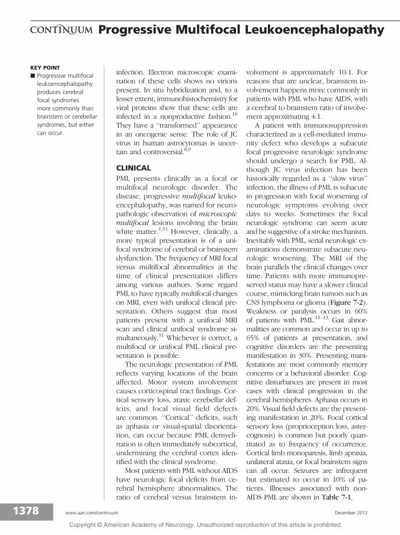
A patient with immunosuppressioncharacterized as a cell-mediated immu-nity defect who develops a subacutefocal progressive neurologic syndromeshould undergo a search for PML. Al-though JC virus infection has beenhistorically regarded as a ‘‘slow virus’’infection, the illness of PML is subacutein progression with focal worsening ofneurologic symptoms evolving overdays to weeks. Sometimes the focalneurologic syndrome can seem acuteand be suggestive of a strokemechanism.Inevitably with PML, serial neurologic ex-aminations demonstrate subacute neu-rologic worsening. The MRI of thebrain parallels the clinical changes overtime. Patients with more immunopre-served status may have a slower clinicalcourse, mimicking brain tumors such asCNS lymphoma or glioma (Figure 7-2).Weakness or paralysis occurs in 60%of patients with PML.11Y13 Gait abnor-malities are common and occur in up to65% of patients at presentation, andcognitive disorders are the presentingmanifestation in 30%. Presenting mani-festations are most commonly memoryconcerns or a behavioral disorder. Cog-nitive disturbances are present in mostcases with clinical progression in thecerebral hemispheres. Aphasia occurs in20%. Visual field defects are the present-ing manifestation in 20%. Focal corticalsensory loss (proprioception loss, aster-eognosis) is common but poorly quan-titated as to frequency of occurrence.Cortical limb monoparesis, limb apraxia,unilateral ataxia, or focal brainstem signscan all occur. Seizures are infrequentbut estimated to occur in 10% of pa-tients. Illnesses associated with non-AIDS PML are shown in Table 7-1.
KEY POINT
h Progressive multifocalleukoencephalopathyproduces cerebralfocal syndromesmore commonly thanbrainstem or cerebellarsyndromes, but eithercan occur.
1378 www.aan.com/continuum December 2012
Progressive Multifocal Leukoencephalopathy
Copyright @ American Academy of Neurology. Unauthorized reproduction of this article is prohibited.

AIDSAIDS is the most common mechanismof immunosuppression leading to PML.It is estimated that PML affects 1% to 4%of symptomatic AIDS cases. PML can bethe presenting manifestation of AIDS.However, more commonly, PML occurswith low CD4 blood counts (less than200 cells/2L) and occurs later in thecourse of AIDS disease. As an oppor-tunistic infection, PML incidence hasbeen less affected by combination anti-retroviral therapy than other opportu-nistic infections of the nervous system,such as toxoplasmosis or cryptococcalmeningitis (Case 7-1).
LYMPHORETICULARMALIGNANCYThe lymphoreticular malignancies arethe most common non-AIDS-relatedcause of immunosuppression predis-posing to PML. The most common dis-orders are chronic lymphocytic leukemia,Hodgkin disease, and non-Hodgkin lym-phoma. It is interesting that chroniclymphocytic leukemia and most non-Hodgkin lymphomas are generally re-garded as neoplasms of B-cell lineage.Yet all of these disorders are well knownto be associated with opportunistic infec-tions that manifest as disorders of T-cellimmune deficiency, such as toxoplasmo-sis or cryptococcal meningitis. Therefore,the precise immune defects that pre-dispose to PML in these malignanciesremain ill defined.
RHEUMATOLOGIC DISORDERSVirtually any form of rheumatologic dis-order has been reported to be associ-ated with PML. Since these autoimmunedisorders are commonly treated withimmunosuppressive agents, it is unclearwhether the underlying disease or thetreatment is primarily responsible forthe predisposition to PML. In a studylooking at underlying disorders in non-AIDS PML (Table 7-1), the frequency of
connective tissue diseasesVrheumatoidarthritis, systemic lupus erythematosus,dermatomyositis, and vasculitis with meth-otrexate and cyclophosphamideVwas16%. Estimates of PML incidence in pa-tients with rheumatoid arthritis suggestthe frequency is low, approximately 0.4per 100,000 discharges.35
TRANSPLANTATIONPML is an important but rare cause ofneurologic disease in transplant recip-ients. A recent study found the esti-mated incidence of PML in patientsreceiving heart and/or lung transplantoccurring approximately 1.24 perevery 1000 transplant person-years.36
Mean onset of PML symptoms aftertransplantation was 17 months. PML ismost often fatal (84% case fatality) inthis population but is compatible withrecipient survival measured in years(3 of 50 patients). The risk of PML likelyexists throughout the entire posttrans-plant period and should be suspected
KEY POINT
h Progressive multifocalleukoencephalopathyprimarily occurs inimmunosuppressedillnesses of T-celldeficit, ie, AIDS orlymphoreticularmalignancy.
FIGURE 7-2 T2-weighted fluid-attenuatedinversion recovery brain MRIscan of progressive multifocal
leukoencephalopathy (PML). Bifrontal PMLlesions including involvement of the corpuscallosum mimicking glioma or lymphoma.
1379Continuum Lifelong Learning Neurol 2012;18(6):1374–1391 www.aan.com/continuum
Copyright @ American Academy of Neurology. Unauthorized reproduction of this article is prohibited.

and quickly diagnosed because tempo-rary reduction of immunosuppressionmay be compatible with PML improve-ment and long-term patient survival.
The case fatality among patientswho develop PML posttransplantationis high, with death occurring within18 months in most cases. Exceptionsto this occur. In one series, three pa-tients were still alive at 13, 44, and155 months following PML symptomonset.36 Among survivors, all patientshad their immunosuppressive drug reg-imen significantly reduced or withdrawnat the time of diagnosis of PML. No
patient survived without immunosup-pressive medication reduction. Graftrejection is of significant concern withimmunosuppressive drug reductionbut consistent with survival in somecases of PML.
MULTIPLE SCLEROSIS TREATEDWITH NATALIZUMABNatalizumab, a monoclonal antibody di-rected against the !4 integrin molecule,was approved by the US Food andDrug Administration in November2004 for treatment of patients with MSafter a controlled trial.37 By February
TABLE 7-1 Progressive Multifocal Leukoencephalopathy Patientsat Mayo Clinic: Non-AIDS Progressive MultifocalLeukoencephalopathy—Associated Diseases (n=58)
b Lymphoreticular Malignancy (n=32, 55%)
Chronic lymphocytic leukemia (n=14, 24%)
Hodgkin disease (n=6, 10%)
Non-Hodgkin lymphoma (n=12, 21%)
b Connective Tissue Diseases (n=9, 16%)
Rheumatoid arthritis (n=3, 5%)
Systemic lupus erythematosus (n=4, 7%)
Dermatomyositis (n=1, 2%)
Vasculitis with methotrexate and cyclophosphamide (n=1, 2%)
b Organ Transplantation (n=4, 7%)
Renal transplantation
Liver transplantation
Heart transplantation
b Granulomatous Disease (n=5, 9%)
Sarcoidosis
b Other (n=4, 7%)
Cirrhosis
Pulmonary fibrosis
Diabetes mellitus, pulmonary histoplasmosis, and prednisone
b ‘‘Normal Aged’’ Patients With No Immunosuppression (n=4, 7%)
Patients aged 66, 76, and 80 years
1380 www.aan.com/continuum December 2012
Progressive Multifocal Leukoencephalopathy
Copyright @ American Academy of Neurology. Unauthorized reproduction of this article is prohibited.

2005, two patients with MS who hadbeen treated with natalizumab therapywere diagnosed with PML, leading to the
withdrawal of natalizumab from the mar-ket. One patient died.24 The secondpatient, with PML proven by brain biopsy,
Case 7-1A 55-year-old right-handed man had received a diagnosis of HIV disease 8 months earlier. He recentlywent through a series of changes in his antiretroviral drug regimen and had persistent nausea despitemultiple changes of his medications. His CD4 count was 386 cells/2L, and HIV serum quantitationwas less than 400 copies of RNA/mL. For 2 months he had unsteadiness of gait and progressive declinein balance, more prominent on the left side and affecting the left lower extremity. Change incontrol of the left upper extremity with the unsteadiness also occurred. In the prior 1 month he alsonoticed some diplopia.
His examination showed that his gait and station were impaired by marked ataxia. He could standindependently only with support. Ataxia was present in the left upper and lower extremities.Nystagmus occurred on lateral gaze and mild conjugate gaze paresis on the gaze to the left.Extraocular movements were otherwise full, and pupils were normal. Diffuse hyperreflexia waspresent, but plantar responses were flexor. The remainder of the neurologic examinationwas unremarkable.
MRI scan of the head showed increased fluid-attenuated inversion recovery (FLAIR) signal involvingthe cerebellar white matter of the left cerebellar hemisphere and extending into the pons through
the middle cerebellar peduncle (Figure 7-3). Theabnormality produced little mass effect. No contrastenhancement was present on postgadolinium images.Spinal fluid protein concentration was 104 mg/dL with2 nucleated cells/2L. CSF Venereal Disease ResearchLaboratory (VDRL) testing was nonreactive. Results fromthe following tests were negative: cryptococcal antigen,cytology, blood serology for toxoplasmosis, and PCR assaysfor toxoplasmosis and Epstein-Barr virus in the spinal fluid.
PCR of spinal fluid for JC virus was ‘‘indeterminate’’positive. Two of four specimens tested positive, withone each of duplicate specimens run twice showing JCvirusYspecific PCR detection. The diagnosis was PML.
Comment. PML in AIDS typically occurs in patientswho have had HIV disease with more severeimmunosuppression. Usually CD4 counts are less than200 cells/2L. However, immunosuppression does notreliably predict pathology, and PML can occur withnormal CD4 counts. The presentation with cerebellaror brainstem disease is more common in AIDS thannon-AIDS cases. PCR testing for JC virus in the spinalfluid of patients with PML has 76% sensitivity,combining a number of studies. A positive PCR findingabolishes the need for brain biopsy. The indeterminateresult is a reflection of the assay operating at the limits
of detection and is a reason why not all patients with PML test positive by this method. The need for brainbiopsy in PCR-negative cases depends on clinical circumstances, including localization of the lesion, socialsituation, and therapeutic options. This patient was treated with cidofovir and significantly improved.34
Whether the improvement was an effect of cidofovir, the immunologic restoration by the cART regimen(which was stable throughout the PML course), or both is open to interpretation.
FIGURE 7-3 T2-weighted fluid-attenuatedinversion recovery brain MRI scanof progressive multifocal
leukoencephalopathy (PML). Left cerebellar andpontine PML lesion.
1381Continuum Lifelong Learning Neurol 2012;18(6):1374–1391 www.aan.com/continuum
Copyright @ American Academy of Neurology. Unauthorized reproduction of this article is prohibited.

had an unstable clinical course. Hedeveloped severe clinical deficits andIRIS when natalizumab was stopped.25
Another case of PML related to natali-zumab was recognized in a patientbeing treated for Crohn disease. Hedied of a progressive syndrome, whichwas initially diagnosed as astrocytoma.A rereview of neuropathology revealedPML.33 The long duration of therapyand prior immunosuppressant expo-sure has been consistent with othercases of PML. In transplantation, wherethe date of immunosuppression can beaccurately determined, PML occurs late(usually longer than 1 year) after im-munosuppressants are introduced.
Since that time, natalizumab has beenreintroduced to the market with a closemonitoring system and a registry ofpatients administered by Biogen Idec,and with data available to physiciansonline for assessment of risk to patients(medinfo.biogenidec.com/medinfo).Several aspects have emerged from thedata about these patients.23 The in-cidence of PML is dependent on theduration of natalizumab use and sub-stantially increased after 24 monthsof use. Incidence of confirmed PML inpatients who have had 24 or moreinfusions of natalizumab is now esti-mated to be one case of PML per 1000patients.23 As of May 1, 2012, Biogen-Idec reported 242 cases of natalizumab-associated PML. Of these, 20% havedied. Those who have survived expe-rienced varying degrees of disability.Seropositivity for JC virus in the bloodincreases incidence of natalizumab-associated PML to as much as oneper 100 patients after 2 years of use.Finally, prior exposure to other immu-nosuppressive drugs also increases therisk of PML (Case 7-2).
OTHER DRUGSOther specific immunosuppressant drugsthat can predispose to PML are predni-
sone, methotrexate, cyclophosphamide,and cyclosporine.26 It is also apparentthat newer immunosuppressives, suchas leflunomide, can predispose to de-veloping PML.38 The exact risk with useof these drugs, such as monoclonal anti-bodies targeting lymphocyte trafficking orantagonizing specific cytokines, remainsto be defined on an individual basis.
Mycophenolate mofetil is used fora variety of B- and T-cell disorders. Itwas initially developed as a posttrans-plant antirejection drug. However, thedrug is used off-label for many otherautoimmune disorders because of itsfavorable side-effect profile. The pres-ence of PML in patients treated withmycophenolate mofetil has been re-ported. Online resources may help withrisk assessment (www.gene.com/gene/products/information/cellcept/), and thissubject has been covered in a recentreview.26
Rituximab is a therapeutic monoclo-nal anti-CD20 antibody that targets Bcells selectively and removes them fromthe circulation. This monoclonal anti-body has been used to treat a varietyof B-cell neoplasms (eg, chronic lym-phocytic leukemia, lymphoma) and auto-immune diseases, specifically rheumatoidarthritis, and off-label for systemic lupuserythematosus. PML has been shown tooccur in rituximab-treated patients.27,39
It is estimated that the risk is approx-imately one per 25,000 patients withrheumatoid arthritis treated with ritux-imab.27 Systemic lupus erythematosusmay have a higher propensity to PML.27
Another monoclonal anti-CD11a anti-body named efalizumab was marketedfor the treatment of refractory psoria-sis. It was withdrawn from the UnitedStates market as of June 2009 becauseof PML occurring in patients treatedwith this drug. The frequency wasestimated to be four of 46,000 patientstreated.40 However, whether the rareoccurrence of a usually fatal neurologic
KEY POINT
h Exogenousimmunosuppressivetreatments such asnatalizumab andrituximab havebeen linked toprogressive multifocalleukoencephalopathyoccurrence.
1382 www.aan.com/continuum December 2012
Progressive Multifocal Leukoencephalopathy
Copyright @ American Academy of Neurology. Unauthorized reproduction of this article is prohibited.

infection should prohibit drugs suchas this from use in all cases is uncertain.
Screening by serologic means al-lows one to determine whether a pa-tient carries latent JC virus.30 SincePML is regarded as a reactivation in-fection, this serologic testing has beenproposed as a screening tool so thatthese immunosuppressives are usedonly with great caution in seropositiveindividuals. No reliable method is avail-able to detect PML before symptoms.A positive JC virus serology predictspredisposition to infection. CSF PCR
techniques or brain biopsy currentlyare the only methods to confirm PML.
SARCOIDOSISSystemic sarcoidosis is associated withdefects in cell and humoral immunity,with a granulomatous pathologic reac-tion. Many reports have been publishedof PML occurring in patients with sar-coidosis. However, it is unclear whetherthe predisposition to PML is part of theprimary disease or a result of the im-munosuppressive treatment often usedchronically to treat the disease, such as
Case 7-2A 54-year-old right-handed woman was diagnosed with MS after experiencing diplopia. She hadan abnormal multifocal MRI scan reported by her local neurologist. Spinal fluid showed absentoligoclonal banding, normal IgG index, normal protein concentration, and 3 white cells. Threeyears later, natalizumab was started for treatment of multiple sclerosis. One year after starting hernatalizumab therapy, she developed progressive weakness of the left hand, which progressed andspread during 1 month so that she essentially had left hemiplegia. Her treatment with natalizumabhad been discontinued at the beginning of her worsening symptoms.
MRI scan of the head showed an enlarging lesionof T2 hyperintensity involving the white matterin the right cerebral hemisphere, extending fromthe deep white matter up to the superficial cortex,and involving the precentral gyrus and premotorsubcortical white matter of the right hemisphere(Figure 7-4). This worsened despite 4 days of IVmethylprednisone 1 g/d.
Spinal fluid testing was repeated and showedglucose concentration of 71 mg/dL, proteinconcentration of 48 mg/dL, and no white blood cells;PCR was positive for JC virus.
Comment. The patient developed PML after startingnatalizumab therapy. A progressive neurologic deficit,with subcortical white matter imaging abnormalitiesthat are worsening subacutely, would strongly suggesta diagnosis of PML. PML needs to be considered withhigh index of suspicion in patients on natalizumabtherapy. The patient received three plasma exchangesto shorten the period in which natalizumab remainedactive. She was at increased risk for developing IRIS,which subsequently needed treatment. IRIS ismore common and more severe in patients withnatalizumab-associated PML than in patients withHIV-associated PML.
FIGURE 7-4 T2-weighted fluid-attenuatedinversion recovery brain MRI scanof progressive multifocal
leukoencephalopathy (PML). Right frontal largePML lesion with tiny left frontal lesions.
1383Continuum Lifelong Learning Neurol 2012;18(6):1374–1391 www.aan.com/continuum
Copyright @ American Academy of Neurology. Unauthorized reproduction of this article is prohibited.

prednisone or methotrexate. Some pa-tients with sarcoidosis who develop PMLare on no active treatment. In eithercase, PML is an infrequent opportunisticinfection in patients with sarcoidosis,although incidence data are lacking. Ina series of non-AIDS PML cases, sarcoid-osis was present in 9% of the patients(Table 7-1).
RADIOLOGIC FEATURESAn MRI scan of the head is more sen-sitive than a CT scan for detecting abnor-malities related to PML (Table 7-2).41 Anormal MRI head scan would argueagainst PML as the cause of neurologicdeficits. Rarely, patients with PML haveclinically detectable neurologic deficitsand no MRI abnormalities. MRI abnor-malities are typically localized to thesubcortical white matter at the gray-white junction (Figure 7-2, Figure 7-4,and Figure 7-5A, B, and C). The lesionsshow increased T2 signal, no or littlemass effect, and minimal or noenhancement after gadolinium admin-istration (Figure 7-5C and D). Thesecharacteristics have exceptions withPML. Any cerebral lobe is potentiallyvulnerable to disease. Some authorshave suggested that occipital PML is
more common.41 Presumably some ofthe variability of the presenting syn-drome represents the neurologic deficiteffect on the patient’s functions of dailyliving. Any area of the white matterareas of the brainstem can be affected.More common is involvement of thecerebellum. A focal abnormality on MRIscan in the brainstem would suggest PMLwhen little mass effect and no contrastenhancement are present.
DIAGNOSTIC TESTSPCR has been used to amplify JC virusDNA from the spinal fluid of patientswith PML. Current studies of JC virusshow that CSF detection is specific forpathologically significant PML.19,20,42,43
All studies have been retrospective anal-yses of spinal fluid specimens of patientswith either pathologically confirmed orclinically suspected PML, based on MRIand coincident underlying immunosup-pressive illness. Sensitivity of detectionis not 100%. Specifically, combined datafrom published studies show that 76%were correctly diagnosed by this assay.It is unclear why false negatives occur.The most likely explanation is low abun-dance of virus DNA in spinal fluid. Otherpossible explanations include storage
KEY POINTS
h Radiologic evaluationshould be done by MRIscan in suspected cases.
h The principal MRIfinding is T2 orfluid-attenuatedinversion recoverysubcortical whitematter demyelination.
h PCR for JC virus inthe spinal fluid is only70% to 75% sensitivein progressive multifocalleukoencephalopathycases.
TABLE 7-2 Brain MRI Findings
b MRI T2 and fluid-attenuated inversion recovery are most sensitive
b Lesions typically localize to subcortical white matter more commonly thanperiventricular regions
b Gadolinium nonenhancing or minimally enhancing (unless immunereconstitution inflammatory syndrome is present)
b Almost always abnormal
b Regional expression:
Frontal equals occipital
Unifocal greater than multifocal
Cerebral greater than brainstem
1384 www.aan.com/continuum December 2012
Progressive Multifocal Leukoencephalopathy
Copyright @ American Academy of Neurology. Unauthorized reproduction of this article is prohibited.

and handling of specimens, small vol-ume of spinal fluid assay, inhibitors inthe spinal fluid, and loss of DNA dur-ing CSF concentration. Combining datafrom studies,19,43,44 the false-positive rateidentifying CSF specimens from AIDSpatients without PML or other controlswas only 2%. Sensitivity of 75% has beenborne out in larger studies.42 If PML issuspected and PCR for JC virus is neg-ative, the recommendation is to repeat
the assay and consider a laboratory capa-ble of detecting low copy numbers ofJC virus DNA.
The other spinal fluid chemistry andcell count findings in patients with PMLare typically nonspecific. Minimal pleocy-tosis occurs, but the finding is usually lessthan 20 cells/2L. More than 20 cells/2Lsuggests the possibility of a different in-fection or restored immunity of the hostwith reaction against JC virus. A spinal
FIGURE 7-5 Brain MRI scan of progressive multifocal leukoencephalopathy (PML). All areT2-weighted fluid-attenuated inversion recovery images except D, which is aT1 postgadolinium image. A, Single superficial subcortical left frontal PML lesion.
B, Multifocal right-greater-than-left subcortical frontal PML lesions. C, D, Symmetric bioccipitalPML lesions that show trace enhancement after gadolinium.
1385Continuum Lifelong Learning Neurol 2012;18(6):1374–1391 www.aan.com/continuum
Copyright @ American Academy of Neurology. Unauthorized reproduction of this article is prohibited.
fluid pleocytosis can be seen when con-trast enhancement increases on thebrain MRI. Pleocytosis correlates withthe neuropathologic finding of perivas-cular lymphocytic cuffing in the brain.Spinal fluid protein is only moderatelyelevated, usually less than 100 mg/dL.Culture for JC virus is unrevealing.
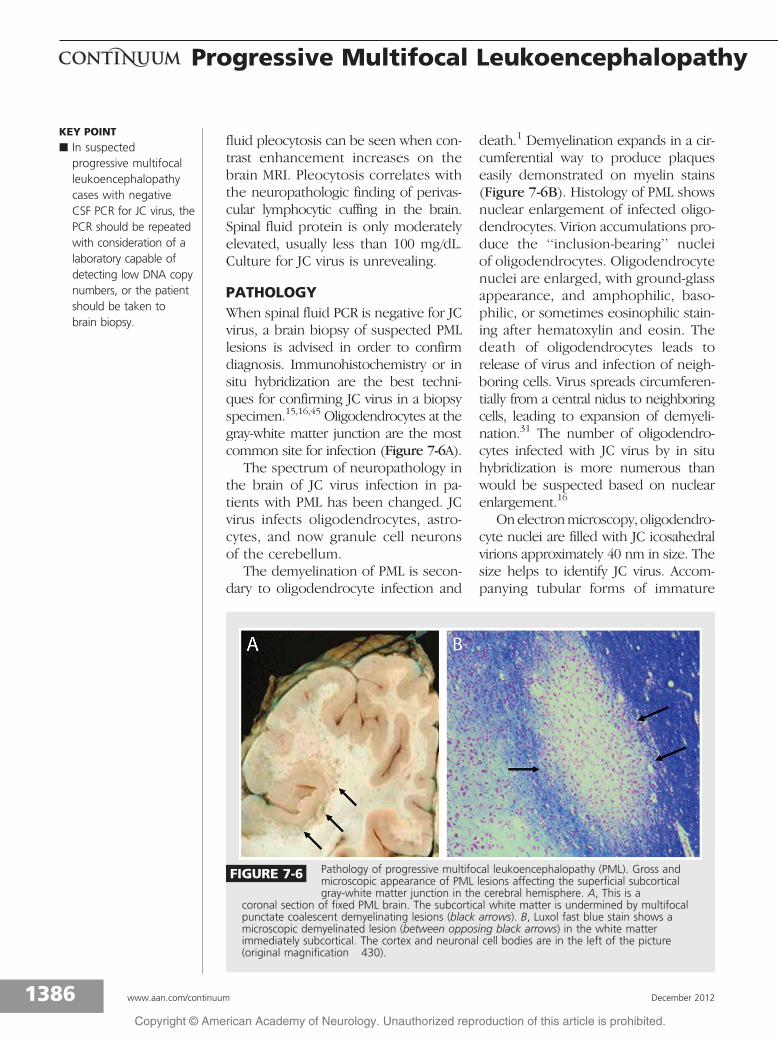
PATHOLOGYWhen spinal fluid PCR is negative for JCvirus, a brain biopsy of suspected PMLlesions is advised in order to confirmdiagnosis. Immunohistochemistry or insitu hybridization are the best techni-ques for confirming JC virus in a biopsyspecimen.15,16,45 Oligodendrocytes at thegray-white matter junction are the mostcommon site for infection (Figure 7-6A).
The spectrum of neuropathology inthe brain of JC virus infection in pa-tients with PML has been changed. JCvirus infects oligodendrocytes, astro-cytes, and now granule cell neuronsof the cerebellum.
The demyelination of PML is secon-dary to oligodendrocyte infection and
death.1 Demyelination expands in a cir-cumferential way to produce plaqueseasily demonstrated on myelin stains(Figure 7-6B). Histology of PML showsnuclear enlargement of infected oligo-dendrocytes. Virion accumulations pro-duce the ‘‘inclusion-bearing’’ nucleiof oligodendrocytes. Oligodendrocytenuclei are enlarged, with ground-glassappearance, and amphophilic, baso-philic, or sometimes eosinophilic stain-ing after hematoxylin and eosin. Thedeath of oligodendrocytes leads torelease of virus and infection of neigh-boring cells. Virus spreads circumferen-tially from a central nidus to neighboringcells, leading to expansion of demyeli-nation.31 The number of oligodendro-cytes infected with JC virus by in situhybridization is more numerous thanwould be suspected based on nuclearenlargement.16
Onelectronmicroscopy, oligodendro-cyte nuclei are filled with JC icosahedralvirions approximately 40 nm in size. Thesize helps to identify JC virus. Accom-panying tubular forms of immature
KEY POINT
h In suspectedprogressive multifocalleukoencephalopathycases with negativeCSF PCR for JC virus, thePCR should be repeatedwith consideration of alaboratory capable ofdetecting low DNA copynumbers, or the patientshould be taken tobrain biopsy.
FIGURE 7-6 Pathology of progressive multifocal leukoencephalopathy (PML). Gross andmicroscopic appearance of PML lesions affecting the superficial subcorticalgray-white matter junction in the cerebral hemisphere. A, This is a
coronal section of fixed PML brain. The subcortical white matter is undermined by multifocalpunctate coalescent demyelinating lesions (black arrows). B, Luxol fast blue stain shows amicroscopic demyelinated lesion (between opposing black arrows) in the white matterimmediately subcortical. The cortex and neuronal cell bodies are in the left of the picture(original magnification �430).
1386 www.aan.com/continuum December 2012
Progressive Multifocal Leukoencephalopathy
Copyright @ American Academy of Neurology. Unauthorized reproduction of this article is prohibited.

capsid protein may assemble in thenucleus.
Bizarre astrocytes are a nonlytic JCvirus infection of astrocytes. These cellslook bizarre with enlarged and multi-lobulated nuclei. They look like neoplas-tic cells but do not form frank tumors.Rare reports of gliomas in PML lesionshave occurred.7 More commonly, thePML can be misdiagnosed as glioma be-cause of the cell atypia.33 Bizarre as-trocytes have limited JC virus DNAreplication by in situ hybridization andviral capsid immunohistochemistry.They do not, however, produce signifi-cant virions based on electron micro-scopic analysis.31,45
Double-labeling techniques haveshown that granule cell neurons of thecerebellum are infectible with JC vi-rus.18,46 Granule cell neurons show lyticinfection and cell death with atrophy ofthe granule cell layer of the cerebellum.46
The pathology is associated with lim-ited or no inflammation. This is regarded
to be proportional to the immune stateof the host. More severe immunosup-pression is associated with little or no in-flammation. Patients have had immunereconstitution and developed IRIS in thenervous system in the context of AIDS.47
TREATMENTNo treatments have proven to be ef-fective for PML (Table 7-3). Generalprinciples about treatment includeimprovement in immune status. Anti-viral therapy may promote survival.48
Therapies can be offered if the goalof neurologic stabilization satisfies thepatient’s quality-of-life goals. Althoughthe prognosis of PML is generallydismal, removal of the immunosup-pression influence of an external drugallows the patient’s own immunesystem to clear JC virus from the brain.This is an effective approach but can alsolead to IRIS, which, when it occurs inthe brain after immune restoration, mayneed treatment. IRIS should be treated if
KEY POINT
h No satisfactory antiviraltherapy is availablefor the treatment ofprogressive multifocalleukoencephalopathy.
TABLE 7-3 Treatment Options
b Immune Reconstitution
Stop immunosuppressants
Optimize antiretroviral therapy (in AIDS)
b Nucleoside Analogs (Questionable Effectiveness)
Cytosine arabinoside 2 mg/kg/d for 5 days, single course
Cidofovir 5 mg/kg once weekly for 2 weeks, then every 2 weeks for 2 months
b Treatments Not Likely to Work
Interferon alpha
Topotecan
5-hydroxytryptamine antagonists
Chlorpromazine
Mirtazapine
Risperidone
Mefloquine
1387Continuum Lifelong Learning Neurol 2012;18(6):1374–1391 www.aan.com/continuum
Copyright @ American Academy of Neurology. Unauthorized reproduction of this article is prohibited.
accompanied by neurologic deteriorationwith short-term corticosteroids.
In patients with AIDS, cART shouldbe initiated. If the patient with AIDS isalready receiving cART, therapy shouldbe changed to optimize immune resto-ration and normalization of the CD4count. Cytosine arabinoside has failedin patients with AIDS-related PML.22
For deteriorating patients with PMLwith or without AIDS, cidofovir can beconsidered,34,49 although several stud-ies have suggested it is ineffective.50,51
For patients with PML without AIDS,no effective therapy is available. If thegoal to stabilize neurologic deteriora-tion is acceptable in the clinical con-text of the systemic disease, however,one may consider IV cytosine arabi-noside 2 mg/kg/d for 5 days. A singlenonblinded study showed an approxi-mately 30% response rate in patientsin whom 85% mortality was expectedwithin 1 year.48
Mirtazapine or risperidone have alsobeen suggested as options for treat-ment, but their effectiveness is notyet proven. The receptor for JC virusentry into the cell is identified as asubtype of the serotonin receptors 5-hydroxytryptamine 2A (5-HT2A).
52 Thisis combined with a sialic acidYN-linkedglycoprotein on the cell membrane. Thisreport has led to treatment of PML withpsychotropic medications known toblock the 5-HT2A receptor. Mirtazapineis an antidepressant, and risperidoneis an antipsychotic proposed to be spe-cific for blockade of this receptor. Mir-tazapine has been used anecdotally at15 mg/d to 30 mg/d, in patients withnonYAIDS-related PML.53,54 Thesepatients also had alteration of theirimmunosuppressive regimen and treat-ment with other agents, including cyto-sine arabinoside or cidofovir. Risperidonehas been suggested to be potentiallymore potent.55 None of this form of ther-apy blocking the serotonin receptor has
been successful, although no blinded pro-spective trials have been published.
Mefloquine has been suggested tobe potentially helpful based on in vitroscreening of compounds with activityagainst JC virus. Mefloquine hydrochlor-ide was used in several cases of rituximab-associated PML.27 However, a recentclinical trial was stopped for lack ofdemonstrable efficacy.56
Interferon alpha has been rarely re-ported to have some success in treat-ing PML; however, the patients weretreated with multiple agents, makingthese reports difficult to interpret.Current consensus is that interferonalpha is not helpful in the treatment ofPML.18 Interferon beta also seems to beof no help.57 Camptothecin and top-otecan, two DNA topoisomerase inhib-itor drugs, have antiviral activity againstJC virus. These drugs are antineoplasticagents. Few case reports of treating PMLhave been published.58 No series of PMLpatients treated suggest that these drugsare successful.
IMMUNE RECONSTITUTIONINFLAMMATORY SYNDROMEIRIS can occur after withdrawal of theimmunosuppressive agent and afterPML diagnosis. The immune responseis important in clearing JC virus fromthe brain. Therefore, at least limitedinflammation is probably important inneurologic survival.28 IRIS, however, isthought to be injurious to the brain,and many patients need additional treat-ment. Treatment is typically a shortcourse of high-dose IV methylpredniso-lone, usually 1 g/d for 5 days.29 How-ever, universal consensus is lacking onroute, type, dose, or duration of corti-costeroid therapy. Despite treatmentsuccess and survival, PML deficits canbe expected to be permanent.
In the circumstance of natalizumab-associated PML, management of thePML has routinely used plasma exchange
KEY POINTS
h Patients withprogressive multifocalleukoencephalopathyassociated withAIDS should haveoptimization ofantiretroviral therapy.
h Immune reconstitutioninflammatory syndromeshould be treated ifit is associated withsignificant neurologicdeterioration.
1388 www.aan.com/continuum December 2012
Progressive Multifocal Leukoencephalopathy
Copyright @ American Academy of Neurology. Unauthorized reproduction of this article is prohibited.
or immunoabsorption to hasten clear-ance of the drug and shorten the pe-riod in which natalizumab remainsactive (usually several months). Exac-erbation of symptoms and enlarge-ment of lesions on MRI have occurredwithin a few days to a few weeks afterplasma exchange, indicative of IRIS.This syndrome seems to be morecommon and more severe in patientswith natalizumab-associated PML thanin patients with HIV-associated PMLtreated with cART.
PROGNOSISIn general, PML has been regarded asnearly a universally fatal disease. How-ever, more recent experience with PMLsuggests that patients can survive. A pre-AIDS era study showed the 4-month sur-vival rate to be 30%, and the 12-monthsurvival rate 15%.13 In the pre-cART era,AIDS-related PML was fatal in 95% ofpatients in 6 months.46 Institution ofoptimized cART therapy has pro-duced 50% survival at 1 year.50 Nata-lizumab-associated PML has had ahigher survival rate of 80% (medinfo.biogenidec.com/medinfo), although PML-associated deficits are expected to bepermanent. These patients usually alsorequire treatment of IRIS.
REFERENCES1. Astrom KE, Mancall EL, Richardson EP Jr.
Progressive multifocal leuko-encephalopathy;a hitherto unrecognized complication ofchronic lymphatic leukaemia and Hodgkin’sdisease. Brain 1958;81(1):93Y111.
2. Richardson EP Jr. Progressive multifocalleukoencephalopathy. N Engl J Med1961;265:815Y823.
3. Zurhein G, Chou SM. Particles resemblingpapova viruses in human cerebraldemyelinating disease. Science1965;148(3676):1477Y1479.
4. Padgett BL, Walker DL, ZuRhein GM, et al.Cultivation of papovalike virus fromhuman brain with progressive multifocalleucoencephalopathy. Lancet1971;1(7712):1257Y1260.
5. Walker D, Frisque R. The biologyand molecular biology of JC virus. In:Salzman NP, ed. The papovaviridae.New York: Plenum Press, 1986.
6. Weiner LP, Herndon RM, Narayan O, et al.Isolation of virus related to SV40 frompatients with progressive multifocalleukoencephalopathy. N Engl J Med1972;286(8):385Y390.
7. Castaigne P, Rondot P, Escourolle R,et al. [Progressive multifocalleukoencephalopathy and multiplegliomas.] Rev Neurol (Paris)1974;130(9Y10):379Y392.
8. Del Valle L, Azizi SA, Krynska B, et al.Reactivation of human neurotropicJC virus expressing oncogenic proteinin a recurrent glioblastoma multiforme.Ann Neurol 2000;48(6):932Y936.
9. Major EO. From telomeres to T-antigens:many roadsImultiple pathwaysInovelassociations in the search for the origins ofhuman gliomas. Ann Neurol2000;48(6):823Y825.
10. Major EO, Amemiya K, Tornatore CS,et al. Pathogenesis and molecular biology ofprogressive multifocal leukoencephalopathy,the JC virus-induced demyelinating diseaseof the human brain. Clin Microbiol Rev1992;5(1):49Y73.
11. Berenguer J, Miralles P, Arrizabalaga J, et al.Clinical course and prognostic factors ofprogressive multifocal leukoencephalopathyin patients treated with highly activeantiretroviral therapy. Clin Infect Dis2003;36(8):1047Y1052.
12. Berger JR, Pall L, Lanska D, Whiteman M.Progressive multifocal leukoencephalopathyin patients with HIV infection. J Neurovirol1998;4(1):59Y68.
13. Brooks BR, Walker DL. Progressivemultifocal leukoencephalopathy.Neurol Clin 1984;2(2):299Y313.
14. Frisque RJ, Bream GL, Cannella MT.Human polyomavirus JC virus genome.J Virol 1984;51(2):458Y469.
15. Aksamit AJ, Mourrain P, Sever JL,Major EO. Progressive multifocalleukoencephalopathy: investigation ofthree cases using in situ hybridizationwith JC virus biotinylated DNA probe.Ann Neurol 1985;18(4):490Y496.
16. Aksamit AJ, Sever JL, Major EO. Progressivemultifocal leukoencephalopathy: JC virusdetection by in situ hybridization comparedwith immunohistochemistry. Neurology1986;36(4):499Y504.
KEY POINT
h Patients withnatalizumab-associatedprogressive multifocalleukoencephalopathyshould have plasmaexchange to speedremoval of the drug.
1389Continuum Lifelong Learning Neurol 2012;18(6):1374–1391 www.aan.com/continuum
Copyright @ American Academy of Neurology. Unauthorized reproduction of this article is prohibited.

17. Houff SA, Major EO, Katz DA, et al.Involvement of JC virus-infectedmononuclear cells from the bonemarrow and spleen in the pathogenesisof progressive multifocal leukoencephalopathy.N Engl J Med 1988;318(5):301Y305.
18. Koralnik IJ. Progressive multifocalleukoencephalopathy revisited: has thedisease outgrown its name? Ann Neurol2006;60(2):162Y173.
19. McGuire D, Barhite S, Hollander H, Miles M.JC virus DNA in cerebrospinal fluid of humanimmunodeficiency virus-infected patients:predictive value for progressive multifocalleukoencephalopathy. Ann Neurol1995;37(3):395Y399.
20. Telenti A, Marshall WF, Aksamit AJ, et al.Detection of JC virus by polymerasechain reaction in cerebrospinal fluid fromtwo patients with progressive multifocalleukoencephalopathy. Eur J Clin MicrobiolInfect Dis 1992;11(3):253Y254.
21. Ryschkewitsch CF, Jensen PN, Monaco MC,Major EO. JC virus persistence followingprogressive multifocal leukoencephalopathyin multiple sclerosis patients treated withnatalizumab. Ann Neurol2010;68(3):384Y391.
22. Hall CD, Dafni U, Simpson D, et al. Failureof cytarabine in progressive multifocalleukoencephalopathy associated withhuman immunodeficiency virus infection.AIDS Clinical Trials Group 243 Team.N Engl J Med 1998;338(19):1345Y1351.
23. Clifford DB, De Luca A, Simpson DM, et al.Natalizumab-associated progressivemultifocal leukoencephalopathy inpatients with multiple sclerosis: lessons from28 cases. Lancet Neurol 2010;9(4):438Y446.
24. Kleinschmidt-DeMasters BK, Tyler KL.Progressive multifocal leukoencephalopathycomplicating treatment with natalizumaband interferon beta-1a for multiple sclerosis.N Engl J Med 2005;353(4):369Y374.
25. Langer-Gould A, Atlas SW, Green AJ, et al.Progressive multifocal leukoencephalopathyin a patient treated with natalizumab. NEngl J Med 2005;353(4):375Y381.
26. Berger JR, Houff S. Opportunistic infectionsand other risks with newer multiple sclerosistherapies. Ann Neurol 2009;65(4):367Y377.
27. Clifford DB, Ances B, Costello C, et al.Rituximab-associated progressive multifocalleukoencephalopathy in rheumatoidarthritis. Arch Neurol 2011;68(9):1156Y1164.
28. Berger JR. Steroids for PML-IRIS: adouble-edged sword? Neurology2009;72(17):1454Y1455.
29. Tan K, Roda R, Ostrow L, et al. PML-IRISin patients with HIV infection: clinicalmanifestations and treatment with steroids.Neurology 2009;72(17):1458Y1464.
30. Bozic C, Richman S, Plavina T, et al.Anti-John Cunningham virus antibodyprevalence in multiple sclerosis patients:baseline results of STRATIFY-1. Ann Neurol2011;70(5):742Y750.
31. Aksamit AJ Jr. Progressive multifocalleukoencephalopathy: a review of thepathology and pathogenesis. Microsc ResTech 1995;32(4):302Y311.
32. Major EO, Amemiya K, Elder G, Houff SA.Glial cells of the human developingbrain and B cells of the immune systemshare a common DNA binding factor forrecognition of the regulatory sequencesof the human polyomavirus, JCV.J Neurosci Res 1990;27(4):461Y471.
33. Van Assche G, Van Ranst M, Sciot R, et al.Progressive multifocal leukoencephalopathyafter natalizumab therapy for Crohn’sdisease. N Engl J Med 2005;353(4):362Y368.
34. Razonable RR, Aksamit AJ, Wright AJ,Wilson JW. Cidofovir treatment ofprogressive multifocal leukoencephalopathyin a patient receiving highly activeantiretroviral therapy. Mayo Clin Proc2001;76(11):1171Y1175.
35. Calabrese LH, Molloy ES. Progressivemultifocal leucoencephalopathy in therheumatic diseases: assessing the risks ofbiological immunosuppressive therapies.Ann Rheum Dis 2008;67(suppl 3):iii64Yiii65.
36. Mateen FJ, Muralidharan R, Carone M,et al. Progressive multifocalleukoencephalopathy in transplantrecipients. Ann Neurol 2011;70(2):305Y322.
37. Rudick RA, Stuart WH, Calabresi PA, et al.Natalizumab plus interferon beta-1a forrelapsing multiple sclerosis. N Engl J Med2006;354(9):911Y923.
38. Rahmlow M, Shuster EA, Dominik J, et al.Leflunomide-associated progressivemultifocal leukoencephalopathy.Arch Neurol 2008;65(11):1538Y1539.
39. Carson KR, Evens AM, Richey EA, et al.Progressive multifocal leukoencephalopathyafter rituximab therapy in HIV-negativepatients: a report of 57 cases from theResearch on Adverse Drug Events andReports Project. Blood 2009;113(20):4834Y4840.
40. Korman BD, Tyler KL, Korman NJ.Progressive multifocal leukoencephalopathy,efalizumab, and immunosuppression: a
1390 www.aan.com/continuum December 2012
Progressive Multifocal Leukoencephalopathy
Copyright @ American Academy of Neurology. Unauthorized reproduction of this article is prohibited.

cautionary tale for dermatologists. ArchDermatol 2009;145(8):937Y942.
41. Whiteman ML, Post MJ, Berger JR, et al.Progressive multifocal leukoencephalopathyin 47 HIV-seropositive patients: neuroimagingwith clinical and pathologic correlation.Radiology 1993;187(1):233Y240.
42. Bossolasco S, Calori G, Moretti F, et al.Prognostic significance of JC virus DNAlevels in cerebrospinal fluid of patientswith HIV-associated progressive multifocalleukoencephalopathy. Clin Infect Dis2005;40(5):738Y744.
43. Weber T, Turner RW, Frye S, et al.Progressive multifocal leukoencephalopathydiagnosed by amplification of JCvirus-specific DNA from cerebrospinalfluid. AIDS 1994;8(1):49Y57.
44. Aksamit AJ. Cerebrospinal fluid in thediagnosis of central nervous system infections.In: Roos KL, ed. Central nervous systeminfectious diseases and therapy. New York,NY: Marcel Dekker, Inc, 1997:731Y745.
45. Aksamit AJ Jr. Nonradioactive in situhybridization in progressive multifocalleukoencephalopathy. Mayo Clin Proc1993;68(9):899Y910.
46. Du Pasquier RA, Corey S, Margolin DH,et al. Productive infection of cerebellargranule cell neurons by JC virus in anHIV+ individual. Neurology2003;61(6):775Y782.
47. Clifford DB, Yiannoutsos C, Glicksman M,et al. HAART improves prognosis inHIV-associated progressive multifocalleukoencephalopathy. Neurology1999;52(3):623Y625.
48. Aksamit AJ. Treatment of non-AIDSprogressive multifocal leukoencephalopathywith cytosine arabinoside. J Neurovirol2001;7(4):386Y390.
49. Viallard JF, Lazaro E, Ellie E, et al.Improvement of progressive multifocalleukoencephalopathy after cidofovirtherapy in a patient with a destructive
polyarthritis. Infection 2007;35(1):33Y36.
50. De Luca A, Ammassari A, Pezzotti P,et al. Cidofovir in addition toantiretroviral treatment is not effectivefor AIDS-associated progressive multifocalleukoencephalopathy: a multicohortanalysis. AIDS 2008;22(14):1759Y1767.
51. Marra CM, Rajicic N, Barker DE, et al.A pilot study of cidofovir for progressivemultifocal leukoencephalopathy in aids.AIDS 2002;16(13):1791Y1797.
52. Elphick GF, Querbes W, Jordan JA, et al.The human polyomavirus, JCV, usesserotonin receptors to infect cells.Science 2004;306(5700):1380Y1383.
53. Owczarczyk K, Hilker R, Brunn A, et al.Progressive multifocal leucoencephalopathyin a patient with sarcoidosisVsuccessfultreatment with cidofovir and mirtazapine.Rheumatology (Oxford) 2007;46(5):888Y890.
54. Vulliemoz S, Lurati-Ruiz F, Borruat FX,et al. Favourable outcome of progressivemultifocal leucoencephalopathy in twopatients with dermatomyositis. J NeurolNeurosurg Psychiatry 2006;77(9):1079Y1082.
55. Kast RE, Focosi D, Petrini M, Altschuler EL.Treatment schedules for 5-HT2a blocking inprogressive multifocal leukoencephalopathyusing risperidone or ziprasidone. BoneMarrow Transplant 2007;39(12):811Y812.
56. Clifford D, Nath A, Cinque P, et al.Mefloquine treatment in patients withprogressive multifocal leukoencephalopathy.Neurology 2011;76:A28.
57. Nath A, Venkataramana A, Reich DS, et al.Progression of progressive multifocalleukoencephalopathy despite treatmentwith beta-interferon. Neurology2006;66(1):149Y150.
58. Royal W 3rd, Dupont B, McGuire D, et al.Topotecan in the treatment of acquiredimmunodeficiency syndrome-relatedprogressive multifocal leukoencephalopathy.J Neurovirol 2003;9(3):411Y419.
1391Continuum Lifelong Learning Neurol 2012;18(6):1374–1391 www.aan.com/continuum
Copyright @ American Academy of Neurology. Unauthorized reproduction of this article is prohibited.