promoting fracture healing - asht 2017_htrc... · fractures rebecca j saunders pt/cht curtis...
TRANSCRIPT

Fractures
Rebecca J Saunders PT/CHT
Curtis National Hand CenterBaltimore, MD
October 6-8, 2017
Promoting fracture healingOnce anatomic reduction takes
place there is a balancing act between providing stability and mobility
Joints need to be splinted in a “close‐pack” position to maintain ligament length and provide maximal congruency
Distal joint motion encourages tendon gliding while decreasing edema and scarring
Epiphysis Physis Metaphysis Diaphysis Periosteum Endosteum
Cortical bone – compact bone◦ Concentrated in diaphysis of long bone◦ Houses osteons (contain osteocytes)◦ 80% of skeletal mass
Cancellous bone – trabecular or spongy) ◦ 20% skeletal mass◦ Concentrated at epiphysis and
metaphysis of long bones◦ Metabolic turnover is greater than for
cortical; usually faster healing
Energy absorbed by bone
mechanical & structural failure
Loss of Continuity of Bone
vascular disruption at fracture site
soft tissue injury

Location in Bone◦Diaphyseal◦Metaphyseal◦Articular
Depth of fracture: complete, incomplete Angle: transverse, oblique, spiral, longitudinal, Complexity: simple, comminuted or crushed Closed versus Open Intra articular vs. extra articular Avulsion
Inflammatory Phase (0-2 weeks)◦ Accumulation of a hematoma between fracture ends under elevated periosteum
◦ Bone necrosis (osteocytes lose nutrition)◦ Proliferation of fibroblasts and osteoblasts◦ Invasion of leukocytes and macrophages
Hematoma organizes forming fibrin scaffold
External carilagenous callous forms from periosteum
Internal callous forms from endosteum
Gradual increase in stability toward clinical union
New bone and oteogenic cells bridge fracture site
Occurs over prolonged period of time
Process of continuous bone resorption and formation
Bone is remodeled as oteoclasts reabsorb callous
Can be influenced by stress
Primary Healing: ◦ No Callus
formation◦ Direct apposition
of bone ends with compression◦ Rigid fixation
(substitute for callus)
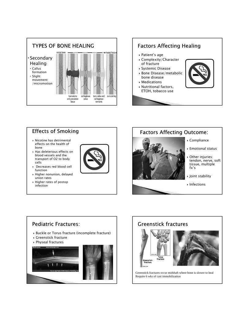
Secondary Healing: Callus
formation Slight
movement /micromotion
Patient’s age Complexity/Character
of fracture Systemic Disease Bone Disease/metabolic
bone disease Medications Nutritional factors,
ETOH, tobacco use
Nicotine has detrimental effects on the health of bone
Has deleterious effects on blood vessels and the transport of O2 to body cells
Decreases red blood cell function
Higher nonunion, delayed union rates
Higher rates of postop infection
Compliance
Emotional status
Other injuries: tendon, nerve, soft tissue, multiple fx’s
Joint stability
Infections
Buckle or Torus fracture (incomplete fracture) Greenstick fracture Physeal fractures
Greenstick fractures occur midshaft where bone is slower to healRequire 6 wks of cast immobilization
Pediatric Fractures
(http://davidlnelson.md/articles/Fracture_Physeal_Fx.htm)
Salter –Harris Fracture Classification system
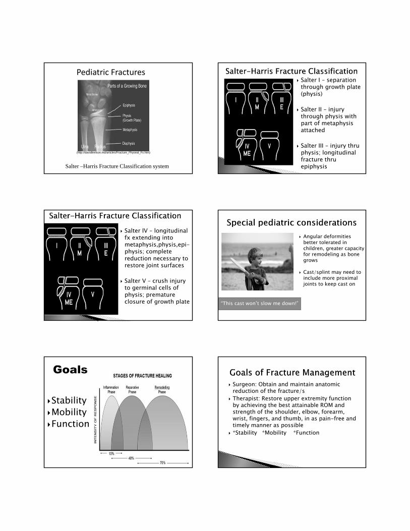
Salter I – separation through growth plate (physis)
Salter II – injury through physis with part of metaphysis attached
Salter III – injury thru physis; longitudinal fracture thru epiphysis
Salter IV – longitudinal fx extending into metaphysis,physis,epi-physis; complete reduction necessary to restore joint surfaces
Salter V – crush injury to germinal cells of physis; premature closure of growth plate “This cast won’t slow me down!”
Angular deformities better tolerated in children, greater capacity for remodeling as bone grows
Cast/splint may need to include more proximal joints to keep cast on
StabilityMobilityFunction
Surgeon: Obtain and maintain anatomic reduction of the fracture/s
Therapist: Restore upper extremity function by achieving the best attainable ROM and strength of the shoulder, elbow, forearm, wrist, fingers, and thumb, in as pain-free and timely manner as possible
*Stability *Mobility *Function
Therapists should have understanding of:◦ Fracture stability/alignment◦ Operative procedures performed◦ Potential dysfunction at uninvolved joints◦ Appropriate timing for ROM, splinting,
strengthening◦ Balancing act between protection and applying
controlled stress for motion and strength
◦ ***Motion or stress to the bone promotes bone healing.
Edema control Pain management
Protection AROM PROM Sensory eval and desens.
Functional activities
Strengthening
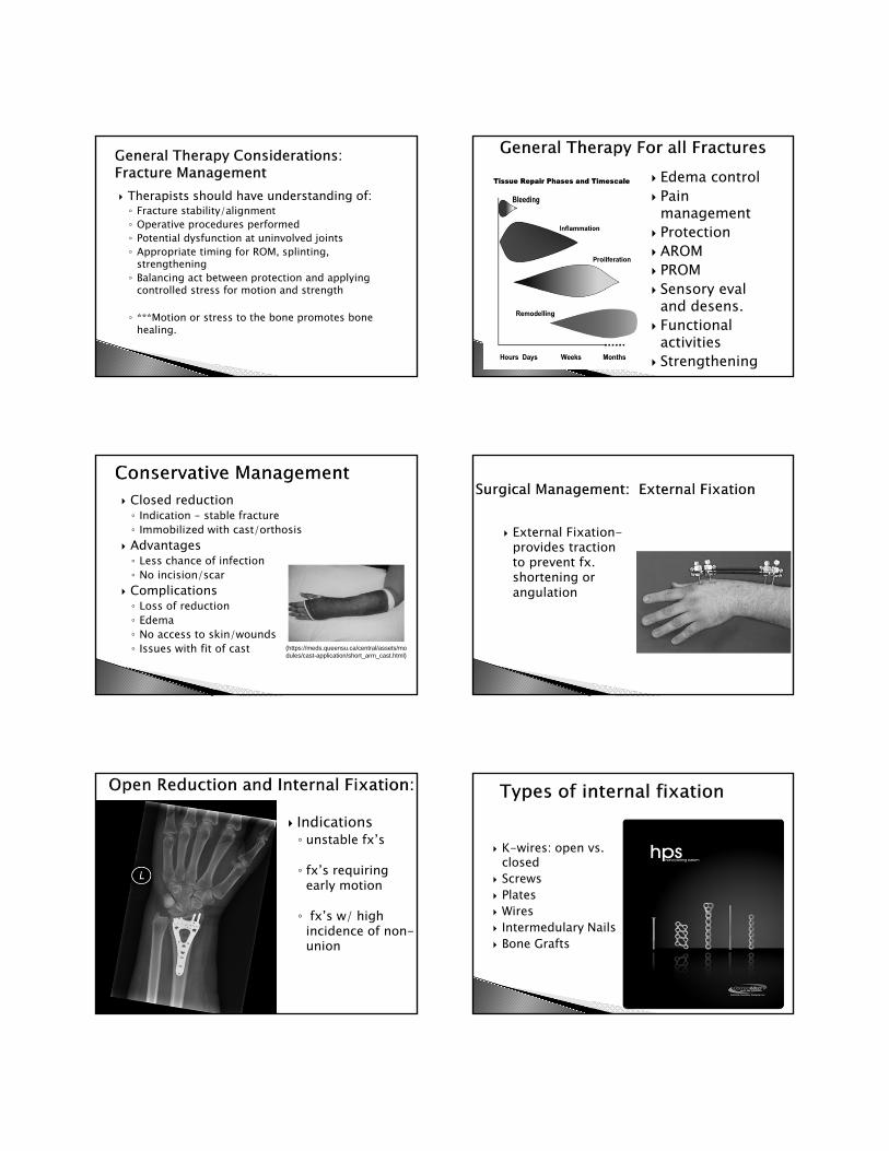
Closed reduction◦ Indication - stable fracture◦ Immobilized with cast/orthosis
Advantages◦ Less chance of infection◦ No incision/scar
Complications◦ Loss of reduction◦ Edema◦ No access to skin/wounds◦ Issues with fit of cast (https://meds.queensu.ca/central/assets/mo
dules/cast-application/short_arm_cast.html)
External Fixation-provides traction to prevent fx. shortening or angulation
Indications◦ unstable fx’s
◦ fx’s requiring early motion
◦ fx’s w/ high incidence of non-union
K-wires: open vs. closed
Screws Plates Wires Intermedulary Nails Bone Grafts
Soft tissue injury
Periosteal stripping
Hardware irritation
Adhesions
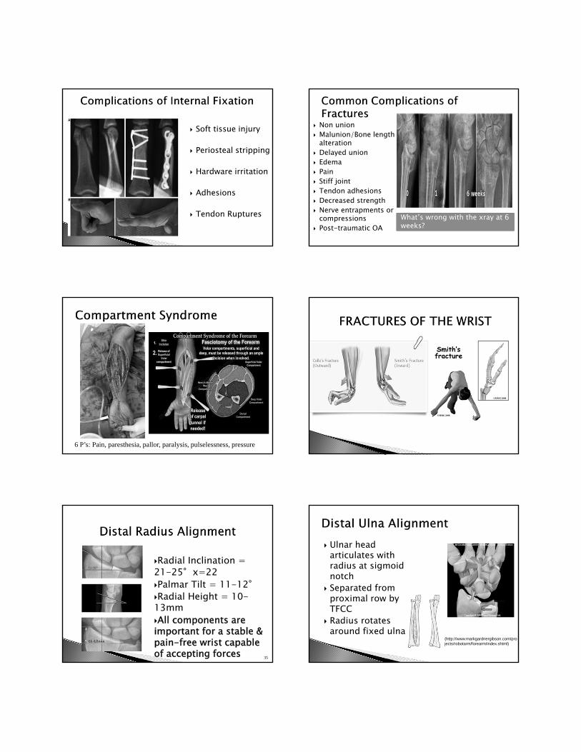
Tendon Ruptures What’s wrong with the xray at 6 weeks?
Non union Malunion/Bone length
alteration Delayed union Edema Pain Stiff joint Tendon adhesions Decreased strength Nerve entrapments or
compressions Post-traumatic OA
6 P’s: Pain, paresthesia, pallor, paralysis, pulselessness, pressure
Radial Inclination = 21-25°x=22 Palmar Tilt = 11-12°Radial Height = 10-13mm All components are important for a stable & pain-free wrist capable of accepting forces 35
Ulnar head articulates with radius at sigmoid notch
Separated from proximal row by TFCC
Radius rotates around fixed ulna
(http://www.markgardnergibson.com/projects/robotarm/forearm/index.shtml)
Reduction in distal radius length alters the force distribution through the radius and the ulna to the proximal row.◦ Normal 80% radius and 20% ulna◦ The ulnar side of wrist is not intended to be the
primary weight-bearing joint ◦ Positive ulnar variance has a greater chance of TFCC
injuries ◦ Ulnar sided wrist pain
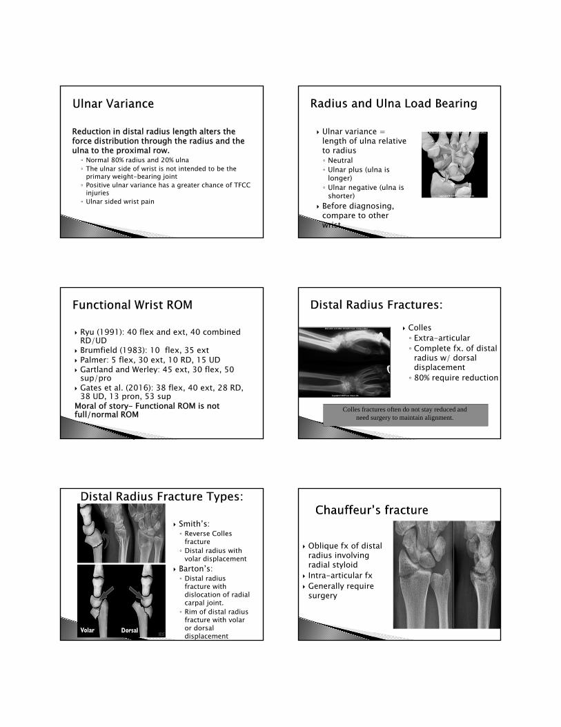
Ulnar variance = length of ulna relative to radius◦ Neutral◦ Ulnar plus (ulna is
longer)◦ Ulnar negative (ulna is
shorter) Before diagnosing,
compare to other wrist
Ryu (1991): 40 flex and ext, 40 combined RD/UD
Brumfield (1983): 10 flex, 35 ext Palmer: 5 flex, 30 ext, 10 RD, 15 UD Gartland and Werley: 45 ext, 30 flex, 50
sup/pro Gates et al. (2016): 38 flex, 40 ext, 28 RD,
38 UD, 13 pron, 53 supMoral of story- Functional ROM is not full/normal ROM
Colles◦ Extra-articular◦ Complete fx. of distal
radius w/ dorsal displacement◦ 80% require reduction
Colles fractures often do not stay reduced and need surgery to maintain alignment.
Smith’s: ◦ Reverse Colles
fracture◦ Distal radius with
volar displacement Barton’s: ◦ Distal radius
fracture with dislocation of radial carpal joint.◦ Rim of distal radius
fracture with volar or dorsal displacement
Oblique fx of distal radius involving radial styloid
Intra-articular fx Generally require
surgery
Fracture stability/alignment – access x-rays as able Operative procedures performed – access op report Potential dysfunction at uninvolved joints Appropriate timing for ROM, orthoses, and
strengthening Balancing act between protection and applying
controlled stress for motion and strength◦ Motion or stress to the bone promotes bone healing
Importance of good patient history, understanding patient, and empowering through education
Orthoses for stability, protection◦ Cross-education during immobilization phase
Edema control Pain management Range of motion – shoulder to fingers Reestablish independent wrist extension Sensorimotor training Restore function Patient education – anatomy and pathology, help
patients accept responsibility for their part in the rehab process
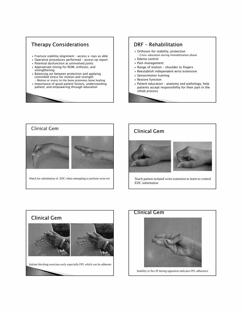
Watch for substitution of EDC when attempting to perform wrist ext Teach patient isolated wrist extension to learn to control EDC substitution
Initiate blocking exercises early especially FPL which can be adherent
Inability to flex IP during oppostion indicates FPL adherence
Strengthening FES – mm. weakness
and retraining Dynamic splinting Work
simulation/hardening Activity Modification
Stiffness, swelling, pain Malunion/radial shortening DRUJ dysfunction RSD/CRPS Median nerve compression Radiocarpal arthritis Weakness Carpal instability Shoulder stiffness
Radial shortening is the most common, disabling deformity◦ Slight shortening
changes axial forces across wrist◦ > 6mm affects flexion,
UD, and pronation, and grip ◦ < 6mm affects forearm
rotation◦ Dorsal tilt >10°
decreases wrist flexion(http://www.handtoelbow.com/images/photos/large/u
lna1.jpg)
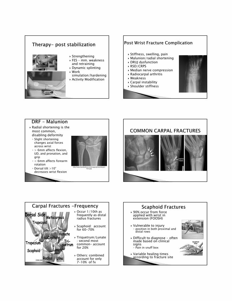
Occur 1/10th as frequently as distal radius fractures
Scaphoid: account for 60-70%
Triquetrum/Lunate: second most common- account for 20%
Others: combined account for only 7-10% of fx
90% occur from force applied with wrist in extension (FOOSH)
Vulnerable to injury ◦ position in both proximal and
distal rows
Difficult to diagnose - often made based on clinical signs◦ Pain in snuff box
Variable healing times according to fracture site
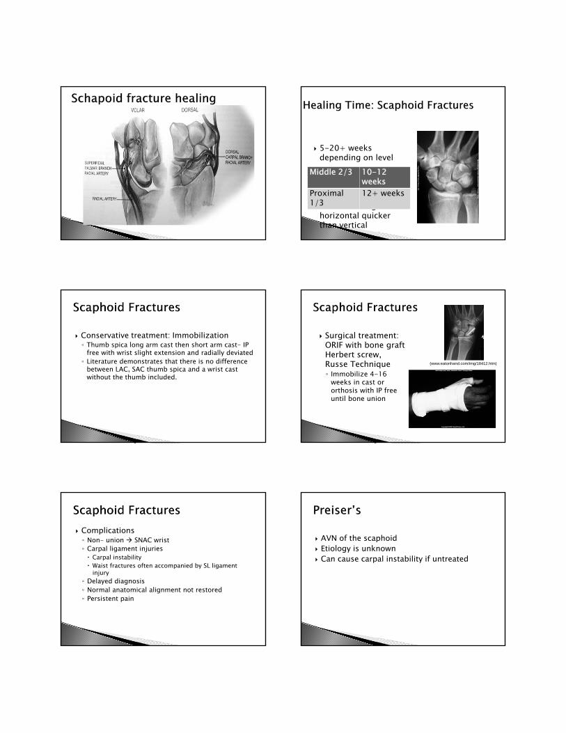
5-20+ weeks depending on level
◦ poor vascular supply Direction of fracture
affects healing-horizontal quicker than vertical
Middle 2/3 10-12 weeks
Proximal 1/3
12+ weeks
Conservative treatment: Immobilization◦ Thumb spica long arm cast then short arm cast- IP
free with wrist slight extension and radially deviated◦ Literature demonstrates that there is no difference
between LAC, SAC thumb spica and a wrist cast without the thumb included.
Surgical treatment: ORIF with bone graft Herbert screw, Russe Technique◦ Immobilize 4-16
weeks in cast or orthosis with IP free until bone union
(www.eatonhand.com/img/18412.htm)
Complications◦ Non- union SNAC wrist◦ Carpal ligament injuries Carpal instability Waist fractures often accompanied by SL ligament
injury◦ Delayed diagnosis◦ Normal anatomical alignment not restored◦ Persistent pain
AVN of the scaphoid Etiology is unknown Can cause carpal instability if untreated

AVN of lunate Associated with negative
ulnar variance Varying degrees of
disability – central wrist pain, limited extension, weak grip
Point tenderness over dorsal aspect of lunate
Immobilization leveling and/or vascularization procedures salvage procedures
Non- union Carpal ligament
injuries◦ Carpal instability
Delayed diagnosis Normal anatomical
alignment not restored
Persistent pain SLAC wrist
SLAC WristS scaphoidL lunateA advanced C collapse
Triquetrum: 2nd most common. FOOSH Lunate: Can result in avascular necrosis
(Kienbocks disease) Trapezium: often associated with
fractures/dislocations involving thumb Pisiform/Hamate: trauma over ulnar/volar
wrist, proximal hypothenar eminence Capitate: rare because this carpal is in a
centrally located and protected position Trapezoid: rare; crush or high energy impact
EDEMA CONTROL Splinting for stability and
protection Motion Modalities Pain management-watch for
increased sympathetic reaction, TENS
Function
Focus on wrist and thumb ROM, and composite flexion
May require protective splint between exercise post cast removal
Functional ADL’s encouraged
Static progressive or dynamic splinting for wrist flexion and extension or composite motion
Grip strengthening Wrist strengthening within pain free range Work hardening depending on RTW status
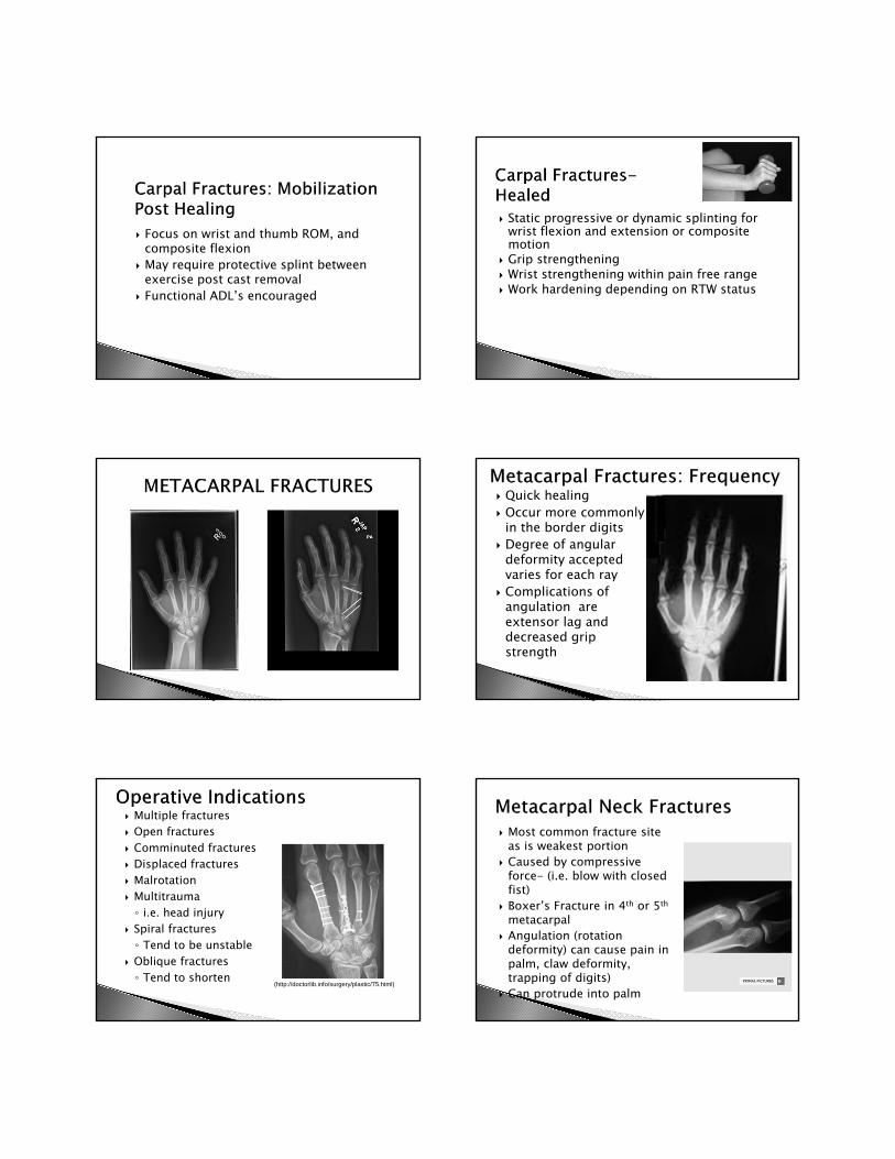
Quick healing Occur more commonly
in the border digits Degree of angular
deformity accepted varies for each ray
Complications of angulation are extensor lag and decreased grip strength
Multiple fractures Open fractures Comminuted fractures Displaced fractures Malrotation Multitrauma◦ i.e. head injury
Spiral fractures◦ Tend to be unstable
Oblique fractures◦ Tend to shorten
(http://doctorlib.info/surgery/plastic/75.html)
Most common fracture site as is weakest portion
Caused by compressive force- (i.e. blow with closed fist)
Boxer’s Fracture in 4th or 5th
metacarpal Angulation (rotation
deformity) can cause pain in palm, claw deformity, trapping of digits)
Can protrude into palm
If including the MP’s in the orthosis, the MP’s need to be immobilized in flexion to keep the collateral ligaments lengthened
Goal is to achieve alignment without rotation or shortening
Shortening of 3-5 mm can produce intrinsic/extrinsic imbalance
Malrotation= finger overlap
Angulation tolerated better in 4/5th MC
(Cindy Glaenzer PT, CHT)
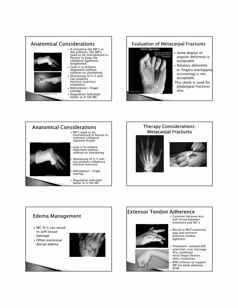
Some degree of angular deformity is acceptable
Rotatory deformityie: fingers overlapping(scissoring) is not acceptable
This check is used for phalangeal fractures also
MP’s need to be immobilized in flexion to maintain collateral ligament length
Goal is to achieve alignment without rotation or shortening
Shortening of 3-5 mm can produce imbalance intrinsic/extrinsic
Malrotation= finger overlap
Angulation tolerated better in 4/5th MC
MC fx’s can result in soft tissue damage
Often excessive dorsal edema
Common because less soft tissue between extensors and MC’s
Result in MCP extension lags and extrinsic extensor tendon tightness
Treatment: isolated EDC exercises, scar massage, FES, combined wrist/finger flexion, other modalities
RME orthosis to support MP ext while allowing ROM

MP joint contractures can occur following metacarpal head and neck fractures
Proper positioning in splint crucial, at least 60⁰ MP flexion; if unable to obtain correct position readjust in a few days
“Hand Fractures can be complicated by deformity from no treatment, stiffness from overtreatment, and both deformity and stiffness from poor treatment”
Alfred Swanson
Type varies◦ Oblique, transverse,
spiral
Spiral/oblique tend to be unstable
Often cause soft tissue adherence
Comminution = more soft tissue damage
Reduction and immobilization
Remember that the Extensor tendons and FDS/FDP are in close proximity to the proximal phalanx tendon adhesions easily develop
Surgery:◦ Percutaneous Pins◦ Screws◦ Plates
Can start motion earlier due to stability of fixation
Addition of scar management
Soft tissue involvement
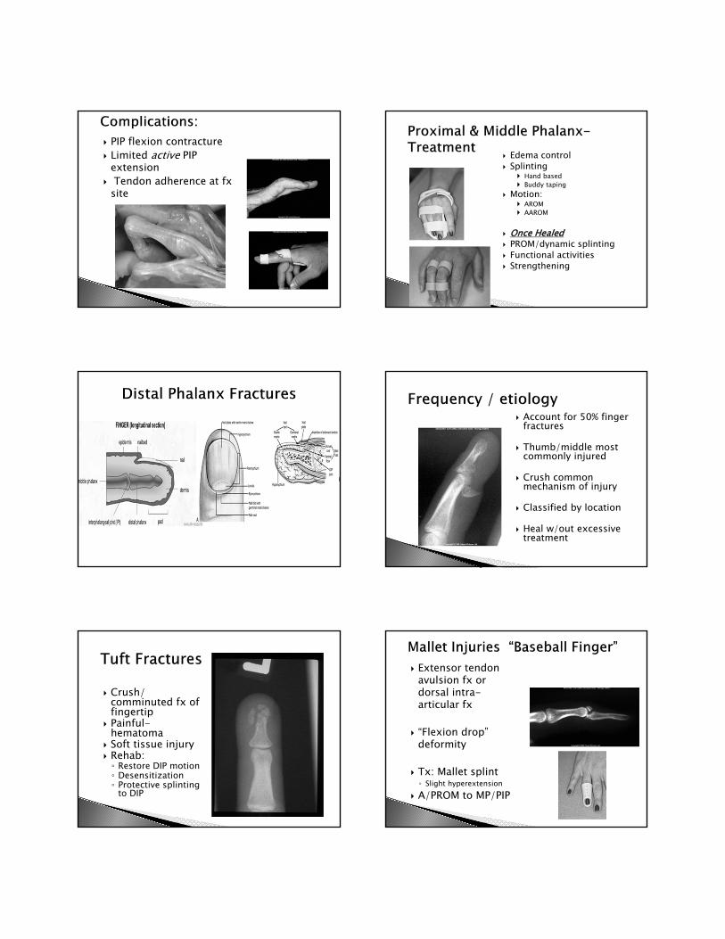
PIP flexion contracture Limited active PIP
extension Tendon adherence at fx
site
Edema control Splinting
Hand based Buddy taping
Motion: AROM AAROM
Once Healed PROM/dynamic splinting Functional activities Strengthening
Account for 50% finger fractures
Thumb/middle most commonly injured
Crush common mechanism of injury
Classified by location
Heal w/out excessive treatment
Crush/ comminuted fx of fingertip
Painful-hematoma
Soft tissue injury Rehab:◦ Restore DIP motion◦ Desensitization◦ Protective splinting
to DIP
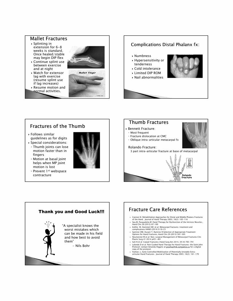
Extensor tendon avulsion fx or dorsal intra-articular fx
“Flexion drop” deformity
Tx: Mallet splint◦ Slight hyperextension
A/PROM to MP/PIP
Splinting in extension for 6-8 weeks is standard. Once healed/stable may begin DIP flex
Continue splint use between exercise and at night
Watch for extensor lag with exercise (resume splint use if lag increases)
Resume motion and normal activities.
Numbness Hypersensitivity or
tenderness Cold intolerance Limited DIP ROM Nail abnormalities
Follows similar guidelines as for digits
Special considerations: ◦ Thumb joints can lose
motion faster than in fingers◦ Motion at basal joint
helps when MP joint motion is lost◦ Prevent 1st webspace
contracture
Bennett Fracture: ◦ Most frequent◦ Fracture dislocation at CMC◦ Oblique intra-articular metacarpal fx
Rolando Fracture:3 part intra-articular fracture at base of metacarpal
“A specialist knows the worst mistakes which can be made in his field and how best to avoid them”
- Nils Bohr
Cannon N: Rehabilitation Approaches for Distal and Middle Phalanx Fractures of the Hand. Journal of Hand Therapy 2003; 16(2): 105-116
Seu,M, Pasqualette,M :Hand Therapy for Dysfunction of the Intrinsic Muscles:, Hand Clin 28 (2012) 87–100
Kollitz M, Hammert WC et al: Metacarpal fractures: treatment and complications HAND (2014) 9:16–23
Sammer DM, Husain T, Ramirez R:Selection of Appropriate Treatment Options for Hand Fractures, Hand Clin 29 (2013) 501–505
Neumeister M et al: Non-surgical Management of Metacarpal Fractures Clin Plastic Surg 41 2014 p451-461
Suh N et al: Carpal Fractures J Hand Surg Am 2014, 39 (4) 785-791 Lalonde D et al: Pain Guided Hand Therapy for Hand Fractures: the Saint John
Protocol: contact Amanda Higgins at [email protected] for a digital copy of the protocol.
Feehan, L, Early Controlled Mobilization of Potentially Unstable Extra-articular Hand Fractures. Journal of Hand Therapy 2003; 16(2): 161-170

Slade,J, MD, et al, (eds), Hand Clinics: Scaphoid Fractures, Volume 17, No. 4, November 2001
Berger,R et al, (eds), Hand Clinics: External Fixation, Philadelphia, WB Saunders, Volume 9, No. 4, November 1993
Freeland A, Lindley, S, (eds), Hand Clinics: Hand Fractures and Dislocations, Philadelphia, WB Saunders, Volume 22, No. 3, August 2006.
Fischgrund, J, (ed), Orthopaedic Knowledge Update, Chapter 2, Fracture Repair and Bone Grafting, AAOS, 2008, pgs 13-19.
LaStayo, P, Winters, K, et al, Fracture Healing: Bone Healing, Fracture Management, and Current Concepts Related to the hand. Journal of Hand Therapy 2003; 16(2): 81-92
Altman, E. (2016). The ulnar side of the wrist: Clinically relevant anatomy and biomechanics. Journal of Hand Therapy, 29, 111-122.
Bain, G. I., MacLean, S. B., Yeo, C. J., Perilli, E., & Lichtman, D. M. (2016). The etiology and pathogenesis of Kienböck disease. Journal of Wrist Surgery, 5, 248-254.
Brogan, D. M., Richard, M. J., Ruch, D., & Kakar, S. (2015). Management of severely comminuted distal radius fractures. Journal of Hand Surgery, 40A, 1905-1914.
Capo, J. T., Rossy, W., Henry, P., Maurer, R. J., Naidu, S., & Chen, L. (2009). External fixation of distal radius fractures: Effect of distraction and duration. Journal of Hand Surgery, 34A, 1605-1611.
Carlson, M. G. (Ed.). (2012). Elite athlete’s hand and wrist injury [Special issue]. Hand Clinics, 28(3).
Chung, K. C. (Ed.). (2012). Current concepts in the treatment of distal radius fractures [Special issue]. Hand Clinics, 28(2).
Dell, P. C., Dell, R. B., & Griggs, R. (2011). Management of carpal fractures and dislocations. In T. M. Skirven, A. L. Osterman, J. M. Fedorczyk, & P. C. Amadio (Eds.), Rehabilitation of the Hand and Upper Extremity (6th ed., pp. 988-1001). Philadelphia, PA: Mosby.
Jørgsholm, P., Thomsen, N. O., Björkman, A., Besjakov, J., & Abrahamsson, S. (2010). The incidence of intrinsic and extrinsic ligament injuries in scaphoid wrist fractures. Journal of Hand Surgery, 35A, 368–374.
Karagiannopoulos, C., & Michlovitz, S. (2016). Rehabilitation strategies for wrist sensorimotor control impairments: From theory to practice. Journal of Hand Therapy, 29, 154-165.
Kim, J. K., Kim, D. J., & Yun, Y. (2016). Natural history and factors associated with ulnar-sided wrist pain in distal radial fractures treated by plate fixation. Journal of Hand Surgery, 41E, 727-731.
Lawton, J. N. (Ed.). (2013). Management of hand fractures [Special issue]. Hand Clinics, 29(4). Lenoir, H., Coulet, B., Lazerges, C., Mares, O., Croutzet, P., & Chammas, M. (2012). Idiopathic
avascular necrosis of the scaphoid: 10 new cases and a review of the literature. Indications for Preiser’s disease. Orthopaedics & Traumatology: Surgery & Research, 98, 390-397
Magnus, C. R., Arnold, C. M., Johnston, G., Dal-Bello Haas, V., Basran, J., Krentz, J. R., & Farthing, J. P. (2013). Cross-education for improving strength and mobility after distal radius fractures: A randomized controlled trial. Archives of Physical Medicine and Rehabilitation, 94, 1247-1255.
Malick, A., & Prem, H. (2017). Physeal injuries in children. Surgery, 35, 10-17. Mathews, A. L., & Chung, K. C. (2015). Management of complications of distal radius fractures.
Hand Clinics, 31, 205-215. Means, K. R., Saunders, R. J., & Graham, T. J. (2011). Pathophysiology and surgical
management of the stiff hand. In T. M. Skirven, A. L. Osterman, J. M. Fedorczyk, & P. C. Amadio (Eds.), Rehabilitation of the Hand and Upper Extremity (6th ed., pp. 885-893). Philadelphia, PA: Mosby.
Medoff, R. J. (2010). Essential radiographic evaluation for distal radius fractures. In D. J.
Medoff, R. J. (2011). Distal radius fractures: Classification and management. In T. M. Skirven, A. L. Osterman, J. M. Fedorczyk, & P. C. Amadio (Eds.), Rehabilitation of the Hand and Upper Extremity (6th ed., pp. 941-948). Philadelphia, PA: Mosby.
Michlovitz, S., & Festa, L. (2011). Therapist’s management of distal radius fractures. In T. M. Skirven, A. L. Osterman, J. M. Fedorczyk, & P. C. Amadio (Eds.), Rehabilitation of the Hand and Upper Extremity (6th ed., pp. 28-35). Philadelphia, PA: Mosby.
Monaco, N. A., Dwyer, L., Ferikes, A. J., & Lubahn, J. D. (2016). Hand surgeon reporting of tendon rupture following distal radius volar plating. HAND, 11, 278-286.
Packham, T. L., Ball, P. D., MacDermid, J. C., Bain, J. R., & DalCin, A. (2016). A scoping review of applications and outcomes of traction orthoses for the management of intra-articular fractures and fracture dislocations in the hand. Journal of Hand Therapy, 29, 246-268.
Reichel, L. M., Bell, B. R., Michnick, S. M., & Reitman, C. A., (2012). Radial styloid fractures. Journal of Hand Surgery, 37A, 1726-1741.
Rosenthal, E. A., & Elhassan, B. T. (2011). The extensor tendons: Evaluation and surgical management. In T. M. Skirven, A. L. Osterman, J. M. Fedorczyk, & P. C. Amadio (Eds.), Rehabilitation of the Hand and Upper Extremity (6th ed., pp. 487-520). Philadelphia, PA: Mosby.
Schwartz, D. A. (2012). Static progressive orthoses for the upper extremity: A comprehensive literature review. HAND, 7, 10-17.
Sendher, R., & Ladd, A. L. (2013). The scaphoid. Hand Clinics, 44, 107-120. Shin, E. K. (2011). Fractures: General principles of surgical management. In T. M. Skirven, A. L.
Osterman, J. M. Fedorczyk, & P. C. Amadio (Eds.), Rehabilitation of the Hand and Upper Extremity (6th ed., pp. 351-360). Philadelphia, PA: Mosby.
Strauch, R.J. (2011). Scapholunate advanced collapse and scaphoid nonunion advanced
Szekeres, M., MacDermid, J. C., Birmingham, T., Grewal, R., & Lalone, E. (2017). The effect of therapeutic whirlpool and hot packs on hand volume during rehabilitation after distal radius fracture: A blinded randomized controlled trial. HAND, 12, 265-271.
Tsang, P., Walton, D., Grewal, R., & MacDermid, J. (2017). Validation of the QuickDASH and DASH in patients with distal radius fracture through agreement analysis. Archives of Physical Medicine and Rehabilitation.
Weinstock-Zlotnick, G., Page, C., Ghomrawi, H. M., & Wolff, A. L. (2015). Responsiveness of three Patient Report Outcome (PRO) measures in patients with hand fractures: A preliminary cohort study. Journal of Hand Therapy, 28, 403-411.
Boundless. (2016). Supply of Blood and Nerves to Bone. In Boundless Anatomy and Physiology. Retrieved from https://www.boundless.com/physiology/textbooks/boundless-anatomy-and-physiology-textbook/skeletal-system-6/introduction-to-bone-71/supply-of-blood-and-nerves-to-bone-436-9154/
When designated, images are courtesy of Primal Pictures
References – Images