property evaluations of dry-cast reconstituted bacterial ... · carbohydrate polymers 93 (2013)...
TRANSCRIPT
Px
CRa
b
c
a
ARRAA
KBXPBSH
1
ttailtlatird(2W
H9
0h
Carbohydrate Polymers 93 (2013) 144– 153
Contents lists available at SciVerse ScienceDirect
Carbohydrate Polymers
j ourna l ho me pag e: www.elsev ier .com/ locate /carbpol
roperty evaluations of dry-cast reconstituted bacterial cellulose/tamarindyloglucan biocomposites
layton F. de Souzaa,b, Neoli Lucyszyna,1, Marco A. Woehla, Izabel C. Riegel-Vidotti a,edouane Borsalib, Maria Rita Sierakowskia,∗
BioPol, Departamento de Química, Universidade Federal do Paraná (UFPR), P.O. 19081, Curitiba 81531-980, PR, BrazilCentre de Recherche sur les Macromolécules Végétales (CERMAV) Domaine Universitaire de Grenoble-St Martin d’Hères, 601 rue de la chimie, CERMAV-CNRS, BP 53, 38041 Grenobleedex 9, France
r t i c l e i n f o
rticle history:eceived 30 October 2011eceived in revised form 30 March 2012ccepted 24 April 2012vailable online 4 May 2012
a b s t r a c t
We describe the mechanical defibrillation of bacterial cellulose (BC) followed by the dry-cast generationof reconstituted BC films (RBC). Xyloglucan (XGT), extracted from tamarind seeds, was incorporated intothe defibrillated cellulose at various compositions, and new films were created using the same process.Microscopy and contact angle analyses of films revealed an increase in the microfibre adhesion, a reducedpolydispersity in the diameters of the microfibrils and increased hydrophobic behaviour as a functionof %XGT. X-ray diffraction analysis revealed changes to the crystallographic planes of the RBC and the
eywords:acterial celluloseyloglucanolysaccharide interactionsiocompositeurface properties
biocomposite films with preferential orientation along the (1 1 0) plane. Compared with BC, RBC/XGT bio-composite with 10% XGT exhibited improvement in its thermal properties and in Young’s modulus. Theseresults indicated a reorganisation of the microfibres with mechanical treatment, which when combinedwith hydrocolloids, can create cellulose-based materials that could be applied as scaffolding for tissueengineering and drug release.
ydrophilicity
. Introduction
The versatility of biopolymers allows for new applications inhe food, pharmaceutical and biotechnology industries. The struc-ures and functionalities of polysaccharides, proteins and lipidsllow their utilisation in biomimetic and nanotechnology systems,ncluding biosensors, transistors or mechanical modifiers. Cellu-ose is one of the most studied biopolymers, and several sources ofhis biopolymer have been evaluated. The backbone chains of cel-ulose, consisting of long linear chains of 1,4-ˇ-d-glucopyranose,re organised by hydrogen bonds that form a hard network struc-ure. This linearity yields multiple elementary fibrils that aggregatento larger bundles, which can contain crystalline and amorphousegions. The degrees of crystallinity and the crystal dimensions areependent on the origin of the cellulose or on the modification
chemical or physical) to which it was submitted (Guo & Catchmark,012; Sugiyama, Vuong, & Chanzy, 1991; Tischer, Sierakowski,estfahl, & Tischer, 2010; Wong, Kasapis, & Tan, 2009; Woodcock &∗ Corresponding author. Tel.: +55 41 33613260; fax: +55 41 33613186.E-mail address: [email protected] (M.R. Sierakowski).
1 Present address: Curso de Licenciatura em Química, Escola de Educac ão eumanidades, Pontifícia Universidade Católica do Paraná (PUCPR) Curitiba 80215-01, PR, Brazil.
144-8617 © 2012 Elsevier Ltd. ttp://dx.doi.org/10.1016/j.carbpol.2012.04.062
Open access under the Elsevier OA license.
© 2012 Elsevier Ltd.
Sarko, 1980). Specifically, cellulose from bacterial sources exhibitshigher crystallinity and has distinct advantages over cellulose fromother sources. These advantages include high purity (pretreatmentis not required for the extraction of lignin or hemicelluloses) andhigh surface area when compared with plant cellulose (Guo &Catchmark, 2012). Therefore, the great interest in bacterial cel-lulose (BC) is justified by its many potential applications. Theseapplications include medical devices (Nge, Nogi, Yano, & Sugiyama,2010) and wound dressings to prevent infection caused by bacterialor fungal agents that can occur as skin heals (Czaja, Young, Kawecki,& Brown, 2007; Jonas & Farah, 1998).
Many of the functional properties of cellulose depend on itscapacity to interact with diverse molecules or macromolecules ofvarying polarity. The adsorption and adhesion phenomena dependon the organisation of glucan chains located at the surface of the cel-lulose microfibrils (Mazeau, 2011). The association of hydrophilicstructures, including hydrocolloids such as xyloglucan, with cel-lulose can modify the mechanical and chemical properties of thecellulose (Zhou, Rutland, Teeri, & Brumer, 2007). The structuraldescription of the cellulose microfibril surfaces or that of cel-lulose associated with other molecules can be performed using
Open access under the Elsevier OA license.
several techniques. Recent studies include atomic force microscopy(AFM), scanning electron microscopy (SEM), contact angle (CA)measurements and small- and wide-angle X-ray diffraction (SAXSand WAXS) (Castro et al., 2011; Elazzouzi-Hafraoui et al., 2008;
drate P
HWipo
sc2sbbtm
siRaCMSE1dwHMA
twchcotmnaoat
agitbipacTwcms
2
2
f
C.F. de Souza et al. / Carbohy
uang, Chen, Lin, Hsu, & Chen, 2010; Klechkovskaya et al., 2003;oehl et al., 2010). The results have provided information regard-
ng the morphology on various scales, the hydrophilicity-relatedroperties, the microfibril dimensions and the relative crystallinityf these cellulose samples.
Recent advances in computer modelling and surface force analy-is have improved our understanding of the cellulose chains and theellulose–xyloglucan interactions at the molecular level (Mazeau,011; Zhang, Brumers, Agren, & Tu, 2011). In vitro, these complextructures are important to many applications particularly in theiotech industries. Some studies have demonstrated that cellulose-ased films, after processing, show selective permeability, allowinghe passage of water vapour while preventing the passage of
icroorganisms (Chang & Zhang, 2011; Klemm et al., 2009).A commercial xyloglucan product isolated from tamarind
eeds (Tamarindus indica) has important applications, especiallyn pharmaceutical formulations to obtain gels (Salazar-Montoya,amos-Ramírez, & Delgado-Reyes, 2002; Yamanaka et al., 2000),nd as a drug vehicle for controlled-release systems (Burgalassi,hetoni, Panichi, Boldrini, & Saettone, 2000; Coviello, Matricardi,arianecci, & Alhaique, 2007; Jó, Petri, Beltramini, Lucyszyn, &
ierakowski, 2010; Kawasaki et al., 1999; Miyazaki, Kawasaki,ndo, & Attwood, 2001). This biopolymer is formed by a,4-ˇ-d-glucopyranosyl backbone partially substituted with 1,6-˛--xylopyranosyl side chains, some of which are further substitutedith 1,2-˛-d-galactopyranosyl residues (Freitas et al., 2005;ayashi, 1989; Jó, Petri, Valenga, Lucyszyn, & Sierakowski, 2009;uller et al., 2011; Stupp et al., 2008; York, Harvey, Guillen,
lbersheim, & Darvill, 1993).In addition, because xyloglucan (XG) has storage or struc-
ural functions in plants, its association with cellulose has beenidely studied in relation to biosynthesis and the growth pro-
ess of higher plant organisms (Zhou et al., 2007). These studiesave enabled new discoveries and applications based on theellulose–xyloglucan interaction. For example, the developmentf XG as a molecular anchor to tether chemical functionalityo cellulose opened new possibilities for industrial plant fibre
odification in which it is used as a dispersing agent or to buildanostructured surfaces (Muller et al., 2011; Zhou et al., 2007). Inll cases, the cellulose–xyloglucan interaction is slightly dependentn its structural details as observed by Lopez et al. (2010). Theuthors observed that the adsorption is dependent on Mw and onhe side chains.
The development of biocomposites from cellulose and otherdditives has been widely explored. However, the methodolo-ies typically used include a pretreatment of the cellulose, whichnvolves a rigorous process of solubilisation and reconstruction ofhe cellulose membrane. Thus, the purpose of this research is to useacterial cellulose (BC) in the development of films by first mechan-
cally defibrillating and reconstructing the cellulose via a dry-castrocess to produce RBC. In addition, the inclusion of tamarind XG as
substitute for defibrillated BC pulp was explored at varying per-entages to produce biocomposite films with distinct properties.he structural, morphological, thermal and mechanical stabilitiesere evaluated using several techniques. The results obtained
an promote new methods for the production of cellulose-basedaterials for use in, among other applications, tissue engineering
upport and drug release.
. Materials and methods
.1. Polysaccharide sources
Prior to the dry-cast process, the xyloglucan was obtainedrom T. indica seeds by aqueous extraction from tamarind kernel
olymers 93 (2013) 144– 153 145
powder (TKP, Balasanka Mills, India) with an approximately55–65% recovery of the polysaccharide (Menon et al., 2010). Thechemical composition of the TKP used in the present work has beenpreviously reported (Jó et al., 2009, 2010).
The TKP was dispersed in distilled water at 30 ◦C and stirredovernight, then sonicated for 30 min using a solid 25-mm probe(VCX 750, Sonics & Materials, Inc., Newton, CT, USA). The insolu-ble fraction was separated by centrifugation at 6311 × g, and thesoluble fraction was precipitated with the addition of commer-cial ethanol (98%, Dipalcool Distribuidora de Álcool Ltda, AlmiranteTamandaré, PR, Brazil), washed with acetone and dried in an ovenat 50 ◦C. Then, the purified xyloglucan (XGT) was characterisedby gel permeation chromatography (GPC), in a Viscotek 270 DualDetector GPC, with PWxl columns (2500, 4000 and 6000 g/mol), at30 ◦C using 0.1 mol/L NaNO3 with 0.02% (w/v) NaN3 as an eluentand a dn/dc increment of 0.144.The never-dried bacterial cellulose(BC) cultured from Acetobacter xylinum was donated by MembracelProdutos Tecnológicos Ltda®, located at Almirante Tamandaré, PR,Brazil. Because the BC was received in the presence of hypochloriteand acetic acid, neutralisation was performed, and the membraneswere dialysed against distilled water. The conductivity and pHwere monitored until constant values (∼4.8 �S/cm and ∼6.8) wereobtained. Finally, the material was washed several times with ultra-pure water. For comparative purposes, dried commercial BC fromthe same company was used as a control in all experiments.
2.2. Achievement of dry-cast cellulose biocomposites
The never-dried BC (99 wt% of water) (Klemm et al., 2009) wassubmitted to mechanical treatment to yield a pulp using a blademixer over 30 min. To this, 2 L ultrapure water was added for each20 g of wet BC.
Various volumes of an aqueous XGT solution were incorporatedinto the pulp to yield final compositions of 10, 20 or 30 wt%.
The dispersions, deposited on polypropylene petri dishes, wereoven dried for 48 h at 37 ◦C to yield RBC or biocomposite films.
2.3. Biocomposite characterisations
2.3.1. Scanning electron microscopy (SEM) and atomic forcemicroscopy (AFM) analysis
The SEM images were obtained using a JEOL JSM-6360LV micro-scope (JEOL Ltd., Tokyo, Japan) at 10 kV and at a magnification of10,000×. All samples were covered by a thin layer of gold (<10 nm)to improve the conductivity of the surface.
The AFM analyses were performed on an Agilent microscope(Agilent Technologies, Santa Clara, CA, USA) using Pico Imagesoftware (Agilent Technologies, Santa Clara, CA, USA). The tap-ping mode images were obtained with Vistaprobes® (NanoscienceInstruments, Inc., Phoenix, AZ, USA) silicon tips (nominal springof 48 N/m and resonance frequency of ∼180 kHz), and the scanningwas over 2.0 �m × 2.0 �m and 5.0 �m × 5.0 �m. The data treatmentand presentation were accomplished with the help of GwyddionSoftware (Czech Metrology Institute).
2.3.2. Contact angle (CA) analysisThe CA analysis was obtained in a DataPhysics GmbH ten-
siometer (Filderstadt, Germany), model OCA 15plus. A study of thehydrophilicity on the surface of the biocomposites was performedusing the sessile drop method with ultrapure water as the solvent.The measurements were conducted at 25 ◦C using a 500-�L Hamil-ton syringe (Bonaduz, Switzerland) and a needle with an internal
diameter of 1.37 mm, an external diameter of 1.65 mm and a lengthof 38.1 mm.The surface free energy was obtained using Young’s equationwith the contact angle results. The calculation was performed with
1 rate P
Sw
2
i(41ua
n
C
iiii2
m(
C
TuA2f
c
(
i
2
(ssweq
2
iiats3
3
3d
mpm
46 C.F. de Souza et al. / Carbohyd
CA 20 DataPhysics software (San Jose, CA, USA) and the resultsere obtained by averaging at least three measurements.
.3.3. X-ray diffraction (XRD)The X-ray diffraction patterns of the BC, RBC and the biocompos-
te films were analysed with a Shimadzu XRD-7000 diffractometerKyoto, Japan) with Ni-filtered Cu K� radiation (� = 0.15418 nm,0 kV, 30 mA). The diffractograms were obtained in a 2�-range from0◦ to 30◦ at a scanning rate of 1.5◦/min. The d-spacing was obtainedsing Bragg’s law and the crystal size (CS) using Scherrer’s equations follows:
� = 2d · sin �
S = 0.9 · �
FWHM · cos �
n which n is an integer, � is the wavelength of the incident wave, ds the spacing between the plane lattices, � is the angle between thencident ray and the scattering planes and the FWHM (in radians)s the width of the peak at half the maximum height (Castro et al.,011; Elazzouzi-Hafraoui et al., 2008).
The crystallinity index (CrI, %) was calculated using the followingethod described by Hermans, Hermans, Vermaas, and Weidinger
1948):
rI (%) = Acryst
Atotal× 100
he Acryst is the sum of the crystalline area and Atotal is the total areander the diffractograms (Poletto, Zattera, Forte, & Santana, 2012).ll calculations were performed using Fityk software (Wojdyr,010), and the peaks were deconvoluted using the Gaussian peakunction.
The contribution of the (1 1 0) plane to the diffractograms wasalculated using the following equation:
1 1 0)contribution = A(1 1 0)
Acryst× 100%
n which A(1 1 0) is the area of the deconvoluted (1 1 0) peak.
.3.4. Thermogravimetric analysis (TG)The experiments were carried out using a Setaram TGA92
Setaram, France) with heating rate of 5.0 ◦C/min under a N2 atmo-phere (gas flow rate of 40 cm3/min). Approximately 30 mg of eachample was placed into a quartz cell, and its thermal degradationas measured from 20 to 500 ◦C. To avoid interference from the
quipment, a background analysis was performed using an emptyuartz cell under the same conditions.
.3.5. Mechanical propertiesTensile testing was performed in an Instron 5565 tensile test-
ng system (Norwood, MA, USA) following the procedures outlinedn the ASTM method D 882-95a. The tests were carried out in
humidity-controlled room with 50% relative humidity. Five-to-en replicates of 10 mm in width and 30 mm in gage length (gripeparation) were tested for each sample at a cross-head speed of
mm/min.
. Results and discussion
.1. Physicochemical parameters of XGT and the preparation ofry-cast biocomposites
The physicochemical characteristics of XGT as determined by aulti-detector GPC are provided in detail in Table S1 of the sup-
lementary material. The number- and weight-average molarass were determined to be 416.29 × 10³ and 214.79 × 10³ g/mol,
olymers 93 (2013) 144– 153
respectively, which differ from the results of Freitas, Gorin, Neves,and Sierakowski (2003), Freitas et al. (2005) and Jó et al. (2009) whorelated higher values of 1.00 × 10⁶ g/mol and 653.00 × 10³ g/mol,respectively. These higher Mw results can be attributed to boththe formation of supramolecular aggregates and the method ofpurification (Freitas et al., 2005; Picout, Ross-Murphy, Errington, &Harding, 2003). The physicochemical characteristics (see Table S1)are in good agreement with the results obtained by Picout et al.(2003), Freitas et al. (2005) and Muller et al. (2011), indicating goodresults for the GPC parameters, which demonstrate good solubili-sation and low aggregation with the given purification conditions.
The reconstituted bacterial cellulose (RBC) and RBC/XGT bio-composites were used to form films via a dry-cast process. A schemefor the membrane production is proposed in Fig. 1. This processattempted the development of a material with similar characteris-tics of commercial BC and with new properties after XGT inclusion.This process attempted the development of a material with prop-erties similar to commercial BC. Thus far, several projects havedemonstrated synergism between xyloglucan and cellulose froma variety of sources, morphologically and chemically changing thecellulose matrix (Lopez et al., 2010; Zhou et al., 2007).
3.2. Scanning electron microscopy (SEM) and contact angle (CA)measurements
To determine the morphological aspects and surface effects ofRBC and RBC/XGT films, SEM and CA analyses were performed.Visually, according to the images shown in Fig. 1, the BC andRBC/XGT exhibited the same appearance with a good dispersionand homogeneity. In contrast, the SEM images (Fig. 2) exhib-ited less homogeneous surfaces, with regions of high associationbetween the cellulose microfibrils and a loss of porous regions(voids), indicating the adsorption of XGT, which provided a morecompact association of the RBC microfibrils. However, all biocom-posites exhibited the same “mesh-like” structure as others obtainedthrough different drying processes. For example, reconstitutedcellulose created using a freeze-drying process was observed bySokolnicki, Fisher, Harrah, and Kaplan (2006), reconstituted cel-lulose created in the presence of chitin was reported by Liang,Zhang, and Xu (2007), and starch/reconstituted BC composites weredescribed by Woehl et al. (2010). The properties of the films orcomposites obtained are dependent upon the conditions of thedrying process. Therefore, a variety of features and morphologiescan be achieved for different applications. Some authors obtainedbiofilters and scaffolds from cellulose-based materials using afreeze-drying process (Barroso, Temtem, Hussain, Aguiar-Ricardo,& Roque, 2010; Nge et al., 2010; Sokolnicki et al., 2006). Othersinserted xyloglucan in situ into the production of the bacterial cellu-lose and observed different associations and ribbon sizes accordingto the xyloglucan concentration, which yielded a cellulose surfacenetwork with a variety of characteristics (Zhou et al., 2007).
The CA images and results obtained for the RBC and RBC/XGTfilms are shown in Fig. 2 and Table 2, respectively. As previously dis-cussed, the SEM results indicated variations in the aggregation andporosity of the biocomposite surfaces as a function of XGT inclusion.In addition, the CA analysis revealed changes to the hydrophilicityof the surfaces. Based on the CA results, the surface free energy wasobtained for the biocomposites, and the relationship between thesurface free energy and the degree of XGT substitution was found,with 10% > 20% > 30%.
The native celluloses are formed by I� and I� allomorphs. Inbacterial cellulose, the I� allomorph occurs preferentially (Attala
& Vanderhart, 1984). This allomorph is more hydrophilic than I�due to its structure in which the hydroxyl groups are more suscep-tible to hydrogen interactions (Mazeau, 2011; Tischer et al., 2010).So, the surface of cellulose is a favourable environment for the
C.F. de Souza et al. / Carbohydrate Polymers 93 (2013) 144– 153 147
se (RB
itart
Fs
Fig. 1. Scheme for the production of biofilms of reconstituted bacterial cellulo
ntercalation of XGT, which also explains the loss of polarity on
he surface. Fig. 2(c) and (d) shows regions with no evident fibrils,part from sub-micrometre-sized round structures that may rep-esent regions of XGT accumulation, which could also contribute tohe changes in the hydrophilicity of the films.ig. 2. SEM and CA images of biofilms of reconstituted bacterial cellulose – RBC (a) aubstitution: 10% (b), 20% (c) and 30% (d). The arrows indicate aggregation zones.
C) and RBC/tamarind xyloglucan (XGT) biocomposites by a dry-cast process.
3.3. Atomic force microscopy (AFM)
Through the topographic analysis of 5.0 �m × 5.0 �m imagesobtained by tapping-mode AFM (Fig. 3), the roughness values forall of the studied films could be calculated, and the results are
nd RBC/tamarind xyloglucan (XGT) biocomposites with varying degrees of XGT

148 C.F. de Souza et al. / Carbohydrate Polymers 93 (2013) 144– 153
Table 1Crystallinity index (CrI)a, crystallite size (CS)a and surface properties of biofilms of bacterial cellulose (BC), reconstituted bacterial cellulose (RBC), and RBC/tamarind xyloglucan(XGT) biocomposites with varying degrees of XGT substitution (%).
Samples CrI (%) CS1 0 0 (nm) CS0 1 0 (nm) CS1 1 0 (nm) Roughness (nm)b Contact angle(±0.1) (◦)c
Surface free energy(±0.1) (mN/m)d
BC 83.27 2.82 3.81 5.63 6.77 (±0.83) 25.8 66.4RBC 89.67 3.58 4.27 6.02 6.87 (±0.92) 11.9 71.2RBC/XGT 10% 76.18 4.52 5.42 6.02 10.23 (±1.69) 45.8 56.1RBC/XGT 20% 82.76 4.36 5.25 6.03 10.10 (±1.40) 49.4 54.1RBC/XGT 30% 79.90 4.33 5.21 6.05 10.97 (±1.86) 58.7 48.6
a Using deconvoluted Gaussian curves on X-ray diffractograms.b Root mean square values (rms) using 5.0 �m × 5.0 �m tapping-mode AFM images.c By OCA 15plus tensiometer measurements using sessil drop method.d By SCA 20 DataPhysics software using Young’s equation method.
Fig. 3. AFM tapping-mode images (5.0 �m × 5.0 �m) of biofilms of reconstituted bacterial cellulose – RBC (a) and RBC/tamarind xyloglucan (XGT) biocomposites with varyingd
prtsa
2tac
egrees of substitution: 10% (b), 20% (c) and 30% (d).
resented in Table 1. In the presence of XGT the biocomposites areougher than the controls (BC or RBC). Moreover, the association ofhe RBC microfibrils changes as a function of the hydrocolloid inclu-ion. The dispersion of the XGT was evident in a variety of cellulosessociations.
A cross-sectional analysis of the topographic AFM-images at
.0 �m × 2.0 �m (Fig. S1 in the supplementary material) indicateshat no modification occurred in the microfibril width, which haslateral size distribution between 50 and 100 nm, following theasting process. The dimensions of the blends are in agreement
with the cellulose values obtained through a different process(Elazzouzi-Hafraoui et al., 2008; Klechkovskaya et al., 2003; Tischeret al., 2010). Furthermore, the RBC allowed more ribbon asso-ciations, and the inclusion of XGT caused a reduction in thepolydispersity of the microfibril diameters.
The more homogeneous organisation of the microfibrils was
observed by AFM, which could explain the change in thehydrophilicity of the biocomposites. The round-shaped featuresincreasing in number from Fig. 3(b)–(d) may result from clustersof XGT that are not associated with the RBC fibrils. This result is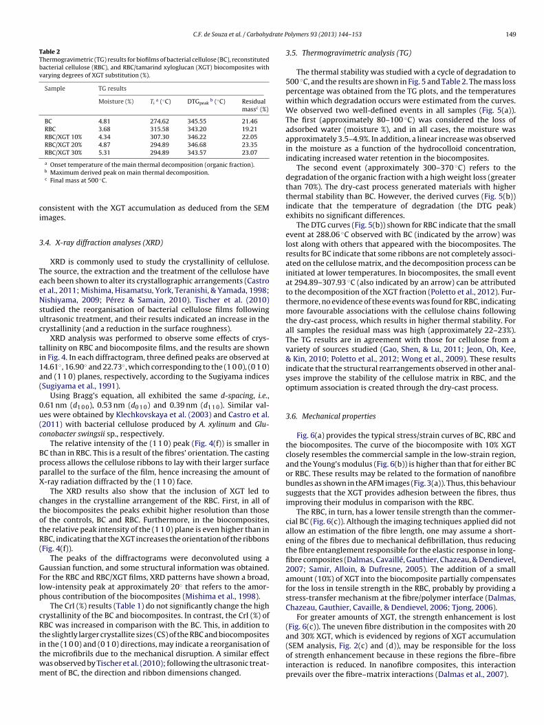
C.F. de Souza et al. / Carbohydrate P
Table 2Thermogravimetric (TG) results for biofilms of bacterial cellulose (BC), reconstitutedbacterial cellulose (RBC), and RBC/tamarind xyloglucan (XGT) biocomposites withvarying degrees of XGT substitution (%).
Sample TG results
Moisture (%) Tia (◦C) DTGpeak
b (◦C) Residualmassc (%)
BC 4.81 274.62 345.55 21.46RBC 3.68 315.58 343.20 19.21RBC/XGT 10% 4.34 307.30 346.22 22.05RBC/XGT 20% 4.87 294.89 346.68 23.35RBC/XGT 30% 5.31 294.89 343.57 23.07
a Onset temperature of the main thermal decomposition (organic fraction).
ci
3
TeeNsuc
ti1a(
0u(c
BppX
ctotR(
GFlp
cRtitwm
(SEM analysis, Fig. 2(c) and (d)), may be responsible for the loss
b Maximum derived peak on main thermal decomposition.c Final mass at 500 ◦C.
onsistent with the XGT accumulation as deduced from the SEMmages.
.4. X-ray diffraction analyses (XRD)
XRD is commonly used to study the crystallinity of cellulose.he source, the extraction and the treatment of the cellulose haveach been shown to alter its crystallographic arrangements (Castrot al., 2011; Mishima, Hisamatsu, York, Teranishi, & Yamada, 1998;ishiyama, 2009; Pérez & Samain, 2010). Tischer et al. (2010)
tudied the reorganisation of bacterial cellulose films followingltrasonic treatment, and their results indicated an increase in therystallinity (and a reduction in the surface roughness).
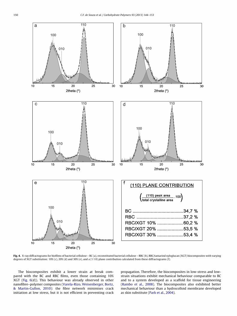
XRD analysis was performed to observe some effects of crys-allinity on RBC and biocomposite films, and the results are shownn Fig. 4. In each diffractogram, three defined peaks are observed at4.61◦, 16.90◦ and 22.73◦, which corresponding to the (1 0 0), (0 1 0)nd (1 1 0) planes, respectively, according to the Sugiyama indicesSugiyama et al., 1991).
Using Bragg’s equation, all exhibited the same d-spacing, i.e.,.61 nm (d1 0 0), 0.53 nm (d0 1 0) and 0.39 nm (d1 1 0). Similar val-es were obtained by Klechkovskaya et al. (2003) and Castro et al.2011) with bacterial cellulose produced by A. xylinum and Glu-onobacter swingsii sp., respectively.
The relative intensity of the (1 1 0) peak (Fig. 4(f)) is smaller inC than in RBC. This is a result of the fibres’ orientation. The castingrocess allows the cellulose ribbons to lay with their larger surfacearallel to the surface of the film, hence increasing the amount of-ray radiation diffracted by the (1 1 0) face.
The XRD results also show that the inclusion of XGT led tohanges in the crystalline arrangement of the RBC. First, in all ofhe biocomposites the peaks exhibit higher resolution than thosef the controls, BC and RBC. Furthermore, in the biocomposites,he relative peak intensity of the (1 1 0) plane is even higher than inBC, indicating that the XGT increases the orientation of the ribbonsFig. 4(f)).
The peaks of the diffractograms were deconvoluted using aaussian function, and some structural information was obtained.or the RBC and RBC/XGT films, XRD patterns have shown a broad,ow-intensity peak at approximately 20◦ that refers to the amor-hous contribution of the biocomposites (Mishima et al., 1998).
The CrI (%) results (Table 1) do not significantly change the highrystallinity of the BC and biocomposites. In contrast, the CrI (%) ofBC was increased in comparison with the BC. This, in addition tohe slightly larger crystallite sizes (CS) of the RBC and biocompositesn the (1 0 0) and (0 1 0) directions, may indicate a reorganisation of
he microfibrils due to the mechanical disruption. A similar effectas observed by Tischer et al. (2010); following the ultrasonic treat-ent of BC, the direction and ribbon dimensions changed.olymers 93 (2013) 144– 153 149
3.5. Thermogravimetric analysis (TG)
The thermal stability was studied with a cycle of degradation to500 ◦C, and the results are shown in Fig. 5 and Table 2. The mass losspercentage was obtained from the TG plots, and the temperatureswithin which degradation occurs were estimated from the curves.We observed two well-defined events in all samples (Fig. 5(a)).The first (approximately 80–100 ◦C) was considered the loss ofadsorbed water (moisture %), and in all cases, the moisture wasapproximately 3.5–4.9%. In addition, a linear increase was observedin the moisture as a function of the hydrocolloid concentration,indicating increased water retention in the biocomposites.
The second event (approximately 300–370 ◦C) refers to thedegradation of the organic fraction with a high weight loss (greaterthan 70%). The dry-cast process generated materials with higherthermal stability than BC. However, the derived curves (Fig. 5(b))indicate that the temperature of degradation (the DTG peak)exhibits no significant differences.
The DTG curves (Fig. 5(b)) shown for RBC indicate that the smallevent at 288.06 ◦C observed with BC (indicated by the arrow) waslost along with others that appeared with the biocomposites. Theresults for BC indicate that some ribbons are not completely associ-ated on the cellulose matrix, and the decomposition process can beinitiated at lower temperatures. In biocomposites, the small eventat 294.89–307.93 ◦C (also indicated by an arrow) can be attributedto the decomposition of the XGT fraction (Poletto et al., 2012). Fur-thermore, no evidence of these events was found for RBC, indicatingmore favourable associations with the cellulose chains followingthe dry-cast process, which results in higher thermal stability. Forall samples the residual mass was high (approximately 22–23%).The TG results are in agreement with those for cellulose from avariety of sources studied (Gao, Shen, & Lu, 2011; Jeon, Oh, Kee,& Kin, 2010; Poletto et al., 2012; Wong et al., 2009). These resultsindicate that the structural rearrangements observed in other anal-yses improve the stability of the cellulose matrix in RBC, and theoptimum association is created through the dry-cast process.
3.6. Mechanical properties
Fig. 6(a) provides the typical stress/strain curves of BC, RBC andthe biocomposites. The curve of the biocomposite with 10% XGTclosely resembles the commercial sample in the low-strain region,and the Young’s modulus (Fig. 6(b)) is higher than that for either BCor RBC. These results may be related to the formation of nanofibrebundles as shown in the AFM images (Fig. 3(a)). Thus, this behavioursuggests that the XGT provides adhesion between the fibres, thusimproving their modulus in comparison with the RBC.
The RBC, in turn, has a lower tensile strength than the commer-cial BC (Fig. 6(c)). Although the imaging techniques applied did notallow an estimation of the fibre length, one may assume a short-ening of the fibres due to mechanical defibrillation, thus reducingthe fibre entanglement responsible for the elastic response in long-fibre composites (Dalmas, Cavaillé, Gauthier, Chazeau, & Dendievel,2007; Samir, Alloin, & Dufresne, 2005). The addition of a smallamount (10%) of XGT into the biocomposite partially compensatesfor the loss in tensile strength in the RBC, probably by providing astress-transfer mechanism at the fibre/polymer interface (Dalmas,Chazeau, Gauthier, Cavaille, & Dendievel, 2006; Tjong, 2006).
For greater amounts of XGT, the strength enhancement is lost(Fig. 6(c)). The uneven fibre distribution in the composites with 20and 30% XGT, which is evidenced by regions of XGT accumulation
of strength enhancement because in these regions the fibre–fibreinteraction is reduced. In nanofibre composites, this interactionprevails over the fibre–matrix interactions (Dalmas et al., 2007).
150 C.F. de Souza et al. / Carbohydrate Polymers 93 (2013) 144– 153
F bacted ution
pXn&i
ig. 4. X-ray diffractograms for biofilms of bacterial cellulose – BC (a), reconstitutedegrees of XGT substitution: 10% (c), 20% (d) and 30% (e), and a (1 1 0) plane contrib
The biocomposites exhibit a lower strain at break com-ared with the BC and RBC films, even those containing 10%
GT (Fig. 6(d)). This behaviour was already observed in otheranofibre–polymer composites (Varela-Rizo, Weisenberger, Bortz,Martin-Gullon, 2010): the fibre network minimises cracknitiation at low stress, but it is not efficient in preventing crack
rial cellulose – RBC (b), RBC/tamarind xyloglucan (XGT) biocomposites with varying calculated from these diffractograms (f).
propagation. Therefore, the biocomposites in low-stress and low-strain situations exhibit mechanical behaviour comparable to BC
and to a system developed as a scaffold for tissue engineering(Rambo et al., 2008). The biocomposites also exhibited bettermechanical behaviour than a hydrocolloid membrane developedas skin substitute (Park et al., 2004).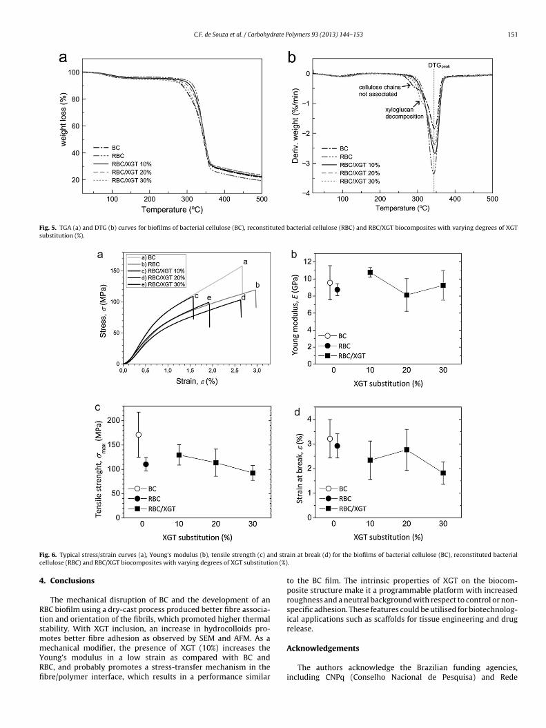
C.F. de Souza et al. / Carbohydrate Polymers 93 (2013) 144– 153 151
Fig. 5. TGA (a) and DTG (b) curves for biofilms of bacterial cellulose (BC), reconstituted bacterial cellulose (RBC) and RBC/XGT biocomposites with varying degrees of XGTsubstitution (%).
F nd strc on (%)
4
RtsmmYRfi
ig. 6. Typical stress/strain curves (a), Young’s modulus (b), tensile strength (c) aellulose (RBC) and RBC/XGT biocomposites with varying degrees of XGT substituti
. Conclusions
The mechanical disruption of BC and the development of anBC biofilm using a dry-cast process produced better fibre associa-ion and orientation of the fibrils, which promoted higher thermaltability. With XGT inclusion, an increase in hydrocolloids pro-otes better fibre adhesion as observed by SEM and AFM. As a
echanical modifier, the presence of XGT (10%) increases theoung’s modulus in a low strain as compared with BC andBC, and probably promotes a stress-transfer mechanism in thebre/polymer interface, which results in a performance similar
ain at break (d) for the biofilms of bacterial cellulose (BC), reconstituted bacterial.
to the BC film. The intrinsic properties of XGT on the biocom-posite structure make it a programmable platform with increasedroughness and a neutral background with respect to control or non-specific adhesion. These features could be utilised for biotechnolog-ical applications such as scaffolds for tissue engineering and drugrelease.
Acknowledgements
The authors acknowledge the Brazilian funding agencies,including CNPq (Conselho Nacional de Pesquisa) and Rede
1 rate P
NCcc
A
fj
R
A
B
B
C
C
C
C
D
D
E
F
F
G
G
H
H
H
J
J
J
J
K
52 C.F. de Souza et al. / Carbohyd
anobiotec/CAPES-Brazil for financial support, and DFIS/UFPR andERMAV for analysis support. The authors are grateful to Membra-el Produtos Tecnológicos Ltda (Brazil) for providing the bacterialellulose samples.
ppendix A. Supplementary data
Supplementary data associated with this article can beound, in the online version, at http://dx.doi.org/10.1016/.carbpol.2012.04.062.
eferences
ttala, R. H., & Vanderhart, D. L. (1984). Native cellulose: A composite of two distinctcrystalline forms. Science, 223(4633), 283–285.
arroso, T., Temtem, M., Hussain, A., Aguiar-Ricardo, A., & Roque, A. C. A. (2010).Preparation and characterization of a cellulose affinity membrane for humanimmunoglobulin G (IgG) purification. Journal of Membrane Science, 348(1–2),224–230.
urgalassi, S., Chetoni, P., Panichi, L., Boldrini, E., & Saettone, M. F. (2000).Xyloglucan as a novel vehicle for timolol: Pharmacokinetics and pressure low-ering activity in rabbits. Journal Ocular Pharmacology and Therapeutics, 16(6),497–509.
astro, C., Zuluaga, R., Puteaux, J.-L., Caro, G., Mondragon, I., & Ganán, P. (2011). Struc-tural characterization of bacterial cellulose produced by Gluconobacter swingsiisp. from Colombian agroindustrial wastes. Carbohydrate Polymers, 84(1), 96–102.
hang, C., & Zhang, L. (2011). Cellulose-based hydrogels: Present status and appli-cation prospects. Carbohydrate Polymers, 84(1), 40–53.
oviello, T., Matricardi, P., Marianecci, C., & Alhaique, F. (2007). Polysaccharidehydrogels for modified release formulations. Journal of Controlled Release, 119(1),5–24.
zaja, W. K., Young, D. J., Kawecki, M., & Brown, R. M., Jr. (2007). The future prospectsof microbial cellulose in biomedical applications. Biomacromolecules, 8(1),1–12.
almas, F., Cavaillé, J.-Y., Gauthier, C., Chazeau, L., & Dendievel, R. (2007). Viscoelasticbehavior and electrical properties of flexible nanofiber filled polymer nanocom-posites. Influence of processing conditions. Composites Science and Technology,67(5), 829–839.
almas, F., Chazeau, L., Gauthier, C., Cavaille, J. Y., & Dendievel, R. (2006). Largedeformation mechanical behavior of flexible nanofiber filled polymer nanocom-posites. Polymer, 47(8), 2802–2812.
lazzouzi-Hafraoui, S., Nishiyama, Y., Puteaux, J.-L., Heux, L., Dubreuil, F., &Rochas, C. (2008). The shape and size distribution of crystalline nanoparti-cles prepared by acid hydrolysis of native cellulose. Biomacromolecules, 9(1),57–65.
reitas, R. A., Gorin, P. A. J., Neves, J., & Sierakowski, M.-R. (2003). A rheologicaldescription of mixtures of a galactoxyloglucan with high amylose and waxycorn starches. Carbohydrate Polymers, 51(1), 25–32.
reitas, R. A., Martin, S., Santos, G. L., Valenga, F., Buckeridge, M. S., Reicher, F., et al.(2005). Physico-chemical properties of seed xyloglucans from different sources.Carbohydrate Polymers, 60(4), 507–514.
ao, Q., Shen, X., & Lu, X. (2011). Regenerated bacterial cellulose fibers prepared bythe NMMO·H2O process. Carbohydrate Polymers, 83(3), 1253–1256.
uo, J., & Catchmark, J. M. (2012). Surface area and porosity of acid hydrolyzedcellulose nanowhiskers and cellulose produced by Gluconacetobacter xylinus.Carbohydrate Polymers, 87(2), 1026–1037.
ayashi, T. (1989). Xyloglucan in the primary cell wall. Annual Review of Plant Phys-iology and Plant Molecular Biology, 40, 139–166.
ermans, P. H., Hermans, J. J., Vermaas, D., & Weidinger, A. J. (1948). Deformationmechanism of cellulose gels. IV. General relationship between orientation of thecrystalline and that of the amorphous portion. Journal of Polymer Science, 3(1),1–9.
uang, H.-C., Chen, L.-C., Lin, S.-B., Hsu, C.-P., & Chen, H.-H. (2010). In situ modifi-cation of bacterial cellulose network structure by adding interfering substancesduring fermentation. Bioresource Technology, 101(15), 6084–6091.
eon, J.-H., Oh, I.-K., Kee, C.-D., & Kin, S.-J. (2010). Bacterial cellulose actuator withelectrically driven bending deformation in hydrated condition. Sensors and Actu-ators B: Chemical, 146, 307–313.
ó, T. A., Petri, D. F. S., Beltramini, L. M., Lucyszyn, N., & Sierakowski, M.-R. (2010).Xyloglucan nano-aggregates: Physico-chemical characterisation in buffer solu-tion and potential application as a carrier for camptothecin, an anti-cancer drug.Carbohydrate Polymers, 82(2), 355–362.
ó, T. A., Petri, D. F. S., Valenga, F., Lucyszyn, N., & Sierakowski, M.-R. (2009). Thin filmsof xyloglucans for BSA adsorption. Materials Science and Engineering C, 29(2),631–637.
onas, R., & Farah, L. F. (1998). Production and application of microbial cellulose.Polymer Degradation and Stability, 59(1–3), 101–106.
awasaki, N., Ohkura, R., Miyazaki, S., Uno, Y., Sugimoto, S., & Attwood, D. (1999).Thermally reversible xyloglucan gels as vehicles for oral drug delivery. Interna-tional Journal of Pharmaceutics, 181(2), 227–234.
olymers 93 (2013) 144– 153
Klechkovskaya, V. V., Baklagina Yu, G., Stepina, N. D., Khripunov, A. K., Buffat, P. A.,Suvorova, E. I., et al. (2003). Structure of cellulose Acetobacter xylinum. Crystal-lography Reports, 48(5), 755–762.
Klemm, D., Schumann, D., Kramer, F., Heßler, N., Koth, D., & Sultanova, B. (2009).Nanocellulose materials—Different cellulose, different functionality. Macro-molecular Symposia, 280(1), 60–71.
Liang, S., Zhang, L., & Xu, J. (2007). Morphology and permeability of cellulose/chitinblend membranes. Journal of Membrane Science, 287(1), 19–28.
Lopez, M., Bizot, H., Chambat, G., Marais, M.-F., Zykwinska, A., Ralet, M.-C.,et al. (2010). Enthalpic studies of xyloglucan–cellulose interactions. Biomacro-molecules, 11(6), 1417–1428.
Mazeau, K. (2011). On the external morphology of native cellulose microfibrils.Carbohydrate Polymers, 84(1), 524–532.
Menon, V., Prakash, G., & Rao, M. (2010). Enzymatic hydrolysis and ethanol pro-duction using xyloglucanase and Debaromyces hansenii from tamarind kernelpowder: Galactoxyloglucan predominant hemicellulose. Journal of Biotechnol-ogy, 148(4), 233–239.
Mishima, T., Hisamatsu, M., York, W. S., Teranishi, K., & Yamada, T. (1998).Adhesion of �-d-glucans to cellulose. Carbohydrate Research, 308(3–4), 389–395.
Miyazaki, N., Kawasaki, N., Endo, K., & Attwood, D. (2001). Oral sustained deliveryof theophylline from thermally reversible xyloglucan gels in rabbits. Journal ofPharmacy and Pharmacology, 53(9), 1185–1191.
Muller, F., Manet, S., Jean, B., Chambat, G., Boué, F., Heux, L., et al. (2011).SANS measurements of semifexible xyloglucan polysaccharide chains inwater reveal their self-avoiding statistics. Biomacromolecules, 12(9), 3330–3336.
Nge, T. T., Nogi, M., Yano, H., & Sugiyama, J. (2010). Microstructure and mechan-ical properties of bacterial cellulose/chitosan porous scaffold. Cellulose, 17(2),349–363.
Nishiyama, Y. (2009). Structure and properties of the cellulose microfibril. Journal ofWood Science, 55(4), 241–249.
Park, S. N., Kim, J. K., & Suh, H. (2004). Evaluation of antibiotic-loadedcollagen–hyaluronic acid matrix as a skin substitute. Biomaterials, 25(17),3689–3698.
Pérez, S., & Samain, D. (2010). Structure and engineering of celluloses. Advances inCarbohydrate chemistry and Biochemistry, 64, 25–116.
Picout, D. R., Ross-Murphy, S. B., Errington, N., & Harding, S. E. (2003). Pressurecell assisted solubilization of xyloglucans: Tamarind seed polysaccharide anddetarium gum. Biomacromolecules, 4(3), 799–807.
Poletto, M., Zattera, A. J., Forte, M. M. C., & Santana, R. M. C. (2012). Thermal decom-position of wood: Influence of wood components and cellulose crystallite size.Bioresource Technology, 109, 148–153.
Rambo, C. R., Recouvreux, D. O. S., Carminatti, C. A., Pitlovanciv, A. K., Antonio, R. V., &Porto, L. M. (2008). Template assisted synthesis of porous nanofibrous cellulosemembranes for tissue engineering. Materials Science & Engineering C: Biomimeticand Supramolecular Systems, 28(4), 549–554.
Salazar-Montoya, J. A., Ramos-Ramírez, E. G., & Delgado-Reyes, V. A. (2002). Changesof the dynamic properties of tamarind (Tamarindus indica) gel with differentsaccharose and polysaccharide concentrations. Carbohydrate Polymers, 49(4),387–391.
Samir, M., Alloin, F., & Dufresne, A. (2005). Review of recent research into cellu-losic whiskers, their properties and their application in nanocomposite field.Biomacromolecules, 6(2), 612–626.
Sokolnicki, A. M., Fisher, R. J., Harrah, T. P., & Kaplan, D. L. (2006). Permeabil-ity of bacterial cellulose membranes. Journal of Membrane Science, 272(1–2),15–27.
Stupp, T., Freitas, R. A., Sierakowski, M.-R., Deschamps, F. C., Wisniewski,A., Jr., & Biavatti, M. W. (2008). Characterization and potential uses ofCopaifera langsdorfii seeds and seed oil. Bioresource Technology, 99(7), 2659–2663.
Sugiyama, J., Vuong, R., & Chanzy, H. (1991). Electron diffraction study on the twocrystalline phases occurring in native cellulose from an algal cell wall. Macro-molecules, 24(9), 4168–4175.
Tjong, S. C. (2006). Structural and mechanical properties of polymer nanocompos-ites. Materials Science & Engineering R: Reports, 53(3–4), 73–197.
Tischer, P. C. S. F., Sierakowski, M.-R., Westfahl, H., Jr., & Tischer, C. A. (2010).Nanostructural reorganization of bacterial cellulose by ultrasonic treatment.Biomacromolecules, 11(5), 1217–1224.
Varela-Rizo, H., Weisenberger, M., Bortz, D. R., & Martin-Gullon, I. (2010). Frac-ture toughness and creep performance of PMMA composites containing microand nanosized carbon filaments. Composites Science and Technology, 70(7),1189–1195.
Woehl, M. A., Canestraro, C. D., Mikowski, A., Sierakowski, M.-R., Ramos,L. P., & Wypych, F. (2010). Bionanocomposites of thermoplastic starchreinforced with bacterial cellulose nanofibres: Effect of enzymatic treat-ment on mechanical properties. Carbohydrate Polymers, 80(3), 866–873.
Wojdyr, M. (2010). Fityk: A general-purpose peak fitting program. Journal of AppliedCrystallography, 43(5–1), 1126–1128.
Wong, S.-S., Kasapis, S., & Tan, Y. M. (2009). Bacterial and plant cellulose mod-
ification using ultrasound irradiation. Carbohydrate Polymers, 77(2), 280–287.Woodcock, C., & Sarko, A. (1980). Packing analysis of carbohydrates and polysac-charides. 11. Molecular and crystal structure of native ramie cellulose.Macromolecules, 13(5), 1183–1187.
drate P
Y
Y
Zhang, Q., Brumers, H., Agren, H., & Tu, Y. (2011). The adsorption of xyloglucan on cel-lulose: Effects of explicit water and side chain variation. Carbohydrate Research,
C.F. de Souza et al. / Carbohy
amanaka, S., Yuguchi, Y., Urakawa, H., Kajiwara, K., Shirakawa, M., & Yamatoya, K.(2000). Gelation of tamarind seed polysaccharide xyloglucan in the presence of
ethanol. Food Hydrocolloids, 14(2), 125–128.ork, W. S., Harvey, L. K., Guillen, R., Albersheim, P., & Darvill, A. G. (1993).Structural analysis of tamarind seed xyloglucan oligosaccharides using �-galactosidase digestion and spectroscopic methods. Carbohydrate Research, 248,285–301.
olymers 93 (2013) 144– 153 153
346(16), 2595–2602.Zhou, Q., Rutland, M. W., Teeri, T. T., & Brumer, H. (2007). Xyloglucan in cellulose
modification. Cellulose, 14(6), 625–641.