prospects of bacterial and plant protein-based immunotoxins for
TRANSCRIPT

Abstract. Bacterial- and plant-derived immunotoxins havedocumented potential for treatment of cancer. We discussAnthrax toxin, ribosome inactivating-toxins, such as saporinand ricin, and ADP-ribosylating toxins such as Diphtheriatoxin and Pseudomonas exotoxin, with focus on the latter,which has been most thoroughly investigated. Regardingtheir potential as anticancer agents, critical issues such asimmunogenicity and toxicity are outlined. We describedifferent generations of immunotoxins, the pathways for thedelivery of the cytotoxic ‘warheads’, molecular parametersmodulating efficacy, and combination therapy with otheranticancer agents. Finally, we discuss deimmunizationstrategies based on the removal of B- and T-cell epitopesfrom the cytotoxic component, and highlight promisingclinical proof-of-concept studies.
Development of anticancer agents is based on the hallmarksof cancer and the identification of corresponding moleculartargets (1, 2). The hallmarks of cancer are properties such asconstitutive proliferative signaling, de-regulation of cellularenergetics, resistance to cell death, evasion of growthsuppressors, avoidance of immune destruction, enabling ofreplicative immortality, tumor-promoting inflammation,activation of invasion and metastasis, genomic instability andmutation, as well as induction of tumor angiogenesis. Basedon these properties, in addition to cytotoxic agents, target-specific therapies were established. Thus, some types of
cancer can be medicated by inhibition of driver mutations (3,4) and others might be treated in the future by induction ofsynthetic lethality (5, 6). Interference with tumorangiogenesis has resulted in clinical benefit for several typesof cancer, especially in combination with chemotherapy (7,8). Activation of an antitumor immune response by antibody-based therapy is another promising approach withdocumented clinical benefit (9, 10). However, targetedtherapies are often hampered by the activation ofcompensatory pathways leading to treatment resistance (11,12). Paul Ehrlich brought-up the idea of a “magic bullet” fora disease-relevant antigen which does not hit normal cells ofthe human body (13). Bacteria- or plant-derived toxins, suchas Pseudomonas exotoxin (PE), Diphtheria toxin (DT), ricin,saporin and others, can inhibit protein synthesis and induceapoptosis efficiently by only a few intracellularly-processedmolecules (14). Since it is not possible to define a therapeuticwindow for these toxins, directing these agents to tumors withappropriate ligands is necessary; such targeting moieties canbe cytokines or antibody-related entities.
In the present review, we discuss prospects, critical issuesand clinical proof-of-concept studies for these powerfulagents. We focus on toxins exerting their function throughcleavage and inactivation of kinases of the mitogen-activatedprotein kinase (MAPK) family, N-glycosidase or adenosinedi-phosphate (ADP)-ribosylation activities. Progress is basedon the development of new formats for targetingimmunotoxins (ITs) (Figure 1) and understanding of thetrafficking pathways of ITs (Figure 2).
Anthrax Toxin-based Approaches
The Anthrax toxin derived from Bacillus anthracis is thecausative agent of anthrax in animals and humans. It iscomposed of protective antigen (PA, 83 kDa), lethal factor(LF, 90 kDa) and edema factor (EF, 89 kDa) (Figure 3A). Itbelongs to a family of toxins in which LF or EF must be
25
Correspondence to: Ulrich H. Weidle, Roche Pharma Research andEarly Development (pRED), Roche Diagnostics GmbH, D-82372Penzberg, Germany. E-mail: [email protected]
Key Words: Anthrax toxin, deimmunization, diphthamide, diphtheriatoxin, eukaryotic elongation factor 2, pseudomonas exotoxin, proof-of-concept studies, ricin, saporin, ribosome inactivating proteins,review.
CANCER GENOMICS & PROTEOMICS 11: 25-38 (2014)
Review
Prospects of Bacterial and Plant Protein-based Immunotoxins for Treatment of Cancer
ULRICH H. WEIDLE1, GEORG TIEFENTHALER1, CHRISTIAN SCHILLER2,ELISABETH H. WEISS2, GUY GEORGES1 and ULRICH BRINKMANN1
1Roche Pharma Research and Early Development (pRED), Roche Diagnostics GmbH, Penzberg, Germany;2Department of Biology II, Ludwig Maximilians University, Munich, Germany
1109-6535/2014

combined with PA in order to elicit toxicity (15-17). Theproteolytic activation of PA has been exploited for tumortherapy. PA can bind to either of two receptors: tumorendothelial marker 8 (TEM8) and capillary morphogenesisprotein 2 (CMG 2) (18, 19). Cell surface proteases, such asfurin, cleave PA into an N-terminal PA63 (63 kDa) and a C-terminal PA20 (20 kDa) fragment. Receptor-associatedPA63 oligomerizes into a ring-shaped heptamer, which canbind up to three molecules of LF or EF in any combination(20). The resulting complexes are referred to as lethal toxinor edema toxin. They are internalized via clathrin-dependentreceptor-mediated endocytosis and undergo a conformationalchange, which leads to the insertion of the complex into themembrane of the endosome and finally delivery of LF and EFinto the cytoplasm. Once in the cytoplasm, LF can functionas a zinc metalloprotease, which can inactivate MAPKthrough cleavage. EF is a Ca/calmodulin-activated adenylatecyclase (AC) involved in the elevation of intracellularadenosine mono-phosphate (AMP) or cyclic adenosine mono-phosphate (cAMP) and thus interferes with the balance ofintracellular signaling. Replacement of the furin cleavage siteof PA with cleavage sites for proteases which areoverexpressed in tumors, such as matrix metalloproteinase-2(MMP2) and MMP9, allows tumor-specific activation of PA.Making use of LF and MMP-activated PA, antitumor activitywas observed not only in tumor xenografts derived fromhuman melanomas bearing the V600E v-Raf murine sarcomaviral oncogene homolog B (BRAF) mutation, but also inother tumor xenograft models with non-mutated BRAF (21).Furthermore, Anthrax toxin can inhibit tumor angiogenesis byinterference with endothelial proliferation, migration and tubeformation for which MAPK plays an essential role (22, 23).Overexpression of urokinase-type plasminogen activator(uPA) in many types of tumors has also been exploited in thiscontext by replacing the furin cleavage site of PA with anuPA-cleavage sequence (24). The specific processing of PAhas also been used for the delivery of other toxins, such asthe ADP-ribosylating moiety of PE. Cleavage site-modifiedPA in combination with a recombinant fusion protein,consisting of amino acids 1-254 and the ADP-ribosylationdomain of PE, referred to as FP59, was shown to beactivated on the surface of tumor cells and to mediateefficient tumor cell binding activity in vitro and in vivo (25).This combination of proteins selectively kills MMP-overexpressing tumor cells, whereas no toxic effect wasnoted in non-tumorigenic cells. Combined inhibition oftumor growth and angiogenesis by LF has been documentedby independent investigations (26, 27). Critical issues ofAnthrax-based toxins, as described above, are the expressionof MMPs and uPA in non-malignant tissues, and the lack ofa tumor-specific targeting component challenges thefeasibility to translate these agents into clinically-validatedanticancer agents.
Ribosome-inactivating Protein (RIP)-based Immunotoxins
RIPs are a family of toxins derived from plants, bacteria,fungi or algae with N-glycosidase activity which inactivateribosomes by cleaving an N-glycosidic bond of a specificadenosine residue (A4324) within the 28-S rRNA subunit,thereby rendering ribosomes unable to interact witheukaryotic elongation factor-2 (eEF2), and thus inhibitingprotein synthesis (28). Type I RIPs are single-chain proteinswith enzymatic activity as described above, while type IIRIPs are heterodimeric proteins which consist of anenzymatically active A-chain linked to a B-chain with lectinproperties (29-32). Saporin, pokeweed antiviral protein (PAP)and gelonin are prototypic class I RIPs, while ricin and abrinare typical class II members (Figures 3B and C). Ricin hasbeen thoroughly investigated. It binds to cell surface galactoseor N-acetylglucosamine residues on glycoproteins orglycolipids through the B-chain, is internalized byendocytosis and is routed backwards from the Golgiapparatus to the endoplasmic reticulum (ER). In thiscompartment, the disulfide bond linking the two chains isreduced and the catalytic A-chain is translocated into thecytoplasm to excert its N-glycosidase activity. The 28-S RNAmodification compromises ribosomes to interact with eEF2,resulting in the inhibition of protein synthesis and ultimatelycell death (33) (Figure 2). In contrast, the class I RIP saporindoes not seem to rely on Golgi-mediated retrograde transportand is probably translocated from the endosomes into thecytoplasm (32). Ricin-based conjugates directed againstcluster of differentiation-5 (CD5), CD19, CD22, CD25 andCD30 were evaluated clinically in several hematologicalmalignancies. Here, several complete and partial responseswere observed, however, vascular leak syndrome emerged asa dose-limiting toxicity (34-36). In order to reduce non-specific uptake by receptor-associated carbohydrates,especially by reticuloendothelial cells in the liver, theenzymatically-active A-chain was chemically de-glycosylated(dgA). With regard to saporin, impressive in vitro and in vivoefficacy of saporin-based ITs was observed in several modelsof hematological and solid tumors (37). CD2, CD3, CD5,CD7, CD19, CD20, CD22, CD30, CD38, CD80 and CD86were explored as targets for hematological malignancies (37).Epidermal growth factor receptor (EGFR), fibroblast growthfactor receptor (FGFR), CUB- domain containing protein 1(CDCP1), prostate-specific membrane protein (PSMA), high-molecular weight melanoma-associated antigen (HMW-MAA) and activated leukocyte cell adhesion molecule(ALCAM) were evaluated for the treatment of solid tumors(37). Depending on the target, corresponding ligands orantibody-derived entities were conjugated or genetically fusedwith saporin. A cleavable adapter inserted between EGF andsaporin was shown to reduce non-specific cytotoxicity of this
CANCER GENOMICS & PROTEOMICS 11: 25-38 (2014)
26
IT without affecting in vitro cytotoxicity and N-glycosidaseactivity (38). Further optimization can be achieved byintroducing cytosolic cleavable peptides, membrane transferpeptides and endosomal cleavage peptides. Tumorangiogenesis-related saporin-based ITs were also evaluated.Endosialin (CD248), expressed in stromal cells, endothelialcells and pericytes of various tumors and TEM8, an integrin-like surface protein which is up-regulated in tumor-associated
blood vessels, were efficiently targeted by saporin ITs (39,40). Saporin ITs were evaluated in clinical trials in a limitednumber of patients (36) with Hodgkin’s lymphoma, non-Hodgkin’s lymphoma and B-cell lymphoma (37). Nine partialresponses and 13 stabilization of disease were observed. Theformation of antibodies against the toxin component, vascularleak syndrome and hepatotoxicity were identified as criticalparameters for successful clinical application of these types
Weidle et al: Immunotoxins in Oncology (Review)
27
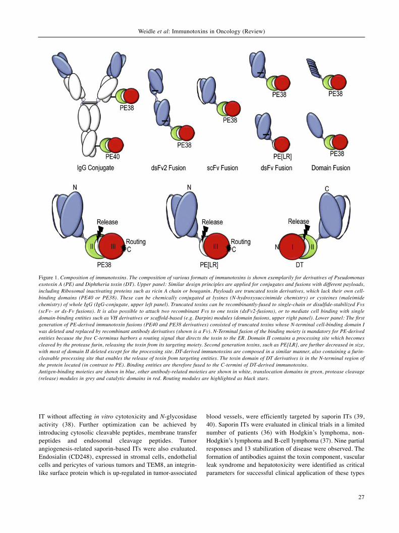
Figure 1. Composition of immunotoxins. The composition of various formats of immunotoxins is shown exemplarily for derivatives of Pseudomonasexotoxin A (PE) and Diphtheria toxin (DT). Upper panel: Similar design principles are applied for conjugates and fusions with different payloads,including Ribosomal inactivating proteins such as ricin A chain or bouganin. Payloads are truncated toxin derivatives, which lack their own cell-binding domains (PE40 or PE38). These can be chemically conjugated at lysines (N-hydroxysuccinimide chemistry) or cysteines (maleimidechemistry) of whole IgG (IgG-conjugate, upper left panel). Truncated toxins can be recombinantly-fused to single-chain or disulfide-stabilized Fvs(scFv- or ds-Fv fusions). It is also possible to attach two recombinant Fvs to one toxin (dsFv2-fusions), or to mediate cell binding with singledomain-binding entities such as VH derivatives or scaffold-based (e.g. Darpin) modules (domain fusions, upper right panel). Lower panel: The firstgeneration of PE-derived immunotoxin fusions (PE40 and PE38 derivatives) consisted of truncated toxins whose N-terminal cell-binding domain Iwas deleted and replaced by recombinant antibody derivatives (shown is a Fv). N-Terminal fusion of the binding moiety is mandatory for PE-derivedentities because the free C-terminus harbors a routing signal that directs the toxin to the ER. Domain II contains a processing site which becomescleaved by the protease furin, releasing the toxin from its targeting moiety. Second generation toxins, such as PE[LR], are further decreased in size,with most of domain II deleted except for the processing site. DT-derived immunotoxins are composed in a similar manner, also containing a furin-cleavable processing site that enables the release of toxin from targeting entities. The toxin domain of DT derivatives is in the N-terminal region ofthe protein located (in contrast to PE). Binding entities are therefore fused to the C-termini of DT-derived immunotoxins.Antigen-binding moieties are shown in blue, other antibody-related moieties are shown in white, translocation domains in green, protease cleavage(release) modules in grey and catalytic domains in red. Routing modules are highlighted as black stars.

of ITs. Other RITs are based on gelonin (rGel) which doesnot bind efficiently to cell surfaces (41). A vascularendothelial growth factor (VEGF121)-rGel fusion protein wasshown to inhibit vascular endothelial growth factor receptor2 (VEGFR2)-mediated angiogenesis and as a consequenceprostate cancer progression in an in vivo model by targetingosteoblasts, osteoclasts and vasculature simultaneously (42,43). B-Lymphocyte stimulator (BLys-rGel) fusion proteintargeting malignant B-cells expressing the B-Lymphocytestimulator BLys receptors, B cell activating factor receptor(BAFF-R), transmembrane activator and calcium-modulatingcyclophilin ligand (CAML) interactor (TACI), and B-cellmaturation antigen (BCMA) (44) of diffuse large B-celllymphoma, mediated growth inhibition in vitro and in vivo byup-regulation of B-cell lymphoma-associated X (BAX) andcaspase 3, inhibition of signal transducer and activator oftranscription-3 (STAT3) signaling and down-regulation of theinterleukin-6 (IL6) receptor (45). Recombinant rGel-single-chain (scFv) fusion proteins targeting human epidermalgrowth factor receptor-2 (HER2), efficiently inhibited growthof a model of ovarian xenograft tumor (46). Another approachis based on the overexpression of FGF-inducible protein 14(Fn14), the cell surface receptor for human tumor necrosisfactor-like weak inducer of apoptosis (TWEAK) in varioustypes of human tumors (47). A recombinant fusion proteincomposed of rGel and a humanized, dimeric single-chainantibody directed against Fn14 was highly cytotoxic to apanel of melanoma cells and significantly inhibited growth ofMDA-MB-435 xenograft tumors (48). VB6-845, an anti-epithelial cellular adhesion molecule (EpCAM) IT containinga T-cell epitope-depleted variant of the plant toxin bouganin(Figure 3D) showed in vitro potency greater than manycommonly used chemotherapeutic agents, and in tumorxenograft models the majority of the mice treated with thistype of molecule were tumor-free at the end of the study (49).
DToxin-based ITs
DT (50, 51) is the prototype of an ADP-ribosylating proteinwhich is secreted by pathogenic strains of the bacteriumCorynebacterium diphtheria and binds to heparin-bindingepidermal growth factor precursor (HBEGF) on the cellmembrane. A single molecule of DT can be lethal to the hostcell (50). DT consists of 535 amino acids and is composed offragments A and B (Figure 4B). Fragment A mediates thecytotoxic enzymatic activity, fragment B confers cell entryand is sub-divided into a translocation domain (T) and areceptor-binding region (R). The catalytic domain (C) islocated at the N-terminus at amino acids residues 1-193 and iscomposed of 7α-helices and 8β-strands; the T-domain and theR-domain are located at the C-terminus. The T-domain(amino acids 202-378) has nine α-helices and is required fortranslocating the C-domain from the endosome to the cytosol.
The R-domain consists of amino acids 386-535, comprises of10β-strands and binds to HBEGF on the cell membrane. Afterbinding, DT is cleaved by furin or furin-like proteases,leaving the two protein chains still linked by a disulfide bondbetween cysteines 186 and 201 (14), and finally undergoesreceptor-mediated endocytosis. In the endosome, at an acidicpH, the loop connecting the T- and C-domains is cleaved byfurin resulting in a conformational change of the T-domain,which is able to insert into the endosomal membrane due tothe exposure of hydrophobic amino acids. After thesubsequent formation of a channel to translocate the C-domain into the cytosol, the disulfide bond linking the C- andT-domains is reduced, releasing the free C-domain. Once inthe cytosol, the C-domain exerts its toxic activity bytransferring ADP-ribose from nicotinamide dinucleotide(NAD) to diphthamide, a modified histidine residue atposition 699 of eEF2{2-[3-carboxyamido-3-(trimethylamino)propyl]histidine (His699)} (14, 50, 51)resulting in translation inhibition and ultimately cell death(Figure 2). Cytotoxic fusion proteins have been constructedpartially or entirely by removing the R-region and substitutingit with ligands specific for receptors overexpressed on tumorcells. This manipulation does not alter ligand binding to itsreceptor, internalization and endocytosis of the IT,translocation into the cytosol and subsequent ADP-ribosylation of eEF2. Fusion of the ligand to the C-terminusof DT was shown to be the most successful strategy. Themost advanced approaches are targeting of the IL2 receptorby denileukin diftitox (Ontak) and the granulocyte-macrophage colony stimulating factor receptor by DT388-granulocyte macrophage-colony stimulating factor (GM-CSF). Denileukin Diftitox was constructed by the geneticfusion of a truncated form of DT, composed of 389 aminoacids of DT followed by the sequence of human IL2, whichreplaces the receptor-binding domain (52-54). This IT wasapproved for the treatment of advanced cutaneous T-celllymphoma (CTCL) in 1999. A 30% overall response rate anda 10% complete response rate were observed. However,serious side-effects, such as infusion reactions, vascular leaksyndrome and loss of visual acuity were noted (53, 54). InDT388 GM-CSF, human GM-CSF was fused to the truncatedDT toxin (DT388), thus replacing the natural receptorbinding-domain (55, 56). Thirty-one patients withchemotherapy-refractory acute myeloid leukemia (AML)were treated, and one complete remission and two partialresponses were seen after i.v. infusion of DT388 GM-CSF.On the other hand, in two patients, liver failure and transienthepatic encephalopathy were observed possibly due to releaseof cytokines from Kupffer cells.
Bispecific DT-based ITs have also been described (57,58). One of them targets the human IL13 receptor and uPAreceptor, the other one, HER2 and EpCAM. These bispecificITs were more active in vivo than the monospecific ones.
CANCER GENOMICS & PROTEOMICS 11: 25-38 (2014)
28

PE and Variants
Pseudomonas aeruginosa exotoxin A (PE) is one of thevirulence factors of this bacterial strain and is a secretedbacterial toxin composed of 613 amino acids. Similar to DT,it is a member of the family of ADP-ribosylating toxins. X-ray crystallographic structure analysis revealed threedomains (59). Domain I is the receptor binding-domain andis subdivided into nonsequential, but structurally adjacent
domains, Ia and Ib. Via this domain I, PE binds to itsreceptor, CD91, also called α2-macroglobulin receptor/low-density lipoprotein receptor-related protein (α2MR/LRP).Domain II has a crucial function in translocation, anddomain III is the catalytic subunit, which inactivates eEF2through ADP-ribosylation (60-62) (Figure 2 and 4A). Aftersecretion, PE is processed by carboxypeptidases, removingthe C-terminal lysine residue, exposing the ER localizationsignal (REDL). After binding to CD91, PE is internalized by
Weidle et al: Immunotoxins in Oncology (Review)
29
Figure 2. How immunotoxins enter and kill cells. The mode by which immunotoxins enter and kill cells is similar for most attached payloads. Shownare the subsequent steps of entry pathways for Pseudomas exotoxin (PE), Diphteria toxin (DT) and Ribosomal-inactivating proteins (RIP) (e.g. bouganin-derived immunotoxins), which inhibit the protein synthesis of eukaryotic cells. Step 1: Immunotoxins (applies to all payloads) bind to cell surfaceantigens and hence accumulate on target cells. Target antigen specificity determines the degree of accumulation on desired (cancer) cells and potentialundesired accumulation on antigen-expressing normal cells. Step 2: Internalization of immunotoxins (frequently via clathrin-coated pits) is triggered byinternalization of the bound target antigen. Internalization is necessary for subsequent entry into the cell. Therefore, non-internalizing antigens makepoor targets for immunotoxin therapy. Step 3: Once internalized, toxins need to be released from their targeting moieties to evade degradation and toenable their independent route within the cell. In the case of recombinant fusion proteins, release from targeting domains is achieved by proteolyticcleavage in endosomal compartments (e.g. via furin for DT, PE and bouganin fusions). Payloads of conjugates can also become released by proteolysisor disulfide reduction. Step 4: Intracellular routing and distribution of processed toxin domains are no longer influenced by bound target antigen. PEderivatives contain a C-terminal routing signal by means of which they enter the endoplasmic reticulum (ER), and subsequently translocate directly intothe cytoplasm. Translocation of RIP derivatives also follows the same route. DT derivatives can translocate directly into the cytoplasm from endosomalcompartments. Step 5: Toxins are in fact enzymes that inactivate intracellular targets. Protein synthesis inhibition by PE and DT is a consequence ofADP-ribosylation of eukaryotic elongation factor-2 (eEF2) at its diphthamide residue, leading to cell death. RIPs block protein synthesis and kill cellsby cleaving ribosomal rRNA and hence inactivating the ribosome. eEF2, rRNA, ribosomal RNA; TGN, trans-Golgi network.

receptor-mediated endocytosis in clathrin-coated pits. Withinthe acidic endocytic compartment, PE dissociates from itsreceptor and is cleaved into two fragments by theendoprotease furin. In the trans Golgi network the REDLsequence binds to the lysine-aspartic acid-glutamic acid-leucine (KDEL) intracellular sorting receptor. The C-terminal fragment complex of domain III and part ofdomain II is transported to the ER, probably due to theREDL sequence. Subsequently, the toxin translocates to thecytoplasm, mediated by sequences located in domain II,where it ADP-ribosylates eEF2 (Figure 2). Several residuesin domain III were shown to have a crucial function incatalysis, such as Glu 553, His 440, Tyr 481 and Tyr 470(63). Diphthamide of eEF2 has been identified as crucial forthe ADP-ribosylation function of PE (64, 65). The ADP-ribosyl group is derived from NAD+ and is transferred to theN3 atom of the imidazole moiety of diphthamide. NAD+ iscleaved to produce nicotinamide, which is released, and anADP ribosyl oxycarbenium containing a positively charged
ribosyl group that reacts with N3 of the imidazole moiety ofdiphthamide (66, 67). Several lines of evidence point to adual mode of action of PE, including inhibition of proteinsynthesis and induction of apoptosis. However, it is notprecisely known how ADP-ribosylation of eEF2 andinduction of apoptosis are interconnected. In cell linesderived from hematological malignancies, hallmarks ofapoptosis such as cleavage of poly(ADP)-ribose polymeraseand DNA laddering, were noted (68, 69). In essence it wasshown that PE-mediated killing is often facilitated, but notcompletely dependent, on apoptosis. More recently it wasshown that PE-mediated apoptosis is dependent on B cellleukemia 2 (BCL2) homologous antagonist/killer (BAK) andis preceded by the degradation of myeloid cell factor-1(MCL-1) (70). Regarding killing efficacy, it was shown thatas few as 1000 molecules of PE per cell can induce celldeath in vitro, and 400-750 molecules per cell can mediatetumor regression in vivo (71). Using labeled PE, it wasshown that less than 1% of the internalized PE may
CANCER GENOMICS & PROTEOMICS 11: 25-38 (2014)
30
Figure 3. Structure of Anthrax toxin, saporin, ricin and bouganin. The proteins or protein assembly are displayed as ribbons with a color coderanging from N- (blue) to C-terminus (red). Structural data from the Protein DataBank (PDB, Oct. 2013) (124) were extracted and superimposed(125) when required to reveal the functional motifs. A: Anthrax toxin has three components: the pro-antigen protein PA83 (pdbcode: 1ACC)recognizes its receptor and a 20-kDa fragment is cleaved from PA83’s amino terminus. The remaining receptor-bound portion of the pro-antigen,PA63 (pdbcode: 3KWV), assembles into a ring-shaped oligomer forming a translocase pore. The exposed new PA63 N-terminal part binds up to threeto four lethal factor proteins (LF, pdcode: 1JKY) edema factor proteins (EF, pdbcode: 1Y0V) and mediates endocytosis. Both EF and LF exhibitenzymatic activity, leading to cellular death. B, C and D: The ribosome-inactivating proteins (RIPs), saporin (B, pdbcode: 1QI7), ricin (C, pdbcode:2AAI), and bouganin (D, pdbcode: 3CTK). They are structurally very similar with respect to their enzymatic domain. The type II RIPs such as ricinhave a B chain with lectin-like properties conferring cytotoxicity in addition to the catalytic domain a. Amino- and carboxy-termini of the proteinsare marked with N and C.

ultimately reach the cytosol and based on observations withDT, one single toxin molecule in the cytosol may besufficient to kill a cell (72, 73).
PE derivatives. Recombinant immunotoxins are chimericmolecules that fuse antibody-based moieties to fragments ofPE (Figure 1). The most frequently used are PE38 and PE40(14, 61), truncated versions of PE, consisting of amino acids253-334 and 318-613 or amino acids 381-613, respectively.Recently, a smaller version with a more extensive deletion indomain II, referred to as PE[LR] was described. PE[LR]encompasses amino acids 274 to 285 and 394 to 613 (62)(Figures 4C and D). Several deimmunized ITs are derived
from this version and are described under de-immunized ITslater in more detail.
PE-based recombinant ITs. First-generation ITs wereantibodies chemically-coupled to toxins with oftendisappointing efficacy in animal models due to non-specifickilling of normal cells. In the second generation, a modifiedversion of PE with a deleted cell-binding region waschemically attached to antibodies. These molecules were muchbetter tolerated in animals and some of them were evaluated inphase I studies in patients with cancer. Some clinical activitywas observed, but these agents were abandoned due toheterogeneity of composition and low tumor penetrance due
Weidle et al: Immunotoxins in Oncology (Review)
31
Figure 4. Structure of Pseudomonas exotoxin variants and Diphtheria toxin. A: The Pseudomonas aeruginosa exotoxin (pdbcode: 1IKQ) containsthree successive domains for receptor binding (blue), translocation into the cell (green), and enzymatic activity (red). B: Diphteria toxin (pdbcode:1SGK) also consists of three domains with similar functions compared to PE toxin but in the reverse order. C: A shortened variant of thePseudomonas exotoxin (PE38) is coupled to a single-chain variable domain fragment (scFv) anti-mesothelin targeting moiety. The immunoglobulinvariable domains (cyan and violet) are linked together and to PE38 with flexible linkers (pink). The PE38 translocation domain (green) contains theprotease cleavage site (black) and is linked directly to the catalytic domain (red) (101). D: Modified PE38 includes one mutation removing a T-cellepitope (magenta) and eight mutations removing B cell epitopes (orange). Four B-cell epitopes are located on the shown protein surface. Thestructural models were generated based on available structure data of domains or entities and are assembled and minimized using DiscoveryStudio40(124, 125). Amino- and carboxy-termini of the proteins are marked with N and C.

to their large size (61) (Figure 1). In the third generation ofrecombinant ITs the cell attachment region of PE was replacedby scFv in which heavy chain variable domain (VH) and lightchain variable domain (VL) are either connected by a linkeror antibody Fv in which VH and VL domains are linked bydisulfide bond stabilized Fv (dsFv). The antibody moiety isfollowed by the translocation and cell killing domains. Inanother version of recombinant ITs, the cell-binding region ofthe IT was replaced by a ligand for a receptor overexpressedon tumor cells. Transforming growth factor-α (TGFα), IL2,GM-CSF, IL4, IL13 are ligands for which ITs were evaluated,with excellent in vivo efficacy (61). However, it should be keptin mind that these agents were evaluated in immune-compromised mice, bypassing the immunogenicity issues ofthe ITs under evaluation. Targets for scFv- or dsFv-basedrecombinant ITs were CD22, CD25, Lewis Y, mesothelin andothers (61). Some of these agents are discussed in greaterdetail later. Lewis Y is a glycoprotein-based antigenoverexpressed in adenocarcinomas. However, vascular leaksyndrome and gastric and renal toxicity have hampered furtherdevelopment of a recombinant IT targeted to Lewis Y (74, 75).The specific subunit of the IL3 receptor (IL-3Rα, CD123) isstrongly expressed on various leukemia blasts and stem cells.There is evidence that relapsed AML is due to leukemic stemcells which express CD123 (76). A recombinant IT[26292(Fv)-PE38] killed CD123+ cells of lines TF1, Molm-13 and Molm-14 with an inhibitory concentration 50 (IC50) of40 nM (76). More recently, a recombinant IT targetingmalignant B-cells expressing receptor tyrosine kinase-likeorphan receptor 1 (ROR-1) was evaluated (77). ROR-1 wasshown to be overexpressed in chronic lymphocytic leukemiaand mantle cell lymphoma (78, 79). The correspondingrecombinant IT consisting of the VH and VL fragments of anantibody to ROR1 and PE38 was partially internalized bychronic lymphocytic leukemia and mantle cell leukemia cellsand induced apoptosis in the latter which expressed ROR-1 invitro with IC50 between 16 pM and 16 nM, paralleling ROR-1 expression levels among these cell lines, but did not affectROR-1-negative cell lines (77). H22scFv-PE is a human scFv-based fusion protein directed against CD64 (80) with theability to kill AML-derived cell lines and primary cells derivedfrom patients with AML. In a U937 cell-based severecombined immunodeficient (SCID) mouse xenograft modelfor AML, the IT eliminated CD64+ cells in mouse organs, andtreatment prolonged overall survival.
Parameters Affecting Recombinant IT-based Cytotoxicity
Since many steps are involved from binding of the IT to thecell surface until its final release into the cytoplasm andsubsequent cell killing, several parameters can have animpact on the efficacy of IT-based cytotoxicity. Antigen
density on the cell surface, selectivity of the target, affinity ofthe antibody to its target antigen and stability of theantibody-antigen complexes are crucial parameters forselectivity and efficacy. PE-based recombinant IT targetingCD19, CD22 and mesothelin showed better efficacy afterimproving affinity which resulted in longer cell surfaceretention (81, 82). Another parameter affecting efficacy ofrecombinant ITs is the epitope recognized by thecorresponding antibodies. Recombinant ITs directed againstdifferent epitopes of an antigen showed significantdifferences in their in vitro potency (81-83). A correlationbetween target antigen density and apoptosis induction hasbeen observed for several recombinant ITs (77, 82-85).However, downstream effector molecules such as BCL2levels and the levels of other anti-apoptotic proteins such asMCL1, survivin and inhibitor of apoptosis proteins werereported to modulate the efficacy of recombinant ITs.Enhanced killing of CD22+ leukemia cells was observed fora combination therapy with BCL2 inhibitor ABT-737 (86,87). Along these lines, it was shown that low BAKexpression could cause resistance to recombinant ITtreatment and that combining recombinant IT with TNFα-related apoptosis-inducing ligand or their agonists was ableto alleviate resistance (88). A further parameter, modulatingthe efficacy of recombinant ITs is the internalization rate oftheir complexes. This is supported by a comparative analysisof recombinant ITs directed against CD19 and CD22 (89).All steps of the intracellular routing and processingpotentially influence efficacy. In addition, it was recentlyshown that the insulin receptor negatively regulatesintracellular processing of a recombinant IT directed againstmesothelin (90). siRNA knock-down of insulin receptorenhanced the cytotoxic activity of native PE and mesothelin-targeted recombinant IT in several human cell lines, but didnot affect the response to cytotoxic agents such as TNFα-related apoptosis-inducing ligand (TRAIL), etoposide orcycloheximide. Making use of recombinant IT constructswhich are targeted to the endocytic recycling compartmentversus those which are delivered through the late endosomes,it was shown that the trafficking route via specific organelleswas an important factor modulating the efficacy of LMB2, arecombinant IT composed of PE38 and an Fv directedagainst the IL2 receptor (91).
Deimmunization
Recombinant ITS are highly immunogenic proteins (92).Thus, removal of B- and T-cell epitopes is a prerequisite forreducing their immunogenicity (92). In general, thegeneration of high-affinity antibodies is initiated by theinteraction of cell surface immunoglobulin on B-cells withcorresponding B-cell epitopes of the antigen. Thesecomplexes are subsequently internalized and processed, and
CANCER GENOMICS & PROTEOMICS 11: 25-38 (2014)
32

peptides derived from these antigens associated with majorhistocompatibility complex (MHC) class II molecules arepresented to CD4+ helper T-cells, which are essential for thegeneration of high-affinity antibodies (93, 94). B-Cellepitopes are mostly discontinuous and conformation-dependent (95-97). In contrast, T-cell epitopes arecontinuous epitopes which are displayed on MHCmolecules. These peptide-MHC complexes in turn are boundby T-cell receptors (98, 99). Since T-cells are essential forantibody responses to proteins, removal of T-cell epitopesalso has to be part of de-immunization strategies. In the caseof PE, B-cell epitopes were identified by using a panel ofantibodies derived from immunized mice. These epitopeswere also recognized by human antibodies present in thesera of patients treated with PE38-based recombinant ITs(100). A mutual competition assay making use of the nativeantigen and 60 monoclonal antibodies involving pair-wisecompetitions was used to identify seven major epitopegroups and 13 subgroups (101). The exact location of theepitopes was determined by introducing 41 individualmutations by alanine replacement of bulky amino acids andsubsequent loss of binding analyzing a panel of monoclonalantibodies. Finally, the thus-identified epitopes of PE38were combined into one molecule, with the final moleculecontaining eight amino acids replacements. The resultingRIT(HA22-8X) targeting CD22 on leukemia cells showedsimilar anti-tumor activity as the non-mutated HA22 inxenograft models (102). HA22-8X induced much lowerantibody responses compared to HA22 after repeated i.v.injections into mice. Immunogenicity was further reducedby removing a large part of PE38 domain II, resulting inHA22-LR-8M (103). HA22-LR-8M did not induceantibodies in mice when given repeatedly by i.v. injectionsand did not induce a secondary response when administeredto mice previously exposed to HA22. HA22-LR-8Mmaintained cytotoxicity against CD22+ leukemia cells andanti-tumoral activity in xenograft models. Furthermodification resulted in HA22-LR-010 (104). Fvs derivedfrom B-cells of patients with antibodies against recombinantITs were isolated and a phage display library containing therecombinant IT-binding Fvs was established. Based on theepitope information thus obtained, HA-22-LR-010 wasconstructed. HA-22-LR-01 possesses low reactivity tohuman anti-sera while maintaining in vitro and in vivocytotoxicity (104). Furthermore, an immunodominant T-cellepitope in PE-based recombinant ITs was identified andeliminated. This was achieved by incubation of peripheralblood mononuclear cells with an IT to stimulate T-cellactivation, subsequent re-stimulation to overlapping peptidesderived from P38, and quantitation of the responses in anIL2-enzyme-linked immunospot assay. The identifiedepitope was recognized by T-cells of 46% (23/50) of thedonors. A mesothelin-targeting recombinant IT (SS-
LR/GGS/8M) with greatly reduced immunogenicity afterremoval of the B-cell epitopes, as described above, wasfunctionally evaluated (105) (Figure 4C and D).
Examples of Proof-of-concept Studies with PE-based Recombinant ITs
In the following, we highlight promising preclinical andclinical studies of PE-based ITs targeting CD22 andmesothelin. CD22 is a lineage restricted differentiationantigen expressed on B-cells and B-cell derivedmalignancies, which is internalized after binding byrecombinant ITs (106). A disulfide-bridged Fv geneticallyfused to PE38, referred to as BL22 or CAT-3888, was foundto be cytotoxic to cells. This required as few as 350 CD22-binding sites per cell and induced tumor regression in CD22+xenografts (107, 108). The agent was also evaluated inpatients with hairy cell leukemia by i.v. administration everyother day three times in adults and every other day three (orsix) times in children, with cycles repeated every 21 or 28days. In a phase II study the overall response rate was 72%,including 47% complete responses (109). The dose-limitingtoxicity (DLT) was completely reversible hemolytic uremicsyndrome. Since BL22 was less active in malignancies withlower numbers of anti- CD22 binding sites, such as chroniclymphocyte leukemia, and with rapidly proliferating cells,such as acute lymphoblastic leukemia, HA22 (moxetumumabpasudotox) was generated (110). HA22 has a 14-fold higheraffinity for CD22 due to a lower off-target rate and was moretoxic towards hairy cell leukemia and chronic lymphocyticleukemia (110), but had similar animal toxicity andpreclinical antitumor efficacy. A corresponding phase I studyin patients with hairy cell leukemia, chronic lymphocyticleukemia, non Hodgkins lymphoma and acute lymphocyticleukemia is ongoing.
Another promising approach relies on recombinant ITsdirected against mesothelin, a cell-specific receptor which ishighly overexpressed in malignant mesothelioma, as well aspancreatic, ovarian and lung adenocarcinomas (111). Theattraction of this target for recombinant IT based approachesis the dramatic overexpression of mesothelin in tumors ascompared to normal mesothelial cells that line the pleura,peritoneum and pericardium (112). The IT SS1P consists ofan anti- mesothelin Fv fused to PE38. SS1P has high affinityto mesothelin (Kd=0.72 nM), killed mesothelin -positivetumor cells efficiently and caused regression of mesothelin-expressing tumor xenografts (105, 112-114). In suchxenografts, combination treatment with SS1P and paclitaxelresulted in increased antitumor activity with completeregression (115, 116). The increased efficacy was shown tobe based on altered tumor architecture and inhibition ofmesothelin shedding, and was restricted to paclitaxel-sensitive cells (115, 116). The most promising clinical results
Weidle et al: Immunotoxins in Oncology (Review)
33

were noted in clinical phase I studies evaluating acombination of SS1P with pemetrexed and cisplatin asfrontline therapy in patients with advanced, unresectablemesothelioma. Of the 14 patients treated at all dose levels,seven had a partial response, three had stable disease andfour had progressive disease (117). Of note, of the sevenpatients evaluable at the maximal tolerated dose, five had apartial response, one patient had stable disease and only onehad progressive disease. Edema, hypo-albuminemia, fatigueand pleuritis were the main side-effects. A recombinant ITreferred to as SS1-LR/GCS/8M, directed against mesothelinand re-engineered for high-affinity, low off-target toxicityand reduced antigenicity was described (82). Reduced off-target toxicity such as vascular leak syndrome was achievedby deletion of a large part of domain II of PE whilemaintaining the 11 residue furin cleavage site and includinga Gly-Gly-Ser linker at the C-terminus of the furin cleavagesite. Dramatically reduced immunogenicity was achieved inmice by introducing the above mentioned eight mutationsthat silence B-cell epitopes into domain III (82).
Critical Issues
Immunogenicity of recombinant ITs is a critical issue forrepeated use. Based on numerous clinical studies, theincidence of an immune response after a single cycle is 0-40% for hematological tumors and 50-100% for solid tumors(118). Removal of B- and T-cell epitopes andimmunosuppressive treatment are important approaches toaddress this issue. Another common toxicity problem inpatients treated with recombinant ITs is vascular leaksyndrome caused by binding of recombinant ITs toendothelial cells, and is characterized by edema, hypo-albuminemia and hypotension (119, 120). It was shown thatthe truncation of a large part of domain II of PE in amesothelin-directed IT prevented this kind of toxicity (82).Hepatotoxicity caused by binding of basic amino acids tonegatively-charged hepatic cells is another issue (119-121).Substitutions in the Fv part of the recombinant IT mightsolve this problem. Kidney toxicity was frequently observedafter treatment of patients with recombinant ITs. Theunderlying mode of action has not been resolved yet, butcould be due to the rapid kidney filtration of the recombinantITs because of their small size, since kidney toxicity has notbeen observed with whole antibody-based recombinant ITs.Proteinuria, hematuria and creatinine release are hallmarksof kidney toxicity. Cytokine release syndrome with fever,chills, hypotension and bone pain was also frequentlyobserved (121). In addition to these general toxicities, target-mediated side-effects were noted. With a recombinant IT-targeting Lewis Y antigen, severe gastritis and renal toxicitywere observed. This is due to antigen expression by normalcells of the stomach and tubular cells of the kidney,
respectively (61, 122). Hepatotoxicity was noticed with aHER2-directed, PE-based recombinant IT (123) due to theexpression of HER2 on normal hepatocytes revealed byimmunohistochemistry.
Concluding RemarksAs outlined above, considerable progress was made by ‘taming’ITs for cancer treatment. The impact of antigen density,internalization of antigen-recombinant IT complexes, efficacyof intracellular processing, including delivery into the cytosol,and the status of cancer cells with respect to expression of anti-apoptotic proteins needs further investigation for each IT andtumor type under consideration. Diphthamide modification ofeEF2 seems to be a prerequisite for efficacy of PE- and DT-based recombinant ITs, but other additional parameters involvedin modulation of efficacy of recombinant ITs need to beidentified. In addition, involvement of changes of tumorarchitecture induced by chemotherapy or other antitumor agentsand its impact on the in vivo efficacy of recombinant ITsdeserves further investigation. The elimination of off-targeteffects by further genetic engineering of recombinant ITs is apromising avenue, as shown by the elimination of vascular leaksyndrome caused by PE-based recombinant ITs throughdeletion of a large part of domain II of PE (62). The clinicalperformance of deimmunized recombinant ITs with B- and T-cell epitopes removed will be crucial for the future prospects oftreatment of cancer with recombinant ITs.
References
1 Hanahan D and Weinberg RA: Hallmarks of cancer: the nextgeneration. Cell 144: 646-674, 2011.
2 Hanahan D and Coussens LM: Accessories to the crime:Functions of cells recruited to the tumor microenvironment.Cancer Cell 21: 309-322, 2012.
3 Huang T, Karsy M, Zhuge J, Zhong M and Liu D: B-RAF and theinhibitors: from bench to bedside. J Hematol Oncol 6: 30, 2013.
4 Antonicelli A, Cafarotti S, Indini A, Galli A, Russo A, CesarioA, Lococo FM, Russo P, Mainini AF, Bonifati LG, Nosotti M,Santambrogio L, Margaritora S, Granone PM and Dutly AE:EGFR-targeted therapy for non-small cell lung cancer: Focus onEGFR oncogenic mutation. Int J Med Sci 10: 320-330, 2013.
5 Montoni A, Robu M, Pouliot E and Shah GM: Resistance toPARP-inhibitors in cancer therapy. Front Pharmacol 4: 18, 2013.
6 Weidle UH, Maisel D and Eick D: Synthetic lethality-basedtargets for discovery of new cancer therapeutics. CancerGenomics Proteomics 8: 159-171, 2011.
7 Sennino B and McDonald DM: Controlling escape fromangiogenesis inhibitors. Nat Rev Cancer 12: 699-709, 2012.
8 Jayson GC, Hicklin DJ and Ellis LM: Antiangiogenic therapy-evolving view based on clinical trial results. Nat Rev Clin Oncol9: 297-303, 2012.
9 von Boehmer H and Daniel C: Therapeutic opportunities formanipulating T(Reg) cells in autoimmunity and cancer. Nat RevDrug Discov 12: 51-63, 2013.
CANCER GENOMICS & PROTEOMICS 11: 25-38 (2014)
34

10 Alderton GK and Bordon Y: Tumour immunotherapy leukocytestake up the fight. Nat Rev Immunol 12: 237, 2012.
11 Li F, Zhao C and Wang L: Molecular-targeted agentscombination therapy for cancer: Developments and potentials.Int J Cancer, 2013, in press
12 Axelrod M, Gordon VL, Conaway M, Tarcsafalvi A, Neitzke DJ,Gioeli D and Weber MJ: Combinatorial drug screening identifiescompensatory pathway interactions and adaptive resistancemechanisms. Oncotarget 4: 622-635, 2013.
13 Piro A, Tagarelli A, Tagarelli G, Lagonia P and Quattrone A:Paul Ehrlich: The Nobel Prize in physiology or medicine 1908.Int Rev Immunol 27: 1-17, 2008.
14 Shapira A and Benhar I: Toxin-based therapeutic approaches.Toxins 2: 2519-2583, 2010.
15 Ascenzi P, Visca P, Ippolito G, Spallarossa A, Bolognesi M andMontecucco C: Anthrax toxin: A tripartite lethal combination.FEBS Lett 531: 384-388, 2002.
16 Collier RJ and Young JA: Anthrax toxin. Annu Rev Cell DevBiol 19: 45-70, 2003.
17 Young JA and Collier RJ: Anthrax toxin: receptor binding,internalization, pore formation, and translocation. Annu RevBiochem 76: 243-265, 2007.
18 Bradley KA, Mogridge J, Mourez M, Collier RJ and Young JA:Identification of the cellular receptor for anthrax toxin. Nature414: 225-229, 2001.
19 Scobie HM, Rainey GJ, Bradley KA and Young JA: Humancapillary morphogenesis protein 2 functions as an anthrax toxinreceptor. Proc Natl Acad Sci USA 100: 5170-5174, 2003.
20 Adkins I, Holubova J, Kosova M and Sadilkova L: Bacteria andtheir toxins tamed for immunotherapy. Curr Pharm Biotechnol13: 1446-1473, 2012.
21 Liu S, Netzel-Arnett S, Birkedal-Hansen H and Leppla SH:Tumor cell-selective cytotoxicity of matrix metalloproteinase-activated anthrax toxin. Cancer Res 60: 6061-6067, 2000.
22 Liu S, Wang H, Currie BM, Molinolo A, Leung HJ, Moayeri M,Basile JR, Alfano RW, Gutkind JS, Frankel AE, Bugge TH andLeppla SH: Matrix metalloproteinase-activated anthrax lethaltoxin demonstrates high potency in targeting tumor vasculature.J Biol Chem 283: 529-540, 2008.
23 Alfano RW, Leppla SH, Liu S, Bugge TH, Meininger CJ,Lairmore TC, Mulne AF, Davis SH, Duesbery NS and FrankelAE: Matrix metalloproteinase-activated anthrax lethal toxininhibits endothelial invasion and neovasculature formation duringin vitro morphogenesis. Mol Cancer Res 7: 452-461, 2009.
24 Liu S, Bugge TH and Leppla SH: Targeting of tumor cells bycell surface urokinase plasminogen activator-dependent anthraxtoxin. J Biol Chem 276: 17976-17984, 2001.
25 Liu S, Aaronson H, Mitola DJ, Leppla SH and Bugge TH:Potent antitumor activity of a urokinase-activated engineeredanthrax toxin. Proc Natl Acad Sci USA 100: 657-662, 2003.
26 Duesbery NS, Resau J, Webb CP, Koochekpour S, Koo HM,Leppla SH and Vande Woude GF: Suppression of RAS-mediatedtransformation and inhibition of tumor growth and angiogenesisby anthrax lethal factor, a proteolytic inhibitor of multiple MEKpathways. Proc Natl Acad Sci USA 98: 4089-4094, 2001.
27 Depeille P, Young JJ, Boguslawski EA, Berghuis BD, Kort EJ,Resau JH, Frankel AE and Duesbery NS: Anthrax lethal toxininhibits growth of and vascular endothelial growth factor releasefrom endothelial cells expressing the human herpes virus 8 viral Gprotein coupled receptor. Clin Cancer Res 13: 5926-5934, 2007.
28 Endo Y, Mitsui K, Motizuki M and Tsurugi K: The mechanismof action of ricin and related toxic lectins on eukaryoticribosomes. The site and the characteristics of the modificationin 28-S ribosomal RNA caused by the toxins. J Biol Chem 262:5908-5912, 1987.
29 Peumans WJ, Hao Q and Van Damme EJ: Ribosome-inactivatingproteins from plants: More than RNA N-glycosidases? FASEB J15: 1493-1506, 2001.
30 Girbés T, Ferreras JM, Arias FJ and Stirpe F: Description,distribution, activity and phylogenetic relationship of ribosome-inactivating proteins in plants, fungi and bacteria. Mini Rev MedChem 4: 461-476, 2004.
31 Hartley MR and Lord JM: Genetics of ribosome-inactivatingproteins. Mini Rev Med Chem 4: 487-492, 2004.
32 Stirpe F: Ribosome-inactivating proteins. Toxicon 44: 371-383,2004.
33 Nielsen K and Boston RS: Ribosome-inactivating proteins: Aplant perspective. Annu Rev Plant Physiol Plant Mol Biol 52:785-816, 2001.
34 Stone MJ, Sausville EA, Fay JW, Headlee D, Collins RH, FiggWD, Stetler-Stevenson M, Jain V, Jaffe ES, Solomon D, LushRM, Senderowicz A, Ghetie V, Schindler J, Uhr JW and VitettaES: A phase I study of bolus versus continuous infusion of theanti-CD19 immunotoxin, IgG-HD37-dgA, in patients with B-cell lymphoma. Blood 88: 1188-1197, 1996.
35 Schnell R, Borchmann P, Staak JO, Schindler J, Ghetie V, VitettaES and Engert A: Clinical evaluation of ricin A-chainimmunotoxins in patients with Hodgkin’s lymphoma. Ann Oncol14: 729-736, 2003.
36 Schnell R, Staak O, Borchmann P, Schwartz C, Matthey B,Hansen H, Schindler J, Ghetie V, Vitetta ES, Diehl V and EngertA: A Phase I study with an anti-CD30 ricin A-chain immunotoxin(Ki-4.dgA) in patients with refractory CD30+ Hodgkin’s and non-Hodgkin’s lymphoma. Clin Cancer Res 8: 1779-1786, 2002.
37 Polito L, Bortolotti M, Pedrazzi M and Bolognesi A:Immunotoxins and other conjugates containing saporin-s6 forcancer therapy. Toxins 3: 697-720, 2011.
38 Heisler I, Keller J, Tauber R, Sutherland M and Fuchs H: Acleavable adapter to reduce nonspecific cytotoxicity ofrecombinant immunotoxins. Int J Cancer 103: 277-282, 2003.
39 Rouleau C, Curiel M, Weber W, Smale R, Kurtzberg L,Mascarello J, Berger C, Wallar G, Bagley R, Honma N, HasegawaK, Ishida I, Kataoka S, Thurberg BL, Mehraein K, Horten B,Miller G and Teicher BA: Endosialin protein expression andtherapeutic target potential in human solid tumors: sarcoma versuscarcinoma. Clin Cancer Res 14: 7223-7236, 2008.
40 Yang MY, Chaudhary A, Seaman S, Dunty J, Stevens J, ElzarradMK, Frankel AE and St Croix B. The cell surface structure oftumor endothelial marker 8 (TEM8) is regulated by the actincytoskeleton. Biochim Biophys Acta 1813: 39-49, 2011.
41 Rosenblum MG, Kohr WA, Beattie KL, Beattie WG, Marks W,Toman PD and Cheung L: Amino acid sequence analysis, geneconstruction, cloning, and expression of gelonin, a toxin derivedfrom Gelonium multiflorum. J Interferon Cytokine Res 15: 547-555, 1995.
42 Mohamedali KA, Ran S, Gomez-Manzano C, Ramdas L, Xu J,Kim S, Cheung LH, Hittelman WN, Zhang W, Waltenberger J,Thorpe PE and Rosenblum MG: Cytotoxicity of VEGF(121)/ rGelon vascular endothelial cells resulting in inhibition of angiogenesisis mediated via VEGFR-2. BMC Cancer 11: 358, 2011.
Weidle et al: Immunotoxins in Oncology (Review)
35

43 Mohamedali KA, Li ZG, Starbuck MW, Wan X, Yang J, Kim S,Zhang W, Rosenblum MG and Navone NM: Inhibition ofprostate cancer osteoblastic progression with VEGF121/rGel, asingle agent targeting osteoblasts, osteoclasts, and tumorneovasculature. Clin Cancer Res 17: 2328-2338, 2011.
44 Lyu MA, Rai D, Ahn KS, Sung B, Cheung LH, Marks JW,Aggarwal BB, Aguiar RC, Gandhi V and Rosenblum MG: TherGel/BLyS fusion toxin inhibits diffuse large B-cell lymphomagrowth in vitro and in vivo. Neoplasia 12: 366-375, 2010.
45 Lyu MA, Sung B, Cheung LH, Marks JW, Aggarwal BB, AguiarRC and Rosenblum MG: The rGel/BLyS fusion toxin inhibitsSTAT3 signaling via down-regulation of interleukin-6 receptorin diffuse large B-cell lymphoma. Biochem Pharmacol 80: 1335-1342, 2010.
46 Cao Y, Marks JD, Marks JW, Cheung LH, Kim S and RosenblumMG: Construction and characterization of novel, recombinantimmunotoxins targeting the Her2/neu oncogene product: In vitroand in vivo studies. Cancer Res 69: 8987-8995, 2009.
47 Winkles JA: The TWEAK-Fn14 cytokine-receptor axis:Discovery, biology and therapeutic targeting. Nat Rev DrugDiscov 7: 411-425, 2008.
48 Zhou H, Ekmekcioglu S, Marks JW, Mohamedali KA, Asrani K,Phillips KK, Brown SA, Cheng E, Weiss MB, Hittelman WN,Tran NL, Yagita H, Winkles JA and Rosenblum MG: TheTWEAK receptor Fn14 is a therapeutic target in melanoma:immunotoxins targeting Fn14 receptor for malignant melanomatreatment. J Invest Dermatol 133: 1052-1062, 2013.
49 Cizeau J, Grenkow DM, Brown JG, Entwistle J and MacDonaldGC: Engineering and biological characterization of VB6-845, ananti-EpCAM immunotoxin containing a T-cell epitope-depletedvariant of the plant toxin bouganin. J Immunother 32: 574-584,2009.
50 Potala S, Sahoo SK and Verma RS: Targeted therapy of cancerusing diphtheria toxin-derived immunotoxins. Drug DiscovToday 13: 807-815, 2008.
51 Liu S, Milne GT, Kuremsky JG, Fink GR and Leppla SH:Identification of the proteins required for biosynthesis ofdiphthamide, the target of bacterial ADP-ribosylating toxins ontranslation elongation factor 2. Mol Cell Biol 24: 9487-9497,2004.
52 Frankel AE, Rossi P, Kuzel TM and Foss F: Diphtheria fusionprotein therapy of chemoresistant malignancies. Curr CancerDrug Targets 2: 19-36, 2002.
53 LeMaistre CF, Saleh MN, Kuzel TM, Foss F, Platanias LC,Schwartz G, Ratain M, Rook A, Freytes CO, Craig F, Reuben Jand Nichols JC: Phase I trial of a ligand fusion-protein(DAB389IL-2) in lymphomas expressing the receptor forinterleukin-2. Blood 91: 399-405, 1998.
54 Olsen E, Duvic M, Frankel A, Kim Y, Martin A, Vonderheid E,Jegasothy B, Wood G, Gordon M, Heald P, Oseroff A, Pinter-Brown L, Bowen G, Kuzel T, Fivenson D, Foss F, Glode M,Molina A, Knobler E, Stewart S, Cooper K, Stevens S, Craig F,Reuben J, Bacha P and Nichols J: Pivotal phase III trial of twodose levels of denileukin diftitox for the treatment of cutaneousT-cell lymphoma. J Clin Oncol 19: 376-388, 2001.
55 Frankel AE, Hall PD, Burbage C, Vesely J, Willingham M,Bhalla K and Kreitman RJ: Modulation of the apoptoticresponse of human myeloid leukemia cells to a diphtheria toxingranulocyte-macrophage colony-stimulating factor fusionprotein. Blood 90: 3654-3661, 1997.
56 Kreitman RJ and Pastan I: Recombinant toxins containinghuman granulocyte-macrophage colony-stimulating factor andeither pseudomonas exotoxin or diphtheria toxin killgastrointestinal cancer and leukemia cells. Blood 90: 252-259,1997.
57 Todhunter DA, Hall WA, Rustamzadeh E, Shu Y, Doumbia SOand Vallera DA: A bispecific immunotoxin (DTAT13) targetinghuman IL-13 receptor (IL-13R) and urokinase-type plasminogenactivator receptor (uPAR) in a mouse xenograft model. ProteinEng Des Sel 17: 157-164, 2004.
58 Stish BJ, Chen H, Shu Y, Panoskaltsis-Mortari A and ValleraDA: Increasing anticarcinoma activity of an anti-ERBB2recombinant immunotoxin by the addition of an anti-EpCAMscFv. Clin Cancer Res 13: 3058-3067, 2007.
59 Allured VS, Collier RJ, Carroll SF and McKay DB: Structure ofexotoxin A of Pseudomonas aeruginosa at 3.0-Angstromresolution. Proc Natl Acad Sci USA 83: 1320-1324, 1986.
60 Pastan I and FitzGerald D: Pseudomonas exotoxin: Chimerictoxins. J Biol Chem 264: 15157-15160, 1989.
61 Pastan I, Hassan R, Fitzgerald DJ and Kreitman RJ: Immunotoxintherapy of cancer. Nat Rev Cancer 6: 559-565, 2006.
62 Weldon JE and Pastan I: A guide to taming a toxin-recombinantimmunotoxins constructed from Pseudomonas exotoxin A forthe treatment of cancer. FEBS J 278: 4683-4700, 2011.
63 Wilson BA and Collier RJ: Diphtheria toxin and Pseudomonasaeruginosa exotoxin A: active-site structure and enzymicmechanism. Curr Top Microbiol Immunol 175: 27-41, 1992.
64 Foley BT, Moehring JM and Moehring TJ: Mutations in theelongation factor 2 gene which confer resistance to diphtheriatoxin and Pseudomonas exotoxin A. Genetic and biochemicalanalyses. J Biol Chem 270: 23218-23225, 1995.
65 Ivankovic M, Rubelj I, Matulic M, Reich E and Brdar B: Site-specific mutagenesis of the histidine precursor of diphthamidein the human elongation factor-2 gene confers resistance todiphtheria toxin. Mutat Res 609: 34-42, 2006.
66 Jørgensen R, Merrill AR, Yates SP, Marquez VE, Schwan AL,Boesen T and Andersen GR: Exotoxin A eEF2 complexstructure indicates ADP ribosylation by ribosome mimicry.Nature 436: 979-984, 2005.
67 Jørgensen R, Wang Y, Visschedyk D and Merrill AR: The natureand character of the transition state for the ADP-ribosyltransferase reaction. EMBO Rep 9: 802-809, 2008.
68 Keppler-Hafkemeyer A, Brinkmann U and Pastan I: Role ofcaspases in immunotoxin-induced apoptosis of cancer cells.Biochemistry 37: 16934-16942, 1998.
69 Keppler-Hafkemeyer A, Kreitman RJ and Pastan I: Apoptosisinduced by immunotoxins used in the treatment of hematologicmalignancies. Int J Cancer 87: 86-94, 2000.
70 Du X, Youle RJ, FitzGerald DJ and Pastan I: Pseudomonasexotoxin A-mediated apoptosis is Bak dependent and precededby the degradation of Mcl-1. Mol Cell Biol 30: 3444-3452,2010.
71 Kreitman RJ and Pastan I: Accumulation of a recombinantimmunotoxin in a tumor in vivo: Fewer than 1000 molecules percell are sufficient for complete responses. Cancer Res 58: 968-975, 1998.
72 Bard F, Mazelin L, Péchoux-Longin C, Malhotra V and JurdicP: Src regulates Golgi structure and KDEL receptor-dependentretrograde transport to the endoplasmic reticulum. J Biol Chem278: 46601-46606, 2003.
CANCER GENOMICS & PROTEOMICS 11: 25-38 (2014)
36
73 Yamaizumi M, Mekada E, Uchida T and Okada Y: One moleculeof diphtheria toxin fragment A introduced into a cell can kill thecell. Cell 15: 245-250, 1978.
74 Posey JA, Khazaeli MB, Bookman MA, Nowrouzi A, GrizzleWE, Thornton J, Carey DE, Lorenz JM, Sing AP, Siegall CB,LoBuglio AF and Saleh MN: A phase I trial of the single-chainimmunotoxin SGN-10 (BR96 sFv-PE40) in patients withadvanced solid tumors. Clin Cancer Res 8: 3092-3099, 2002.
75 Damle B, Tay L, Comereski C, Warner W and Kaul S: Influenceof immunogenicity on the pharmacokinetics of BMS-191352, aPseudomonas exotoxin immunoconjugate, in rats and dogs. JPharm Pharmacol 52: 671-678, 2000.
76 Du X, Ho M and Pastan I: New immunotoxins targeting CD123,a stem cell antigen on acute myeloid leukemia cells. JImmunother 30: 607-613, 2007.
77 Baskar S, Wiestner A, Wilson WH, Pastan I and Rader C:Targeting malignant B-cells with an immunotoxin against ROR1.MAbs 4: 349-361, 2012.
78 Rosenwald A, Alizadeh AA, Widhopf G, Simon R, Davis RE,Yu X, Yang L, Pickeral OK, Rassenti LZ, Powell J, Botstein D,Byrd JC, Grever MR, Cheson BD, Chiorazzi N, Wilson WH,Kipps TJ, Brown PO and Staudt LM: Relation of geneexpression phenotype to immunoglobulin mutation genotype inB-cell chronic lymphocytic leukemia. J Exp Med 194: 1639-1647, 2001.
79 Klein U, Tu Y, Stolovitzky GA, Mattioli M, Cattoretti G, HussonH, Freedman A, Inghirami G, Cro L, Baldini L, Neri A, CalifanoA and Dalla-Favera R: Gene expression profiling of B-cellchronic lymphocytic leukemia reveals a homogeneous phenotyperelated to memory B-cells. J Exp Med 194: 1625-1638, 2001.
80 Tur MK, Huhn M, Jost E, Thepen T, Brümmendorf TH andBarth S: In vivo efficacy of the recombinant anti-CD64immunotoxin H22(scFv)-ETA in a human acute myeloidleukemia xenograft tumor model. Int J Cancer 129: 1277-1282,2011.
81 Alderson RF, Kreitman RJ, Chen T, Yeung P, Herbst R, Fox JAand Pastan I: CAT-8015: a second-generation pseudomonasexotoxin A-based immunotherapy targeting CD22-expressinghematologic malignancies. Clin Cancer Res 15: 832-839, 2009.
82 Weldon JE, Xiang L, Zhang J, Beers R, Walker DA, Onda M,Hassan R and Pastan I. A recombinant immunotoxin against thetumor-associated antigen mesothelin reengineered for highactivity, low off-target toxicity, and reduced antigenicity. MolCancer Ther 12: 48-57, 2013.
83 Godal A, Kumle B, Pihl A, Juell S and Fodstad O:Immunotoxins directed against the high-molecular-weightmelanoma-associated antigen. Identification of potent antibodytoxin combinations. Int J Cancer 52: 631-635, 1992.
84 Yazdi PT, Wenning LA and Murphy RM: Influence of cellulartrafficking on protein synthesis inhibition of immunotoxinsdirected against the transferrin receptor. Cancer Res 55: 3763-3771, 1995.
85 Press OW, Martin PJ, Thorpe PE and Vitetta ES: Ricin A-chaincontaining immunotoxins directed against different epitopes onthe CD2 molecule differ in their ability to kill normal andmalignant T-cells. J Immunol 141: 4410-4417, 1988.
86 Decker T, Oelsner M, Kreitman RJ, Salvatore G, Wang QC,Pastan I, Peschel C and Licht T: Induction of caspase-dependentprogrammed cell death in B-cell chronic lymphocytic leukemiaby anti-CD22 immunotoxins. Blood 103: 2718-2726, 2004.
87 Fitzgerald DJ, Moskatel E, Ben-Josef G, Traini R, Tendler T,Sharma A, Antignani A, Mussai F, Wayne A, Kreitman RJ andPastan I: Enhancing immunotoxin cell-killing activity viacombination therapy with ABT-737. Leuk Lymphoma 52: 79-81,2011.
88 Du X, Xiang L, Mackall C and Pastan I: Killing of resistantcancer cells with low Bak by a combination of an antimesothelinimmunotoxin and a TRAIL receptor 2 agonist antibody. ClinCancer Res 17: 5926-5934, 2011.
89 Du X, Beers R, Fitzgerald DJ and Pastan I: Differential cellularinternalization of anti-CD19 and -CD22 immunotoxins resultsin different cytotoxic activity. Cancer Res 68: 6300-6305,2008.
90 Liu XF, FitzGerald DJ and Pastan I: The insulin receptornegatively regulates the action of Pseudomonas toxin-basedimmunotoxins and native Pseudomonas toxin. Cancer Res 73:2281-2288, 2013.
91 Tortorella LL, Pipalia NH, Mukherjee S, Pastan I, Fitzgerald Dand Maxfield FR: Efficiency of immunotoxin cytotoxicity ismodulated by the intracellular itinerary. PLoS One 7: e47320,2012.
92 Nagata S and Pastan I: Removal of B cell epitopes as a practicalapproach for reducing the immunogenicity of foreign protein-based therapeutics. Adv Drug Deliv Rev 61: 977-985, 2009.
93 Parker DC: T-Cell-dependent B-cell activation. Annu RevImmunol 11: 331-360, 1993.
94 Bernard A, Coitot S, Brémont A and Bernard G: T- And B-cellcooperation: A dance of life and death. Transplantation 79: S8-S11, 2005.
95 Van Regenmortel MH: What is a B-cell epitope? Methods MolBiol 524: 3-20, 2009.
96 Barlow DJ, Edwards MS and Thornton JM: Continuous anddiscontinuous protein antigenic determinants. Nature 322: 747-748, 1986.
97 Laver WG, Air GM, Webster RG and Smith-Gill SJ: Epitopeson protein antigens: Misconceptions and realities. Cell 61: 553-556, 1990.
98 De Groot AS, Knopp PM and Martin W: De-immunization oftherapeutic proteins by T-cell epitope modification. Dev Biol122: 171-194, 2005.
99 Parker DC: The functions of antigen recognition in T-cell-dependent B-cell activation. Semin Immunol 5: 413-420, 1993.
100 Onda M, Nagata S, FitzGerald DJ, Beers R, Fisher RJ, VincentJJ, Lee B, Nakamura M, Hwang J, Kreitman RJ, Hassan R andPastan I: Characterization of the B-cell epitopes associated witha truncated form of Pseudomonas exotoxin (PE38) used to makeimmunotoxins for the treatment of cancer patients. J Immunol177: 8822-8834, 2006.
101 Nagata S, Numata Y, Onda M, Ise T, Hahn Y, Lee B and PastanI: Rapid grouping of monoclonal antibodies based on theirtopographical epitopes by a label-free competitive immunoassay.J Immunol Methods 292: 141-155, 2004.
102 Onda M, Beers R, Xiang L, Nagata S, Wang QC and Pastan I:An immunotoxin with greatly reduced immunogenicity byidentification and removal of B cell epitopes. Proc Natl Acad SciUSA 105: 11311-11316, 2008.
103 Onda M, Beers R, Xiang L, Lee B, Weldon JE, Kreitman RJ andPastan I: Recombinant immunotoxin against B-cell malignancieswith no immunogenicity in mice by removal of B-cell epitopes.Proc Natl Acad Sci USA 108: 5742-5747, 2011.
Weidle et al: Immunotoxins in Oncology (Review)
37
104 Liu W, Onda M, Lee B, Kreitman RJ, Hassan R, Xiang L andPastan I: Recombinant immunotoxin engineered for lowimmunogenicity and antigenicity by identifying and silencinghuman B-cell epitopes. Proc Natl Acad Sci USA 109: 11782-11787, 2012.
105 Mazor R, Vassall AN, Eberle JA, Beers R, Weldon JE, VenzonDJ, Tsang KY, Benhar I and Pastan I: Identification andelimination of an immunodominant T-cell epitope inrecombinant immunotoxins based on Pseudomonas exotoxin A.Proc Natl Acad Sci USA 109: E3597-3603, 2012.
106 Kreitman RJ and Pastan I: Antibody fusion proteins: Anti-CD22recombinant immunotoxin moxetumomab pasudotox. ClinCancer Res 17: 6398-6405, 2011.
107 Kreitman RJ: Recombinant immunotoxins for the treatment ofchemoresistant hematologic malignancies. Curr Pharm Des 15:2652-2664, 2009.
108 Kreitman RJ, Margulies I, Stetler-Stevenson M, Wang QC,FitzGerald DJ and Pastan I: Cytotoxic activity of disulfide-stabilized recombinant immunotoxin RFB4(dsFv)-PE38 (BL22)toward fresh malignant cells from patients with B-cellleukemias. Clin Cancer Res 6: 1476-1487, 2000.
109 Kreitman RJ, Stetler-Stevenson M, Margulies I, Noel P,Fitzgerald DJ, Wilson WH and Pastan I: Phase II trial ofrecombinant immunotoxin RFB4(dsFv)-PE38 (BL22) in patientswith hairy cell leukemia. J Clin Oncol 27: 2983-2990, 2009.
110 Salvatore G, Beers R, Margulies I, Kreitman RJ and Pastan I:Improved cytotoxic activity toward cell lines and fresh leukemiacells of a mutant anti-CD22 immunotoxin obtained by antibodyphage display. Clin Cancer Res 8: 995-1002, 2002.
111 Kelly RJ, Sharon E, Pastan I and Hassan R: Mesothelin-targetedagents in clinical trials and in preclinical development. MolCancer Ther 11: 517-525, 2012.
112 Chang K, Pastan I and Willingham MC: Isolation andcharacterization of a monoclonal antibody, K1, reactive withovarian cancers and normal mesothelium. Int J Cancer 50: 373-381, 1992.
113 Chowdhury PS, Viner JL, Beers R and Pastan I: Isolation of ahigh-affinity stable single-chain Fv specific for mesothelin fromDNA-immunized mice by phage display and construction of arecombinant immunotoxin with anti-tumor activity. Proc NatlAcad Sci USA 95: 669-674, 1998.
114 Chowdhury PS and Pastan I: Improving antibody affinity bymimicking somatic hypermutation in vitro. Nat Biotechnol 17:568-572, 1999.
115 Zhang Y, Xiang L, Hassan R, Paik CH, Carrasquillo JA, JangBS, Le N, Ho M and Pastan I: Synergistic antitumor activity oftaxol and immunotoxin SS1P in tumor-bearing mice. ClinCancer Res 12: 4695-4701, 2006.
116 Zhang Y, Xiang L, Hassan R and Pastan I: Immunotoxin andTaxol synergy results from a decrease in shed mesothelin levelsin the extracellular space of tumors. Proc Natl Acad Sci USA104: 17099-17104, 2007.
117 Hassan R, Sharon E, Schuler B, Mallory Y, Zhang Y, Ling A andPastan I: Antitumor activity of SS1P with pemetrexed andcisplatin for front-line treatment of pleural mesothelioma andutility of serum mesothelin as a marker of tumor response. J ClinOncol 29: 15S, (suppl; abstr 7026), 2011.
118 Kreitman RJ: Immunotoxins for targeted cancer therapy. AAPSJ 8: E532-551, 2006.
119 Onda M, Nagata S, Tsutsumi Y, Vincent JJ, Wang Q, KreitmanRJ, Lee B and Pastan I: Lowering the isoelectric point of the Fvportion of recombinant immunotoxins leads to decreasednonspecific animal toxicity without affecting antitumor activity.Cancer Res 61: 5070-5077, 2001.
120 Pastan I, Hassan R, FitzGerald DJ and Kreitman RJ:Immunotoxin treatment of cancer. Annu Rev Med 58: 221-237,2007.
121 Jeyarajah DR and Thistlethwaite JR Jr.: General aspects ofcytokine-release syndrome: timing and incidence of symptoms.Transplant Proc 25: 16-20, 1993.
122 Pastan I, Lovelace ET, Gallo MG, Rutherford AV, Magnani JLand Willingham MC: Characterization of monoclonal antibodiesB1 and B3 that react with mucinous adenocarcinomas. CancerRes 51: 3781-3787, 1991.
123 Pai-Scherf LH, Villa J, Pearson D, Watson T, Liu E, WillinghamMC and Pastan I: Hepatotoxicity in cancer patients receivingERB-38, a recombinant immunotoxin that targets the ERBB2receptor. Clin Cancer Res 5: 2311-2315, 1999.
124 Berman HM, Westbrook J, Feng Z, Gilliland G, Bhat TN,Weissig H, Shindyalov IN and Bourne PE: The Protein DataBank (www.pdb.org). Nucleic Acids Res 28: 235-242, 2000.
125 Accelrys Software Inc., Discovery Studio Modeling Environment,Release 4.0. Accelrys Software Inc., San Diego, 2013.
Received November 27, 2013Revised December 12, 2013Accepted December 13, 2013
CANCER GENOMICS & PROTEOMICS 11: 25-38 (2014)
38