prosthodontics approach for the fabrication of feeding
TRANSCRIPT

Science Journal of Clinical Medicine 2016; 5(4-1): 31-36
http://www.sciencepublishinggroup.com/j/sjcm
doi: 10.11648/j.sjcm.s.2016050401.16
ISSN: 2327-2724 (Print); ISSN: 2327-2732 (Online)
Prosthodontics Approach for the Fabrication of Feeding Plates in Cleft Palate Patients
Shajahan P. A., Rohit Raghavan, Ritha Bos, Geethprasad T. S
Department Of Prosthodontics, Royal Dental College, Chalissery, Palakkad
Email address: [email protected] (Shajahan P. A.), [email protected] (R. Raghavan), [email protected] (R. Bos),
[email protected] (Geethprasad T. S)
To cite this article: Shajahan P. A., Rohit Raghavan, Ritha Bos, Geethprasad T. S. Prosthodontics Approach for the Fabrication of Feeding Plates in Cleft Palate
Patients. Science Journal of Clinical Medicine. Special Issue: Clinical Conspectus on Cleft Deformities. Vol. 5, No. 4-1, 2016, pp. 31-36.
doi: 10.11648/j.sjcm.s.2016050401.16
Received: December 23, 2015; Accepted: March 8, 2016; Published: May 19, 2016
Abstract: Cleft lip and palate is most common congenital anomalies of the craniofacial region. Cleft lip and palate is an
anomaly that affects several systems and functions which includes the facial growth, dentition, speech, hearing and genetic
aspects because of the complex mode of inheritance. Cleft lip and palate forms a part of many syndromicand non-syndromic
disorders like the Pierre-Robin sequence, etc. Early intervention provides a positive impact on the development of the infants
with clefts. Adequate knowledge of the appliances which are available and the impression procedures which should be
followed, leads to a better understanding and coordination of the efforts of the various specialties which are involved in cleft
lip and palate care.
Keywords: Cleft Lip, Cleft Palate, Feeding Plate, Impression Materials
1. Introduction
Clefts lip and palate are considered as a common
congenital abnormalities of the orofacial region. Cleft palate
can be defined as a furrow in the palatal vault. This condition
can affect the children and their families in many ways. It is
estimated that the overall global prevalence of oro-facial
clefts is one affected individual in every 800 new born
babies. Cleft palate may be inherited as an autosomal
dominant condition. Family history in first degree
consanguinity increases the risk by a factor of 20 percent.
When a child is born with a cleft, maintenance of nutritional
balance, which is necessary for growth, development, and the
infant’s preparation for corrective final surgery, is a priority
[1]. The oro-nasal communication due to the defect poses
great problems for the newborn in suckling and also affects
speech and the overall physical and mental growth of the
child [2]. The rehabilitation of infants having cleft primarily
involves the defect closure. So the first stage of management
would be the fabrication of a feeding plate or passive
maxillary obturator[3]. The crucial step in fabrication of any
appliance or obturator is the impression procedure. Patient
positioning, tray, and impression material selection are the
important factors to consider in any impression procedure.
This is done because the cleft creates an opening in the roof
of the mouth, and thus the infant are not able produce the
necessary pressure in the oral cavity to suck[3].One of the
main problems with conventional preoperative maxillary
orthopedic appliances in infants with cleft palate is
synchronization of the soft palate extension with the activity
of the muscles surrounding the defect during swallowing.
When the appliance cannot adapt to the changing
morphology of the defect, both the tolerance of the patient
and the ideal seal are jeopardized [4].So the first stage of
management would be the fabrication of a feeding plate or
passive maxillary obturator. The crucial step in fabrication of
any appliance or obturator is the impression procedure.
Patient positioning, tray, and impression material selection
are the important factors to consider in any impression
procedure.
2. Developmental Pathogenesis
Development of the lip and palate entails a complex
series of events that require close coordination of
programmes for cell migration, growth, differentiation, and

32 Shajahan P. A. et al.: Prosthodontics Approach for the Fabrication of Feeding Plates in Cleft Palate Patients
apoptosis. Neural crest cells, which delaminate from the
neural folds, contribute to and migrate through
mesenchymal tissue into the developing craniofacial region
where, by the 4th week of human embryonic development,
they participate in formation of the frontonasal prominence,
the paired maxillary processes, and the paired mandibular
processes, which surround the primitive oral cavity [3, 4].
Formation of the nasal placodes (ectodermal thickenings)
by the end of the 4th week of embryogenesis divides the
lower portion of the frontonasal prominence into paired
medial and lateral nasal processes. By the end of the 6th
week of development, merging of the medial nasal
processes with one another and with the maxillary processes
on each side leads to formation of the upper lip and the
primary palate. Immediately before completion of these
processes, the lateral nasal process has a peak of cell
division that renders it susceptible to teratogenic insults,
and any disturbance in growth at this critical time can lead
to failure of the closure mechanism.
3. Etiology of Cleft Lip and Palate
� Most common etiological factor is genetic
� Detrimental forces can interfere with cell formation,
replication or migration and produce craniofacial
malformation including cleft lip and palate
� Environmental factors like maternal epilepsy,
alcoholism, smoking
� Drug interactions like steroids, diazepam, phenytoin,
accutane and folic acid deficiency also disturb
metabolic rate and cellular activity and may alter
normal development
� As a part of many syndromes, including Down’s
syndrome and Treacher Collin’s syndrome
4. Problems of Cleft Lip and Palate
Patients
a) Social and emotional problems
b) Diet and nutritional problems
c) Dental, ENT and Speech problems
5. Steps in the Fabrication of Appliance
• Impression of the defect
• Fabrication of the appliance
• Insertion of the appliance
• Activation of the appliance
This article deals with the impression materials and
impression methods used to fabricate a prosthesis.
• The Impression
The quality of a cleft lip and palate impression depends on
two factors
� Complete inclusion of the lateral maxillary segments
with a good reproduction of the mucobuccal fold
� Adequate extension of the impression into the cleft area.
The impression must extend into the nasal chamber and
every available undercut. It is these undercuts that provide
the retention capability of the appliance.
Materials Used
Impression materials which are used commonly are
irreversible hydrocolloid impression material, putty
elastomeric impression material, impression compound [5].
According to a study, alginate and cartridge delivery
silicones provided excellent replication of the surface detail.
Though cartridge delivery systems were expected to be better
in neonatal cleft impressions due to better mixing and
reduced chances of cross infection. The use of fast setting
chromatic alginates has been suggested in these cases.
Alginates however have poor tear strength which usually
tear on removal, especially when alginate extrudes deep into
the cleft undercuts. The rate of force application during
removal improves tear strength and hence, a quick snap
removal has been suggested.
The impression compound has also been in use for the
impressions of infants with oral clefts [5]. The advantage of
its use in infants with oral clefts are, that it can be removed
before it sets in case of any emergency and it has better
resistance to tearing as compared to other impression
materials. Impression compound is a thermoplastic material
and is usually heated in a water bath in a piece of cloth at
around 60°C. This can lead to problems, as overheating can
lead to scalding or burns in infants, the leaching out of
volatile components of the compound can be harmful to the
infants and the use of a water bath may compromise sterility.
Here Preliminary impression of the palate can also be made
with an impression compound and low fusing impression
compound (green stick). First the impression compound and
green stick was soften in warm water and kneaded. With a
finger impression material carried into the baby’s mouth and
pressed the material against the hard palate and into the
buccal and labial vestibules, while the baby was held in prone
position, in order to prevent aspiration in the event of
vomiting and asphyxiation due to airway obstruction.
In silicone materials the best results with least flow were
obtained with the addition of cure silicones. The
condensation cure silicones were messier to handle and
difficult to mix.
Impression methods
� Pre-prosthodontic considerations
All impressions were taken with the infant fully awake
without anesthesia. Instructions were given to the parents in
the visit prior to impression taking not to feed their baby for
at least two hours [6]. If the impression will be taken in the
first visit, it was advised to wait about two hours after the last
meal to prevent the infant from vomiting during the
procedure. Adequate suction apparatus should be available as
a safety precaution, in the event that a piece of alginate is
torn from the impression as it is withdrawn. This can be
easily and quickly removed with a broad suction tip, avoiding
the possibility of aspiration of the fragment
� Patient’s Position During Impression Making[6]
A number of positions have been adopted for cleft palate

Science Journal of Clinical Medicine 2016; 5(4-1): 31-36 33
impression making in infants, including prone, facedown,
upright and even upside down. The best view and access is
obtained when the infant is lying back in a horizontal
position. As the tray is inserted, some of the material will
be expressed forward to envelop the premaxillary area.
When the tray has been seated properly, the baby is raised
to a sitting position. It is during this stage that assistance,
usually by the mother, is quite necessary. The baby will
now be crying quite actively, which is the best indicator for
the operator that the airway is clear.
� Selection of the impression tray
The tray size is a very important factor when impressions
of the cleft lip and palate are concerned .A set of perforated
custom acrylic trays of different shapes and sizes, both
unilateral and bilateral, can be easily made from different size
casts, or size and shape can be roughly estimated and trays
individually trimmed and perforated with a large round bur.
Prefabricated tray are used for impression making as it can be
adjusted according to size of cleft. Rimming of the entire tray
with utility wax has been suggested to provide an additional
bulk of material laterally, to avoid the sharp edges of the tray
and also to provide a posterior dam to prevent the material
from seeping posteriorly [7]. Shatkin and Stark have
described the use of wax as impression trays in cleft lip and
palate patients .Ice cream sticks can also be used to carry
materials for infant impressions.
� Preliminary impression making
Before taking impression, deeper undercuts of cleft
should be blocked with wet gauze piece tied in suture thread
so as to prevent unnecessary flow of material into cleft. The
impression material is selected according to the case.
Usually color-timed alginate impression material (fig 1) is
used. This lets the operator know exactly when the mix is
ready for placement, so that the impression will be ready for
withdrawal within 15 to 20 seconds after placement of the
tray[5,7]
. Usually one scoop of powder, lightly packed, is
sufficient for most of these young children. The water
should be slightly warmer than room temperature to
accelerate setting. As the mix is first briskly spatulated, it
turns a dark purple color, gradually lightening to pink. The
tray is loaded at this time. The mix should feel rather thick
in consistency, and is usually placed in the tray so that most
of the bulk is in the center. This insures that there is enough
material to provide a good impression of the important
undercut areas in the cleft site. When the mix becomes
white, the operator has approximately 30 seconds before it
will be completely set. Withdrawal of the impression is
accomplished by the fast snapping action typically used for
impression removal [8]. Or else if using elastomeric
impression material in putty consistency or impression
compound of the cleft in infants, the materials can be
supported with the fingers and placed in the patient’s mouth
till the material sets. After which the cast is poured in stone
on which a custom acrylic tray was prepared. Before the
fabrication of custom tray (fig. 2) the cast is blocked with
wax if any undercuts are present. The tray was smoothened
and polished to avoid rough areas.
Fig. 1. Preliminary Impression.
� Secondary impression making
The putty wash impression (fig 3) can produce accurate
impressions with good reproduction of the details and its
biggest advantage is its greater tear strength and the
possibility of making multiple casts with the same impression
[8]. After taking impression, cast was prepared and
unnecessary undercuts on the cast was blocked with stone
plaster.
Fig. 2. Customized Special Tray.
Fig. 3. Final Impression.
34 Shajahan P. A. et al.: Prosthodontics Approach for the Fabrication of Feeding Plates in Cleft Palate Patients
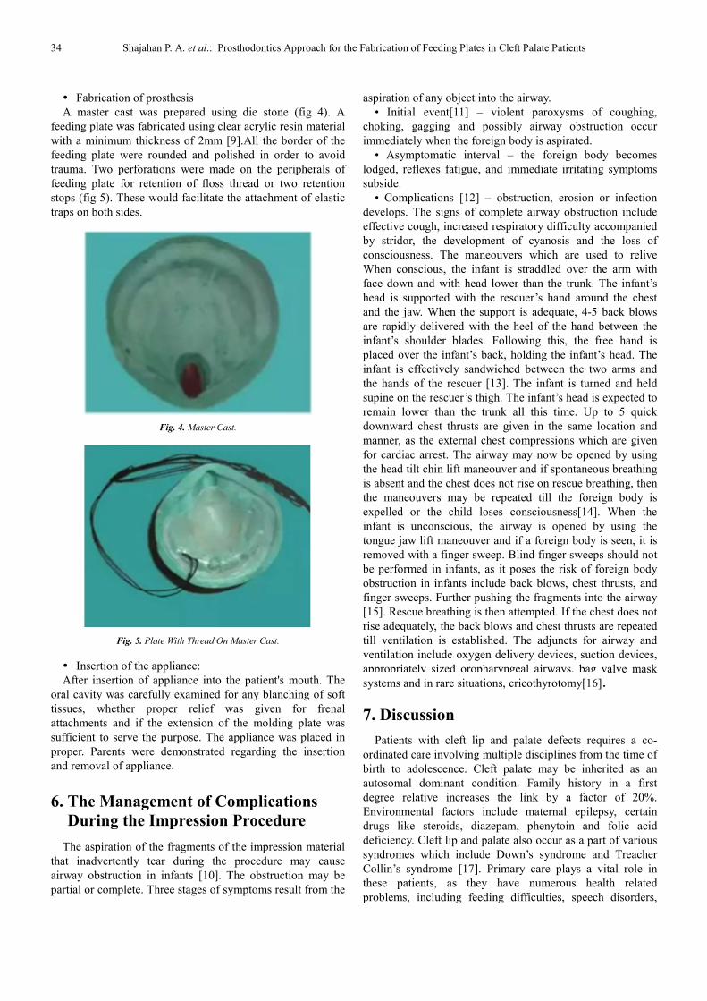
� Fabrication of prosthesis
A master cast was prepared using die stone (fig 4). A
feeding plate was fabricated using clear acrylic resin material
with a minimum thickness of 2mm [9].All the border of the
feeding plate were rounded and polished in order to avoid
trauma. Two perforations were made on the peripherals of
feeding plate for retention of floss thread or two retention
stops (fig 5). These would facilitate the attachment of elastic
traps on both sides.
Fig. 4. Master Cast.
Fig. 5. Plate With Thread On Master Cast.
� Insertion of the appliance:
After insertion of appliance into the patient's mouth. The
oral cavity was carefully examined for any blanching of soft
tissues, whether proper relief was given for frenal
attachments and if the extension of the molding plate was
sufficient to serve the purpose. The appliance was placed in
proper. Parents were demonstrated regarding the insertion
and removal of appliance.
6. The Management of Complications
During the Impression Procedure
The aspiration of the fragments of the impression material
that inadvertently tear during the procedure may cause
airway obstruction in infants [10]. The obstruction may be
partial or complete. Three stages of symptoms result from the
aspiration of any object into the airway.
• Initial event[11] – violent paroxysms of coughing,
choking, gagging and possibly airway obstruction occur
immediately when the foreign body is aspirated.
• Asymptomatic interval – the foreign body becomes
lodged, reflexes fatigue, and immediate irritating symptoms
subside.
• Complications [12] – obstruction, erosion or infection
develops. The signs of complete airway obstruction include
effective cough, increased respiratory difficulty accompanied
by stridor, the development of cyanosis and the loss of
consciousness. The maneouvers which are used to relive
When conscious, the infant is straddled over the arm with
face down and with head lower than the trunk. The infant’s
head is supported with the rescuer’s hand around the chest
and the jaw. When the support is adequate, 4-5 back blows
are rapidly delivered with the heel of the hand between the
infant’s shoulder blades. Following this, the free hand is
placed over the infant’s back, holding the infant’s head. The
infant is effectively sandwiched between the two arms and
the hands of the rescuer [13]. The infant is turned and held
supine on the rescuer’s thigh. The infant’s head is expected to
remain lower than the trunk all this time. Up to 5 quick
downward chest thrusts are given in the same location and
manner, as the external chest compressions which are given
for cardiac arrest. The airway may now be opened by using
the head tilt chin lift maneouver and if spontaneous breathing
is absent and the chest does not rise on rescue breathing, then
the maneouvers may be repeated till the foreign body is
expelled or the child loses consciousness[14]. When the
infant is unconscious, the airway is opened by using the
tongue jaw lift maneouver and if a foreign body is seen, it is
removed with a finger sweep. Blind finger sweeps should not
be performed in infants, as it poses the risk of foreign body
obstruction in infants include back blows, chest thrusts, and
finger sweeps. Further pushing the fragments into the airway
[15]. Rescue breathing is then attempted. If the chest does not
rise adequately, the back blows and chest thrusts are repeated
till ventilation is established. The adjuncts for airway and
ventilation include oxygen delivery devices, suction devices,
appropriately sized oropharyngeal airways, bag valve mask
systems and in rare situations, cricothyrotomy[16].
7. Discussion
Patients with cleft lip and palate defects requires a co-
ordinated care involving multiple disciplines from the time of
birth to adolescence. Cleft palate may be inherited as an
autosomal dominant condition. Family history in a first
degree relative increases the link by a factor of 20%.
Environmental factors include maternal epilepsy, certain
drugs like steroids, diazepam, phenytoin and folic acid
deficiency. Cleft lip and palate also occur as a part of various
syndromes which include Down’s syndrome and Treacher
Collin’s syndrome [17]. Primary care plays a vital role in
these patients, as they have numerous health related
problems, including feeding difficulties, speech disorders,

Science Journal of Clinical Medicine 2016; 5(4-1): 31-36 35
chronic ear infections and dental & orthodontic problems. It
is the general opinion of the plastic surgeon and
prosthodontists that pre-surgical management should be
undertaken as soon possible after birth. Simplified technique
was put forward by Chang and Wang, for the fabrication of
feeding plate [18]. The procedure described is easy, simple
and minimizes any risks to the infant during the procedure.
Impression procedures in cleft infants pose a unique set of
challenges including the size constraints imposed by the
infant’s oral cavity, anatomical variations associated with the
severity of cleft and a lack of ability of the infant to
cooperate and respond to commands. Various impression
materials have been routinely employed for making
impressions of neonates with oral clefts. An ideal impression
material should exhibit certain characteristic in both clinical
and laboratory settings. As primary impression usually color-
timed alginate impression material is used. This lets the
operator know exactly when the mix is ready for placement,
so that the impression will be ready for withdrawal within 15
to 20 seconds after placement of the tray [5,7]. Usually one
scoop of powder, lightly packed, is sufficient for most of
these young children. The water should be slightly warmer
than room temperature to accelerate setting. As the mix is
first briskly spatulated, it turns a dark purple color, gradually
lightening to pink. The material used for final impression was
rubber base which has the advantage as it reproduces all the
areas of interest with good surface details and resist tearing,
more over its removal is atraumatic to the infant.
Additionally, the material remains dimensionally stable and
permits pouring of multiple casts.
Various techniques have also been used to enhance the
retention of the plate. However, the retention of the obturator
is not that critical, because it can be held in the baby’s mouth
during sucking, swallowing and the resting state by the
tongue and by mouth closure. A regular follow up is required
for the examination of oral mucosa which is very delicate and
easily damaged by the obturator. It is indicated that a check
up every 3-4 weeks is required at the bilateral sides of border
to accommodate the growing arches. A new obturator should
be constructed every three months to accommodate the
enlarged craniofacial sutures at growth. The mother is
advised to hold the infant upright or semi-upright position
while feeding so that the swallowed air can be expelled
during the feeding process [19].
A comprehensive management of children born with cleft
lip and palate is best accomplished by the multidisciplinary
team approach. Prosthodontist plays an important role in the
team. However, prompt intervention by fabrication of feeding
plate can eliminate the immediate problems and can bring
about proper nourishment and prevention of any infections
for the already debilitated infant [20, 21].
8. Conclusion
Adequate knowledge of the appliances and impression
procedures is necessary for the management of patients with
clefts involving lips and palate [22]. The feeding obturator
overcomes the factors that act as a stumbling block in the
milestones of normal development and should be inserted as
early as possible after birth. Feeding plate prosthesis reduce
the stress of both parents and the baby, It can aid nursing,
stimulate oral-facial development, helps develop the palatal
shelves, prevent tongue distortion and nasal septum irritation,
decrease the number of ear infections, expand the collapsed
maxillary segment and promote neonate health which is
important in preparing the baby later for the surgical
procedures[23]. The feeding plate needs to be re-fabricated
regularly due to the overall slow but constant growth of
palate; which offers an interim treatment option till the
palatal defect closed surgically[24, 25].
Reference
[1] Booth PW, Schendel SA, Hausamen JE. 2 nded, Vol. 2. Churchill Livingstone. Philadelphia: Elsevier; 2007. p. 1000-48.
[2] Proffit WR, Fields HW, Ackermann JI, Thomas PM, Tulloch JF.Contemporary Orthodontics. Vol. 74. St. Louis: CV Mosby; 2000. p. 287-8.
[3] Savion I, Huband ML. A feeding obturator for a preterm baby with Piere Robin sequence: J Prosthet Dent2005; 93: 197-200.
[4] American cleft palate-craniofacial association, “Parameters for the evalution and treatment of patients with cleft lip/palate craniofacial anomalies,” The cleft palate craniofacial journal, vol 30, supplement 1, 2000 revised 2009.
[5] Grayson B, Santiago PE, Brecht LE, Cutting CB. Presurgicalnasoalveolarmolding in infants with cleft lip and palate. Cleft Palate Craniofac J 1999; 36: 486-98.
[6] Grayson B, Brecht LE, Cutting CB. Nasoalveolar Molding in Early Management of Cleft Lip and Palate. In: Taylor TD, editor. Clinicalmaxillofacial prosthetics. Chapter 5. Quintessence; 63-84.
[7] Jacobson BN, Rosenstein SW. Early maxillaryorthopedics for
the newborn cleft lip and palate patient: An impression and an
appliance. Angle Orthod 1984; 54: 247-63.
[8] Shah CP, Wong D. Management of children with cleft lip and
palate. CMAJ 1980; 122: 19-24.
[9] Shetye P, Cutting C. Presurgicalnasoalveolarmolding
treatment in cleft lip and palate patients. Cleft J 2005; 1: 4-7.
[10] Rathee M, Hooda A, Tamarkar AK, YadavSPS. Role of
Feeding Plate in Cleft Palate: Case Report and Review of
Literature. TheInternet Journal of Otorhinolaryngology. 2010;
12(1).
[11] Agarwal A, Rana V, Shafi S. A feeding appliance for a
newborn baby with cleft lipand palate. Natl J Maxillofac Surg.
2010; 1: 91-93.
[12] Meharban S. Medical emergencies in children. Sagar
Publications; 2000. p. 31-3.
[13] Behrman RE, Kliegman RM, Jenson BH. Nelson Textbook of
Pediatrics. 17th ed. Saunders; 2004. p. 286-91.

36 Shajahan P. A. et al.: Prosthodontics Approach for the Fabrication of Feeding Plates in Cleft Palate Patients
[14] M. Rathee, A. Hooda, A. Tamarkar & S. Yadav: Role of Feeding Plate in Cleft Palate: Case Report and Review of Literature. The Internet Journal of Otorhinolaryngology 2010; 12: 123-7.
[15] Annie Cole, Patricia Lynch, Rona Slator (2008) A New Grading of Pierre Robin Sequence. The Cleft Palate-Craniofacial Journal 2008, 45; 6; 603-6.
[16] Fraser FC (1970) The Genetics of Cleft Lip and Cleft Palate. Cleft Palate Craniofac J 22: 336–352.
[17] Habel A, Sell D, Mars M. Management of Cleft lip and palate. Arch Dis Child 1996; 74: 360-6.
[18] Chang WC, Wang WN. The early management of lip and palate deformity in infants. Bull School Dent NDMC, 1984; 15: 39-42.
[19] Marriot WM. Infant nutrition. In “Textbook of Infant Feeding for Students and Practitioners of Medicine”, CV Mosby Co, St. Louis, 1930 pp. 119-139.
[20] Turner L, Jacobsen C, HumenczukM, Singhal VK, Moore D, Bell H. Theeffects of lactation education and aprosthetic
obturator appliance onfeeding efficiency in infants withcleft lip and palate. Cleft PalateCraniofac J 2001; 38: 519-24.
[21] M. Rathee, A. Hooda, A.K. Tamarkar,S.P.S. Yadav: Role of Feeding Plate inCleft Palate: Case Report and Reviewof Literature. The Internet Journal ofOtorhinolaryngology. 2010 Volume12 Number 1.
[22] Malik P,Aggrawal A, AhujaR.Feeding Appliance For And Infant With Cleft Lip And Palate. Pakistan Oral and DentalJournal 2012; 32(2): p 264-6.
[23] Gupta R, Singhal P, Mahajan K, Singhal A. FabricatingFeeding Plate In CLPInfants With Two Different Materials:ASeries Of Case Report. JISPP2012; 30(4): p 352-355.
[24] Chandan P, Adlakha V.K, Singh N. Feeeidng Obturator Appliance For An Infant With Cleft Lip And Palate. JISPP2011; 1(29): p 71-3.
[25] D. Vojvodic and V. Jerolimov, “The cleft palate patient: a challenge for prosthetic rehabilitation—clinical report,” Quintessence International, vol. 32, no. 7, pp. 521–524, 2001.