protection of sound enamel and artificial enamel lesions ... · keywords: enamel, demineralisation,...
TRANSCRIPT
Zurich Open Repository andArchiveUniversity of ZurichMain LibraryStrickhofstrasse 39CH-8057 Zurichwww.zora.uzh.ch
Year: 2012
Protection of sound enamel and artificial enamel lesions againstdemineralisation: caries infiltrant versus adhesive
Schmidlin, P R ; Sener, B ; Attin, T ; Wiegand, A
Abstract: OBJECTIVE: To compare the protective potential of a conventional adhesive, a caries infiltrantand a combination of both against acidic challenge in vitro. METHODS: One-hundred-and-fifty discsfrom bovine lower central incisors were fabricated. Seventy-five samples remained untreated, whereas theother half was subjected to a demineralisation process (14 days, acidic buffer, and pH 5) to create artificialenamel lesions. Specimens were then radioactively irradiated, and each 15 sound and demineralised speci-mens were treated with a caries infiltrant (Icon, DMG), an unfilled adhesive (Heliobond, IvoclarVivadent)or a combination of infiltrant and adhesive. Specimens treated with the adhesive followed by a flowablecomposite (TetricEvoFlow, IvoclarVivadent) served as positive control, while untreated specimens servedas negative control. All samples were then subjected to lactic acid for 3 weeks at pH 4. Loss of apatitewas determined using the radiochemical method of liquid scintillation. Data were statistically analysedby Kruskal-Wallis-test, one-way ANOVA and Scheffe’s post hoc tests (p �.05). RESULTS: In both soundenamel and artificial caries lesions, untreated specimens showed the highest rate of apatite loss, whereasenamel treated with the adhesive and the flowable composite showed almost complete protection surfaceagainst dissolution. The caries infiltrant, the adhesive and the combination of both were able to decreaseenamel dissolution, but the adhesive and the combination of adhesive and infiltrant were more effectivethan the infiltrant alone. CONCLUSION: Within the limitations of this in vitro study, the applicationof an adhesive (alone or in combination with the caries infiltrant) is more effective to protect enameldissolution than the infiltrant alone.
DOI: https://doi.org/10.1016/j.jdent.2012.07.003
Posted at the Zurich Open Repository and Archive, University of ZurichZORA URL: https://doi.org/10.5167/uzh-74978Journal ArticleAccepted Version
Originally published at:Schmidlin, P R; Sener, B; Attin, T; Wiegand, A (2012). Protection of sound enamel and artificial enamellesions against demineralisation: caries infiltrant versus adhesive. Journal of Dentistry, 40(10):851-856.DOI: https://doi.org/10.1016/j.jdent.2012.07.003
Protection of sound enamel and artificial enamel lesions against
demineralisation: caries infiltrant versus adhesive
Short title: Enamel dissolution protection by infiltrant and adhesive
PR Schmidlin, B Sener, T Attin, A Wiegand
Clinic of Preventive Dentistry, Periodontology and Cariology, Center for Dental Medicine,
University of Zurich, Plattenstrasse 11, 8032 Zurich, Switzerland
Corresponding author
PD Dr. Patrick R. Schmidlin
Clinic of Preventive Dentistry, Periodontology and Cariology, University of Zurich
Plattenstrasse 11, 8032 Zürich, Switzerland
Email: [email protected]
Phone: 0041-44-6343417
Keywords: Enamel; demineralisation; caries infiltration; liquid scintillation; SEM; In vitro
Protection of sound enamel and artificial enamel lesions against
demineralisation: caries infiltrant versus adhesive
Abstract
Objective: To compare the protective potential of a conventional adhesive, a caries infiltrant
and a combination of both against acidic challenge in vitro.
Methods: One-hundred-and-fifty discs from bovine lower central incisors were fabricated.
Seventy-five samples remained untreated, whereas the other half was subjected to a
demineralisation process (14 days, acidic buffer, pH 5) to create artificial enamel lesions.
Specimens were then radioactively irradiated, and each 15 sound and demineralised
specimens were treated with a caries infiltrant (Icon, DMG), an unfilled adhesive (Heliobond,
Ivoclar Vivadent) or a combination of infiltrant and adhesive. Specimens treated with the
adhesive followed by a flowable composite (Tetric EvoFlow, Ivoclar Vivadent) served as
positive control, while untreated specimens served as negative control. All samples were
then subjected to lactic acid for 3 weeks at pH 4. Loss of apatite was determined using the
radiochemical method of liquid scintillation. Data were statistically analysed by Kruskal-
Wallis-test, one-way ANOVA and Scheffe`s post-hoc tests (p < =.05).
Results: In both sound enamel and artificial caries lesions, untreated specimens showed the
highest rate of apatite loss, whereas enamel treated with the adhesive and the flowable
composite showed almost complete protection surface against dissolution. The caries
infiltrant, the adhesive and the combination of both were able to decrease enamel
dissolution, but the adhesive and the combination of adhesive and infiltrant were more
effective than the infiltrant alone.
Conclusion: Within the limitations of this in vitro study, the application of an adhesive (alone
or in combination with the caries infiltrant) is more effective to protect enamel dissolution than
the infiltrant alone.
Keywords: Enamel, demineralisation, caries infiltration, liquid scintillation, SEM, In vitro Introduction
Pit and fissure sealing with composite resins bonded to enamel is an effective procedure for
caries prevention.6,32 Based on the good clinical results with sealing of these mentioned
predilection sites, approaches have been made to extend this preventive concept to smooth
enamel surfaces.25,26 Among the latter, interproximal areas have the highest risk for caries
development.1,9 The infiltration of caries lesions with low-viscosity light curing resins is
considered as treatment option for non-cavitated lesions, which are not expected to arrest or
remineralise. In contrast to the conventional sealing concept where a resin layer is created
on the surface, caries infiltrants aim to penetrate the porous lesion body thoroughly.7,14
Compared to conventional dental adhesives, caries infiltrants were optimized for rapid
capillary penetration and exhibit a very low-viscosity, low contact angle to enamel and high
surface tension.13 As a consequence, laboratory experiments demonstrated a significantly
deeper penetration in the lesion body than conventional adhesives.11,12 However, in spite of
the deeper penetration of caries lesions, it has not be shown yet that caries infiltration as an
increased capability to prevent progression of demineralisation than conventional sealing of
the lesion.19 While both infiltrants11,18,20 and adhesives16,24 were shown to be effective in
reducing progression of artificial enamel lesions, their protective potential on sound enamel
was not compared so far. This aspect might of interest in areas neighbouring the lesion,
which might be of risk to be affected by demineralisation if the lesion progresses.
Therefore, the aim of the present study was to compare the potential of a conventional
adhesive, a caries infiltrant and a combination of both to protect sound enamel and artificial
enamel lesion against an acidic challenge in vitro. The hypotheses were that protective
capability of the infiltrant and the adhesive do not differ, and that the combination of both is
comparable to the respective treatments alone.
Methods
Specimen preparation
Specimens were prepared from 150 extracted bovine permanent incisors. Only tooth material
free of defects or cracks was selected. The teeth were cleaned and sectioned at the enamel-
cementum junction using a water-cooled cutting wheel (Isomet, Buehler, Illinois, USA). The
labial surface of the teeth was cleaned by brushing during 25 min. Brushing was performed
in a custom-made brushing machine at 2.5 N with a manual toothbrush (ParoM39, ESRO,
Thalwil, Switzerland) and a toothpaste-slurry (6 g Depurdent, Wild SA, Basel, Switzerland; 10
g artificial saliva; 8 drops of a silicone antifoam, Fluka, Art. Nr. 85390, Switzerland). Discs
with a diameter of 7 mm were cut from the mid-labial aspect of each tooth using a custom-
made diamond-coated trephine (80µm, Intensiv SA, Lugano-Grancia, Switzerland). The discs
were then flattened from the bottom to approximately 2 mm in height (Struers, Birmensdorf).
Then, half of the specimens were immersed for 14 days in an acidic buffer containing 3 mM
CaCl2 x 2 H2O, 3 mM KH2PO4, 50 mM acetic acid, 6 µM MHDP, KOH to adjust the initial pH
to 5.0 and traces of thymol.4 This solution was renewed each second day to keep the pH
constant.
The Specimens were irradiated at the "Atominstitut der Österreichischen Universitäten"
(Vienna, Austria) with an exposure time of 85 min to a neutron flow of 1.02 x 10 e12
neutrons/cm2.s, resulting in a ß—-activity of 0.56 Gbq/p.
Treatment procedure
Before the enamel treatment, all surfaces except the top enamel test surface were carefully
sealed with unfilled bonding agent (Heliobond, Ivaoclar-Vivadent, Schaan, Liechtenstein) and
a flowable composite resin (Tetric EvoFlow, Ivoclar Vivadent, Schaan, Liechtenstein).
Therefore, the lateral cutted enamel was carefully etched with 35% phosphoric acid
(UltraEtch 35%, Ultradent Products, South Jordan, USA) and rinsed with distilled water for 60
s before the sealing procedure.
Then, 75 sound and 75 demineralised samples were randomly assigned to five groups (15
samples each) and treated as follows:
1. The enamel surface was etched for 60 s with 35% phosphoric acid (UltraEtch 35%,
Ultradent Products, South Jordan, USA), and then rinsed distilled water for 60 s. After
air drying of the surface, an unfilled adhesive (Heliobond, Ivoclar Vivadent, Schaan,
Liechtenstein) was applied for 20 s with a microbrush, then thinned with mild air (1-2
s) and light cured for 20 s (3M Espe Elipar S10, 3M Espe, Seefeld, Germany).
2. The enamel surface was etched for 2 min with 15% hydrochloric acid (Icon Etch,
DMG, Hamburg, Germany) and then rinsed with water spray for 30 s. Air drying of the
surface was followed by application of ethanol (Icon Dry, DMG, Hamburg, Germany)
for 30 s and additional air drying. Then, the low-viscosity resin infiltrant (Icon Infiltrant,
DMG, Hamburg, Germany) was applied on the surface for 3 min by means of the
sponge applicator provided with the resin infiltration system. After light-curing for 40 s,
the infiltrant was applied for further 60 s and again light-cured for 40 s.
3. Specimens were first treated with the caries infiltrant as described under point 2.
Then, the adhesive was applied and light cured for 20 s.
4. Same treatment as under point 1 but additional application of a flowable composite
resin material (Tetric EvoFlow, Ivoclar Vivadent, Schaan, Liechtenstein), which was
light cured for 40 s (3M Espe Elipar S10, 3M Espe, Seefeld, Germany); positive
control
5. No sealing; negative control
The composition of the adhesive systems and materials based on the manufacturers’
instructions are listed in Table 1.
Acidic challenge and evaluation of mineral loss
All specimens were immersed at 37 °C under constant motion for up to 21 days in 5 ml of
artificial saliva or 5 ml lactic acid (15 µmol/l, pH 4).
After 1, 2, 4, 7, 11, 14 and 21 days, immersion solutions were collected, the weight of each
sample was measured and 1 ml 2N HCl plus 4 ml distilled water were added to an end
volume of 10 ml. 32P was assessed by determining the Cherenkow radiation. For calculation
of mineralised tissue loss standard solutions of 32P and apatite, together with a background
sample were prepared, as described elsewhere.27 In short, the resulting counts per minute
(CPM) were calculated to the date of irradiation, resulting in decays per minute (DPM). The
arithmetic mean of the samples was calculated and the amount of dissolved mineralised
tissue was calculated with the aid of the apatite standard.
Morphological assessment
For the SEM analysis, two additional specimens of each group were prepared for
examination of the surfaces directly after application of the test materials and after 21 d
storage in lactic acid. Specimens were dehydrated in a desiccator device (Optivac, König
Physik, Diemelstadt, Germany) using blue silica gel, sputter-coated with gold and examined
at 10 kV (Zeiss Supra 50 VP, Zeiss, Oberkochen, Germany).
Data presentation and analysis
Statistical analysis was performed with StatView (Version 5, Abacus Concepts Inc., Berkley,
USA).
Cumulative mineral loss in µg apatite was calculated for each group. Normal distribution was
tested using Kolmogorov-Smirnov and Shaprio-Wilk tests.
As data were not normally distributed, non-parametric statistics (Kruskal-Wallis test) were
applied within sound specimens and within specimens with artificial lesions to analyse
possible differences between the groups at the different time points. Non-parametric
statistics were followed by one-way ANOVA, separately for sound and demineralised
enamel, and Scheffe`s post-hoc tests. The level for statistical significance was set at
p < 0.05.
Results
Mineral loss
In both sound enamel and enamel exhibiting artificial lesions, Kruskal-Wallis tests revealed
significant differences between the groups at each time point. Cumulative enamel loss at the
evaluated time points is presented in Figures 1 and 2.
Untreated specimens showed the highest rate of apatite loss over the whole observation
period, whereas enamel treated with the adhesive and the flowable composite showed
almost complete protection surface against dissolution at all time points. The caries infiltrant,
the adhesive and the combination of the infiltrant and the adhesive reduced apatite
dissolution significantly, but the adhesive and the combination of infiltrant and adhesive were
more protective against acid dissolution than the infiltrant alone, irrespective whether sound
or demineralised enamel was treated.
Morphological aspects
The morphological appearances of the sound and predemineralised specimens before and
after storage in lactic acid for 21 days are presented in Figure 3, respectively.
Untreated enamel exposed to lactic acid showed only minute surface changes (Figure 3 B).
Application of the caries infiltrant system induced a severe demineralisation with a
honeycomb appearance of enamel (Figure 3 I). In contrast, the enamel surface appeared
smooth after application of the adhesive or the combination of caries infiltrant and adhesive,
even after storage in lactic acid for 21 days (Figure 3 M/N).
Artificial-caries like lesions revealed a demineralised surface (Figure 3 C). While application
of the caries infiltrant increased/modified demineralisation (Figure 3 K), the adhesive induced
a sealing of the surface (Figure 3 G), which was still present after 21 d storage in lactic acid
(Figure 3 H/P).
Discussion
This study demonstrated that both a caries infiltrant system and a conventional adhesive
protect sound enamel and artificial enamel lesions from further demineralisation. However,
the adhesive and the combination of infiltrant and adhesive were more effective than the
caries infiltrant alone.
In the present study, bovine teeth were used, which are widely used in resin infiltration
tests.16,19 Following the same irradiation protocol as in previous studies,28,29 the cumulative
mineral loss of untreated samples comparing the results of the present investigation revealed
remarkably comparable results with median losses of 580 and 660 µg apatite, respectively.
The samples had the same pretreatment and geometry, but the embedding was slightly
different. In the present study, we used an adhesive embedding of the discs, which revealed
a more reliable lining and sealing of the irradiated samples during the dissolution process.
The median mineral loss in the former study was still 26 µg apatite as compared to nearly
zero in this investigation.
As in previous studies lactic acid was used for the demineralisation regime as it represents
the main organic acid produced by dental plaque bacteria.5,28,29 However, it should be
acknowledged that the study conditions differed from the in vivo situation in that there was no
protective salivary pellicle and that enamel surfaces were in continuous contact with the
acidic challenge.
In this study artificial lesions of about 150 µm depth were used.2 However, although these
lesions were shown to exhibit some of the typical histological structure of intact enamel
caries including the intact surface layer,8 the histological condition of the surface und
subsurface layers is unpredictable in the clinical situation. The surface layer is thicker in
natural (~ 40 to 50 µm and) compared to artificial (~ 20 µm) lesions,3 but it was shown that
hydrochloric acid etching is required not only for natural23 but also for artificial caries lesions2
to remove the surface layer completely and allow for successful penetration of the infiltrant. It
was therefore decided to use the infiltration material as provided in the commercially
available kit, i.e. using hydrochloric acid conditioning material. Heliobond was chosen as a
representative of an unfilled enamel bonding agent as it exhibits potential to penetrate at
least early enamel lesions.10,13,30 However, it has to be considered that natural enamel
lesions might be much deeper than the artificial lesions created in the present study.
Therefore, an incomplete penetration under natural caries conditions is possible.
While resin infiltrants led to a complete, but partially inhomogenous penetration of artificial
lesions, Heliobond was shown to induce the formation of a homogenous surface layer, but
penetrated only the outer part of the lesion.13 The results of the present study indicate that
the superficial penetration and surface coating of the adhesive might be more effective in
protecting enamel dissolution than the penetration of the infiltrant. This observation is also
evident in the morphological evaluation of this study. While the surfaces treated with the
caries infiltrant exhibited a demineralised appearance, the other test groups revealed a
dense adhesive layer, respectively. While the amount of TEGDMA in the resin infiltrant
promotes the penetration of the resin,22 it also increases the susceptibility to degradation
compared to resins containing less TEGDMA 17,31. Moreover, the surface leakage might be a
result of polymerization shrinkage and polymerization stress of the resin.21 However,
surprisingly, the infiltrant alone showed some protection against the acid dissolution even in
sound enamel, which does not represent a substrate for the infiltrant.
However, in both substrates, the combination of both materials was not more effective than
the adhesive alone. At least for artificial enamel lesions it was assumed that the infiltration of
the demineralised subsurface layer and the sealing of the surface might have an additive
effect on the dissolution protection. As the bonding of the adhesive is not impaired on
infiltrated enamel surfaces,33 this observation cannot be explained yet. However, covering
infiltrated lesions with an adhesive layer might be beneficial in terms of surface properties, as
surface roughness of infiltrated lesions is comparatively high 15.
Conclusion
The application of an adhesive (alone or in combination with the caries infiltrant) is more
effective to protect enamel dissolution than the infiltrant alone within the limitations of this in
vitro study.
References
1. Alm A, Wendt LK, Koch G, Birkhed D. Prevalence of approximal caries in posterior
teeth in 15-year-old Swedish teenagers in relation to their caries experience at 3 years
of age. Caries Research 2007;41:392-398.
2. Belli R, Rahiotis C, Schubert EW, Baratieri LN, Petschelt A, Lohbauer U. Wear and
morphology of infiltrated white spot lesions. Journal of Dentistry 2011;39:376-385.
3. Bergman G, Lind PO. A quantitative microradiographic study of incipient enamel caries.
Journal of Dental Research 1966;45:1477-1484.
4. Buskes JA, Christoffersen J, Arends J. Lesion formation and lesion remineralization in
enamel under constant composition conditions. A new technique with applications.
Caries Research 1985;19:490-496.
5. Fejerskov O, Thylstrup A. Pathology of dental caries. In: Thylstrup A, Fejerskov O,
editors. Textbook of Cariology. Copenhagen: Munksgaard; 1996; p. 204-235.
6. Horowitz HS, Heifetz SB, Poulsen S. Retention and effectiveness of a single application
of an adhesive sealant in preventing occlusal caries: final report after five years of a
study in Kalispell, Montana. Journal of the American Dental Association 1977;95:1133-
119.
7. Kielbassa AM, Muller J, Gernhardt CR. Closing the gap between oral hygiene and
minimally invasive dentistry: a review on the resin infiltration technique of incipient
(proximal) enamel lesions. Quintessence International 2009;40:663-681.
8. Moron BM, Comar LP, Wiegand A, Buchalla W, Buzalaf MA. Comparison of cross-
sectional hardness and transverse microradiography of artificial carious enamel lesions
induced by different demineralising solutions and gels. Caries Research 2009;43:474-
483.
9. Menghini G, Steiner M, Marthaler T, Helfenstein U, Brodowski D, Imfeld C, Weber R,
Imfeld T. Caries prevalence among students in 16 Zurich districts in the years 1992 to
2000. Schweizer Monatsschrift für Zahnmedizin 2003;113:267-277.
10. Meyer-Lueckel H, Mueller J, Paris S, Hummel M, Kielbassa AM. The penetration of
various adhesives into early enamel lesions in vitro. Schweizer Monatsschrift für
Zahnmedizin 2005;115:316-323.
11. Meyer-Lueckel H, Paris S. Progression of artificial enamel caries lesions after infiltration
with experimental light curing resins. Caries Research 2008;42:117-124.
12. Meyer-Lueckel H, Paris S. Improved resin infiltration of natural caries lesions. Journal
of Dental Research 2008;87:1112-1116.
13. Meyer-Lueckel H, Paris S, Mueller J, Colfen H, Kielbassa AM. Influence of the
application time on the penetration of different dental adhesives and a fissure sealant
into artificial subsurface lesions in bovine enamel. Dental Materials 2006;22:22-28.
14. Meyer-Lueckel H, Chatzidakis A, Naumann M, Dörfer CE, Paris S. Influence of
application time on penetration of an infiltrant into natural enamel caries. Journal of
Dentistry 2011;39:465-9
15. Mueller J, Yang F, Neumann K, Kielbassa AM. Surface tridimensional topography
analysis of materials and finishing procedures after resinous infiltration of subsurface
bovine enamel lesions. Quintessence International 2011;42:135-147.
16. Mueller J, Meyer-Lueckel H, Paris S, Hopfenmuller W, Kielbassa AM. Inhibition of
lesion progression by the penetration of resins in vitro: influence of the application
procedure. Operative Dentistry 2006;31:338-345.
17. Ortengren U, Wellendorf H, Karlsson S, Ruyter IE. Water sorption and solubility of
dental composites and identification of monomers released in an aqueous environment.
J Oral Rehabil 2001;28:1106-1115.
18. Paris S, Hopfenmuller W, Meyer-Lueckel H. Resin infiltration of caries lesions: an
efficacy randomized trial. Journal of Dental Research 2010;89:823-826.
19. Paris S, Meyer-Lueckel, H. Inhibition of caries progression by resin infiltration in situ.
Caries Research 2010;44:47-54.
20. Paris S, Meyer-Lueckel H. Infiltrants inhibit progression of natural caries lesions in vitro.
Journal of Dental Research 2010;89:1276-1280.
21. Paris S, Meyer-Lueckel H, Colfen H, Kielbassa AM. Resin infiltration of artificial enamel
caries lesions with experimental light curing resins. Dental Materials Journal
2007;26:582-588.
22. Paris S, Meyer-Lueckel H, Colfen H, Kielbassa AM. Penetration coefficients of
commercially available and experimental composites intended to infiltrate enamel
carious lesions. Dental Materials 2007;23:742-748.
23. Paris S, Meyer-Lueckel H, Kielbassa AM. Resin infiltration of natural caries lesions.
Journal of Dental Research 2007;86:662-666.
24. Paris S, Meyer-Lueckel H, Mueller J, Hummel M, Kielbassa AM. Progression of sealed
initial bovine enamel lesions under demineralizing conditions in vitro. Caries Research
2006;40:124-129.
25. Phark JH, Duarte SJ, Meyer-Lueckel H, Paris S. Caries infiltration with resins: a novel
treatment option for interproximal caries. Compendium of Continuing Education in
Dentistry 2009;30:13-17.
26. Schmidlin PR, Besek MJ. Atraumatic tooth separation and proximal sealing: filling the
gap between preventive and restorative dentistry. Practical Procedures & Aesthetic
Dentistry 2003;15:65-69.
27. Schmidlin PR, Beuchat M, Busslinger A, Lehmann B, Lutz F. Tooth substance loss
resulting from mechanical, sonic and ultrasonic root instrumentation assessed by liquid
scintillation. Journal of Clinical Periodontology 2001;28:1058-1066.
28. Schmidlin PR, Gohring TN, Sener B, Lutz, F. Resistance of an enamel-bonding agent
to saliva and acid exposure in vitro assessed by liquid scintillation. Dental Materials
2002;18:343-50.
29. Schmidlin PR, Zehnder M, Zimmermann MA, Zimmermann J, Roos M, Roulet JF.
Sealing smooth enamel surfaces with a newly devised adhesive patch: a radiochemical
in vitro analysis. Dental Materials 2005;21:545-550.
30. Schmidlin PR, Zehnder M, Pasqualetti T, Imfeld T, Besek, M. Penetration of a bonding
agend into de- and remineralized enamel in vitro. Journal of Adhesive Dentistry
2004;6:111-115.
31. Sideridou ID, Karabela MM, Vouvoudi EC. Volumetric dimensional changes of dental
light-cured dimethacrylate resins after sorption of water or ethanol. Dental Materials
2008;24:1131-1136.
32. Splieth CH, Ekstrand KR, Alkilzy, Clarkson J, Meyer-Lueckel H, Martignon S, Paris S,
Pitts NB, Ricketts DN, van Loveren C. Sealants in dentistry: outcomes of the ORCA
Saturday Afternoon Symposium 2007. Caries Research 2010;44:3-13.
33. Wiegand A, Stwarczyk B, Kolakovic M, Hämmerle CH, Attin T, Schmidlin PR. Adhesive
performance of a caries infiltrant on sound and demineralised enamel. Journal of
Dentistry 2011;39:117-121.
Captions
Figure 1
Results of the sealing ability after different treatments of sound enamel specimens
determined as loss of apatite in µg (box-plot illustration; horizontal bars: medians; boxes:
inter-quartile areas; error bars: 10th and 90th percentiles; dots: extreme values). Identical
superscript capitals at the different time points represent no statistically different values.
Figure 2
Results of the sealing ability after different treatments of artificial enamel lesion specimens
determined as loss of apatite in µg (box-plot illustration; horizontal bars: medians; boxes:
inter-quartile areas; error bars: 10th and 90th percentiles; dots: extreme values). Identical
superscript capitals at the different time points represent no statistically different values.
Figure 3
SEM images at 5’000x of representative samples after different treatments of sound enamel
and artificial caries lesions before (baseline) and after 21 days lactic acid exposure.
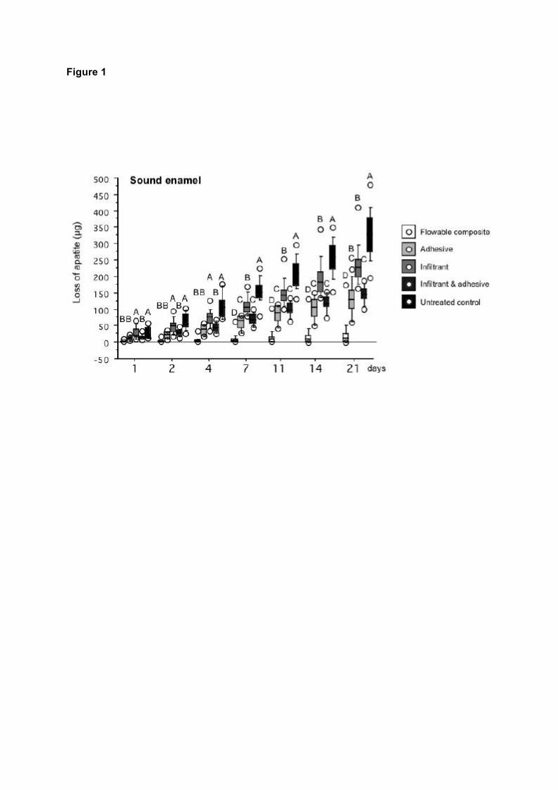
Figure 1
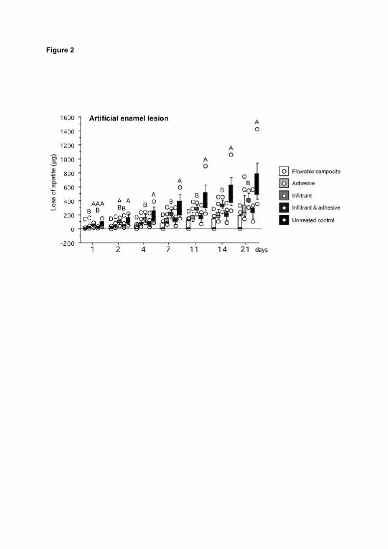
Figure 2
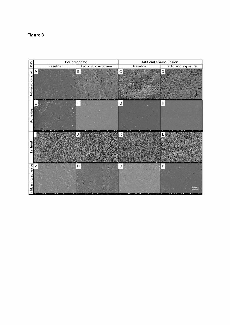
Figure 3
Sound enamel Artificial enamel lesionBaseline BaselineLactic acid exposure Lactic acid exposure
Unt
reat
ed c
ontro
lA
dhes
ive
Infil
trant
Infil
trant
& a
dhes
ive
5000
x
10 !m
A B C D
E F G H
I J K L
M N O P
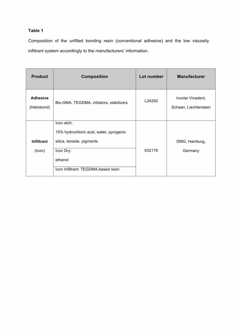
Table 1
Composition of the unfilled bonding resin (conventional adhesive) and the low viscosity
infiltrant system accordingly to the manufacturers’ information.
Product Composition Lot number Manufacturer
Adhesive
(Heliobond) Bis-GMA, TEGDMA, initiators, stabilizers
L24292 Ivoclar Vivadent,
Schaan, Liechtenstein
Infiltrant
(Icon)
Icon etch:
15% hydrochloric acid, water, pyrogenic
silica, tenside, pigments
632178
DMG, Hamburg,
Germany Icon Dry:
ethanol
Icon Infiltrant: TEGDMA-based resin