protective effect of solanum torvum on doxorubicin-induced
TRANSCRIPT

Pharmacologyonline 2: 1192-1204 (2009) Kamble et al.
1192
Protective Effect of Solanum Torvum on Doxorubicin-Induced
Cardiactoxicity in Rats
1Sarika Kamble, 1Mahalaxmi Mohan*, 2Sanjay Kasture
1Department of Pharmacology, M. G. V’s Pharmacy College, Panchavati, �asik,
Maharashtra 422 003, India 2Pinnacle Biomedical Research Institute, Bhopal, 462003, India
Summary
Oxidative stress is the main factor in Doxorubicin (DOX) induced cardiotoxicity. Wistar
rats received either DOX (67.75 mg/kg, i.v, 2 days before sacrifice) or Solanum torvum
extract (100 mg/kg and 300 mg/kg, p.o.) prior to DOX or S. torvum (100 mg/kg and 300
mg/kg, p.o.) extract alone for 4 weeks. Cardiotoxicity was assessed by recording changes
in ECG, heart rate and measuring the levels of cardiac marker enzymes- lactic acid
dehydrogenase (LDH) and creatine phosphokinase (CK-MB). The antioxidant defence
enzymes superoxide dismutase (SOD) and catalase (CAT) for heart tissue and
histopathological changes were also measured at the end of the treatment schedule.
Treatment with S. torvum (100 mg/kg and 300 mg/kg) significantly (p<0.05) reversed the
changes in ECG; decreased the levels of CK-MB and LDH; and increased the anti-
oxidant defence enzyme levels of SOD and CAT. S. torvum treated animals showed a
lesser degree of cellular infiltration in histopathological studies. The results suggest that
S. torvum has the potential in preventing the cardiotoxicity induced by Doxorubicin.
Key words: S. torvum, marker enzymes, antioxidant defence enzyme, doxorubicin
Running title: Solanum torvum on Doxorubicin cardiotoxicity
* - Corresponding author
Email: [email protected]
Ph. No. +91, 9823234514
Fax No. +91, 0253, 2511931

Pharmacologyonline 2: 1192-1204 (2009) Kamble et al.
1193
Introduction
Doxorubicin is an anthracycline glycoside antibiotic that possesses a potent and broad
spectrum antitumour activity against a variety of human solid tumours and
haematological malignancies (1, 2). However its use in chemotherapy has been limited
largely due to its diverse toxicities, including cardiac, renal, hematological and testicular
toxicity (3, 4). Cardiotoxicity induced by DOX is usually mediated through lipid
peroxidation and inhibition of long fatty acid oxidation in cardiac tissues (5-8). Thus,
oxidative stress, lipid peroxidation, and mitochondrial dysfunction have been associated
with DOX induced cardiomyopathy (7).
Solanum torvum Sw. (Solanaceae), commonly known as Turkey berry is an erect spiny
shrub of about 4 m tall, evergreen and widely branched. It is native and found cultivated
in Africa and West Indies (9). The fruits and leaves are widely used in Camerooninan
folk medicine. The plant is cultivated in the tropics for its sharp tasting immature fruits. It
is used in the treatment of stomach pain and skin infections (10). It possesses
antimicrobial (11, 12) antiviral (13), immuno-secretory (14), antiulcer (15), antioxidant
(16), analgesic and anti-inflammatory (17), cardiovascular and anti-platelet aggregation
activities (18). Solanum torvum contains a number of potentially pharmacologically
active chemicals like isoflavonoid sulfate and steroidal glycosides (13, 19), chlorogenone
and neochlorogenone (20) triacontane derivatives (21, 22), 22-β-o-spirostanol
oligoglycosides (23), 26-O-β-glucosidase (24). The free radical scavenging effects of
Solanum torvum on DPPH (2, 2-diphenyl-1-picrylhydrazyl) was measured in-vitro. The
antioxidant properties of flavonoids and their ability to chelate free iron could be
effective in reducing cardiotoxicity of DOX (25). In view of this, since DOX induced
cardiotoxicity is linked to oxidative stress, we have investigated the possible protective
effect of Solanum torvum, against DOX-induced cardiotoxicity in rats.
Materials and Methods
Extract preparation Dried fruits of Solanum torvum Sw. (Solanaceae) were purchased locally and
authenticated by Dr. S. C. Pal, NDMVP Samaj’s College of Pharmacy, Nashik, India.
The voucher specimen has been deposited at Agharkar Research Institute, Pune, India.
Mature fruits were collected, sundried and grounded. The powder obtained (1kg) was
defatted using pet ether (60-800C). The marc was macerated in ethanol for 3-4 days at
room temperature. The filterate was air dried and concentrated under reduced pressure to
obtain 120 g, corresponding to a yield of 12.0 % w/w. The total flavanoid content of
ethanolic extract of S.torvum was found to be 85.26±0.02 µg rutin equivalent/ mg of
extract (26). The total phenolic content of ethanolic extract of S.torvum was found to be
99.52± 0.42 µg gallic acid equivalent/ mg of extract (27). Appropriate concentrations of
the extracts was made in distilled water. The phytoconstituents present in the crude
extract were flavonoids, alkaloids, tannins and saponins (28).
Animals
Laboratory breed Wistar albino rats of either sex weighing between 150-200 g,
maintained under standard laboratory conditions of 25 ± 1°C, and photo period (12 h
dark/12 h light) were used for the experiment.

Pharmacologyonline 2: 1192-1204 (2009) Kamble et al.
1194
Commercial pellet diet (Amrut laboratory rat and mice feed, Sangli, India.) and water
were provided ad libitum. The experiments were carried out according to the guidelines
of the Committee for the Purpose of Control and Supervision of Experiments on Animals
(CPCSEA), New Delhi, India, and approved by the Institutional Animal Ethical
Committee.
Chemicals
Doxorubicin (Oncodria, Sun Pharmaceutical Ind. Ltd. Gujarat, India) was obtained from
the local market. 1,1-diphenyl, 2- picrylhydrazyl (DPPH) were purchased from Sigma-
Aldrich, Mumbai. All chemicals for sensitive biochemical assays were obtained from
Sigma Chemicals Co. India and Hi Media Chemicals, Mumbai, India. Distilled water was
used for biochemical assays. LDH and CK-MB kits were obtained from Aspen
Laboratories Pvt. Ltd.,Baddi; India. and Teco Diagnostics, Anaheim, USA respectively.
Experimental protocol
During the acclimatization period, the baseline ECG was recorded (Chart 5.0 AD
Instrument, Austria). The animals were then randomly divided into the following
experimental groups with 5 animals in each group.
Group 1: Vehicle treated group
Group 2: DOX (67.75 mg/kg, i.v, 2 days before the sacrifice)
Group 3: Solanum torvum (100 mg/kg, p.o.) daily for four weeks
Group 4: Solanum torvum (300 mg/kg, p.o.) daily for four weeks
Group 5: Solanum torvum (100 mg/kg, p.o.) daily for four weeks+ DOX (67.75 mg/kg,
i.v, 2 days before the sacrifice)
Group 6: Solanum torvum (300 mg/kg, p.o.) daily for four weeks + DOX (67.75 mg/kg,
i.v, 2 days before the sacrifice)
At the end of the treatment schedule, the animals were anaesthetized with diethyl ether,
ECG recorded, and then sacrificed by a high dose of diethyl ether. Blood was withdrawn
immediately for enzyme assays and the heart was dissected out and weighed. Heart tissue
was washed with ice-cold 0.9% saline and homogenized quickly with ice cold 0.1 M Tris
HCl buffer (pH 7.5) using Remi homogenizer to give a 10% homogenate.
Electrocardiography (ECG)
ECG was recorded before and after the treatment schedule. For ECG recording (Chart
5.0, ADI Instruments) rats underwent light ether anesthesia. Needle electrodes were
inserted under the skin. For each ECG tracing, QRS complex, QT interval and ST
intervals were measured (29).
Preparation of Serum and tissue homogenate
Blood was collected and allowed to clot. Serum was separated by centrifugation of the
clotted blood at 5000 rpm for 4 min and used for estimation of LDH, CK-MB. Known
amount of tissue was weighed and homogenized in ice cold 0.1 M Tris HCl buffer for
estimation of SOD and CAT.
Antioxidant Parameters
Superoxide dismutase activity (SOD)
Pharmacologyonline 2: 1192-1204 (2009) Kamble et al.
1195
The assay of SOD was based on the ability of SOD to inhibit spontaneous oxidation of
adrenaline to adrenochrome (30, 31). 0.05 ml supernatant was added to 2.0 ml of
carbonate buffer and 0.5 ml of 0.01mM EDTA solution. The reaction was initiated by
addition of 0.5ml of epinephrine and the autooxidation of adrenaline (3x10⁻⁴
M) to
adrenochrome at pH 10.2 was measured by following change in OD at 480 nm. The
change in optical density every minute was measured at 480 nm against reagent blank.
The results are expressed as units of SOD activity (mg/wet tissue). One unit of SOD
activity induced approximately 50% inhibition of adrenaline.
Catalase activity (CAT)
Catalase(CAT): The reaction mixture consisted of 2 ml phosphate buffer (pH 7.0), 0.95
ml of hydrogen peroxide (0.019M) and 0.05ml of supernatant in a final volume of 3 ml.
Absorbance were recorded at 240 nm every 10 sec for 1 min. One unit of CAT was
defined as the amount of enzyme required to decompose 1 µmol of peroxide per min, at
25 oC at pH 7.0. The results were expressed as units of CAT U/g of wet tissue (32).
Cardiac marker enzymes
Blood samples were collected from each animal and the serum obtained by centrifugation
was used for determination of LDH (LDH kit-Aspen Laboratories Pvt. Ltd.,Baddi; India.)
and CK-MB (Teco Diagnostics, Anaheim, USA).
Histopathological examination
The hearts were excised and immediately fixed in 10% buffered formalin. The tissue
specimen was embedded in paraffin after being dehydrated in alcohol and subsequently
cleared with xylene. Five-micrometer thick serial histological sections were obtained
from the paraffin blocks and stained with hematoxylin and eosin. The sections were
examined under light microscope and photomicrographs were taken.
Statistical analysis
All data were expressed as the mean ± SEM. For statistical analysis of the data, group
means were compared by one-way analysis of variance (ANOVA) followed by Dunnett’s
test, P<0.05 was considered significant.
Results
ECG, heart rate and heart weight
Animals treated with DOX (67.75 mg/kg, i.v, 2 days before sacrifice) showed a
significant (p<0.05) prolongation of QT interval and ST interval as compared to vehicle
treated animals. Treatment with S. torvum extract (100 mg/kg and 300 mg/kg) alone has
not shown any significant change as compared to vehicle treated group. S. torvum extract
(100 and 300 mg/kg) significantly (p<0.05) decreased the prolongation of QT and ST
intervals in animals treated with DOX as compared to DOX treated group. There was a
significant increase in heart rate and a significant decrease in heart weight in DOX treated
animals which was prevented by S. torvum extract (100 and 300 mg/kg) treatment for
four weeks (Table 1).

Pharmacologyonline 2: 1192-1204 (2009) Kamble et al.
1196
Table 1:- Effect of ethanolic extract of Solanum torvum on ECG changes, heart rate and
heart weight in DOX treated animals
Sr
no
Treatment group
(mg/kg)
QT interval
(mV )
ST changes
(mV )
Heart rate
( beats/min)
Heart weight per
100 gm BW
(gm)
1 Control 0.06±0.001 0.05±0.01 334.2±2.32 0.45±0.013
2 DOX (67.75) 0.09±0.006* -0.06±0.01* 397.8±3.22* 0.39±0.003*
3 ST (100) 0.05±0.01 0.03±0.004 331.1±14.7 0.45±0.007
4 ST (300) 0.05±0.002 0.02±0.001 277.4±5.65 0.45±0.003
5 ST (100) +
DOX (67. 75) 0.08±0.002 0.07±0.002# 352.6±3.61# 0.40±0.006#
6 ST (300) +
DOX (67. 75) 0.04±0.005# 0.00±0.000# 272.6±5.21# 0.45±0.004#
F (5,24) 10.72 57.19 44.04 43.39
N=5. The observations are mean ± SEM. * p<0.05 as compared to control and # p<0.05
as compared to DOX (ANOVA followed by Dunnett’s test).
ST = Ethanolic extract of Solanum torvum, DOX= Doxorubicin, BW=Body weight.
Cardiac marker enzymes
Animals treated with DOX (67.75 mg/kg, i.v, 2 days before sacrifice) significantly
increased the levels of cardiac injury markers i.e. serum LDH and CK-MB levels.
Animals treated with S. torvum extract (300 mg/kg) alone has shown a significant
increase in cardiac marker enzymes as compared to control group. Treatment with S.
torvum extract (100 and 300 mg/kg) significantly (p<0.05) decreased the levels of LDH
and CK-MB in DOX treated animals as compared to DOX group (Table 2).
Table 2:- Effect of ethanolic extract of Solanum torvum on cardiac marker enzymes-
CK-MB and LDH in DOX treated animals
Sr no Treatment group (mg/kg) CK-MB (IU/L) LDH (IU/L)
1 Control 255.2± 4.13 83.67±3.36
2 DOX (67.75) 570.4±15.17* 606.5±10.11*
3 ST (100) 266.4± 5.04 90.25±7.55
4 ST (300) 423.3± 32.5* 251.9±97.24*
5 ST (100)+DOX (67.75) 302.6±1.96# 98.33±2.20#
6 ST (300)+DOX (67.75) 474.8± 32.5# 503.6±45.01#
F (5,24) 51.84 27.2
N=5. The observations are mean ± SEM. * p<0.05 as compared to control and # p<0.05
as compared to DOX (ANOVA followed by Dunnett’s test).
ST = Ethanolic extract of Solanum torvum, DOX= Doxorubicin.
Pharmacologyonline 2: 1192-1204 (2009) Kamble et al.
1197
Antioxidant enzymes
Animals treated with DOX (67.75 mg/kg, i.v, 2 days before sacrifice) significantly
(p<0.05) decreased the levels of SOD and CAT. Animals treated with S. torvum extract
(100 and 300 mg/kg) alone has significantly increased the levels of CAT as compared to
vehicle treated group. Treatment with S. torvum extract (100 and 300 mg/kg) in DOX
treated animals significantly (p<0.05) increased the levels of SOD and CAT as compared
to DOX group (Table 3).
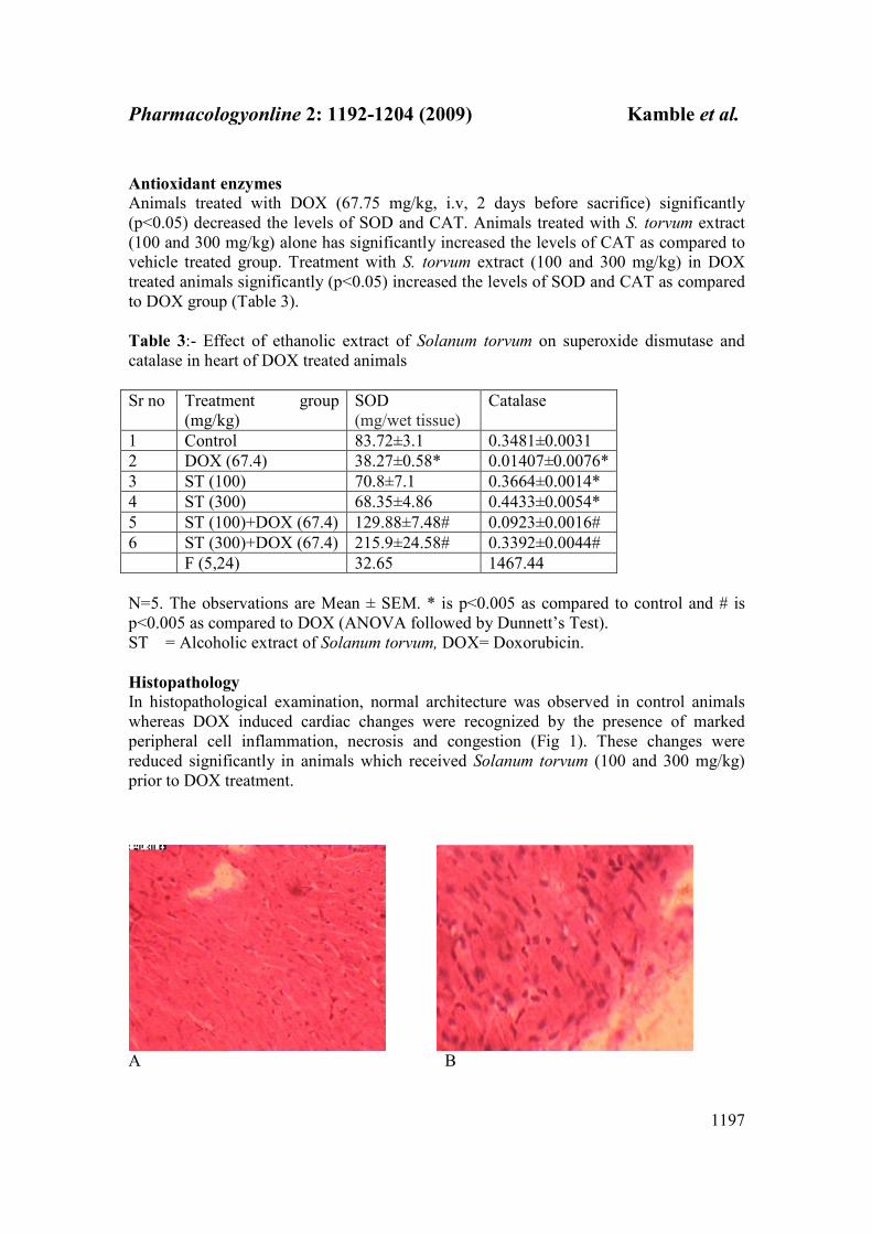
Table 3:- Effect of ethanolic extract of Solanum torvum on superoxide dismutase and
catalase in heart of DOX treated animals
Sr no Treatment group
(mg/kg)
SOD
(mg/wet tissue)
Catalase
1 Control 83.72±3.1 0.3481±0.0031
2 DOX (67.4) 38.27±0.58* 0.01407±0.0076*
3 ST (100) 70.8±7.1 0.3664±0.0014*
4 ST (300) 68.35±4.86 0.4433±0.0054*
5 ST (100)+DOX (67.4) 129.88±7.48# 0.0923±0.0016#
6 ST (300)+DOX (67.4) 215.9±24.58# 0.3392±0.0044#
F (5,24) 32.65 1467.44
N=5. The observations are Mean ± SEM. * is p<0.005 as compared to control and # is
p<0.005 as compared to DOX (ANOVA followed by Dunnett’s Test).
ST = Alcoholic extract of Solanum torvum, DOX= Doxorubicin.
Histopathology
In histopathological examination, normal architecture was observed in control animals
whereas DOX induced cardiac changes were recognized by the presence of marked
peripheral cell inflammation, necrosis and congestion (Fig 1). These changes were
reduced significantly in animals which received Solanum torvum (100 and 300 mg/kg)
prior to DOX treatment.
A B
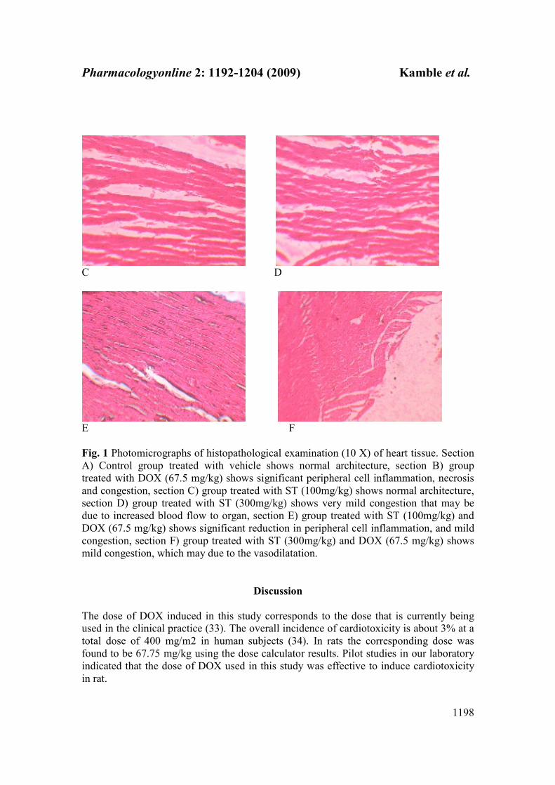
Pharmacologyonline 2: 1192-1204 (2009) Kamble et al.
1198
C D
E F
Fig. 1 Photomicrographs of histopathological examination (10 X) of heart tissue. Section
A) Control group treated with vehicle shows normal architecture, section B) group
treated with DOX (67.5 mg/kg) shows significant peripheral cell inflammation, necrosis
and congestion, section C) group treated with ST (100mg/kg) shows normal architecture,
section D) group treated with ST (300mg/kg) shows very mild congestion that may be
due to increased blood flow to organ, section E) group treated with ST (100mg/kg) and
DOX (67.5 mg/kg) shows significant reduction in peripheral cell inflammation, and mild
congestion, section F) group treated with ST (300mg/kg) and DOX (67.5 mg/kg) shows
mild congestion, which may due to the vasodilatation.
Discussion
The dose of DOX induced in this study corresponds to the dose that is currently being
used in the clinical practice (33). The overall incidence of cardiotoxicity is about 3% at a
total dose of 400 mg/m2 in human subjects (34). In rats the corresponding dose was
found to be 67.75 mg/kg using the dose calculator results. Pilot studies in our laboratory
indicated that the dose of DOX used in this study was effective to induce cardiotoxicity
in rat.

Pharmacologyonline 2: 1192-1204 (2009) Kamble et al.
1199
The mechanism of DOX induced cardiomyopathy is not completely understood, but
several hypotheses have been postulated which include inhibition of nucleic acid (35),
protein synthesis (36), release of vasoactive amines (37), alterations in sarcolemmal Ca++
transport (38), alterations in membrane bound enzymes (39), abnormalities in
mitochondria and lysosomal alterations (40) and an imbalance of myocardial electrolytes
(41). Data derived from several laboratories suggest that Ca ++ transport abnormalities
occur in cardiac (42, 43) tissues exposed to DOX and that the myocardium eventually
becomes overloaded with calcium (44, 45). This conclusion is based on total tissue
calcium levels in hearts from treated animals and the observation of calcium phosphate
accumulations in necrotic cells. The generation of free radical by DOX in the form of
DOX semiquinone also has been suggested to play a major role in its cardiotoxic effects
(46) by increasing oxygen free radical activity (47) and inducing the peroxidation of
unsaturated lipids within the membranes (48). Semiquinones are unstable under aerobic
conditions thereby generating superoxide anion radicals.
The ECG changes induced by DOX consisted of prolongation of QT interval, ST interval
and widening of QRS complex. These ECG changes have been partly explained by
Jensen (49). Whereas the QRS-intervals are directly related to cell depolarization, the QT
interval is an expression of the late repolarization phase; the anthracyclines specifically
prolong the later phase by disturbing the ion flux across the cellular membrane. It has
been found that the above parameters are the most reliable ECG parameters for the
assessment of DOX-induced cardiotoxicity (29). S. torvum extract (100 and 300 mg/kg)
significantly decreased the prolongation of QT and ST intervals and heart rate in DOX
treated groups. The heart weight was significantly decreased in DOX treated group which
was prevented by S. torvum extract (100 and 300 mg/kg) treatment.
Administration of DOX to rats increased cardiotoxicity manifested by elevation in the
levels of cardiac injury markers i.e. serum LDH and CK-MB levels. Our results are in
good agreement with those previously reported (50, 51). However treatment with S.
torvum extracts (100 and 300 mg/kg) in DOX treated group has resulted in reversal of
cardiac enzyme activities (Table 2). The increase in cardiac marker levels in serum
suggests an increased leakage of these enzymes from mitochondria as a result of toxicity
induced by treatment with DOX.
The heart is particularly susceptible to free radical injury, because it contains less free
radical detoxifying substances than do metabolic organs like liver or kidney (52, 53).
Moreover DOX is known to have a higher affinity for cardiolipin, a major phospholipid
component of the mitochondrial membrane in heart cells resulting in selective
accumulation of doxorubicin inside cardiac cells (54).
The cardioprotective mechanism also appears to be through modulation of various anti-
oxidant parameters thereby improving the overall antioxidant defence of the myocardial
tissue. Free radical scavenging enzymes such as catalase, superoxide dismustase are the
first line cellular defense enzymes against oxidative injury, decomposing O2 and H2O2
before their interaction to form the more reactive hydroxyl radical (OH .
). The
equilibrium between these enzymes is an important process for the effective removal of
oxygen stress in intracellular organelles (55). In our study, a decrease in concentration of
cardiac SOD and CAT levels in DOX treated group was observed. S. torvum extract (100
and 300 mg/kg) treatment significantly reversed the changes in antioxidant levels induced
by DOX treatment. The fall in SOD levels may be due to the involvement of superoxide

Pharmacologyonline 2: 1192-1204 (2009) Kamble et al.
1200
free radical in myocardial cell damage. A decrease in the activity of SOD can result in the
decreased removal of superoxide ion, which can be harmful to the myocardium (56).
There is a general agreement that flavonoids act as scavengers of reactive oxygen species
(57). Flavonoids have been found to protect heart from DOX induced cardiotoxicity
when co-administered with DOX in mice (58), which suggests that these compounds are
potential cardioprotectors against DOX, induced chronic cardiotoxicity. The antioxidant
properties of Solanum torvum could be attributed to the presence of flavonoid
phytoconstituent in it.
Cardiac histopathological features induced by DOX treatment was observed as significant
peripheral cell inflammation, necrosis and congestion. Pretreatment with Solanum torvum
extract reversed these abnormal changes. Thus, in conclusion the above data suggests that
Solanum torvum which is rich in flavonoids has the potential in preventing the
cardiotoxic effects induced by doxorubicin.
Acknowledgement
The authors are grateful to Prin.V. M. Aurangabadkar for providing the necessary
laboratory facilities.
References
1. Blum RH, Carter SK. A new drug with significant clinical activity. Ann
Intern Med 1974; 80:249-56.
2. Calabresi P. Chabner BA. Chemotherapy of neoplastic diseases. In: Gilman
AG, Rall TW, Nies AS, Taylor P, eds. The Pharmacological Basis of
Therapeutics NY: Pergamon Press Inc, 1990; 1203-1263.
3. Gillick J, Giles S, Bannigan J, Puri, P. Cell death in the early adriamycin rat
Model. Pediatr Surg Int 2002; 18:576-80.
4. Yilmaz S, Atessahin A, Sahna E, Karahan I, Ozer S. Protective effect
lycopene on adriamycin- induced cardiotoxicity and nephrotoxicity.
Toxicology 2006; 218:164-71.
5. Abdel-Aleem S, El-merzabani MM, Sayed-Ahmed MM, Taylor DA, Lowe
JE. Acute and chronic effects of adriamycin on fatty acid oxidation in
isolated cardiac myocytes. J Mol Cell Cardiol 1997; 29:789-97.
6. Doroshow JH. Anthracycline and anthracenediones. In: Chabner BA, Longo
DL, eds. Cancer Chemotherapy and Biotherapy. Philadelphia, USA:
Lippincott Raven Publishers 1996;409-433.
7. Nohl H, Gille L, Stanick K. The exogenous NADH dehydrogenase of heart
mitochondria is the key enzyme responsible for selective cardiotoxicity of
anthracyclines. Z Naturforsch 1998; 53:279-85.
8. Paulson DJ. Carnitine deficiency-induced cardiomyopathy. Mol Cell
Biochem 1998; 180:33-41.

Pharmacologyonline 2: 1192-1204 (2009) Kamble et al.
1201
9. Adjanohoun JE, Aboubakar N, Dramane K, Ebot ME, Ekpere JA,
Enoworock EG, Foncho D, Gbile ZO, Kamanyi A, Kamoukom J, Keeta A,
Mbenkum T, Mbi CM, Mbielle AL, Mbome IL, Mubiru NK,Naney WL,
Nkongmeneck B, Satabie B, Sofowa A, Tanze V, Wirmum CK. Traditional
medicine and pharmacopeia-contribution to ethnobotanical and floristic
studies in Cameroon. In: CNPMS. Porto-Novo, Benin 1996; 50–52.
10. Siemonsma JS, Piluek K. Plant resources of South-East Asia 8 (PROSEA).
Indonesia: Bogor 1994.
11. Ajaiyeoba EO. Comparative phytochemical and antimicrobial studies of
Solanum macrocarpum and Solanum torvum leaves. Fitoterapia
1999;70:184– 86.
12. Chah KF, Muko KN, Oboegbulem SI. Antimicrobial activity of methanolic
extract of Solanum torvum fruit. Fitoterapia 2000; 71:187–89.
13. Arthan D, Svasti J, Kittakoop P, Pittayakhachonwut D, Tanticharoen M,
Thebtaranonth Y. Antiviral isoflavonoid sulfate and steroidal glycosides
From the fruits of Solanum torvum. Phytochemistry 2002; 59:459–63.
14. Israf DA, Lajis NH, Somchit MN, Sulaiman MR. Enhancement of
ovalbumin-specific IgA responses via oral boosting with antigen co-
administered with an aqueous Solanum torvum extract. Life Science 2004;
75:397–06.
15. Telesphore B, Nguelefack CB, Feumebo G, Ateufack PW, Simplice T,
Albert D, Atsamo PT, Albert K, et al. Anti-ulcerogenic properties of the
aqueous and methanol extracts from the leaves of Solanum torvum Swartz
(Solanaceae) in rats. J Ethnopharmacol 2008; 119 (1):135-40.
16. Sivapriya M, Srinivas L. Isolation and purification of a novel antioxidant
protein from the water extract of Sundakai (Solanum torvum) seeds. Food
Chemistry 2007; 104:510–17.
17. Ndebia EJ, Kamga R, Nchunga-Anye NB. Analgesic and anti-inflammatory
properties of aqueous extract from leaves of Solanum torvum (Solanaceae).
AJTCAM 2007; 4:240–44.
18. Nguelefack TB, Feumebo CB, Watcho GAP, Tatsimo S, Atsamo AD, Tane
P, Kamanyi A, et al. Anti- ulcerogenic properties of the aqueous and
methanol extracts from the leaves of Solanum torvum Swartz (Solanaceae)
in rats. J. Ethnopharmacol 2008; 119 (1):135-40.
19. Yahara S, Yamashita T, Nozawa N, Nohara T. Steroidal glycosides from
Solanum torvum. Phytochemistry 1996; 43:1069–74.
20. Carobot, CA, Blunden G, Patel VA. Chlorogenone and neochlorogenone
from the unripe fruits of Solanum torvum. Phytochemistry 1991; 30:1339–
41.
21. Mahmood U, Agrawal PK, Thakur RS. Torvonin-A, a spirostane saponin
from Solanum torvum leaves. Phytochemistry 1985 24:2456–57.
Pharmacologyonline 2: 1192-1204 (2009) Kamble et al.
1202
22. Mahmood U, Shukla YN, Thakur RS. 1983 Non-alkaloidal constituents
fromSolanum torvum leaves. Phytochemistry 1983; 22:167–70.
23. Iida Y, Yanai Y, Ono M, Ikeda T, Nohara T. 2005 Three unusual 22-β-O-
23-hydroxy-(5α)-spirostanol glycosides from the fruits of Solanum torvum.
Chemical & Pharmaceutical Bulletin 2005; 53:1122–25.
24. Arthan D, Kittakoop P, Esen A, Svasti J. Furostanol glycoside 26-O-β
glucosidase from the leaves of Solanum torvum. Phytochemistry 2006;
67:27–33.
25. Vaclavıkova R, Kondrova E, Ehrlichova DM, Boumendjel A, Kovar J,
Stopka P, et al. The effect of flavonoid derivatives on doxorubicin transport
and metabolism. Bioorganic & Medicinal Chemistry 2008; 16:2034–42.
26. Woisky R, Salatino A. Analysis of Propolis: some parameters and
Procedures for chemical quality control. J. Agri.Res.1998; 37: 99-105.
27. Slinkard K, Singleton VL. Total Phenol analyses: Automation and
comparison with manual methods. Am. J. Enol. Vitic. 1977; 8: 49-55.
28. Kokate CK. Practical Pharmacognosy, 3rd edn., Vallabh Prakashan, New
Delhi 1994;107-09.
29. Danesi R, Tacca MD, Soldan G. Measurement of the S [alpha]-T segment as
the most reliable electrocardiogram parameter for the assessment of
adriamycin-induced cardiotoxicity in the rat. Journal of Pharmaceutical
Methods 1986; 16: 251–59.
30. Niehaus WG, Samuelsson B. Formation of malondialdehyde from
phospholipids arachidonate during microsomal lipid peroxidation. Eur J
Biochem 1968; 6:126-30.
31. Jiang ZY, Hunt JY, Wolff SP. Detection of lipid hydroperoxides using the
fox method. Anal Biochem 1992; 200:384-89.
32. Beers RF and Sizer IW. A spectrophotometric method for measuring the
breakdown of hydrogen peroxide by catalase. J Biological Chem 1952; 115:
133-40.
33. Chabner BA, Ryan DP, Paz-Ares L, Garcia-Carbonevo R, Calabresi P.
Antineoplastic agents. In: Hardman, JG, Limbird LE, Gilman AG eds,
Goodman and Gilman’s the Parmacological Basis of Therapeutics.
McGraw-Hill Companies Inc., USA 2001; 1389–459.
34. Tallaj JA, Veronica F, Rayburn BK, Pinderski L, Benza RL, Pamboukian S,
Foley B, Bourge RC. Response of Doxorubicin-induced cardiomyopathy to
the current management stratergy of heart failure. J Heart Lung Transplant
2005; 24:2196-200.
35. Arena E, Bionda F, DAlessndro N, Dusoncher L, Gebbia N, Gerbasi R.
DNA, RNA protein synthesis in heart, liver and brain of mice treated with
denaturation of adriamycin. Int Res Commun Syst Med Sci 1984; 2:10543–
61.

Pharmacologyonline 2: 1192-1204 (2009) Kamble et al.
1203
36. Buja LM, Ferrans VJ, Mayer RJ, Roberts WC, Hinderson ES. Cardiac
ultrastructural changes induced by daunorubucin therapy. Cancer 1973;
32:771–78.
37. Bristow MR, Sagemen WS, Scot RH, Billingham ME, Bowden RE, Kernoff
RS, et al. Acute and chronic cardiovascular effects of doxorubicin in the
dog: the cardiovascular pharmacology of drug-induced histamine release.
Cardiovasc Pharmacol 1980; 2:487–15.
38. Singal PK, Pierce GN. Adriamycin stimulates Ca2+
- binding and lipid
peroxidation but depress myocardial function. American Journal of
Physiology, 1986; 250: H419–24.
39. Singal PK, Panagia V. Direct effect of adriamycin on the rat heart
sarcolemma. Res Commun Chem Pathol Pharmacol 1984; 43:67–77.
40. Singal PK, Segstro RU, Singh RP, Kutryk MJ. Changes in lysosomal
morphology and enzyme activities during development of adriamycin-
induced cardiomyopathy. Canadian Journal of Cardiology 1985; 1:139–47.
41. Oslan HM, Young DM, Prieur DJ, LeRoy AF, Reagan RL. Electrolyte and
oxidation products of certain lipids. J Biol Chem 1974; 174: 257–64.
42. Villani F, Piccinini F, Merelli P, Favalli L. Influence of adriamycin on
calcium exchangeability in cardiac muscle and its modification by ouabain.
Biochem Pharmacol 1978; 27: 985-87.
43. Caroni P, Villani F, Carafoli E. The cardiotoxic antibiotic doxorubicin
inhibits the Na+/Ca
+ exchange of dog heart sarcolemmal vesicles. FEBS
Letters 1981; 130:184-86.
44. Olson HM, Capen CC. Subacute cardiotoxicity of Adriamycin in the rat:
Biochemical and ultrastructural investigations. Laboratory Investigation
1977; 37: 386-94.
45. Mettler FP, Young DM, Ward JM. Adriamycin-induced cardiotoxicity
(cardiomyopathy and congestive heart failure) in rats. Cancer Research
1977;37: 2705-13.
46. Bachur NR, Gordon SL, Gee MV, Kon H. NADPH-cytochrome P450
reductase activation of quinone anticancer agents to free radicals.
Proceedings of the National Academy of Sciences USA 1979; 76:954-57.
47. Lee V, Randhawa AK, Singhal PK. Adriamycin induced myocardial
dysfunction in vitro is mediated by free radicals. American Journal of
Physiology 1991; 261: H989–95.
48. Myers CE, Mcguire WP, Liss RH, Ifrim I, Grotzinger K, Young RC.
Adriamycin: the role of lipid peroxidation in cardiac toxicity and tumor
response. Science 1977; 19:165–67.
49. Jensen RA, Acton EM, Peters JH. Doxorubicin cardiotoxicity in the rat:
Comparision of electrocardiogram, transmembrane potential, and structural
effects. J Cardiovasc Pharmacol 1984;6:186-200.

Pharmacologyonline 2: 1192-1204 (2009) Kamble et al.
1204
50. Van aker SA, Kramer K, Voest EE, Grimbergen JA, Zharg J, Van-Der-
Viigh WJ, et al. Doxorubicin induced cardiotoxicity monitored by ECG in
freely moving mice. A new model to test potential protectors. Cancer
Chemotherapy and Pharmacology 1996; 38:95-101.
51. Venkatesan N. Curcumin attenuation of acute adriamycin myocardial
toxicity in rats. Br J Pharmacol 1998; 124:425-27.
52. Olson RD, Boerth RC, Gerber JG, Nies AS. Mechanism of adriamycin
cardiotoxicity: evidence for oxidative stress. Life Sciences 1981; 29:1393-
01.
53. Olson RD, Mushlin PS. Doxorubicin cardiotoxicity: analysis of prevailing
hypotheses. FASEB Journal 1990; 4:3076–86.
54. Goormaghtigh E, Ruysschaert JM. Anthracycline glycosides-membrane
interactions. Biochimie et Biophysica Acta 1984; 779: 271-88.
55. Sharma M, Kishore K, Gupta SK, Joshi S, Arya D. Cardioprotective
potential of Ocimum sanctum in isoproterenol induced myocardial infarction
in rats. Mol Cell Biochem 2001; 225:75-83.
56. Liu J, Simon LM, Philips JR, Robin ED: Superoxide dismutase (SOD)
activity in hypoxic mammalian systems. J Appl Physiol 1977; 42:107-10.
57. Haenen GRMM, Janssen FP, Bast A. The antioxidant properties of five (O-β
hydroxyethyl) rutosides of the flavonoid mixture Venoruton. Phlebol Suppl
1993; 1:10-17.
58. Van Acker SA, Voest EE, Beems DB, Madhuizen HT, De Jong J, Bast A,
et. al. Cardioprotective properties of O-(beta-hydroxyethyl)-rutosides in
doxirubcin-pretreated BALB/ c mice. Cancer Research, 53, 4603-4607.Yeh
ET. (2006) Cardiotoxicity induced by chemotherapy and antibody therapy.
Annu Rev Med 1993; 57:485–98.