protective effects of green tea extract against hepatic tissue injury
TRANSCRIPT

Hindawi Publishing CorporationEvidence-Based Complementary and Alternative MedicineVolume 2012, Article ID 740671, 10 pagesdoi:10.1155/2012/740671
Research Article
Protective Effects of Green Tea Extract against Hepatic TissueInjury in Streptozotocin-Induced Diabetic Rats
Ali Akbar Abolfathi,1 Daryoush Mohajeri,2 Ali Rezaie,3 and Mehrdad Nazeri4
1 Department of Biological Science, Ahar Branch, Islamic Azad University, Ahar, Iran2 Department of Pathobiology, Tabriz Branch, Islamic Azad University, Tabriz, Iran3 Department of Clinical Science, Tabriz Branch, Islamic Azad University, Tabriz, Iran4 Young Researchers Club, Tabriz Branch, Islamic Azad University, Tabriz, Iran
Correspondence should be addressed to Ali Akbar Abolfathi, [email protected]
Received 21 October 2011; Revised 22 November 2011; Accepted 29 November 2011
Academic Editor: Olumayokun A. Olajide
Copyright © 2012 Ali Akbar Abolfathi et al. This is an open access article distributed under the Creative Commons AttributionLicense, which permits unrestricted use, distribution, and reproduction in any medium, provided the original work is properlycited.
Although diabetic hepatopathy is potentially less common, it may be appropriate for addition to the list of target organ conditionsrelated to diabetes. This study was designed to evaluate the hepatoprotective properties of green tea extract (GTE) in STZ-induceddiabetes in rats. Wistar rats were made diabetic through single injection of STZ (75 mg/kg i.p.). The rats were randomly divided intofour groups of 10 animals each: Group 1, healthy control; Group 2, nondiabetics treated with GTE administered orally (1.5%, w/v);Group 3, diabetics; Group 4, diabetics treated with GTE (1.5%, w/v) for 8 weeks. Serum biomarkers were assessed to determinehepatic injury. Malondialdehyde (MDA) and reduced glutathione (GSH) contents were measured to assess free radical activityin the liver tissue. Hepatic antioxidant activities of glutathione peroxidase (GSH-Px), superoxide dismutase (SOD), and catalase(CAT) were also determined. The biochemical findings were matched with histopathological verifications. Liver MDA content andserum levels of ALT, AST, ALP, and bilirubin in Group 3 significantly increased compared to Group 1 (P < 0.05) and significantlydecreased in Group 4 compared to Group 3 (P < 0.05). Serum albumin level and GSH, SOD, CAT, and GSH-Px contents of the liverin Group 3 were significantly decreased compared to Group 1 (P < 0.05) and were significantly increased in Group 4 compared toGroup 3 (P < 0.05). Histopathologically, the changes were in the same direction with biochemical findings. This study proved thehepatoprotective activity of GTE in experimentally induced diabetic rats.
1. Introduction
Type 1 and type 2 diabetes mellitus (T2DM) are influencedby different genetic factors, but individuals with eitherof them are prone to developing complications includingnephropathy, retinopathy, peripheral neuropathy, and hyper-tension [1, 2]. Diabetes, by most estimates, is now the mostcommon cause of liver disease in the USA and liver diseaseis an important cause of death in type 2 diabetes [3]. Thus,patients with diabetes have a high prevalence of liver diseaseand patients with liver disease have a high prevalence ofdiabetes. Virtually the entire spectrum of liver disease is seenin patients with type 2 diabetes. This includes abnormalliver enzymes, nonalcoholic fatty liver disease (NAFLD),cirrhosis, hepatocellular carcinoma, and acute liver failure
[4]. Chronic mild elevations of transaminases are frequentlyfound in type 2 diabetic patients. However, when the liverfails, there is no equivalent form of management, such ashemodialysis or retinal photo-coagulation. Thus, althoughdiabetic hepatopathy is potentially less common, it may beappropriate for addition to the list of target organ conditionsrelated to diabetes, such as glomerulopathy, retinopathy,and neuropathy. However, annual screening for liver diseasemight be accomplished by means of a simple biochemicalanalyte such as alanine aminotransferase [5].
Oxidative stress is currently suggested as a mechanismunderlying diabetes and diabetic complications [6]. Freeradicals are continually produced in the body as the resultof normal metabolic processes and interaction with environ-mental stimuli. Under physiological conditions, a wide range
2 Evidence-Based Complementary and Alternative Medicine
of antioxidant defences protects against the adverse effects offree radical production in vivo [6]. Oxidative stress resultsfrom an imbalance between radical-generating and radical-scavenging systems, that is, increased free radical productionor reduced activity of antioxidant defences or both thesephenomena. In diabetes, protein glycation and glucoseautoxidation may generate free radicals, which in turncatalyse lipid peroxidation [7, 8]. Moreover, disturbancesof antioxidant defence systems in diabetes were shown:alteration in antioxidant enzymes [9], impaired glutathionemetabolism [10], and decreased ascorbic acid levels [11, 12].However, an increased oxidative stress in vivo has neverbeen clearly demonstrated. Several studies in humans andanimal models, using thiobarbituric acid reactive substances(TBARS) assay [13–18], have shown increased lipid per-oxidation in membranes and lipoproteins in the diabeticstate. However, Lipid peroxidation and antioxidant statusof hepatic tissue were studied by Feillet-Coudray et al. inexperimental diabetes [19].
Antidiabetic agents have generally been shown todecrease the levels of serum biomarker of hepatic injury[20], but these agents can produce serious side effects [21].Natural remedies from medicinal plants are considered to beeffective and safe alternative treatment for hyperglycemia andliver toxicity. There is a growing interest in herbal remediesbecause of their effectiveness, minimal side effects in clinicalexperience, and relatively low cost. Herbal drugs or theirextracts are prescribed widely, even when their biologicalactive compounds are unknown [22]. For example, beneficialeffect of freshly prepared aqueous extracts of Psidiumguajava, Momordica charantia, and Coccinia indica leaves andtheir combination in STZ-induced diabetes rats has beenproved by Rafiq et al. [23]. Similarly, antidiabetic activityof hydroethanolic extracts of Nymphaea stellata flowershas been documented in alloxan-induced diabetic rats byRajagopal and Sasikala [24]. Therefore, studies with plantextracts are useful to know their efficacy and mechanism ofaction and safety.
Green tea (leaves of Camellia sinensis, Theaceae) is a pop-ular beverage in East Asia and also used as a herbal remedyin Europe and North America. An increased consumptionof green tea may reduce the risk of liver disease. Population-based clinical studies have shown that men who drink morethan 10 cups of green tea per day are less likely to developdisorders of the liver. Green tea also seems to protect theliver from the damaging effects of toxic substances such asalcohol [25]. Green tea is considered to be anti inflammatory,antioxidative, antimutagenic, and anticarcinogenic [26, 27],and can prevent cardiac disorders. Epidemiologically, it hasbeen suggested that green tea consumption prevents type 2diabetes. [28, 29]. Tsuneki et al. demonstrated that greentea produces an antihyperglycemic effect in STZ diabeticmice [30]. Crespy and Williamson [31] reported that greentea extract (GTE) displays antioxidants and free radicalsscavenger properties.
Considering the antihyperglycemic properties andantioxidant activities of green tea, this study was designedto evaluate the hepatoprotective effects of green tea extractin streptozotocin-induced diabetes of rats. In this context,
green tea can rightly be mentioned as a plant of considerableinterest.
2. Materials and Methods
2.1. Chemicals. Streptozotocin was from Sigma (St. Louis,MO, USA). All other chemicals used were of analytical grade.All chemicals used in this study were of analytical grade andobtained from Nanjing Jiancheng Bioengineering Institute,Nanjing, China.
2.2. Green Tea Extract (GTE). The green tea extract (GTE)was made according to Maity et al. [32], by soaking 15 g ofinstant green tea powder in 1 L of boiling distilled water for5 min. The solution was filtered to make 1.5% GTE. Thissolution was provided to rats as their sole source of drinkingwater.
2.3. Induction of Diabetes Mellitus. Diabetes was induced byintravenous injection of streptozotocin (Sigma, St. Louis,Mo, USA) into the tail vein at a dose of 65 mg/kg bodyweight. STZ was extemporaneously dissolved in 0.1 M coldsodium citrate buffer, pH 4.5. After 18 h, animals with fastingblood glucose levels greater than 16.5 mmol/L were consid-ered diabetic and then included in this study [33]. Fastingblood glucose was estimated by using one touch glucometer(Accu-Chek sensor) of Roche Diagnostics, Germany.
2.4. Animal Treatment. This experimental study was carriedout in Islamic Azad University Research Center and allprocedures and works on animals were conducted underAnimal Rights Monitoring Committee of the Islamic AzadUniversity Research Center.
Forty healthy male Wistar rats (about 180–200 g bodyweight) were purchased from Animal House, Islamic AzadUniversity. All animals were conditioned at room tempera-ture at a natural photoperiod for 1 week before experimentexecution. A commercial balanced diet and tap water adlibitum were provided. The duration of experiment was 8weeks. The rats were randomly divided into 4 groups (10rats each) as follows: Group 1, healthy control rats receiveddistilled water as sole drinking source; Group 2 non-diabeticrats GTE (1.5%, w/v) was given in drinking water; Group3, diabetic and no treatment was given of beginning ofexperiment; Group 4, diabetic rats were treated with weretreated with GTE (1.5%, w/v) as sole drinking source.
2.5. Biochemical Factors Evaluation. At the end of experi-ment, blood samples were collected from the retro-orbitalplexus and the sera prepared through centrifuging at 2500×gfor 15 minutes at 30◦C. Serum biomarkers of liver func-tion including alanine aminotransferase (ALT), aspartateaminotransferase (AST) [34], alkaline phosphatase (ALP)[35], albumin [36], and total bilirubin [37] were measuredusing commercially available kits. Aminotransferases (ALTand AST) were measured to determine the concentrationof intracellular hepatic enzymes that have leaked into thecirculation and serve as a marker of hepatocyte injury.
Evidence-Based Complementary and Alternative Medicine 3
ALP and bilirubin were measured to assess biliary function.Albumin was measured to reflect liver synthetic function.
2.6. Measurement of Antioxidant Activity. The rat’s liverwas removed immediately and washed in normal salineand homogenate 10% prepared in 1.15% w/v of potassiumchloride. The homogenate was centrifuged in 7000×g for 10minutes at 4◦C and supernatant was used for measurementof oxidative stress by estimation of reduced glutathione(GSH) and determination of malondialdehyde (MDA) aswell as antioxidant enzymes (AOEs) such as superoxide dis-mutase (SOD), catalase (CAT), and glutathione peroxidase(GSH-Px). GSH, MDA, SOD, CAT, and GSH-Px were mea-sured by using commercially available kits according to themanufacturer’s protocol (Nanjing Jiancheng BioengineeringInstitute, Nanjing, China).
Reduced glutathione (GSH) content was determinedaccording to Sedlak and Lindsay [38]. GSH reacts with 5,5′-dithiobis-2-nitrobenzoic acid, and the absorbance spectraof the product have a maximum absorbance at 410 nm.The results were expressed as μmol/g wt. Liver homogenateMDA levels were expressed as nmol MDA per mg proteinand antioxidant activity was expressed as arbitrary unitsper mg protein. Degree of lipid peroxidation in liver tissuehomogenates was determined in terms of thiobarbituric acidreactive substances (TBARSs) formation by following theprotocol of Esterbauer and Cheeseman [39]. SOD activitywas measured by Nishikimi et al. method [40] and wasmodified by Kakkar et al. method [41]. Each unit of SODactivity was determined as required enzyme concentrationfor prohibition of creation color at 1 minute, under studyconditions. CAT activity was measured by Claiborne method[42] and was based on hydrogen peroxide breakdown. GSH-Px activity was measured by Rotruck et al. method [43] andwas expressed as micromole of GSSG/minute/milligram ofprotein, based on the below reaction:
2H2O + GSSG −→ H2O2 + 2GSH (1)
2.7. Statistical Analysis. The Statistical Package for SocialSciences (SPSS Inc., Chicago, IL, USA), version 13.0, wasused for statistical analysis. All data are presented as mean± SEM. Before statistical analysis, all variables were checkedfor normality and homogeneity of variance by using theKolmogorov-Smirnoff and Levene tests, respectively. Thedata obtained were tested by ANOVA followed by Tukey’spost hoc multiple comparison test. P < 0.05 was consideredstatistically significant.
2.8. Microscopic Studies. The animals of different groupswere sacrificed under light anesthesia (diethyl ether) 1 dayafter the end of the treatment. A small piece of hepatictissue from the anterior portion of the left lateral lobewas removed for histological analysis. The sample was fixedby immersing it in 10% neutral-buffered formalin. Thesample was then embedded in paraffin, sliced into 5 μmsections, and stained with hematoxylin-eosin for blindedhistological assessment. The degree of liver tissue injurywas evaluated semiquantitatively according to the method
20
0
40
60
80
100
120
140
160
180
1 2 3 4
FBS
(mg/
dL)
Groups
a
b
c
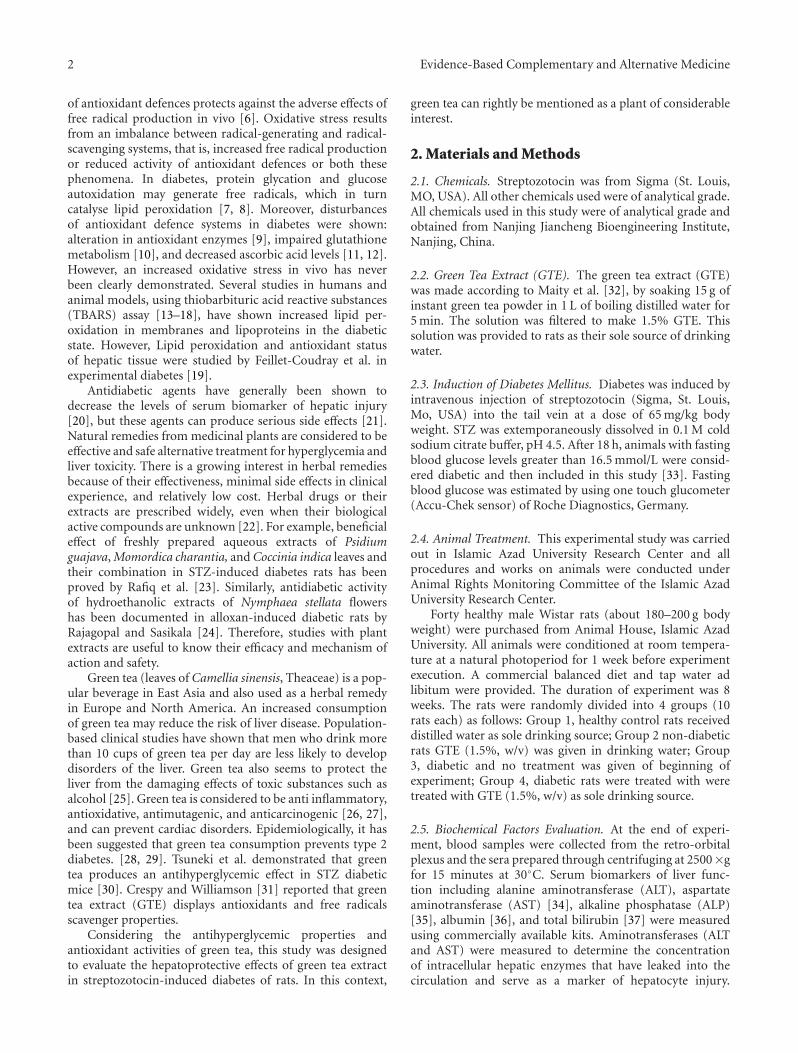
Figure 1: Comparison of the effect of GTE (1.5%, w/v) on bloodglucose levels among the experimental groups (mean± SEM). ∗P <0.05, a,bcompared to Group 1, ccompared to Group 3.
0
50
100
150
200
250
1 2 3 4
AST
(U
/L)
Groups
a
b
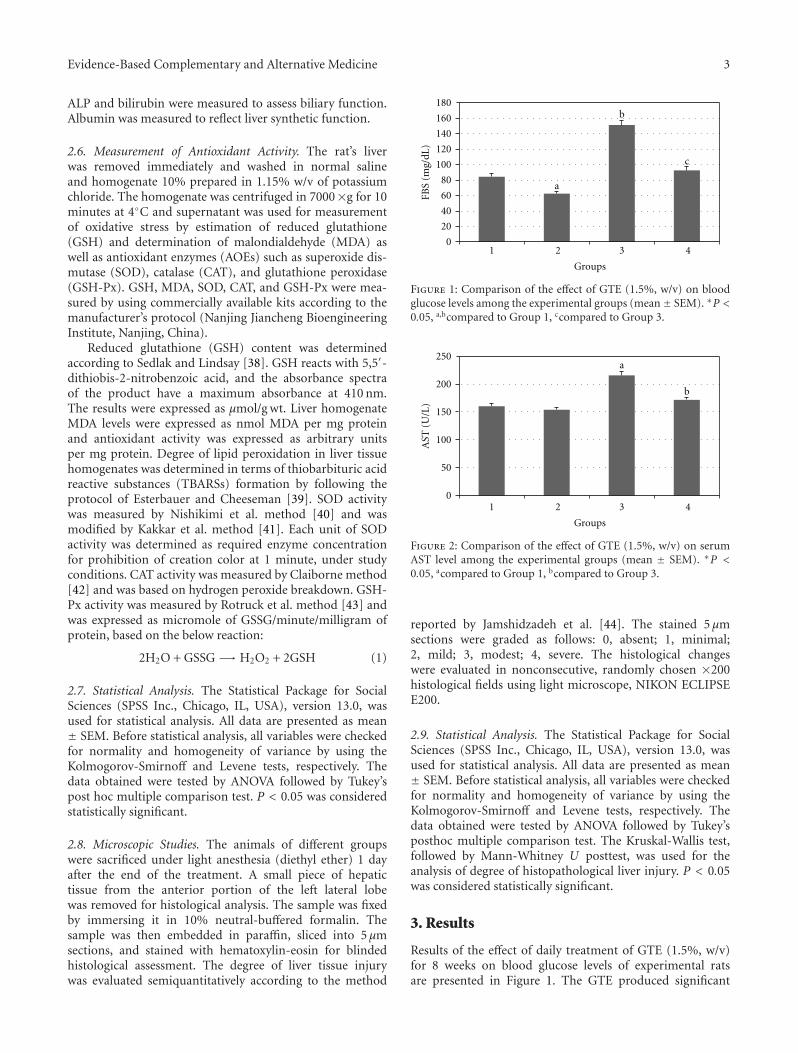
Figure 2: Comparison of the effect of GTE (1.5%, w/v) on serumAST level among the experimental groups (mean ± SEM). ∗P <0.05, acompared to Group 1, bcompared to Group 3.
reported by Jamshidzadeh et al. [44]. The stained 5 μmsections were graded as follows: 0, absent; 1, minimal;2, mild; 3, modest; 4, severe. The histological changeswere evaluated in nonconsecutive, randomly chosen ×200histological fields using light microscope, NIKON ECLIPSEE200.
2.9. Statistical Analysis. The Statistical Package for SocialSciences (SPSS Inc., Chicago, IL, USA), version 13.0, wasused for statistical analysis. All data are presented as mean± SEM. Before statistical analysis, all variables were checkedfor normality and homogeneity of variance by using theKolmogorov-Smirnoff and Levene tests, respectively. Thedata obtained were tested by ANOVA followed by Tukey’sposthoc multiple comparison test. The Kruskal-Wallis test,followed by Mann-Whitney U posttest, was used for theanalysis of degree of histopathological liver injury. P < 0.05was considered statistically significant.
3. Results
Results of the effect of daily treatment of GTE (1.5%, w/v)for 8 weeks on blood glucose levels of experimental ratsare presented in Figure 1. The GTE produced significant
4 Evidence-Based Complementary and Alternative Medicine
20
0
40
60
80
100
120
140
160
1 2 3 4
ALT
(U
/L)
Groups
a
b
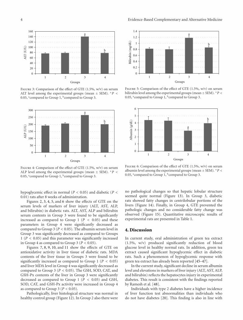
Figure 3: Comparison of the effect of GTE (1.5%, w/v) on serumALT level among the experimental groups (mean ± SEM). ∗P <0.05, acompared to Group 1, bcompared to Group 3.
0
50
100
150
200
250
300
1 2 3 4
ALP
(U
/L)
Groups
a
b
Figure 4: Comparison of the effect of GTE (1.5%, w/v) on serumALP level among the experimental groups (mean ± SEM). ∗P <0.05, acompared to Group 1, bcompared to Group 3.
hypoglycemic effect in normal (P < 0.05) and diabetic (P <0.01) rats after 8 weeks of administration.
Figures 2, 3, 4, 5, and 6 show the effects of GTE on theserum levels of markers of liver injury (ALT, AST, ALP,and bilirubin) in diabetic rats. ALT, AST, ALP and bilirubinserum contents in Group 3 were found to be significantlyincreased as compared to Group 1 (P < 0.05) and theseparameters in Group 4 were significantly decreased ascompared to Group 3 (P < 0.05). The albumin serum level inGroup 3 was significantly decreased as compared to Groups1 (P < 0.05) and this parameter was significantly increasedin Group 4 as compared to Group 3 (P < 0.05).
Figures 7, 8, 9, 10, and 11 show the effects of GTE onantioxidative activity in liver tissue of diabetic rats. MDAcontents of the liver tissue in Groups 3 were found to besignificantly increased as compared to Group 1 (P < 0.05)and liver MDA level in Group 4 was significantly decreased ascompared to Group 3 (P < 0.05). The GSH, SOD, CAT, andGSH-Px contents of the liver in Group 3 were significantlydecreased as compared to Group 1 (P < 0.05) and GSH,SOD, CAT, and GSH-Px activity were increased in Group 4as compared to Group 3 (P < 0.05).
Pathologically, liver histological structure was normal inhealthy control group (Figure 12). In Group 2 also there were
0.2
0
0.4
0.6
0.8
1
1.2
1.4
1 2 3 4
Bili
rubi
n (
mg/
dL)
Groups
a
b
Figure 5: Comparison of the effect of GTE (1.5%, w/v) on serumbilirubin level among the experimental groups (mean± SEM). ∗P <0.05, acompared to Group 1, bcompared to Group 3.
1
0
2
3
4
5
6
1 2 3 4
Alb
um
in (
g/dL
)
Groups
a
b
Figure 6: Comparison of the effect of GTE (1.5%, w/v) on serumalbumin level among the experimental groups (mean± SEM). ∗P <0.05, acompared to Group 1, bcompared to Group 3.
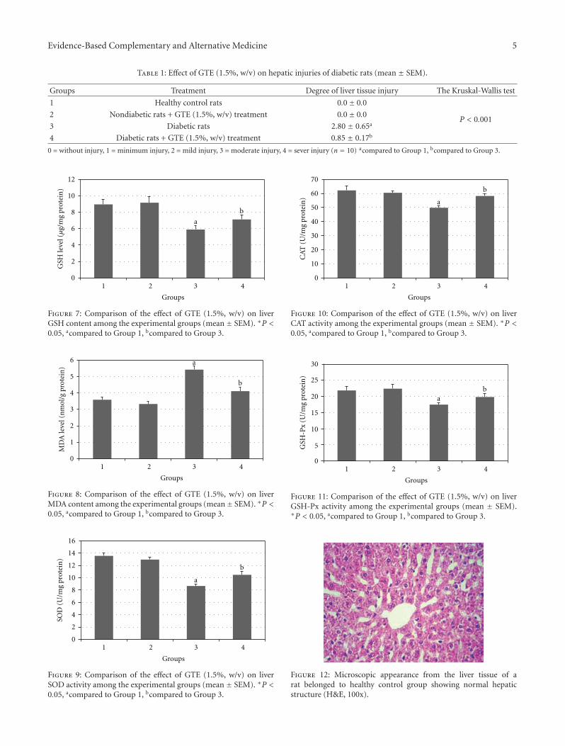
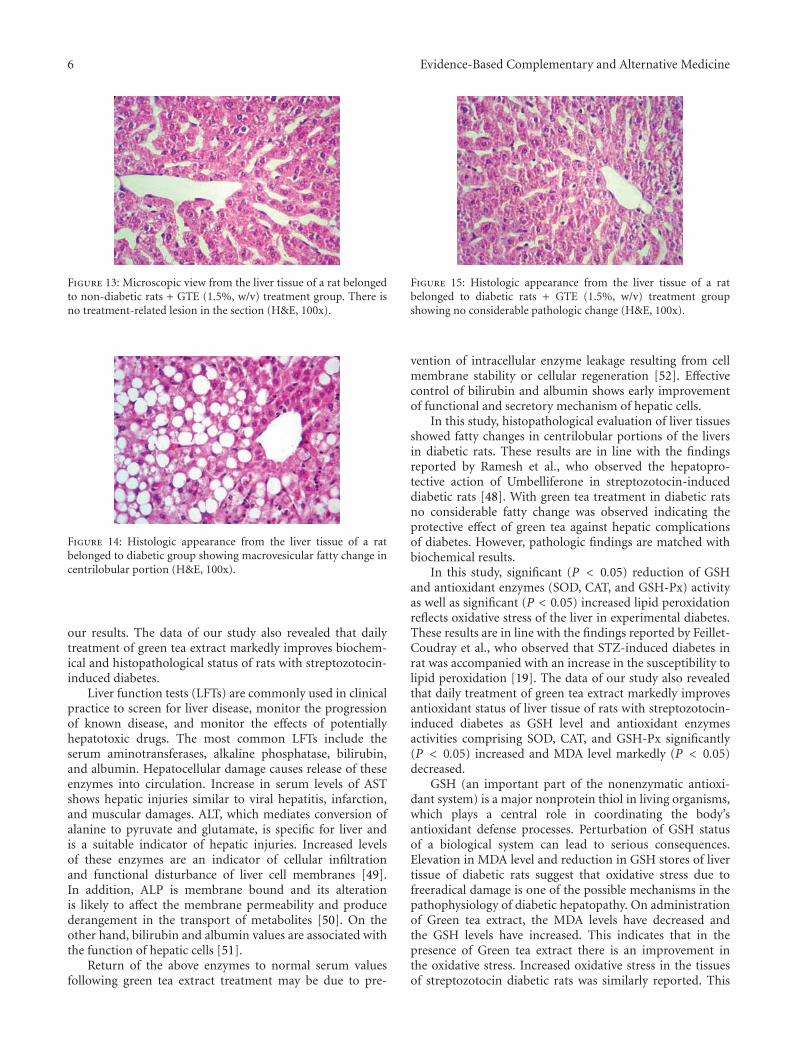
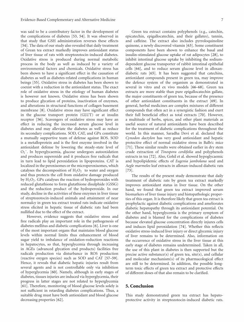
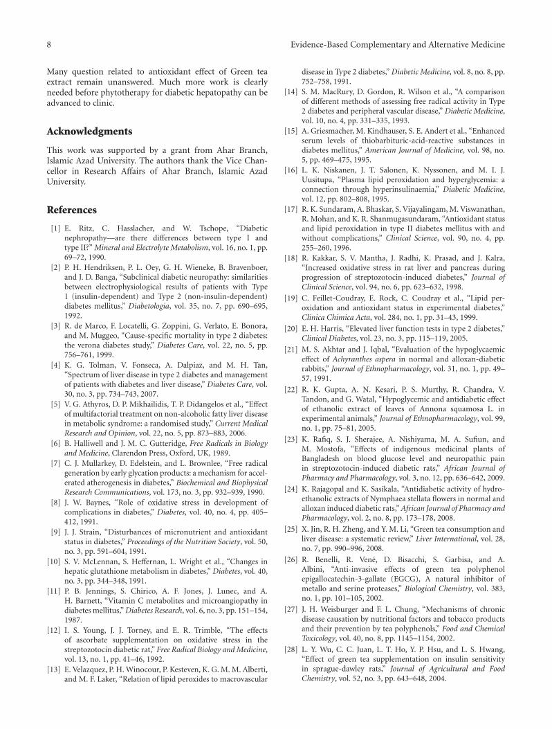
no pathological changes so that hepatic lobular structureseemed quite normal (Figure 13). In Group 3, diabeticrats showed fatty changes in centrilobular portions of thelivers (Figure 14). Finally, in Group 4, GTE prevented thepathologic changes and no considerable fatty change wasobserved (Figure 15). Quantitative microscopic results ofexperimental rats are presented in Table 1.
4. Discussion
In current study, oral administration of green tea extract(1.5%, w/v) produced significantly reduction of bloodglucose level in healthy normal rats. In addition, green teaextract caused significant hypoglycemic effect in diabeticrats. Such a phenomenon of hypoglycemic response withgreen tea extract has already been reported [45–47].
In the current study, significant decline in serum albuminlevel and elevations in markers of liver injury (ALT, AST, ALP,and bilirubin) reflects the hepatocytes injury in experimentaldiabetes. This result is consistent with the findings reportedby Ramesh et al. [48].
Individuals with type 2 diabetes have a higher incidenceof liver function test abnormalities than individuals whodo not have diabetes [20]. This finding is also in line with
Evidence-Based Complementary and Alternative Medicine 5
Table 1: Effect of GTE (1.5%, w/v) on hepatic injuries of diabetic rats (mean ± SEM).
Groups Treatment Degree of liver tissue injury The Kruskal-Wallis test
1 Healthy control rats 0.0± 0.0
2 Nondiabetic rats + GTE (1.5%, w/v) treatment 0.0± 0.0P < 0.001
3 Diabetic rats 2.80 ± 0.65a
4 Diabetic rats + GTE (1.5%, w/v) treatment 0.85 ± 0.17b
0 = without injury, 1 = minimum injury, 2 = mild injury, 3 = moderate injury, 4 = sever injury (n = 10) acompared to Group 1, bcompared to Group 3.
2
0
4
6
8
10
12
1 2 3 4
Groups
GSH
leve
l (µ
g/m
g pr
otei
n)
a
b
Figure 7: Comparison of the effect of GTE (1.5%, w/v) on liverGSH content among the experimental groups (mean ± SEM). ∗P <0.05, acompared to Group 1, bcompared to Group 3.
1
0
2
3
4
5
6
1 2 3 4
MD
A le
vel (
nm
ol/g
pro
tein
)
Groups
a
b
Figure 8: Comparison of the effect of GTE (1.5%, w/v) on liverMDA content among the experimental groups (mean± SEM). ∗P <0.05, acompared to Group 1, bcompared to Group 3.
2
0
4
6
8
12
14
10
16
1 2 3 4
SOD
(U
/mg
prot
ein
)
Groups
a
b
Figure 9: Comparison of the effect of GTE (1.5%, w/v) on liverSOD activity among the experimental groups (mean ± SEM). ∗P <0.05, acompared to Group 1, bcompared to Group 3.
10
0
20
30
40
50
60
70
1 2 3 4
CA
T (
U/m
g pr
otei
n)
Groups
a
b
Figure 10: Comparison of the effect of GTE (1.5%, w/v) on liverCAT activity among the experimental groups (mean ± SEM). ∗P <0.05, acompared to Group 1, bcompared to Group 3.
5
0
10
15
20
25
30
1 2 3 4
Groups
GSH
-Px
(U/m
g pr
otei
n)
ab
Figure 11: Comparison of the effect of GTE (1.5%, w/v) on liverGSH-Px activity among the experimental groups (mean ± SEM).∗P < 0.05, acompared to Group 1, bcompared to Group 3.
Figure 12: Microscopic appearance from the liver tissue of arat belonged to healthy control group showing normal hepaticstructure (H&E, 100x).
6 Evidence-Based Complementary and Alternative Medicine
Figure 13: Microscopic view from the liver tissue of a rat belongedto non-diabetic rats + GTE (1.5%, w/v) treatment group. There isno treatment-related lesion in the section (H&E, 100x).
Figure 14: Histologic appearance from the liver tissue of a ratbelonged to diabetic group showing macrovesicular fatty change incentrilobular portion (H&E, 100x).
our results. The data of our study also revealed that dailytreatment of green tea extract markedly improves biochem-ical and histopathological status of rats with streptozotocin-induced diabetes.
Liver function tests (LFTs) are commonly used in clinicalpractice to screen for liver disease, monitor the progressionof known disease, and monitor the effects of potentiallyhepatotoxic drugs. The most common LFTs include theserum aminotransferases, alkaline phosphatase, bilirubin,and albumin. Hepatocellular damage causes release of theseenzymes into circulation. Increase in serum levels of ASTshows hepatic injuries similar to viral hepatitis, infarction,and muscular damages. ALT, which mediates conversion ofalanine to pyruvate and glutamate, is specific for liver andis a suitable indicator of hepatic injuries. Increased levelsof these enzymes are an indicator of cellular infiltrationand functional disturbance of liver cell membranes [49].In addition, ALP is membrane bound and its alterationis likely to affect the membrane permeability and producederangement in the transport of metabolites [50]. On theother hand, bilirubin and albumin values are associated withthe function of hepatic cells [51].
Return of the above enzymes to normal serum valuesfollowing green tea extract treatment may be due to pre-
Figure 15: Histologic appearance from the liver tissue of a ratbelonged to diabetic rats + GTE (1.5%, w/v) treatment groupshowing no considerable pathologic change (H&E, 100x).
vention of intracellular enzyme leakage resulting from cellmembrane stability or cellular regeneration [52]. Effectivecontrol of bilirubin and albumin shows early improvementof functional and secretory mechanism of hepatic cells.
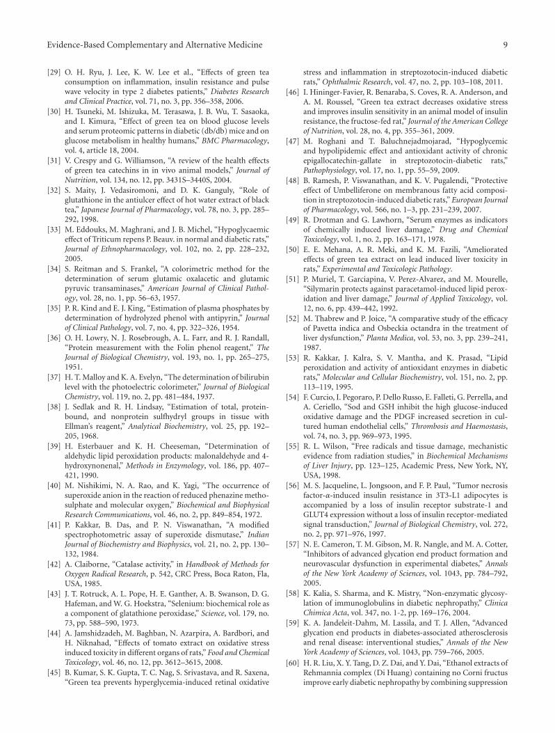
In this study, histopathological evaluation of liver tissuesshowed fatty changes in centrilobular portions of the liversin diabetic rats. These results are in line with the findingsreported by Ramesh et al., who observed the hepatopro-tective action of Umbelliferone in streptozotocin-induceddiabetic rats [48]. With green tea treatment in diabetic ratsno considerable fatty change was observed indicating theprotective effect of green tea against hepatic complicationsof diabetes. However, pathologic findings are matched withbiochemical results.
In this study, significant (P < 0.05) reduction of GSHand antioxidant enzymes (SOD, CAT, and GSH-Px) activityas well as significant (P < 0.05) increased lipid peroxidationreflects oxidative stress of the liver in experimental diabetes.These results are in line with the findings reported by Feillet-Coudray et al., who observed that STZ-induced diabetes inrat was accompanied with an increase in the susceptibility tolipid peroxidation [19]. The data of our study also revealedthat daily treatment of green tea extract markedly improvesantioxidant status of liver tissue of rats with streptozotocin-induced diabetes as GSH level and antioxidant enzymesactivities comprising SOD, CAT, and GSH-Px significantly(P < 0.05) increased and MDA level markedly (P < 0.05)decreased.
GSH (an important part of the nonenzymatic antioxi-dant system) is a major nonprotein thiol in living organisms,which plays a central role in coordinating the body’santioxidant defense processes. Perturbation of GSH statusof a biological system can lead to serious consequences.Elevation in MDA level and reduction in GSH stores of livertissue of diabetic rats suggest that oxidative stress due tofreeradical damage is one of the possible mechanisms in thepathophysiology of diabetic hepatopathy. On administrationof Green tea extract, the MDA levels have decreased andthe GSH levels have increased. This indicates that in thepresence of Green tea extract there is an improvement inthe oxidative stress. Increased oxidative stress in the tissuesof streptozotocin diabetic rats was similarly reported. This
Evidence-Based Complementary and Alternative Medicine 7
was said to be a contributory factor in the development ofthe complications of diabetes [53, 54]. It was observed inthat study that GSH administration reverses these effects[54]. The data of our study also revealed that daily treatmentof Grean tea extract markedly improves antioxidant statusof liver tissue of rats with streptozotocin-induced diabetes.Oxidative stress is produced during normal metabolicprocess in the body as well as induced by a variety ofenvironmental factors and chemicals. Oxidative stress hasbeen shown to have a significant effect in the causation ofdiabetes as well as diabetes-related complications in humanbeings [55]. Oxidative stress in diabetes has been shown tocoexist with a reduction in the antioxidant status. The exactrole of oxidative stress in the etiology of human diabetesis however not known. Oxidative stress has been shownto produce glycation of proteins, inactivation of enzymes,and alterations in structural functions of collagen basementmembrane [8]. Oxidative stress may have significant effectin the glucose transport protein (GLUT) or at insulinreceptor [56]. Scavengers of oxidative stress may have aneffect in reducing the increased serum glucose level indiabetes and may alleviate the diabetes as well as reduceits secondary complications. SOD, CAT, and GPx constitutea mutually supportive team of defense against ROS. SODis a metalloprotein and is the first enzyme involved in theantioxidant defense by lowering the steady-state level ofO2
–. In hyperglycaemia, glucose undergoes autooxidationand produces superoxide and it produces free radicals thatin turn lead to lipid peroxidation in lipoproteins. CAT islocalized in the peroxisomes or the microperoxisomes, whichcatalyses the decomposition of H2O2 to water and oxygenand thus protects the cell from oxidative damage producedby H2O2. GPx catalyses the reaction of hydroperoxides withreduced glutathione to form glutathione disulphide (GSSG)and the reduction product of the hydroperoxide. In ourstudy, decline in the activities of these enzymes in liver tissueof streptozotocin-induced animals and attainment of nearnormalcy in green tea extract treated rats indicate oxidativestress elicited in hepatic tissue of diabetic rats had beennullified due to the effect of the extract.
However, evidence suggests that oxidative stress andfree radicals play an important role in the pathogenesis ofdiabetes mellitus and diabetic complications [6]. Liver is oneof the most important organs that maintains blood glucoselevels within normal limits thus enhancement of bloodsugar yield to imbalance of oxidation-reduction reactionsin hepatocytes, so that, hyperglycemia through increasingin AGEs (advanced glycation end products) facilities freeradicals production via disturbance in ROS production(reactive oxygen species) such as SOD and CAT [57–59].Hence, it reveals that diabetic hepatic injuries result fromseveral agents and is not controllable only via inhibitionof hyperglycemia [60]. Namely, although in early stages ofdiabetes, tissues injuries are induced via hyperglycemia, theirprogress in latter stages are not related to hyperglycemia[61]. Therefore, monitoring of blood glucose levels solely isnot sufficient in retarding diabetes complications. Thus, asuitable drug must have both antioxidant and blood glucosedecreasing properties [62].
Green tea extract contains polyphenols (e.g., catechin,epicatechin, epigallocatechin, and their gallates), tannin,and caffeine. The extract also includes pyrroloquinolinequinone, a newly discovered vitamin [63]. Some constituentcomponents have been shown to enhance the basal andinsulin-stimulated glucose uptake of rat adipocytes [28], toinhibit intestinal glucose uptake by inhibiting the sodium-dependent glucose transporter of rabbit intestinal epithelialcells [64], and to reduce serum glucose level in alloxan-diabetic rats [65]. It has been suggested that catechins,antioxidant compounds present in green tea, may improvethe defence system of the organism as demonstrated inseveral in vitro and ex vivo models [66–68]. Green teaextracts are more stable than pure epigallocatechin gallate,the major constituents of green tea, because of the presenceof other antioxidant constituents in the extract [69]. Ingeneral, herbal medicines are complex mixtures of differentcompounds that often act in a synergistic fashion and exerttheir full beneficial effect as total extracts [70]. However,a multitude of herbs, spices, and other plant materials asuseful source of natural antioxidants have been describedfor the treatment of diabetic complications throughout theworld. In this manner, Saradha Devi et al. declared thatCynodon dactylon has very good antioxidant and hepaticprotective effect of normal oxidative stress in Balb/c mice[71]. These similar results were obtained earlier in dry stemcrude extraction of Tinospora cordifolia and polyphenolsextracts in tea [72]. Also, Gohil et al. showed hypoglycaemicand hypolipidemic effects of Eugenia jambolana seed andAegle marmelos leaf extracts in alloxan-induced diabetic rats[73].
The results of the present study demonstrate that dailytreatment of diabetic rats by green tea extract markedlyimproves antioxidant status in liver tissue. On the otherhand, we found that green tea extract improved serumbiomarkers of liver tissue injury and histopathologic proper-ties of this organ. It is therefore likely that green tea extract isprophylactic against diabetic complications and amelioratesdiabetic hepatopathy through its antioxidant potential. Onthe other hand, hyperglycemia is the primary symptom ofdiabetes and is blamed for the complications of diabetesbecause elevated glucose concentration directly injures cellsand induces lipid peroxidation [74]. Whether this reflectsoxidative stress-induced liver injury or direct glycemic injuryof liver remains to be determined. Also, information onthe occurrence of oxidative stress in the liver tissue at thisearly stage of diabetes remains undetermined. Taken in all,the use of this plant in diabetes is then supported but theprecise active substance(s) of green tea, site(s), and cellularand molecular mechanism(s) of its pharmacological effectare still to be determined. In addition, the possible long-term toxic effects of green tea extract and protective effectsof different doses of that also remain to be clarified.
5. Conclusion
This study demonstrated green tea extract has hepato-protective activity in streptozotocin-induced diabetic rats.
8 Evidence-Based Complementary and Alternative Medicine
Many question related to antioxidant effect of Green teaextract remain unanswered. Much more work is clearlyneeded before phytotherapy for diabetic hepatopathy can beadvanced to clinic.
Acknowledgments
This work was supported by a grant from Ahar Branch,Islamic Azad University. The authors thank the Vice Chan-cellor in Research Affairs of Ahar Branch, Islamic AzadUniversity.
References
[1] E. Ritz, C. Hasslacher, and W. Tschope, “Diabeticnephropathy—are there differences between type I andtype II?” Mineral and Electrolyte Metabolism, vol. 16, no. 1, pp.69–72, 1990.
[2] P. H. Hendriksen, P. L. Oey, G. H. Wieneke, B. Bravenboer,and J. D. Banga, “Subclinical diabetic neuropathy: similaritiesbetween electrophysiological results of patients with Type1 (insulin-dependent) and Type 2 (non-insulin-dependent)diabetes mellitus,” Diabetologia, vol. 35, no. 7, pp. 690–695,1992.
[3] R. de Marco, F. Locatelli, G. Zoppini, G. Verlato, E. Bonora,and M. Muggeo, “Cause-specific mortality in type 2 diabetes:the verona diabetes study,” Diabetes Care, vol. 22, no. 5, pp.756–761, 1999.
[4] K. G. Tolman, V. Fonseca, A. Dalpiaz, and M. H. Tan,“Spectrum of liver disease in type 2 diabetes and managementof patients with diabetes and liver disease,” Diabetes Care, vol.30, no. 3, pp. 734–743, 2007.
[5] V. G. Athyros, D. P. Mikhailidis, T. P. Didangelos et al., “Effectof multifactorial treatment on non-alcoholic fatty liver diseasein metabolic syndrome: a randomised study,” Current MedicalResearch and Opinion, vol. 22, no. 5, pp. 873–883, 2006.
[6] B. Halliwell and J. M. C. Gutteridge, Free Radicals in Biologyand Medicine, Clarendon Press, Oxford, UK, 1989.
[7] C. J. Mullarkey, D. Edelstein, and L. Brownlee, “Free radicalgeneration by early glycation products: a mechanism for accel-erated atherogenesis in diabetes,” Biochemical and BiophysicalResearch Communications, vol. 173, no. 3, pp. 932–939, 1990.
[8] J. W. Baynes, “Role of oxidative stress in development ofcomplications in diabetes,” Diabetes, vol. 40, no. 4, pp. 405–412, 1991.
[9] J. J. Strain, “Disturbances of micronutrient and antioxidantstatus in diabetes,” Proceedings of the Nutrition Society, vol. 50,no. 3, pp. 591–604, 1991.
[10] S. V. McLennan, S. Heffernan, L. Wright et al., “Changes inhepatic glutathione metabolism in diabetes,” Diabetes, vol. 40,no. 3, pp. 344–348, 1991.
[11] P. B. Jennings, S. Chirico, A. F. Jones, J. Lunec, and A.H. Barnett, “Vitamin C metabolites and microangiopathy indiabetes mellitus,” Diabetes Research, vol. 6, no. 3, pp. 151–154,1987.
[12] I. S. Young, J. J. Torney, and E. R. Trimble, “The effectsof ascorbate supplementation on oxidative stress in thestreptozotocin diabetic rat,” Free Radical Biology and Medicine,vol. 13, no. 1, pp. 41–46, 1992.
[13] E. Velazquez, P. H. Winocour, P. Kesteven, K. G. M. M. Alberti,and M. F. Laker, “Relation of lipid peroxides to macrovascular
disease in Type 2 diabetes,” Diabetic Medicine, vol. 8, no. 8, pp.752–758, 1991.
[14] S. M. MacRury, D. Gordon, R. Wilson et al., “A comparisonof different methods of assessing free radical activity in Type2 diabetes and peripheral vascular disease,” Diabetic Medicine,vol. 10, no. 4, pp. 331–335, 1993.
[15] A. Griesmacher, M. Kindhauser, S. E. Andert et al., “Enhancedserum levels of thiobarbituric-acid-reactive substances indiabetes mellitus,” American Journal of Medicine, vol. 98, no.5, pp. 469–475, 1995.
[16] L. K. Niskanen, J. T. Salonen, K. Nyssonen, and M. I. J.Uusitupa, “Plasma lipid peroxidation and hyperglycemia: aconnection through hyperinsulinaemia,” Diabetic Medicine,vol. 12, pp. 802–808, 1995.
[17] R. K. Sundaram, A. Bhaskar, S. Vijayalingam, M. Viswanathan,R. Mohan, and K. R. Shanmugasundaram, “Antioxidant statusand lipid peroxidation in type II diabetes mellitus with andwithout complications,” Clinical Science, vol. 90, no. 4, pp.255–260, 1996.
[18] R. Kakkar, S. V. Mantha, J. Radhi, K. Prasad, and J. Kalra,“Increased oxidative stress in rat liver and pancreas duringprogression of streptozotocin-induced diabetes,” Journal ofClinical Science, vol. 94, no. 6, pp. 623–632, 1998.
[19] C. Feillet-Coudray, E. Rock, C. Coudray et al., “Lipid per-oxidation and antioxidant status in experimental diabetes,”Clinica Chimica Acta, vol. 284, no. 1, pp. 31–43, 1999.
[20] E. H. Harris, “Elevated liver function tests in type 2 diabetes,”Clinical Diabetes, vol. 23, no. 3, pp. 115–119, 2005.
[21] M. S. Akhtar and J. Iqbal, “Evaluation of the hypoglycaemiceffect of Achyranthes aspera in normal and alloxan-diabeticrabbits,” Journal of Ethnopharmacology, vol. 31, no. 1, pp. 49–57, 1991.
[22] R. K. Gupta, A. N. Kesari, P. S. Murthy, R. Chandra, V.Tandon, and G. Watal, “Hypoglycemic and antidiabetic effectof ethanolic extract of leaves of Annona squamosa L. inexperimental animals,” Journal of Ethnopharmacology, vol. 99,no. 1, pp. 75–81, 2005.
[23] K. Rafiq, S. J. Sherajee, A. Nishiyama, M. A. Sufiun, andM. Mostofa, “Effects of indigenous medicinal plants ofBangladesh on blood glucose level and neuropathic painin streptozotocin-induced diabetic rats,” African Journal ofPharmacy and Pharmacology, vol. 3, no. 12, pp. 636–642, 2009.
[24] K. Rajagopal and K. Sasikala, “Antidiabetic activity of hydro-ethanolic extracts of Nymphaea stellata flowers in normal andalloxan induced diabetic rats,” African Journal of Pharmacy andPharmacology, vol. 2, no. 8, pp. 173–178, 2008.
[25] X. Jin, R. H. Zheng, and Y. M. Li, “Green tea consumption andliver disease: a systematic review,” Liver International, vol. 28,no. 7, pp. 990–996, 2008.
[26] R. Benelli, R. Vene, D. Bisacchi, S. Garbisa, and A.Albini, “Anti-invasive effects of green tea polyphenolepigallocatechin-3-gallate (EGCG), A natural inhibitor ofmetallo and serine proteases,” Biological Chemistry, vol. 383,no. 1, pp. 101–105, 2002.
[27] J. H. Weisburger and F. L. Chung, “Mechanisms of chronicdisease causation by nutritional factors and tobacco productsand their prevention by tea polyphenols,” Food and ChemicalToxicology, vol. 40, no. 8, pp. 1145–1154, 2002.
[28] L. Y. Wu, C. C. Juan, L. T. Ho, Y. P. Hsu, and L. S. Hwang,“Effect of green tea supplementation on insulin sensitivityin sprague-dawley rats,” Journal of Agricultural and FoodChemistry, vol. 52, no. 3, pp. 643–648, 2004.
Evidence-Based Complementary and Alternative Medicine 9
[29] O. H. Ryu, J. Lee, K. W. Lee et al., “Effects of green teaconsumption on inflammation, insulin resistance and pulsewave velocity in type 2 diabetes patients,” Diabetes Researchand Clinical Practice, vol. 71, no. 3, pp. 356–358, 2006.
[30] H. Tsuneki, M. Ishizuka, M. Terasawa, J. B. Wu, T. Sasaoka,and I. Kimura, “Effect of green tea on blood glucose levelsand serum proteomic patterns in diabetic (db/db) mice and onglucose metabolism in healthy humans,” BMC Pharmacology,vol. 4, article 18, 2004.
[31] V. Crespy and G. Williamson, “A review of the health effectsof green tea catechins in in vivo animal models,” Journal ofNutrition, vol. 134, no. 12, pp. 3431S–3440S, 2004.
[32] S. Maity, J. Vedasiromoni, and D. K. Ganguly, “Role ofglutathione in the antiulcer effect of hot water extract of blacktea,” Japanese Journal of Pharmacology, vol. 78, no. 3, pp. 285–292, 1998.
[33] M. Eddouks, M. Maghrani, and J. B. Michel, “Hypoglycaemiceffect of Triticum repens P. Beauv. in normal and diabetic rats,”Journal of Ethnopharmacology, vol. 102, no. 2, pp. 228–232,2005.
[34] S. Reitman and S. Frankel, “A colorimetric method for thedetermination of serum glutamic oxalacetic and glutamicpyruvic transaminases,” American Journal of Clinical Pathol-ogy, vol. 28, no. 1, pp. 56–63, 1957.
[35] P. R. Kind and E. J. King, “Estimation of plasma phosphates bydetermination of hydrolyzed phenol with antipyrin,” Journalof Clinical Pathology, vol. 7, no. 4, pp. 322–326, 1954.
[36] O. H. Lowry, N. J. Rosebrough, A. L. Farr, and R. J. Randall,“Protein measurement with the Folin phenol reagent,” TheJournal of Biological Chemistry, vol. 193, no. 1, pp. 265–275,1951.
[37] H. T. Malloy and K. A. Evelyn, “The determination of bilirubinlevel with the photoelectric colorimeter,” Journal of BiologicalChemistry, vol. 119, no. 2, pp. 481–484, 1937.
[38] J. Sedlak and R. H. Lindsay, “Estimation of total, protein-bound, and nonprotein sulfhydryl groups in tissue withEllman’s reagent,” Analytical Biochemistry, vol. 25, pp. 192–205, 1968.
[39] H. Esterbauer and K. H. Cheeseman, “Determination ofaldehydic lipid peroxidation products: malonaldehyde and 4-hydroxynonenal,” Methods in Enzymology, vol. 186, pp. 407–421, 1990.
[40] M. Nishikimi, N. A. Rao, and K. Yagi, “The occurrence ofsuperoxide anion in the reaction of reduced phenazine metho-sulphate and molecular oxygen,” Biochemical and BiophysicalResearch Communications, vol. 46, no. 2, pp. 849–854, 1972.
[41] P. Kakkar, B. Das, and P. N. Viswanathan, “A modifiedspectrophotometric assay of superoxide dismutase,” IndianJournal of Biochemistry and Biophysics, vol. 21, no. 2, pp. 130–132, 1984.
[42] A. Claiborne, “Catalase activity,” in Handbook of Methods forOxygen Radical Research, p. 542, CRC Press, Boca Raton, Fla,USA, 1985.
[43] J. T. Rotruck, A. L. Pope, H. E. Ganther, A. B. Swanson, D. G.Hafeman, and W. G. Hoekstra, “Selenium: biochemical role asa component of glatathione peroxidase,” Science, vol. 179, no.73, pp. 588–590, 1973.
[44] A. Jamshidzadeh, M. Baghban, N. Azarpira, A. Bardbori, andH. Niknahad, “Effects of tomato extract on oxidative stressinduced toxicity in different organs of rats,” Food and ChemicalToxicology, vol. 46, no. 12, pp. 3612–3615, 2008.
[45] B. Kumar, S. K. Gupta, T. C. Nag, S. Srivastava, and R. Saxena,“Green tea prevents hyperglycemia-induced retinal oxidative
stress and inflammation in streptozotocin-induced diabeticrats,” Ophthalmic Research, vol. 47, no. 2, pp. 103–108, 2011.
[46] I. Hininger-Favier, R. Benaraba, S. Coves, R. A. Anderson, andA. M. Roussel, “Green tea extract decreases oxidative stressand improves insulin sensitivity in an animal model of insulinresistance, the fructose-fed rat,” Journal of the American Collegeof Nutrition, vol. 28, no. 4, pp. 355–361, 2009.
[47] M. Roghani and T. Baluchnejadmojarad, “Hypoglycemicand hypolipidemic effect and antioxidant activity of chronicepigallocatechin-gallate in streptozotocin-diabetic rats,”Pathophysiology, vol. 17, no. 1, pp. 55–59, 2009.
[48] B. Ramesh, P. Viswanathan, and K. V. Pugalendi, “Protectiveeffect of Umbelliferone on membranous fatty acid composi-tion in streptozotocin-induced diabetic rats,” European Journalof Pharmacology, vol. 566, no. 1–3, pp. 231–239, 2007.
[49] R. Drotman and G. Lawhorn, “Serum enzymes as indicatorsof chemically induced liver damage,” Drug and ChemicalToxicology, vol. 1, no. 2, pp. 163–171, 1978.
[50] E. E. Mehana, A. R. Meki, and K. M. Fazili, “Amelioratedeffects of green tea extract on lead induced liver toxicity inrats,” Experimental and Toxicologic Pathology.
[51] P. Muriel, T. Garciapina, V. Perez-Alvarez, and M. Mourelle,“Silymarin protects against paracetamol-induced lipid perox-idation and liver damage,” Journal of Applied Toxicology, vol.12, no. 6, pp. 439–442, 1992.
[52] M. Thabrew and P. Joice, “A comparative study of the efficacyof Pavetta indica and Osbeckia octandra in the treatment ofliver dysfunction,” Planta Medica, vol. 53, no. 3, pp. 239–241,1987.
[53] R. Kakkar, J. Kalra, S. V. Mantha, and K. Prasad, “Lipidperoxidation and activity of antioxidant enzymes in diabeticrats,” Molecular and Cellular Biochemistry, vol. 151, no. 2, pp.113–119, 1995.
[54] F. Curcio, I. Pegoraro, P. Dello Russo, E. Falleti, G. Perrella, andA. Ceriello, “Sod and GSH inhibit the high glucose-inducedoxidative damage and the PDGF increased secretion in cul-tured human endothelial cells,” Thrombosis and Haemostasis,vol. 74, no. 3, pp. 969–973, 1995.
[55] R. L. Wilson, “Free radicals and tissue damage, mechanisticevidence from radiation studies,” in Biochemical Mechanismsof Liver Injury, pp. 123–125, Academic Press, New York, NY,USA, 1998.
[56] M. S. Jacqueline, L. Jongsoon, and F. P. Paul, “Tumor necrosisfactor-α-induced insulin resistance in 3T3-L1 adipocytes isaccompanied by a loss of insulin receptor substrate-1 andGLUT4 expression without a loss of insulin receptor-mediatedsignal transduction,” Journal of Biological Chemistry, vol. 272,no. 2, pp. 971–976, 1997.
[57] N. E. Cameron, T. M. Gibson, M. R. Nangle, and M. A. Cotter,“Inhibitors of advanced glycation end product formation andneurovascular dysfunction in experimental diabetes,” Annalsof the New York Academy of Sciences, vol. 1043, pp. 784–792,2005.
[58] K. Kalia, S. Sharma, and K. Mistry, “Non-enzymatic glycosy-lation of immunoglobulins in diabetic nephropathy,” ClinicaChimica Acta, vol. 347, no. 1-2, pp. 169–176, 2004.
[59] K. A. Jandeleit-Dahm, M. Lassila, and T. J. Allen, “Advancedglycation end products in diabetes-associated atherosclerosisand renal disease: interventional studies,” Annals of the NewYork Academy of Sciences, vol. 1043, pp. 759–766, 2005.
[60] H. R. Liu, X. Y. Tang, D. Z. Dai, and Y. Dai, “Ethanol extracts ofRehmannia complex (Di Huang) containing no Corni fructusimprove early diabetic nephropathy by combining suppression
10 Evidence-Based Complementary and Alternative Medicine
on the ET-ROS axis with modulate hypoglycemic effect inrats,” Journal of Ethnopharmacology, vol. 118, no. 3, pp. 466–472, 2008.
[61] M. D. Vestra and P. Fioretto, “Diabetic nephropathy: renalstructural studies in type 1 and type 2 diabetic patients,”Internatrional Congress Series, vol. 1253, pp. 163–169, 2003.
[62] B. Ramesh and K. V. Pugalendi, “Impact of umbelliferone (7-hydroxycoumarin) on hepatic marker enzymes in streptozo-tocin diabetic rats,” Indian Journal of Pharmacology, vol. 38,no. 3, pp. 209–210, 2006.
[63] T. Kasahara and T. Kato, “A new redox-cofactor vitamin formammals,” Nature, vol. 422, no. 6934, p. 832, 1993.
[64] Y. Kobayashi, M. Suzuki, H. Satsu et al., “Green tea polyphe-nols inhibit the sodium-dependent glucose transporter ofintestinal epithelial cells by a competitive mechanism,” Journalof Agricultural and Food Chemistry, vol. 48, no. 11, pp. 5618–5623, 2000.
[65] M. C. Sabu, K. Smitha, and R. Kuttan, “Anti-diabetic activityof green tea polyphenols and their role in reducing oxidativestress in experimental diabetes,” Journal of Ethnopharmacol-ogy, vol. 83, no. 1-2, pp. 109–116, 2002.
[66] N. Salah, N. J. Miller, G. Paganga, L. Tijburg, G. P. Bolwell,and C. Rice-Evans, “Polyphenolic flavanols as scavengers ofaqueous phase radicals and as chain-breaking antioxidants,”Archives of Biochemistry and Biophysics, vol. 322, no. 2, pp.339–346, 1995.
[67] J. Terao, M. Piskula, and Q. Yao, “Protective effect ofepicatechin, epicatechin gallate, and quercetin on lipid perox-idation in phospholipid bilayers,” Archives of Biochemistry andBiophysics, vol. 308, no. 1, pp. 278–284, 1994.
[68] W. Wang and M. T. Goodman, “Antioxidant property ofdietary phenolic agents in a human LDL-oxidation ex vivomodel: interaction of protein binding activity,” NutritionResearch, vol. 19, no. 2, pp. 191–202, 1999.
[69] M. Kaszkin, K. F. Beck, W. Eberhardt, and J. Pfeilschifter,“Unravelling green tea’s mechanisms of action: more thanmeets the eye,” Molecular Pharmacology, vol. 65, no. 1, pp. 15–17, 2004.
[70] D. Locvv and M. Kaszkin, “Approaching the problem ofbioequivalence of herbal medicinal products,” PhytotherapyResearch, vol. 16, no. 8, pp. 705–711, 2002.
[71] K. M. Saradha Devi, S. Annapoorani, and K. Ashokkumar,“Hepatic antioxidative potential of ethyl acetate fraction ofCynodon dactylon in Balb/c mice,” Journal of Medicinal PlantResearch, vol. 5, no. 6, pp. 929–996, 2011.
[72] B. Frei and J. V. Higdon, “Antioxidant activity of teapolyphenols in vivo: evidence from animal studies,” Journal ofNutrition, vol. 133, no. 10, pp. 327–328, 2003.
[73] T. Gohil, N. Pathak, N. Jivani, V. Devmurari, and J. Patel,“Treatment with extracts of Eugenia jambolana seed andAegle marmelos leaf extracts prevents hyperglycemia andhyperlipidemia in alloxan induced diabetic rats,” AfricanJournal of Pharmacy and Pharmacology, vol. 4, no. 5, pp. 270–275, 2010.
[74] G. Davı, A. Falco, and C. Patrono, “Lipid peroxidation indiabetes mellitus,” Antioxidants and Redox Signaling, vol. 7, no.1-2, pp. 256–268, 2005.