protective role of cathepsin l in mouse skin carcinogenesis
TRANSCRIPT
MOLECULAR CARCINOGENESIS 51:352–361 (2012)
Protective Role of Cathepsin L in MouseSkin Carcinogenesis
Fernando Benavides,1* Carlos Perez,1 Jorge Blando,1 Oscar Contreras,1 Jianjun Shen,1 Lisa M. Coussens,2
Susan M. Fischer,1 Donna F. Kusewitt,1 John DiGiovanni,1 and Claudio J. Conti1
1Department of Molecular Carcinogenesis, The University of Texas M. D. Anderson Cancer Center, Science-Park,Smithville, Texas2Cancer Research Institute, University of California-San Francisco, San Francisco, California
Lysosomal cysteine protease cathepsin L (CTSL) is believed to play a role in tumor progression and is considered a
marker for clinically invasive tumors. Studies from our laboratory using the classical mouse skin carcinogenesis model,with 7,12-dimethyl-benz[a]anthracene (DMBA) for initiation and 12-O-tetradecanoylphorbol-13-acetate (TPA) for pro-motion, showed that expression of CTSL is increased in papillomas and squamous cell carcinomas (SCC). We also
carried out carcinogenesis studies using Ctsl-deficient nackt (nkt) mutant mice on three different inbred backgrounds.Unexpectedly, the multiplicity of papillomas was significantly higher in Ctsl-deficient than in wild-type mice on twounrelated backgrounds. Topical applications of TPA or DMBA alone to the skin of nkt/nkt mice did not induce papil-lomas, and there was no increase in spontaneous tumors in nkt/nkt mice on any of the three inbred backgrounds.
Reduced epidermal cell proliferation in Ctsl-deficient nkt/nkt mice after TPA treatment suggested that they are notmore sensitive than wild-type mice to TPA promotion. We also showed that deficiency of CTSL delays terminal differ-entiation of keratinocytes, and we propose that decreased elimination of initiated cells is at least partially responsible
for the increased papilloma formation in the nackt model. � 2011 Wiley Periodicals, Inc.
Key words: mouse models; proteases; cysteine cathepsins; skin cancer; two-stage carcinogenesis
INTRODUCTION
Cysteine cathepsins, lysosomal proteases that areinvolved in extracellular and intracellular proteindegradation, have multiple roles during cancerdevelopment and progression [1], including modu-lation of tumor cell proliferation, invasion, andapoptosis [2]. Cathepsin L (CTSL) is a widelyexpressed lysosomal protease with roles in epider-mal homeostasis and hair follicle morphogenesis[3–5], tumor invasiveness, angiogenesis [6], andapoptosis [7–9]. Recently, the Nepveu laboratoryidentified a nuclear isoform of CTSL that regulatescell cycle progression through proteolytic process-ing of the CDP/Cux transcription factor [10–12].Although mice and rats possess only one Ctsl gene,humans have two CTSL-like genes (CTSL andCTSL2); however, mouse and rat Ctsl genes aremore closely related to human CTSL2 than tohuman CTSL gene [13,14].
We previously described the recessive nackt (nkt)mutation, which is characterized by partial alope-cia associated with CD4þ T cell deficiency. Thenackt mutation is a 1.6 kilobases deletion in theCtsl gene (Ctslnkt) [15,16] that eliminates exon 7from the Ctsl gene transcript (F. Benavides, unpub-lished data). Ctslnkt/Ctslnkt mice do not generateany of the mature active forms of CTSL, althougha truncated unprocessed CTSL protein is present[17]. Homozygous nackt mutants exhibit pheno-types similar to Ctsl deficient mice [17], while
heterozygous nackt mice are phenotypically indis-tinguishable from wild-type mice. CTSL back-ground activity is comparably low in Ctslnkt/Ctslnkt
and targeted knockout Ctsltm1Cptr/Ctsltm1Cptr mice.Together, these results indicate that nackt is a loss-of-function mutation of the Ctsl gene. Studies withthe nackt model suggested a critical role for CTSLin hair follicle morphogenesis and cycling and inepidermal differentiation [4]. The nackt model wasalso used to demonstrate that CTSL influences theexpression of extracellular matrix components inlymphoid organs [18].Recent studies using the three other Ctsl-
deficient mouse models currently available haveshed light on the role of CTSL in a number ofdevelopmental and pathological processes. Forexample, the first reported Ctsl knockout mice
Abbreviations: CTSL; cathepsin L; DMBA; 7,12-dimethyl-ben-z[a]anthracene; TPA; 12-O-tetradecanoylphorbol-13-acetate; i.p.;intraperitoneal; BrdU; 5-bromo-2-deoxyuridine; IHC, immunohisto-chemistry; SCC; squamous cell carcinoma; .
Jorge Blando and John DiGiovanni present address is DellPediatric Research Institute, University of Texas, Austin, Texas.
*Correspondence to: Department of Molecular Carcinogenesis,The University of Texas M. D. Anderson Cancer Center, Science-Park, 1808 Park Road 1C, PO Box 389, Smithville, TX 78957.
Received 19 January 2011; Revised 1 April 2011; Accepted 7April 2011
DOI 10.1002/mc.20792
Published online 2 May 2011 in Wiley Online Library(wileyonlinelibrary.com).
� 2011 WILEY PERIODICALS, INC.

(Ctsltm1Cptr) exhibited a 60–80% reduction in thenumber of CD4þ T cells in the thymus and periph-ery [19], gingival overgrowth [20], abnormal hairfollicle morphogenesis and cycling [5,21], lateonset dilated cardiomyopathy [22,23], and defectsin adipogenesis and glucose metabolism [24]. Asecond independent Ctsl knockout line (Ctsltm1Alpk)has been described with abnormal skin and defec-tive bone development [25]. The spontaneousrecessive mutation furless (Ctslfs), carrying an enzy-matically inactive CTSL, also exhibits a skin phe-notype [3] and abnormal spermatogenesis [26].We assessed the role of CTSL in multistage skin
carcinogenesis by comparing Ctsl-deficient nacktand wild-type mice. Skin tumorigenesis wasenhanced in nackt compared to wild-type mice,and this trait was influenced by the genetic back-ground. However, loss of CTSL function did notact as an initiator or promoter, since topical 7,12-dimethyl-benz[a]anthracene (DMBA) or 12-O-tetra-decanoylphorbol-13-acetate (TPA) alone did notinduce papillomas in nackt mice. Our results alsoshowed that the absence of CTSL enzymaticactivity in mutant mice delays the onset of kerati-nocyte terminal differentiation and decreases pro-liferation after TPA treatment. These findingssuggest that delayed keratinocyte transit and con-comitant increased retention of initiated cells maybe responsible for the increased tumor suscepti-bility in nackt mice.
MATERIALS AND METHODS
Mouse Strains
SENCARB/Pt.Cg-nkt, DBA/2.Cg-nkt, BALB/c.Cg-nkt, and FVB/N.129S6-Cd4tm1Knw (CD4 knock-out)colonies were maintained under specific pathogenfree (SPF) conditions in our AAALAC accreditedAnimal Facilities at M. D. Anderson Cancer Center,Smithville, Texas. All procedures were conductedin compliance with the Guide for the Care and Useof Laboratory Animals and under an IACUC-approved protocol. FVB/N-CD4 knockout congenicmice were a gift from Lisa M. Coussens from theUniversity of California at San Francisco, San Fran-cisco. We developed SENCARB/Pt.Cg-nkt and DBA/2.Cg-nkt congenic lines, thus placing the mutationon backgrounds with high and moderate skintumor susceptibility, respectively. To develop theselines we used marker assisted ‘‘speed congenic’’breeding strategies [27]. Microsatellite analysis ofN6 mice from the two lines showed that morethan 99% of the alleles in the genome scan werefrom the recipient strain. The BALB/c.Cg-nkt con-genic (N12) line was developed previously [15].
Skin Carcinogenesis and Tissue Collection
Ctslnkt/Ctslnkt (nkt/nkt) age-matched mice pluswild-type littermate controls (male and female)
were used to determine the susceptibility to chemi-cally induced tumorigenesis in three inbred back-grounds (SENCARB/Pt, DBA/2, and BALB/c). In aseparate experiment, FVB/N-Cd4�/� and FVB/Nwild-type mice were also challenged. The two-stagecarcinogenesis protocol was performed by initiat-ing mice (6–8 wk old) as previously described [28]with 10 nmol/200 mL acetone of DMBA (Sigma–Aldrich, St. Louis, MO) (SENCARB/Pt mice) or100 nmol/200 mL acetone of DMBA (DBA/2, BALB/c, and FVB/N backgrounds). Promotion was started2 wk after initiation with twice weekly appli-cations of 1 mg of TPA (Sigma–Aldrich; SENCARB/Pt), 2 mg (DBA/2), or 4 mg (BALB/c and FVB/N) in200 mL of acetone on the dorsal skin. Tumors werecounted weekly and statistical significance fortumor multiplicity was determined by calculationof P values using Wilcoxon Rank Sum test.
Analysis of Cell Proliferation Following Treatments
With TPA
For analysis of epidermal proliferation, groups offour DBA/2-nkt/nkt and four wild-type littermatecontrols mice were treated with a single topicalapplication or twice-weekly applications of TPA(2 mg/200 mL acetone) on the dorsal skin for 2 wkand sacrificed 24 h after the final treatment. Micereceived intraperitoneal (i.p.) injections of 5-bromo-2-deoxyuridine (BrdU; Sigma–Aldrich) at100 mg/g body weight in phosphate-buffered saline(PBS) 30 min prior to euthanasia. Dorsal skin wasfixed in formalin and embedded in paraffin priorto sectioning and staining with hematoxylin andeosin (H&E) and immunohistochemistry (IHC).BrdU incorporation was detected by standardthree-step immunoperoxidase detection usingmouse anti-BrdU monoclonal antibody (Becton-Dickinson Immunocytometry System, Becton-Dickinson, San Jose, CA), biotin F(ab’) rabbit antimouse IgG (Accurate Chemical, Westbury, NY),and Streptavidin Peroxidase (BioGenex, SanRamon, CA). Diaminobenzidine (BioGenex) wasthe chromagen used for visualization. Epidermalcell proliferation (labeling index) was determinedby calculating the percentage of epidermal basalcells positive for BrdU and Ki67. A minimum of4000 basal cells was counted using digital imagesof IHC slides (dorsal skin) captured with the AperioScanScope CS slide scanner (Aperio Technologies,Vista, CA). A fully automated nuclear algorithmprovided with the instrument was adapted tocount BrdU- and Ki67-positive cells in the epider-mis of both wild-type and mutant phenotypes.Statistical significance was determined by calcu-lation of P values using Wilcoxon Rank Sum test.
In Vivo Keratinocyte Transit Studies
Mice were injected i.p. with 100 mg/g bodyweight of BrdU (Sigma–Aldrich) in 200 mL of PBS
PROTECTIVE ROLE OF CATHEPSIN L IN MOUSE SKIN 353
Molecular Carcinogenesis
17 h after the last of four TPA applications (2 mg/200 mL acetone) over a 2 wk period and sacrificed1, 8, and 30 h after BrdU injection as described[29]. Dorsal skin was collected and processed forhistopathology and IHC evaluation as describedabove. BrdU incorporation was quantified asdescribed above with the Aperio ScanScope CSslide scanner. A total of four mice per genotypeper time point were evaluated.
Immunohistochemistry
TPA-treated dorsal skin and papillomas from nkt/nkt and littermate controls were fixed in formalinovernight and then transferred to 70% ethanol.IHC analysis was performed with polyclonal anti-bodies directed against mouse keratins K8, K10,K13, and K14 (Covance Research Products, Rich-mond, CA), involucrin and profilaggrin/filaggrin(BabCo, Richmond, CA), Ki67 (Dako, Carpinteria,CA), Cd31 (PharMingen, San Diego, CA), p21, p27,VEGF, and active caspase-3 (R&D Systems, Minne-apolis, MN) as previously described [4]. Controlreactions without primary antibodies were rou-tinely performed.
Protein Lysates and Western Blotting
Protein was isolated from TPA-treated dorsal epi-dermis and papillomas of mutant and littermatecontrols using RIPA lysis buffer. Protein lysates(25–50 mg) were electrophoresed on 10% Tris–Glycine Gels (Novex, San Diego, CA) and trans-ferred onto nitrocellulose membranes. Westernblot analysis was performed with the followingprimary antibodies: anti-mouse p21, p27, Stat3,VEGF, and caspase-3 (Santa Cruz Biotechnology,Santa Cruz, CA); CTSB, CTSD, and CTSL (R&D andSanta Cruz Biotechnology); and b-actin (Sigma–Aldrich). Binding of antibodies was detected usingECL þ Plus Western blotting detection system(Amersham Pharmacia Biotech UK Limited,Buckinghamshire).
Genotyping of the Polymorphic Variant of the Mouse
Patched (Ptch1) Gene
To differentiate the C57BL/6 (Ptch1B6) from theFVB (Ptch1FVB) allele [30] we used genomic DNAand primers designed to amplify a 179 bp segmentfrom Ptch1 (accession number NT_039589.7) exon23 under standard PCR conditions. The sequencesof the primers were: Ptch1-FOR: 50-GTGGCCG-CAAGCCTTCTCTA-30 and Ptch1-REV: 50-ACCAT-CCTACCTCCCTGTGTTGAC-30. The sequence ofthe amplification products was obtained using theABI-PRISMTM Dye Terminator Cycle SequencingReady Reaction kit and an ABI 3130XL DNAsequencer (Applied Biosystems, Foster City, CA).
RESULTS
CTSL Expression Increases During Two-StageCarcinogenesis
We analyzed the expression of CTSL in early-,mid-, and late-stage papillomas and squamous cellcarcinomas (SCC) from our archives and in normaldorsal skin using polyclonal goat anti mouse CTSL(carboxy terminal) antibody (M-19, sc-6500) forWestern blotting. The tumors were generated inSENCARB/Pt mice using a two-stage skin carcino-genesis protocol [31]. Figure 1A shows that CTSLexpression was markedly increased in papillomasand SCC compared to normal skin. These resultssuggested that CTSL might play a role in tumordevelopment and progression during mouse skintumorigenesis.
Skin Tumorigenesis Is Enhanced in the Absence of CTSL
To determine if the absence of CTSL enzymaticactivity had an effect on skin tumor development,we conducted a two-stage skin carcinogenesisstudy with Ctsl-deficient nkt/nkt mice on SEN-CARB/Pt (very sensitive) and DBA/2 (sensitive)backgrounds and their wild-type littermates.Because SENCARB/Pt-nkt/nkt mice developedsevere dermatitis, experiments using this inbredbackground were terminated at 20 wk and tumormultiplicity (tumors per mouse) was determined;tumor multiplicity in the DBA/2 group was scoredat 30 wk [28].In the SENCARB/Pt group, the first papillomas
appeared in both nkt/nkt and wild-type littermatesat 6 wk of promotion. The tumor incidence wassimilar between the two groups: (100% at 8 wk).However, the tumor multiplicity was higher innkt/nkt mice compared to wild-type littermates.The number of papillomas increased rapidly innkt/nkt mice, reaching an average of 18.1 papillo-mas per mouse (n ¼ 9) at 20 wk, while littermatecontrols developed an average of 8.6 papillomasper mouse (n ¼ 7, P < 0.001) at 20 wk (Figure 1B).In the DBA/2 group, wild-type mice (n ¼ 20;
ungenotyped þ/nkt or þ/þ mice) developed 0.3papillomas per mouse by 19 wk of TPA treatment,but all papillomas regressed after 20 wk. In DBA/2-nkt/nkt mice, the number of papillomas reached anaverage of 3.1 papillomas per mouse at 20 wk(n ¼ 18, P < 0.001). These nkt/nkt papillomas nev-er regressed and less than 1% progressed to SCC.In DBA/2-nkt/nkt mice, the papilloma incidencereached a plateau of 94% at 18 wk. For wild-typemice, before regression of papillomas, the inci-dence was never higher than 38% (Figure 1C andD). Tumors in these DBA/2-nkt/nkt mice generallydisplayed the histology typical of papillomas froma two-stage carcinogenesis protocol, however, weobserved one atypical SCC with basaloid prolifer-ation and follicular differentiation in a nkt/nkt
354 BENAVIDES ET AL.
Molecular Carcinogenesis

mouse. This SCC expressed basal cell marker K14,but not differentiation marker K10, when deter-mined by IHC (Figure 2). We compared the pro-liferation rate (by BrdU labeling) of theses Ctsl-deficient papillomas (at 30 wk of promotion) witharchive papillomas generated in wild-type DBA/2mice using the same protocol and did not findstatistical significant differences (Figure 2).Topical applications of TPA (2 mg twice-weekly
for 20 wk) or DMBA (100 nmol, single dose) alonedid not induce papillomas in DBA/2-nkt/nkt mice(data not shown). No differences were foundbetween tumors in nkt/nkt and wild-type mice(SENCARB/Pt and DBA/2 background) whenexpression of p21, caspase-3, and VEGF, or BrdU-labeling index was compared by IHC (data notshown).
To determine whether the absence of CTSLwas able to overcome the resistance of an inbredstrain to two-stage skin carcinogenesis, we carriedout a similar study using nkt/nkt mice on the rela-tively skin tumor resistant BALB/c background.However, BALB/c-nkt/nkt congenic mice (n ¼ 10)and control littermates (n ¼ 10) did not developpapillomas by 20 wk of promotion (data notshown). In addition, aging studies in SENCARB/Pt,BALB/c, and DBA/2 mice showed no increase inspontaneous skin tumors during an observationperiod of 20 months when nkt/nkt mutants werecompared with their wild-type counterparts(approximately 30 mice for each mutant and con-trol group) (data not shown). This suggests thatCtsl is not acting as a typical tumor suppressorgene in our model.
Figure 1. Two-stage carcinogenesis studies in Ctsl-deficientmice. (A) Western blot of normal skin, early (11 wk), mid (21 wk),and late (35 wk) papillomas and squamous cell carcinomas fromSENCARB/Pt mice showing how CTSL protein expression increasesduring papilloma development (antibody: M-19, sc-6500). (B)Tumor multiplicity of skin tumors after two-stage carcinogenesis inSENCARB/Pt-nkt/nkt mice (n ¼ 9) was statistically highly significantfrom that of wild-type mice (n ¼ 7) at 20 wk (P < 0.001, Wil-coxon Rank Sum test). (C) Tumor multiplicity in DBA/2-nkt/nktmice (n ¼ 18) was statistically highly significant from that of wild-
type mice (n ¼ 20) at 20 wk (P < 0.001). (D) Tumor incidence inDBA/2-nkt/nkt mice reached a plateau of 94% at 18 wk comparedto 38% for wild-type mice (all the papillomas from this groupregressed by week 30). For two-stage carcinogenesis, 6–8 wk oldmice were initiated with DMBA and after 2 wk received repeatedapplications of TPA as described in Materials and Methods Section.The number of papillomas was determined weekly. The tumor inci-dence is defined as the percentage of mice with skin tumors andthe tumor multiplicity is the average number of skin tumors permouse. Mutant nkt/nkt mice (&); wild-type mice (*).
PROTECTIVE ROLE OF CATHEPSIN L IN MOUSE SKIN 355
Molecular Carcinogenesis

Susceptible nkt/nktMice Carry the SCC-Resistant Allele of
Ptch1Considering the close proximity of the patched
homolog 1 (Ptch1) gene (63.6 Mb) and Ctsl(64.4 Mb) on mouse chromosome 13, we geno-typed all our congenic strains carrying the Ctslnkt
mutant allele for the SCC susceptible Ptch1 poly-morphism described by Wakabayashi et al. [30] for
FVB/N mice. Homozygous nkt/nkt mice from allnackt congenic strains carried the resistant B6 allele(PtchB6/threonine), ruling out an enhancementeffect on the two-stage carcinogenesis due to thiscarboxy-terminal polymorphism in the Ptch1 gene.In contrast, the standard BALB/c, DBA/2, and SEN-CARB/Pt inbred strains shared the susceptible allele(PtchFVB/asparagine).
Figure 2. Typical papilloma and atypical SCC with basaloid pro-liferation and follicular differentiation in nkt/nkt mice. Representa-tive immunohistochemistry images of a papilloma (at 30 wk ofpromotion) from Ctsl-deficient DBA/2-nkt/nkt mice induced by thetwo-stage carcinogenesis protocol showing BrdU-labeled nuclei (A)and the absence of CTSL expression (B). Insets show wild-type pap-illomas from DBA/2 mice induced by the two-stage carcinogenesis(from archived samples). Representative hematoxylin and eosin
staining images of an atypical SCC from Ctsl-deficient DBA/2-nkt/nkt mice showing epithelial proliferation composed of basaloidcells and the presence of keratin cysts (arrows) (C and D). Repre-sentative immunohistochemistry staining images showing theexpression of basal cell marker K14 (E) and the absence of expres-sion for the differentiation marker K10 (F) by the basaloid cells.Magnifications at �40 (A, B, C, E, and F) and �100 (D).
356 BENAVIDES ET AL.
Molecular Carcinogenesis

CD4 T-Cell Deficiency Cannot Explain the Increased Skin
Tumorigenesis in nackt Mice
To determine whether low levels of CD4 T-cellsin nackt mice affected skin tumor development inour model, we conducted a two-stage skin carcino-genesis protocol using FVB/N-Cd4�/� and wild-type littermates and assessed the number of papil-lomas per mouse for each genotype. Interestingly,tumor multiplicity at 24 wk was significantlyhigher in FVB/N wild-type, with an average of 14.2papillomas per mouse (n ¼ 8), than in CD4 T-cell-deficient FVB/N mice, with an average of 5.9 papil-lomas per mouse (n ¼ 10; P < 0.001) (Figure 3).Despite the difference in tumor multiplicity,no histological differences were found betweenCd4�/� and wild-type papillomas.
Epidermal Proliferation After TPA Treatment Is Decreased
in nkt/nkt Mice Compared to Wild-Type Littermates
To examine the role of CTSL in tumor promoter-induced epidermal proliferation in vivo, DBA/2-nkt/nkt mutant mice and littermate controls weretreated topically with TPA. Twenty-four hours aftera single application of 3.4 nmol of TPA, the epider-mis of wild-type mice showed increased keratino-cyte proliferation (compared to untreated skin),while the epidermis of Ctsl-deficient mice exhib-ited reduced proliferation compared to treatedwild-type mice (data not shown). Following fourtopical treatments of 3.4 nmol of TPA over a 2-wkperiod, the epidermis of Ctsl-deficient mice still
exhibited significantly reduced keratinocyte pro-liferation compared to wild-type mice (Figure 4Aand B). The number of BrdU and Ki67 positive cellswere reduced approximately 50% in the skin ofDBA/2-nkt/nkt mice when compared to wild-typemice (Figure 4C and D). The epidermal thicknesswas also reduced in Ctsl-deficient mice comparedwith control littermates (Figure 4E). Western blotanalysis of epidermal lysates from these TPA-treated skin samples showed a small increase inp21 protein expression (Figure 4F), which mayexplain, in part, the decreased epidermal prolifer-ation. Interestingly, epidermal cell proliferationmeasured by BrdU labeling in untouched nkt/nktskin did not reveal significant differences withwild-type skin, even though mild epidermal hyper-plasia is one of the pathological features of themutant skin [4].Western blotting and IHC from TPA-treated skin
did not reveal differences in expression of p27,Stat3, or VEGF between wild-type and mutantmice (data not shown). We also examined expres-sion of CTSB and CTSD to rule out compensatoryoverexpression of these lysosomal proteases inCtsl-deficient mice. Moderate overexpression ofCTSD was observed by Western blotting (1.5-fold)and by two-dimensional gel electrophoresis fol-lowed by mass spectrometry (2.8-fold up-regula-tion) (data not shown). No significant differenceswere found in the expression levels of CTSB.
Deficiency of CTSL in the Skin Alters Keratinocyte
Transit After TPA Treatment
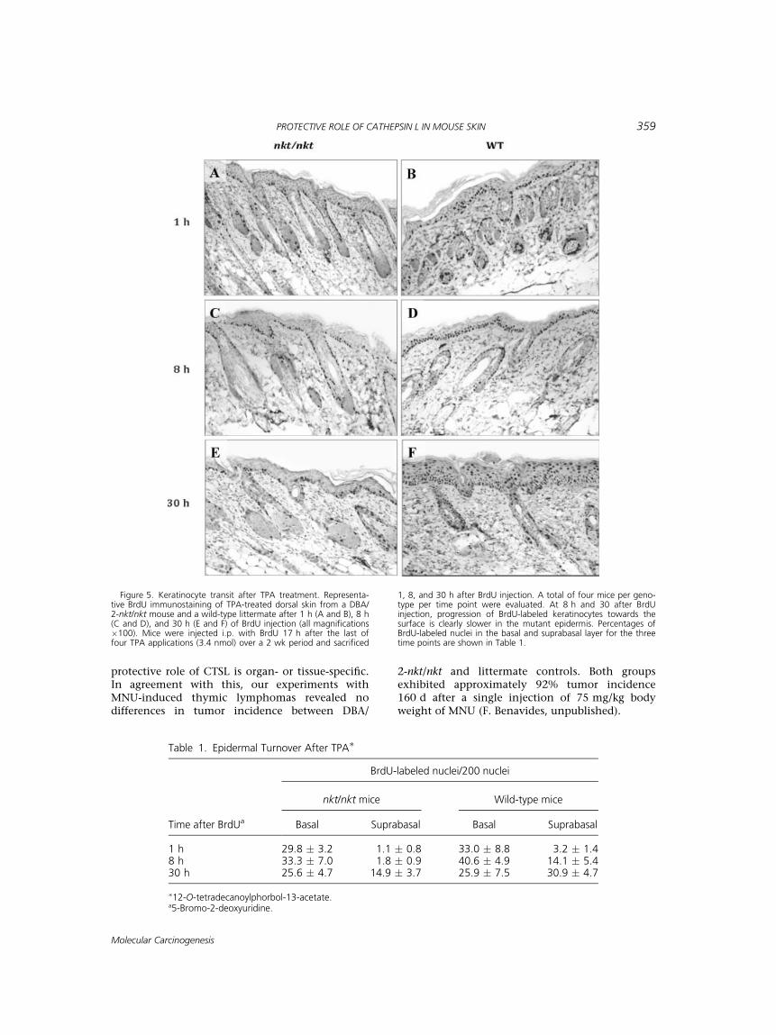
We analyzed keratinocyte transit through theepidermis by following the fate of BrdU-labeledcells at 1, 8, and 30 h after BrdU injection (17 hafter the last TPA application). The transit of kera-tinocytes was delayed in DBA/2-nkt/nkt comparedwith wild-type skin in the three time points ana-lyzed. After 30 h, many labeled keratinocytes ofthe wild-type skin had reached the granular andcornified layers of the epidermis, whereas in themutant skin, basal, and suprabasal keratinocytesretained most of the BrdU-labeled nuclei(Figure 5). Our analysis showed that the absence ofCTSL resulted in delayed terminal differentiationof keratinocytes and decreased epidermal cell turn-over. Because ordered differentiation, upwardmigration, and exfoliation of peripheral keratino-cytes are essential for timely removal of cells har-boring oncogenic mutations, the increased transittime in nkt/nkt mice may lead to retentionof DMBA-initiated keratinocytes and increasedtumorigenesis.
DISCUSSION
Cellular proteases like CTSL promote tumorgrowth, invasion, and metastasis [32], and CTSL is
Figure 3. Two-stage carcinogenesis study in CD4 T-cell deficientmice. Tumor multiplicity of FVB/N-Cd4�/� mice (n ¼ 10) was stat-istically highly significant from that of FVB/N wild-type mice(n ¼ 8) at 20 wk (P < 0.001, Wilcoxon Rank Sum test). Mice 6–8 wk old were initiated with DMBA and after 2 wk receivedrepeated applications of TPA as described in Materials andMethods Section. The number of papillomas was determinedweekly. Tumor multiplicity is the average number of skin tumorsper mouse. FVB/N wild-type mice (&); Cd4�/� mice (*).
PROTECTIVE ROLE OF CATHEPSIN L IN MOUSE SKIN 357
Molecular Carcinogenesis

considered a clinical marker for invasive tumors[33,34]. High levels of CTSL have been found innearly all human epithelial tumors in comparisonwith normal tissues, including breast [35], prostate[36], colorectal tumors [37], and in head and neckSCC [38]. Our finding that CTSL expressionincreased during two-stage skin carcinogenesis isconsistent with previous reports of the strongassociation between high levels of cathepsins andcancer. Thus our finding that nkt/nkt mice, whichdo not express functional CTSL, have enhancedsusceptibility to two-stage skin carcinogenesiscompared to wild-type mice was unexpected. How-ever, a protective role for CTSL in skin cancerwas previously demonstrated using Ctsl�/� micecrossed with the HPV16-transgenic mice [39,40].
In this in vivo model, Ctsl deficiency promotesskin carcinogenesis and lymph node metastasis,with HPV16; Ctsl�/� mice showing early onset ofdysplastic and neoplastic lesions and poorly differ-entiated carcinomas [40]. Our findings are alsosupported by recent reports showing increasedDMBA/TPA-induced skin carcinogenesis in trans-genic mice overexpressing the CTSL inhibitor hur-pin [41] as well as increased intestinal neoplasia inApcMin;furless (Ctsl deficient) mice [42]. Similarunexpected results in two-stage skin carcinogenesiswere obtained with collagenase-2 (MMP8) [43] andId1 [44] knockout mice.On the other hand, Ctsl deficiency in a mouse
model of pancreatic cancer reduces tumorburden and invasiveness [2], suggesting that the
Figure 4. Epidermal proliferation after TPA treatment. Represen-tative BrdU immunostaining of TPA-treated dorsal skin from aDBA/2-nkt/nkt mouse (A) and a wild-type littermate (B) (both mag-nifications �100). Mice were treated with four topical applicationsof 3.4 nmol of TPA over a 2-wk period and sacrificed 24 h afterthe final treatment. Bar graphs show cell proliferation level asmeasured by Ki67 (C) and BrdU (D) indexes, as well as epidermal
thickness (E). The determination of epidermal thickness and label-ing index was performed as described in Materials and MethodsSection. Values represent mean � SD (�P < 0.05). Black bars,mutant mice; gray bars, wild-type littermates. (F) Western blotanalysis of p21 in the epidermis of TPA-treated dorsal skin fromDBA/2-nkt/nkt mice and littermate controls. Protein was normal-ized to b-actin.
358 BENAVIDES ET AL.
Molecular Carcinogenesis
protective role of CTSL is organ- or tissue-specific.In agreement with this, our experiments withMNU-induced thymic lymphomas revealed nodifferences in tumor incidence between DBA/
2-nkt/nkt and littermate controls. Both groupsexhibited approximately 92% tumor incidence160 d after a single injection of 75 mg/kg bodyweight of MNU (F. Benavides, unpublished).
Figure 5. Keratinocyte transit after TPA treatment. Representa-tive BrdU immunostaining of TPA-treated dorsal skin from a DBA/2-nkt/nkt mouse and a wild-type littermate after 1 h (A and B), 8 h(C and D), and 30 h (E and F) of BrdU injection (all magnifications�100). Mice were injected i.p. with BrdU 17 h after the last offour TPA applications (3.4 nmol) over a 2 wk period and sacrificed
1, 8, and 30 h after BrdU injection. A total of four mice per geno-type per time point were evaluated. At 8 h and 30 after BrdUinjection, progression of BrdU-labeled keratinocytes towards thesurface is clearly slower in the mutant epidermis. Percentages ofBrdU-labeled nuclei in the basal and suprabasal layer for the threetime points are shown in Table 1.
Table 1. Epidermal Turnover After TPA�
Time after BrdUa
BrdU-labeled nuclei/200 nuclei
nkt/nkt mice Wild-type mice
Basal Suprabasal Basal Suprabasal
1 h 29.8 � 3.2 1.1 � 0.8 33.0 � 8.8 3.2 � 1.48 h 33.3 � 7.0 1.8 � 0.9 40.6 � 4.9 14.1 � 5.430 h 25.6 � 4.7 14.9 � 3.7 25.9 � 7.5 30.9 � 4.7
�12-O-tetradecanoylphorbol-13-acetate.a5-Bromo-2-deoxyuridine.
PROTECTIVE ROLE OF CATHEPSIN L IN MOUSE SKIN 359
Molecular Carcinogenesis

It was recently reported that keratinocytes fromCtsl knock-out mice show enhanced proliferation,suggesting that CTSL regulates keratinocyte pro-liferation by controlling growth factor recycling[39]. However, in our Ctsl-deficient nackt modelthere was decreased keratinocyte proliferation inTPA-treated skin. The difference in keratinocyteproliferation may be attributable to the differentgenetic backgrounds in these models. The Ctslknock-out model was described mainly in129P2;C57BL/6 or 129P2;C57BL/6;FVB (whencrossed with HPV16 transgenic mice) mixedgenetic backgrounds, whereas the nackt model wasstudied using defined congenic strains in SEN-CARB/Pt, DBA/2, and BALB/c inbred backgrounds.Additionally, when compared with archived papil-lomas generated in wild-type DBA/2 mice, the pro-liferation rate of the Ctsl-deficient papillomas wassimilar, suggesting that the mutant keratinocytesovercome their resistance to TPA.
How CTSL deficiency leads to enhanced skintumorigenesis is unclear. As previously reported byDennemarker et al. [40], the absence of CTSL didnot affect angiogenesis or apoptosis during two-stage carcinogenesis in our model, since IHC andWestern blot analysis using antibodies againstCD31 and activated caspase-3 revealed no signifi-cant differences between mutant and wild-typepapillomas. On the other hand, our data obtainedfrom the TPA-induced epidermal proliferationstudies support the idea that disrupted terminaldifferentiation of the epidermis, together withdecreased turnover of initiated keratinocytes,could be responsible for the enhanced skin tumori-genesis. The influence of keratinocyte turnover onskin carcinogenesis was previously described inK10 [45] and in Cox-1 and Cox-2 knockout [29]mice. In these cases, the exit of keratinocytes fromthe basal layer was accelerated and increased turn-over resulted in decreased tumor formation aftertwo-stage carcinogenesis. Since therapeutics inhib-iting CTSL are being developed to treat humancancer, it is very important to carefully evaluatethe potential of CTSL to promote rather than pre-vent tumorigenesis in different organs.
ACKNOWLEDGMENTS
We thank Lezlee Coghlan, Pamela S. Huskey,April Weiss, Nancy Doradau, Dale Weiss, andDonna Schutz for their assistance with mainten-ance of the mouse strains. We are grateful to Mar-cela Franco, Monica Flores, and John Bartonico fortechnical assistance and Melissa Bracher foradministrative assistance. We also wish to thankKevin Lin for statistical analyses and the Histologyand Tissue Processing Facility Core for the process-ing of samples. This work was supported by PilotProject from NIEHS Center Grant ES07784 to FB
and NIH Grant CA90922 to CJC. This study alsomade use of the Research Animal Support Facility-Smithville, including Genetic Services and MutantMouse Pathology Service, supported by P30CA16672-30 DHHS/NCI Cancer Center SupportGrant (CCSG). FVB/N-CD4 knock-out congenicmice where a gift from Lisa M. Coussens from theUniversity of California at San Francisco, San Fran-cisco, CA.
REFERENCES
1. Mohamed MM, Sloane BF. Cysteine cathepsins: Multi-functional enzymes in cancer. Nat Rev Cancer 2006;6:764–775.
2. Gocheva V, Zeng W, Ke D, et al. Distinct roles for cysteinecathepsin genes in multistage tumorigenesis. Genes Dev2006;20:543–556.
3. Roth W, Deussing J, Botchkarev VA, et al. Cathepsin Ldeficiency as molecular defect of furless: Hyperprolifera-tion of keratinocytes and pertubation of hair folliclecycling. FASEB J 2000;14:2075–2086.
4. Benavides F, Starost MF, Flores M, Gimenez-Conti IB,Guenet JL, Conti CJ. Impaired hair follicle morphogenesisand cycling with abnormal epidermal differentiation innackt mice, a cathepsin L-deficient mutation. Am J Pathol2002;161:693–703.
5. Tobin DJ, Foitzik K, Reinheckel T, et al. The lysosomal pro-tease cathepsin L is an important regulator of keratinocyteand melanocyte differentiation during hair folliclemorphogenesis and cycling. Am J Pathol 2002;160:1807–1821.
6. Joyce JA, Baruch A, Chehade K, et al. Cathepsin cysteineproteases are effectors of invasive growth and angiogene-sis during multistage tumorigenesis. Cancer Cell 2004;5:443–453.
7. Levicar N, Dewey RA, Daley E, et al. Selective suppressionof cathepsin L by antisense cDNA impairs human braintumor cell invasion in vitro and promotes apoptosis. Can-cer Gene Ther 2003;10:141–151.
8. Stoka V, Turk B, Turk V. Lysosomal cysteine proteases:Structural features and their role in apoptosis. IUBMB Life2005;57:347–353.
9. Stoka V, Turk V, Turk B. Lysosomal cysteine cathepsins:Signaling pathways in apoptosis. Biol Chem 2007;388:555–560.
10. Goulet B, Baruch A, Moon NS, et al. A cathepsin L isoformthat is devoid of a signal peptide localizes to the nucleusin S phase and processes the CDP/Cux transcription fac-tor. Mol Cell 2004;14:207–219.
11. Goulet B, Sansregret L, Leduy L, et al. Increased expres-sion and activity of nuclear cathepsin L in cancer cellssuggests a novel mechanism of cell transformation. MolCancer Res 2007;5:899–907.
12. Sullivan S, Tosetto M, Kevans D, et al. Localization ofnuclear cathepsin L and its association with diseaseprogression and poor outcome in colorectal cancer. Int JCancer 2009;125:54–61.
13. Santamaria I, Velasco G, Cazorla M, Fueyo A, Campo E,Lopez-Otin C. Cathepsin L2, a novel human cysteineproteinase produced by breast and colorectal carcinomas.Cancer Res 1998;58:1624–1630.
14. Puente XS, Lopez-Otin C. A genomic analysis of rat pro-teases and protease inhibitors. Genome Res 2004;14:609–622.
15. Benavides F, Giordano M, Fiette L, et al. Nackt (nkt), anew hair loss mutation of the mouse with associated CD4deficiency. Immunogenetics 1999;49:413–419.
16. Benavides F, Venables A, Poetschke Klug H, et al. TheCD4 T cell-deficient mouse mutation nackt (nkt) involves a
360 BENAVIDES ET AL.
Molecular Carcinogenesis

deletion in the cathepsin L (CtsI) gene. Immunogenetics2001;53:233–242.
17. Benavides F, Perez C, Blando J, Guenet JL, Conti CJ. Theradiation-induced nackt (nkt) allele is a loss-of-functionmutation of the mouse cathepsin L gene. J Immunol2006;176:702–703.
18. Lombardi G, Burzyn D, Mundinano J, et al. Cathepsin-Linfluences the expression of extracellular matrix in lym-phoid organs and plays a role in the regulation of thymicoutput and of peripheral T cell number. J Immunol2005;174:7022–7032.
19. Nakagawa T, Roth W, Wong P, et al. Cathepsin L: Criticalrole in Ii degradation and CD4 T cell selection in thethymus. Science 1998;280:450–453.
20. Nishimura F, Naruishi H, Naruishi K, et al. Cathepsin-L, akey molecule in the pathogenesis of drug-induced and I-cell disease-mediated gingival overgrowth: A study withcathepsin-L-deficient mice. Am J Pathol 2002;161:2047–2052.
21. Reinheckel T, Deussing J, Roth W, Peters C. Towardsspecific functions of lysosomal cysteine peptidases: Pheno-types of mice deficient for cathepsin B or cathepsin L. BiolChem 2001;382:735–741.
22. Stypmann J, Glaser K, Roth W, et al. Dilated cardiomyop-athy in mice deficient for the lysosomal cysteine peptidasecathepsin L. Proc Natl Acad Sci USA 2002;99:6234–6239.
23. Petermann I, Mayer C, Stypmann J, et al. Lysosomal, cyto-skeletal, and metabolic alterations in cardiomyopathy ofcathepsin L knockout mice. FASEB J 2006;20:1266–1268.
24. Yang M, Zhang Y, Pan J, et al. Cathepsin L activity con-trols adipogenesis and glucose tolerance. Nat Cell Biol2007;9:970–977.
25. Potts W, Bowyer J, Jones H, et al. Cathepsin L-deficientmice exhibit abnormal skin and bone development andshow increased resistance to osteoporosis following ovari-ectomy. Int J Exp Pathol 2004;85:85–96.
26. Wright WW, Smith L, Kerr C, Charron M. Mice thatexpress enzymatically inactive cathepsin L exhibit abnor-mal spermatogenesis. Biol Reprod 2003;68:680–687.
27. Wakeland E, Morel L, Achey K, Yui M, Longmate J. Speedcongenics: A classic technique in the fast lane (relativelyspeaking). Immunol Today 1997;18:472–477.
28. Stern MC, Benavides F, LaCava M, Conti CJ. Geneticanalyses of mouse skin tumor progression susceptibilityusing SENCAR inbred derived strains. Mol Carcinog 2002;35:13–20.
29. Tiano HF, Loftin CD, Akunda J, et al. Deficiency of eithercyclooxygenase (COX)-1 or COX-2 alters epidermal differ-entiation and reduces mouse skin tumorigenesis. CancerRes 2002;62:3395–3401.
30. Wakabayashi Y, Mao JH, Brown K, Girardi M, Balmain A.Promotion of Hras-induced squamous carcinomas by apolymorphic variant of the Patched gene in FVB mice.Nature 2007;445:761–765.
31. Stern MC, Benavides F, Klingelberger EA, Conti CJ. Allelo-type analysis of chemically induced squamous cell
carcinomas in F(1) hybrids of two inbred mouse strainswith different susceptibility to tumor progression. Carcino-genesis 2000;21:1297–1301.
32. Kane SE, Gottesman MM. The role of cathepsin L inmalignant transformation. Semin Cancer Biol 1990;1:127–136.
33. Tumminello FM, Leto G, Pizzolanti G, et al. Cathepsin D,B and L circulating levels as prognostic markers of malig-nant progression. Anticancer Res 1996;16:2315–2319.
34. Cuvier C, Jang A, Hill RP. Exposure to hypoxia, glucosestarvation and acidosis: Effect on invasive capacity of mur-ine tumor cells and correlation with cathepsin (LþB)secretion. Clin Exp Metastasis 1997;15:19–25.
35. Lah TT, Kalman E, Najjar D, et al. Cells producing cathep-sins D, B, and L in human breast carcinoma and theirassociation with prognosis. Hum Pathol 2000;31:149–160.
36. Friedrich B, Jung K, Lein M, et al. Cathepsins B, H, L andcysteine protease inhibitors in malignant prostate celllines, primary cultured prostatic cells and prostatic tissue.Eur J Cancer 1999;35:138–144.
37. Herszenyi L, Plebani M, Carraro P, et al. The role of cys-teine and serine proteases in colorectal carcinoma. Cancer1999;86:1135–1142.
38. Strojan P, Budihna M, Smid L, et al. Prognostic signifi-cance of cysteine proteinases cathepsins B and L and theirendogenous inhibitors stefins A and B in patients withsquamous cell carcinoma of the head and neck. Clin Can-cer Res 2000;6:1052–1062.
39. Reinheckel T, Hagemann S, Dollwet-Mack S, et al. Thelysosomal cysteine protease cathepsin L regulates kerati-nocyte proliferation by control of growth factor recycling.J Cell Sci 2005;118:3387–3395.
40. Dennemarker J, Lohmuller T, Mayerle J, et al. Deficiencyfor the cysteine protease cathepsin L promotes tumor pro-gression in mouse epidermis. Oncogene 2009;29:1611–1621.
41. Walz M, Kellermann S, Bylaite M, et al. Expression of thehuman Cathepsin L inhibitor hurpin in mice: Skin altera-tions and increased carcinogenesis. Exp Dermatol 2007;16:715–723.
42. Boudreau F, Lussier CR, Mongrain S, et al. Loss of cathep-sin L activity promotes claudin-1 overexpression and intes-tinal neoplasia. FASEB J 2007;21:3853–3865.
43. Balbin M, Fueyo A, Tester AM, et al. Loss of collagenase-2confers increased skin tumor susceptibility to male mice.Nat Genet 2003;35:252–257.
44. Sikder H, Huso DL, Zhang H, et al. Disruption of Id1reveals major differences in angiogenesis between trans-planted and autochthonous tumors. Cancer Cell 2003;4:291–299.
45. Reichelt J, Furstenberger G, Magin TM. Loss of keratin 10leads to mitogen-activated protein kinase (MAPK) acti-vation, increased keratinocyte turnover, and decreasedtumor formation in mice. J Invest Dermatol 2004;123:973–981.
PROTECTIVE ROLE OF CATHEPSIN L IN MOUSE SKIN 361
Molecular Carcinogenesis