protein engineering of candida antarctica lipase a377788/... · 2011-02-21 · protein engineering...
TRANSCRIPT

Protein Engineering of Candida
antarctica Lipase A
Enhancing Enzyme Properties by Evolutionary and Semi-Rational
Methods
Anders G. Sandström

ii
© Anders G. Sandström, Stockholm 2010
Cover picture: The Hand That Shapes, Anders Sandström, 2010.
Photo by Richard Lihammar.
ISBN 978-91-7447-202-8
Printed in Sweden by US-AB, Stockholm 2010
Distributor: Department of Organic Chemistry, Stockholm University

iii
Till min familj
”Strength is not an absolute value. To be strong is
to evolve. Mutability is strength.”
– Trevor Goodchild, Æon Flux: End Sinister

iv

v
Abstract
Enzymes are gaining increasing importance as catalysts for selective
transformations in organic synthetic chemistry. The engineering and design
of enzymes is a developing, growing research field that is employed in
biocatalysis. In the present thesis, combinatorial protein engineering
methods are applied for the development of Candida antarctica lipase A
(CALA) variants with broader substrate scope and increased enantio-
selectivity. Initially, the structure of CALA was deduced by manual
modeling and later the structure was established by X-ray crystallography.
The elucidation of the structure of CALA revealed several biocatalytically
interesting features. With the knowledge derived from the enzyme structure,
enzyme variants were produced via iterative saturation mutagenesis (ISM), a
powerful protein engineering approach. Several of these variants were highly
active and enantioselective towards bulky esters. Furthermore, an
extensively combinatorial protein engineering approach was developed and
investigated. A CALA variant with a spacious substrate binding pocket that
can accommodate an unusually bulky substrate, an ester derivate of the non-
steroidal anti-inflammatory drug (S)-ibuprofen, was obtained with this
approach.

vi
vii
List of Publications
This thesis is based on the following papers, referred to in the text by their
Roman numerals I-V.
I Prediction of the Candida antarctica Lipase A Protein
Structure by Comparative Modeling and Site-Directed
Mutagenesis
Kasrayan, A.; Bocola, M.; Sandström, A. G.; Lavén, G.;
Bäckvall, J.-E. ChemBioChem 2007, 8, 1409–1415.
II X-ray Structure of Candida antarctica Lipase A Shows a
Novel Lid Structure and a Likely Mode of Interfacial
Activation
Ericsson, D. J.; Kasrayan, A.; Johansson, P.; Bergfors, T.;
Sandström, A. G.; Bäckvall, J.-E.; Mowbray, S. L. J. Mol. Biol.
2008, 376, 109–119.
III Directed Evolution of Candida antarctica Lipase A Using an
Episomaly Replicating Yeast Plasmid
Sandström, A. G.; Engström, K.; Nyhlén, J.; Kasrayan, A.;
Bäckvall, J.-E. Protein Eng. Des. Sel. 2009, 22, 413–420.
IV Directed Evolution of an Enantioselective Lipase with Broad
Substrate Scope for Hydrolysis of α-Substituted Esters
Engström, K.; Nyhlén, J.; Sandström, A. G.; Bäckvall, J.-E. J.
Am. Chem. Soc. 2010, 132, 7038–7042.
V Highly Combinatorial Reshaping of the Candida antarctica
Lipase A Substrate Pocket Using an Extremely Condensed
Library
Sandström, A. G.; Wikmark, Y.; Engström, K.; Nyhlén, J.;
Bäckvall, J.-E. Manuscript.
Reprints were made with the kind permission of the publishers

viii
Related papers by the author, but not submitted as part of this
thesis:
VI Influence of δ-Functional Groups on the Enantiorecognition
of Secondary Alcohols by Candida antarctica Lipase B.
Nyhlén, J.; Martín-Matute, B.; Sandström, A. G.; Bocola, M.;
Bäckvall, J.-E. ChemBioChem 2008, 9, 1968–1974.
VII Highly Enantioselective Resolution of β-Amino Esters by
Candida antarctica Lipase A Immobilized in Mesocellular
Foam: Application to Dynamic Kinetic Resolution.
Shakeri, M.; Engström, K.; Sandström, A. G.; Bäckvall, J.-E.
ChemCatChem 2010, 5, 534–538.
ix
Contribution to Publications
I Performed molecular biology experimental, expressed enzyme,
performed activity assay, active-site titration, protein purification
and sequence data analysis. Wrote parts of the paper.
II Designed and performed molecular biology experimental,
expressed enzyme and performed protein purification.
III Designed and performed molecular biology experimental, protein
purification, screening (in part) and sequence data analysis.
Determined kinetic constants. Wrote the paper.
IV The methods from paper III was extended to aromatic substrates.
Practically, I performed minor sequence data analysis.
V Conceived, designed and performed molecular biology
experimental, screening (in part), activity assay, protein
purification and sequence data analysis. Wrote the paper.

x

xi
Table of Contents
Abstract ......................................................................................................... v
List of Publications .................................................................................... vii
Contribution to Publications ..................................................................... ix
Abbreviations ............................................................................................. xiv
Amino Acid Abbreviations ........................................................................ xv
1. Introduction ........................................................................................... 17 1.1 Introduction to Enzymes ................................................................................. 17 1.2 Enzymes as Catalysts in Organic Chemistry ............................................... 17 1.3 Enzymatic Kinetic Resolution ......................................................................... 18 1.4 Lipases and Serine Hydrolases ...................................................................... 19 1.5 Candida antarctica Lipase A ........................................................................... 21 1.6 Protein Engineering .......................................................................................... 22
1.6.1 Natural and Directed Evolution ............................................................. 22 1.6.2 Random Protein Engineering ................................................................. 23 1.6.3 Site-Specific Protein Engineering.......................................................... 24 1.6.4 Semi-Rational Protein Engineering ....................................................... 25
1.7 Objectives .......................................................................................................... 27
2. Determination of the Candida antarctica Lipase A Protein
Structure (Paper I and II) ....................................................................... 28 2.1 Introduction ....................................................................................................... 28 2.2 Recombinant Production of CALA .................................................................. 29 2.3 Manual Structure Modelling ............................................................................ 30
2.3.1 Alanin-Scanning ....................................................................................... 30 2.3.2 Active Site Titration................................................................................. 31
2.4 X-ray Structure ................................................................................................. 33 2.5 Conclusions ........................................................................................................ 35
3. Directed Evolution of Candida antarctica Lipase A for Enhanced
Enantioselectivity (Paper III and IV) .................................................... 36 3.1 Introduction ....................................................................................................... 36 3.2 Preparation of the Episomally Replicating Yeast Expression Vector
pBGP1-CALA ............................................................................................................. 37
xii
3.3 Directed Evolution of CALA for Increased Enantioselective Towards 4-
Nitrophenyl 2-Methylheptanoate .......................................................................... 38 3.3.1 4-Nitrophenyl 2-Methylheptanoate as Model Substrate ................... 38 3.3.2 Selection of Mutable Sites ...................................................................... 38 3.3.3 Production of Libraries ............................................................................ 40 3.3.4 Library Screening .................................................................................... 41 3.3.5 Kinetic Investigation and Model Analysis of Enantioselective
Variants ................................................................................................................ 42 3.4 Directed Evolution of CALA towards 4-Nitrophenyl 2-Phenylpropanoate
.................................................................................................................................... 43 3.4.1 4-Nitrophenyl 2-Phenylpropanoate as Model Substrate ................... 43 3.4.2 Library Screening .................................................................................... 44 3.4.3 Substrate Scope ...................................................................................... 45 3.4.3 Kinetic Resolution of 2-Phenylpropanoates with Different Alcohol
Moieties ................................................................................................................ 46 3.4.4 Models of Enantioselective Enzyme Variants ...................................... 47
3.5 Mechanistic Investigations via Site-Directed Mutagenesis ....................... 48 3.5 Conclusions ........................................................................................................ 49
4. Combinatorial Reshaping of the Substrate Pocket (Paper V) ..... 51 4.1 Introduction ....................................................................................................... 51 4.2 Experimental Outline ....................................................................................... 52 4.3 Results and Discussion .................................................................................... 53
4.3.1 Combinatorial Library Design ................................................................ 53 4.3.2 Mutagenesis and Homologous Recombination ................................... 56 4.3.3 Functional Diversity of the Library ....................................................... 56 4.3.4 Library Screening towards Ibuprofen Ester ........................................ 57 4.3.5 Back Mutations ......................................................................................... 58 4.3.6 Enzyme Models ........................................................................................ 59 4.3.7 Protein Fitness Landscapes .................................................................... 60 4.3.8 Combinatorial Substrate Pocket Sculpting .......................................... 61 4.3.9 Other Considerations .............................................................................. 62
4.4 Conclusions ........................................................................................................ 63
5. Concluding Remarks ............................................................................ 64
Acknowledgments ..................................................................................... 65
References .................................................................................................. 67

xiii
xiv
Abbreviations
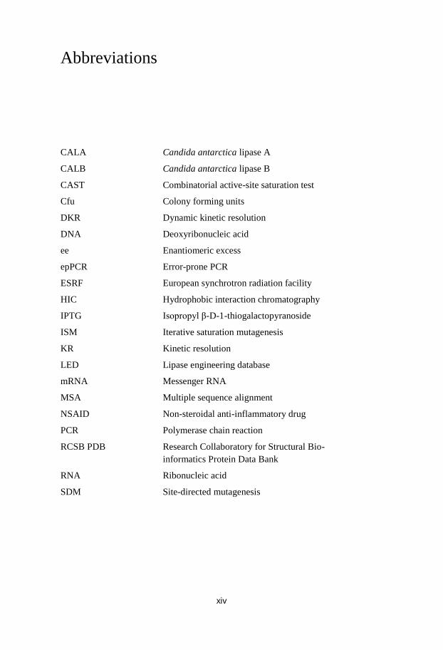
CALA Candida antarctica lipase A
CALB Candida antarctica lipase B
CAST combinatorial active-site saturation test
Cfu colony forming units
DKR dynamic kinetic resolution
DNA deoxyribonucleic acid
ee enantiomeric excess
epPCR
ESRF
HIC hydrophobic interaction chromatography
IPTG
ISM isopropyl β-D-1-thiogalactopyranoside
KR
LED kinetic resolution
mRNA
MSA
NSAID non-steroidal anti-inflammatory drug
PCR polymerase chain reaction
RCSB PDB
RNA ribonucleic acid
SDM site-directed mutagenesis
Candida antarctica lipase A
Candida antarctica lipase B
Combinatorial active-site saturation test
Colony forming units
Dynamic kinetic resolution
Deoxyribonucleic acid
Enantiomeric excess
Error-prone PCR
European synchrotron radiation facility
Hydrophobic interaction chromatography
Isopropyl β-D-1-thiogalactopyranoside
Iterative saturation mutagenesis
Kinetic resolution
Lipase engineering database
Messenger RNA
Multiple sequence alignment
Non-steroidal anti-inflammatory drug
Polymerase chain reaction
Research Collaboratory for Structural Bio-
informatics Protein Data Bank
Ribonucleic acid
Site-directed mutagenesis

xv
Amino Acid Abbreviations
Abbreviation Amino acid name
Three-letter Single-letter
Ala A Alanine
Arg R Arginine
Asn N Aspargine
Asp D Aspartic acid (Aspartate)
Cys C Cystein
Gln Q Glutamine
Glu E Glutamic acid (Glutamate)
Gly G Glycine
His H Histidine
Ile I Isoleucine
Leu L Leucine
Lys K Lysine
Met M Methionine
Phe F Phenylalanine
Pro P Proline
Ser S Serine
Thr T Threonine
Trp W Tryptophan
Tyr Y Tyrosine
Val V Valine

xvi

17
1. Introduction
1.1 Introduction to Enzymes
In 1897 Eduard Buchner discovered that yeast extracts can ferment sugars to
alcohols and that the process was promoted by substances found in the
extract. Wilhelm Kühne had already introduced the term enzyme in 1878, to
describe such „non-living‟ catalysts.1 Enzymes are biocatalysts, and as such
in principle work like other catalysts – it decreases the activation energy via
transition state stabilization, leading to an increased rate of the reaction. It is
essential to understand that catalysts (such as enzymes) never alter a
chemical equilibrium.
Compared to other catalysts, many enzymes show a remarkable
specificity. This specificity is popularly believed to be due to an „induced fit‟
of the enzyme to the shape of the substrate.2 The induced fit mainly
influences the initial binding, and not the catalytic process itself. Yet, at the
same time many enzymes also show a high degree of promiscuity, i.e. they
can catalyze reactions and accept substrates that are not natural substrates for
the enzyme.3 The immense catalytic ability has been mainly explained as a
result of the preorganization found in the active site of enzymes.4-5
The
transition state is stabilized by the electrostatic environment, which is the
main contributor of the lowering of the activation barrier compared to the
corresponding reaction in water. Other disputed hypotheses have been put
forward over the years, which claim that strain, protein dynamics, low
barrier hydrogen bonds or quantum tunneling is the main contributor of
catalytic activity.6 Another popular theory has been the ground state
destabilization idea, i.e. shielding the transition state from solvation effects.7
However, as mentioned above, it is now largely accepted that enzymes work
by transition state stabilization.5,8
Some enzymes, such as carbonic
anhydrase, have reached so called catalytic perfection, where the chemical
reaction occurs so fast that it is only limited by the diffusion of the reactants
entering and leaving the active site.9
1.2 Enzymes as Catalysts in Organic Chemistry
Enzymes have been used by mankind since early history. One of the oldest
applications has been the fermentation of carbohydrates to alcoholic

18
beverages. There is evidence that even Mesopotamian people in 6000 BC
fermented sweet fruits to produce wine.10
The understanding of what
occurred in the fermentation process was of course limited. We now
understand that it is Saccharomyces cerevisiae, bakers‟ yeast, which carries
out an anaerobic oxidation of carbohydrates to form ethanol.
Saccharomyces cerevisiae was used by the Bayer corporation already in
the 1930‟s to form a precursor to ephedrine, L-phenylacetylcarbinol via
whole-cell biotransformation of benzaldehyde.11
Ever since then, the use of
enzymes for biotransformations have slowly but steadily gained momentum.
Enzymes are used in industry either isolated or in living whole-cell systems.
Many energy-efficient processes have been developed using enzymes, as
many enzymes have their temperature optimum at room temperature.12
Enzymes are large polypeptides that are easy to produce with modern
recombinant gene technology. The use of enzymes will most likely increase
in an energy- and resource-conscious world. A vision of the future is the
concept of „microbial cell factories‟, the idea of utilizing genetically
engineered microbes, with entire biosynthetic pathways (catalyzed by
several enzymes) incorporated.13-14
1.3 Enzymatic Kinetic Resolution
Many molecules can exist as non-superimposable mirror images of each
other. Such molecules are considered to be chiral. These „mirror‟ images of
a chiral molecule are called enantiomers. This fundamental discovery was
made in the 19th century by Louis Pasteur who separated the enantiomeric
crystals of sodium ammonium tartrate; the crystal shapes were mirror-
images of each other.15
Enantiomers have the same physical properties provided that they are in
an achiral environment. Biological organisms contain a large quantity of
enantiopure molecules, and therefore constitute chiral environments. Amino
acids and sugars occur predominantly in one enantiomeric form in nature.
Enzymes and cellular receptors are made up of only L-amino acids, thus they
are enantiomerically pure. Nature is pervaded by this homochirality, and it is
essential for the existence of terrestrial life.16
Many modern drugs are chiral, and the two enantiomers of the compound
can often interact with the organism in completely different ways. Methods
for the preparation of enantiopure compounds are thus highly relevant for the
production of pharmaceuticals. One of the methods available is kinetic
resolution (KR).
KR relies on the rate difference between two enantiomers in the
transformation from substrate to product (Scheme 1). KR can be achieved by
the use of a chiral catalyst, for example an enzyme.17

19
Scheme 1. (S)-selective enzymatic kinetic resolution
Enzymes have the advantage of having a defined topology in the active
site where the catalytic reaction occurs. Compared to chiral ligands, the
active site of an enzyme most often has considerably larger defined space
where the chiral recognition occurs. Thus, in many cases, extremely high
enantioselectivity can be obtained.18
The enantioselectivity of the reaction is
defined by the E-value,19
which has been introduced to specify the
selectivity, as
slowk
fastk
E
1.4 Lipases and Serine Hydrolases
Lipases (EC 3.1.1.3) are currently the most used class of enzymes in
chemoenzymatic reactions and kinetic resolutions.20
The reaction catalyzed
by lipases in nature is the hydrolysis of water-insoluble esters such as lipids.
Lipases have a tendency to increase their activity in presence of high lipid
concentration; this is assumed to be caused by a change in enzymatic
conformation when in close contact with a non-polar surface, such as lipid
droplets. This phenomenon is called the interfacial activation. It has been
suggested that a hydrophobic „lid‟ is responsible for this effect; the lid
covers the active site, and swings open and immerses itself in the
hydrophobic media when the lid comes in contact with the lipid phase.21
Lipases have been used for hydrolysis of esters, and for the reverse
reaction, the synthesis of esters in organic solvents. Some lipases can also be
used for acylating amines for the formation of amide bonds.22
Enzymes were
thought to be unstable in organic solvents; however, Klibanov et al.
discovered that that many enzymes are actually stabilized by dry unpolar
solvents.23
The proposed reason is that in these dry solvents the native
conformation is kept and the enzymes do not unfold.
Many lipases display enantioselectivity, a highly useful property. Lipases
have been applied to perform kinetic resolution of many different substrates.
Our research group have used KR to great extent, and also in combination
with transition metal-catalysed racemisation of the chiral substrates, which
has been coined dynamic kinetic resolution (DKR) (for comprehensive
reviews, see refs.17,24-27
). This method has been applied to produce several
interesting compounds in high yields and enantiopurity.28-30
20
In comparison with the oxidoreductases, which are relying on either
expensive cofactors (such as NADPH/NADP+) with regeneration systems or
whole cell-systems, many lipases can be used in vitro without any special
additives.31
All serine hydrolases (which includes lipases, esterases and
serine proteases) work via a similar molecular mechanism.32
Three amino
acid residues, the so called „catalytic triad‟, are key players. An acid residue
(aspartate or glutamate) coordinates to a histidine, which in turn works as a
charge relay residue.33
The histidine withdraws a proton from the
nucleophilic serine. The now activated serine works as the nucleophile
attacking the ester carbonyl, and the formed oxyanion of the tetrahedral
intermediate is stabilized by the so called the oxyanion hole (Figure 1).34
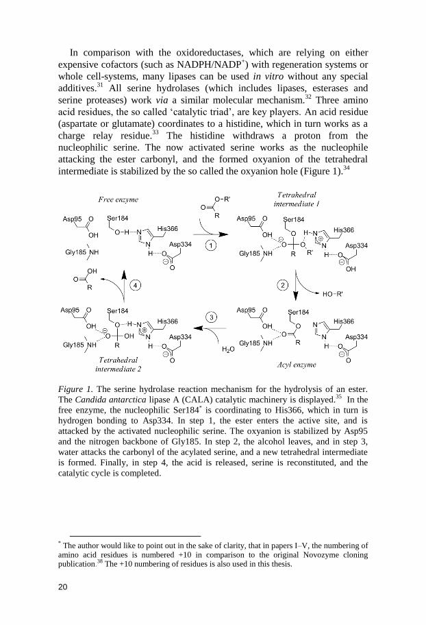
Figure 1. The serine hydrolase reaction mechanism for the hydrolysis of an ester.
The Candida antarctica lipase A (CALA) catalytic machinery is displayed.35
In the
free enzyme, the nucleophilic Ser184* is coordinating to His366, which in turn is
hydrogen bonding to Asp334. In step 1, the ester enters the active site, and is
attacked by the activated nucleophilic serine. The oxyanion is stabilized by Asp95
and the nitrogen backbone of Gly185. In step 2, the alcohol leaves, and in step 3,
water attacks the carbonyl of the acylated serine, and a new tetrahedral intermediate
is formed. Finally, in step 4, the acid is released, serine is reconstituted, and the
catalytic cycle is completed.
* The author would like to point out in the sake of clarity, that in papers I–V, the numbering of amino acid residues is numbered +10 in comparison to the original Novozyme cloning publication.38 The +10 numbering of residues is also used in this thesis.

21
1.5 Candida antarctica Lipase A
Several Japanese research expeditions were sent out in the 1960s‟ to sample
Antarctic soils to examine the microbiological flora. Soil and water samples
were assayed in the McMurdo dry valleys. The yeast Candida antarctica
was found in a sample from lake sediment at 9 m depth, from a hypersaline
lake, Lake Vanda.36
Lake Vanda is perennially covered by ice.37
Researchers at Novo Nordisk A/S (now Novozymes) isolated two lipases
from Candida antarctica. The two lipases, called Candida antarctica lipase
A and B (CALA and CALB, respectively) were both found to be highly
thermostable, and were cloned into Aspergillus oryzae.38
Homology analysis of the CALA and CALB peptide amino acid
sequences and DNA sequences reveals close relationship to the
basidiomycetous fungi Pseudozyma aphidis39
, Kurtzmanomyces sp. I-1140
and Ustilago maydis.41
P. aphidis have highly homologous genes to the two
lipases from C. antarctica. U. maydis, also called corn smut, is a well
studied pathogen found on maize. Pseudozyma aphidis was curiously first
isolated from the faeces of aphids. The Pseudozyma aphidis strain DSMZ
7072542
was used in our laboratory for the in-house isolation of CALA and
CALB. The isolated CALA gene contains a single silent mutation, and the
CALB gene gives rise to two surface located amino acid substitutions that
differ from the original Novozyme publication.38,43
CALB has been used for vast numbers of biotransformations and kinetic
resolutions of many substrates, and is probably the single most used enzyme
for kinetic resolutions. CALA has not found such broad application yet, but
it has some interesting properties that are currently exploited.
CALA is a monomeric 431 amino acid residues single peptide lipase,
weighing 45 kDa, with a pH optimum at 7.44-45
CALA is, as previously
mentioned, highly thermostable, and is claimed to be one of the most
thermostable lipases known.46
Novozymes has produced CALA in
Aspergillus oryzae and is marketing the lipase as Novozyme 735. Regarding
the preference of esters, CALA prefers medium to long chain lengths of the
alcohol and acid moieties.46
CALA is known to exhibit a weak interfacial
activation.45
CALA has also the interesting property that it has an sn-2
preference towards triglycerides.47
In triglycerides, sn-2 is a designation of
the center carbon of the glycerol moiety. The sn-2 preference can be used for
selective substitutions on triglycerides, which could be useful for the
preparation of fat replacement products and in theory, covalently tethered
drug hidden in a triglyceride-like compound.
CALA has found use for the preparation of highly enantiopure β-amino
acids/esters, which holds large promise as building blocks for important drug
candidates, such as specific protease inhibitors.48-50
CALA has also shown
the unusual trait of being able to hydrolyse esters with tertiary alcohol
moieties.51-52
Tertiary alcohols are used as a protective group in synthetic

22
organic chemistry, and the specific removal of such groups can be of great
interest. Enantiopure tertiary alcohols are also interesting, and the
enrichment via kinetic resolution could prove very useful.
In literature, mutational studies of CALA are quite sparse, but some
information can be found in Novozyme patents. One CALA variant has been
reported, with the modifications F145W† and F149W, which is claimed to
have a fourfold increase in the activity towards glycerol tributyrate.53-54
1.6 Protein Engineering
Enzymes are proteins, and as such biopolymers, produced by the cells to
facilitate various molecular processes such as metabolism and replication of
DNA. As with all proteins, their formation is based on the „central dogma‟;
transcription of DNA to produce mRNA, transport of the mRNA to the
ribosome, where the mRNA is translated and the protein is synthesized. The
ribosome is a large RNA-protein complex which synthesizes polypeptides,
using mRNA as a template, and amino acids as building blocks.55
The
polypeptide is processed, and folded into a defined structure, and the protein
is formed.1 The fact that the genetic information is coupled to the protein
phenotype facilitates the adaption of protein properties via the modification
of genetic information. Protein engineering is the deliberate modification of
these properties, by the use of molecular biology techniques. This field is
currently expanding rapidly, and several techniques have been established,
or are in the process of being established.56
1.6.1 Natural and Directed Evolution
The British naturalist Charles Darwin developed his theory of evolution in
the mid-19th century.
57 The theory of evolution can be roughly summarized
as follows:
Diversification: Copy X (parents) into several Y (offspring). Introduce
slight variations in the Ys. Throw away all X.
Selection (natural or non-natural): Only Y that has traits that grant
„survivability‟ are kept, the other Ys are discarded.
Reproduction: Remaining Y (offspring) becomes X (parents). Go to first
step and repeat.
This simple iterative process has created all the variation in natural
biological life, observed so far. „Survivability‟ is an abstract concept; in
† A note on amino acid residue substitutions; F145W, or Phe145Trp, means that
phenylalanine residue no. 145 has been replaced with tryptophan in that particular enzyme variant.

23
biological science the term fitness is used, where it indicates an organisms‟
capacity to replicate its genetic material.58
In non-natural selection, such as
in directed evolution, it can be any arbitrary property that the researcher
selects for.
The refinement and development by breeding and selection of
domesticated livestock, dogs and cultivated grass are based on evolution.
The information carrier in living organisms, the inheritable genetic code, is
DNA. Variation in the genetic code can be introduced by several processes,
such as mutations by exchanges of bases in DNA, or sexual recombination.59
Directed evolution is a method used in protein engineering, where the
power of non-natural selection is utilized to improve desired properties of
proteins. The iterative process, the essence of directed evolution, facilitates
these stepwise improvements.60-63
Molecular biology techniques and
recombinant DNA technologies have steadily improved over the last
decades. Many of these methods have found usage in directed evolution
procedures, where they are used for introducing protein diversity.56,64-66
1.6.2 Random Protein Engineering
One of the first techniques used for directed evolution was the error-prone
PCR (epPCR) technique. It is based on the non-perfect replication of DNA
in the polymerase chain reaction (PCR).67
Misincorporation of nucleotides
occurs over the entire replicated sequence. By altering the concentration of
magnesium and manganese ions it is possible to modify the amount of
erroneously incorporated nucleotides in the replicated DNA.65
It gives rise
to a pool of mutated sequences, a so called „library‟ of mutants. A word on
definition: a mutant gene gives rise to a protein variant.
Classical error-prone PCR for directed evolution requires neither crystal
structure of the protein in question, nor any special knowledge of the
mechanism of the enzyme, or of the active site. The majority of amino acids
found in an enzyme are generally quite far from the active site. Thus, there is
slim chance of hitting a residue involved in substrate binding and that is
influencing activity, and this approach may therefore require screening of
very large libraries.68-69
Another technique that usually does not require prior knowledge of the
structure is the gene (DNA) shuffling techniques.70
A multitude of DNA
shuffling techniques have been developed, such as ITCHY, SCRATCHY
and SCOPE, etc.62,71-72
They all have in common that they are based on
recombining more-or-less homologous sequences, for example homologous
enzymes derived from different species.73
24
1.6.3 Site-Specific Protein Engineering
Site-directed mutagenesis (SDM) is currently one of the most used
mutagenesis methods in protein engineering.74
It is based on the use of
primers, short oligonucleotides used in the polymerase chain reaction (PCR)
step, which are not completely complementary to the sequence being
amplified. The non-complementary nucleotides are introduced in the
amplified sequence. The template sequence is preferably a plasmid, a
circular extra-chromosomal body of DNA (Figure 2).
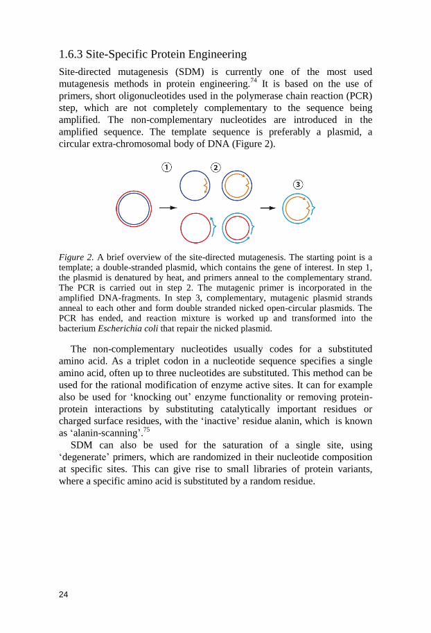
Figure 2. A brief overview of the site-directed mutagenesis. The starting point is a template; a double-stranded plasmid, which contains the gene of interest. In step 1, the plasmid is denatured by heat, and primers anneal to the complementary strand. The PCR is carried out in step 2. The mutagenic primer is incorporated in the amplified DNA-fragments. In step 3, complementary, mutagenic plasmid strands anneal to each other and form double stranded nicked open-circular plasmids. The PCR has ended, and reaction mixture is worked up and transformed into the bacterium Escherichia coli that repair the nicked plasmid.
The non-complementary nucleotides usually codes for a substituted
amino acid. As a triplet codon in a nucleotide sequence specifies a single
amino acid, often up to three nucleotides are substituted. This method can be
used for the rational modification of enzyme active sites. It can for example
also be used for „knocking out‟ enzyme functionality or removing protein-
protein interactions by substituting catalytically important residues or
charged surface residues, with the „inactive‟ residue alanin, which is known
as „alanin-scanning‟.75
SDM can also be used for the saturation of a single site, using
„degenerate‟ primers, which are randomized in their nucleotide composition
at specific sites. This can give rise to small libraries of protein variants,
where a specific amino acid is substituted by a random residue.
25
1.6.4 Semi-Rational Protein Engineering
The size and utility of the protein libraries generated are important
parameters when deciding what protein engineering strategy that should be
pursued.76
Factors such as cost and labor time for screening are reasons to
keep the library size as small as possible.77
Protein engineering methods that
focus on the active site are known to have a higher chance of influencing
catalytic properties.69
These methods generally create small libraries, as only
a few amino acid residues are targeted. One development of SDM was
conceived in the group of Manfred T. Reetz. The technique is called
combinatorial active-site saturation test (CAST), which is based on the
simultaneous randomization of a few amino acid sites, in close sequence
proximity, using one single primer pair.78
Two or three amino acid residues
are generally subjected simultaneously to mutagenesis. The reason for
choosing more than one amino acid to mutate is the potential synergistic
conformational and electrostatic effects that may appear. Amino acid residue
pairs surrounding the active site are usually the target for the saturation.
These active site-focused libraries have been used with good results for the
improvement of activity and enantioselectivity.79
Iterative rounds of
mutagenesis of the active site often give rise to highly synergistic effects.80
CASTing has been used in an iteratively manner (coined iterative saturation
mutagenesis, ISM) by the Reetz group to change diverse properties such as
thermostability and enantioselectivity.81
CASTing (and site-directed
mutagenesis) requires knowledge of the substrate binding, and preferably the
mechanism, of the enzyme and associated amino acids. This knowledge is
often derived from the X-ray structure of the enzyme in question.
The ability to determine the composition of nucleotides at certain
positions when designing primers gives rise to different sets of potentially
encoded amino acids (Figure 3).
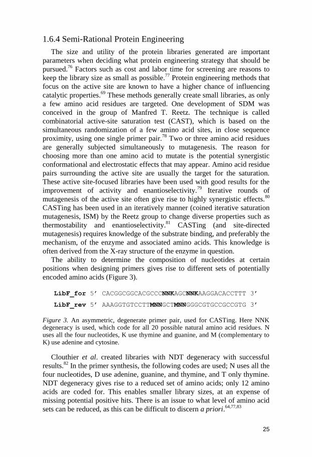
LibF_for 5’ CACGGCGGCACGCCCNNKAGCNNKAAGGACACCTTT 3’
LibF_rev 5’ AAAGGTGTCCTTMNNGCTMNNGGGCGTGCCGCCGTG 3’
Figure 3. An asymmetric, degenerate primer pair, used for CASTing. Here NNK degeneracy is used, which code for all 20 possible natural amino acid residues. N uses all the four nucleotides, K use thymine and guanine, and M (complementary to K) use adenine and cytosine.
Clouthier et al. created libraries with NDT degeneracy with successful
results.82
In the primer synthesis, the following codes are used; N uses all the
four nucleotides, D use adenine, guanine, and thymine, and T only thymine.
NDT degeneracy gives rise to a reduced set of amino acids; only 12 amino
acids are coded for. This enables smaller library sizes, at an expense of
missing potential positive hits. There is an issue to what level of amino acid
sets can be reduced, as this can be difficult to discern a priori.64,77,83
26
Mutational suggestions may not only be rationally deducted from the
three-dimensional structure information, but computational and
bioinformatical based-methods are also used to a high degree. For example,
the degree of amino acid residue conservation derived from a multiple
sequence alignment (MSA) can be used for the elucidation of a residues‟
mutability.84-85
Combinatorial libraries with small sets of amino acid residues
have been used for the generation of consensus libraries with84
or without86-87
phylogenetic bias, for the development of thermostable enzymes. The
structure-based multiple-sequence alignment 3DM database has been used
for suggesting mutational sites and „allowed‟ residues.88
Also,
computationally designed combinatorial libraries have generated broad
functional diversity for fluorescent proteins.89
In these described methods
each mutational site is randomized with a small set of amino acid residues.
Indeed, information from statistical and computational methods assists
modern protein engineering in an increasing extent.90-91
27
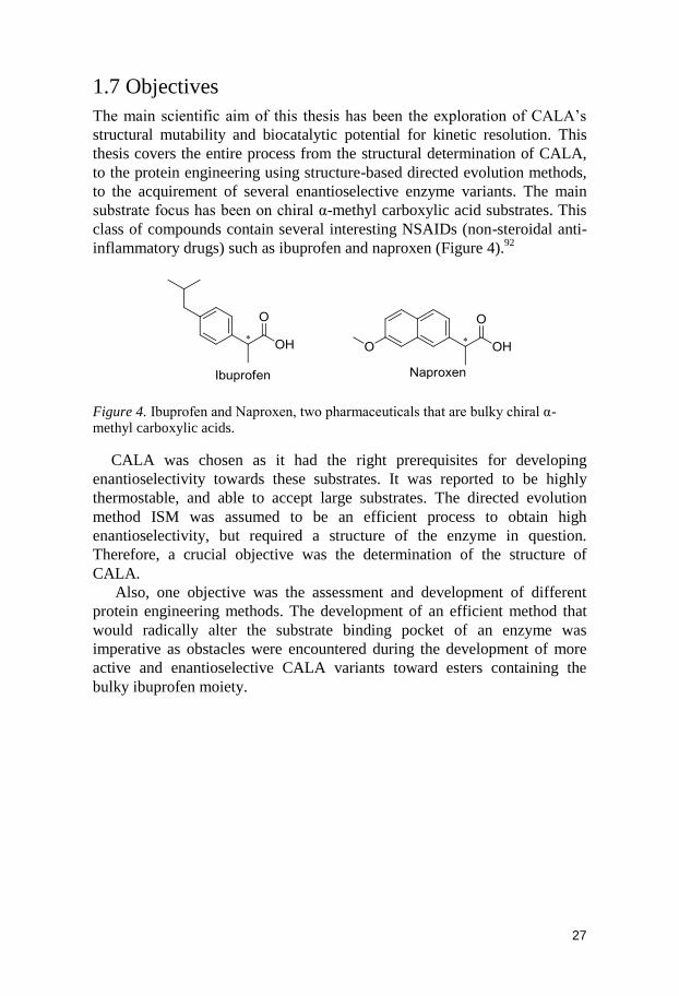
1.7 Objectives
The main scientific aim of this thesis has been the exploration of CALA‟s
structural mutability and biocatalytic potential for kinetic resolution. This
thesis covers the entire process from the structural determination of CALA,
to the protein engineering using structure-based directed evolution methods,
to the acquirement of several enantioselective enzyme variants. The main
substrate focus has been on chiral α-methyl carboxylic acid substrates. This
class of compounds contain several interesting NSAIDs (non-steroidal anti-
inflammatory drugs) such as ibuprofen and naproxen (Figure 4).92
Figure 4. Ibuprofen and Naproxen, two pharmaceuticals that are bulky chiral α-methyl carboxylic acids.
CALA was chosen as it had the right prerequisites for developing
enantioselectivity towards these substrates. It was reported to be highly
thermostable, and able to accept large substrates. The directed evolution
method ISM was assumed to be an efficient process to obtain high
enantioselectivity, but required a structure of the enzyme in question.
Therefore, a crucial objective was the determination of the structure of
CALA.
Also, one objective was the assessment and development of different
protein engineering methods. The development of an efficient method that
would radically alter the substrate binding pocket of an enzyme was
imperative as obstacles were encountered during the development of more
active and enantioselective CALA variants toward esters containing the
bulky ibuprofen moiety.

28
2. Determination of the Candida antarctica Lipase A Protein Structure (Paper I and II)
2.1 Introduction
For the development of an enzyme with increased enantioselectivity,
directed evolution is an excellent approach. It was decided early on to use
CALA for the development of a highly enantioselective lipase towards large
substrates, as it was considered to have the prerequisites necessary for the
project.
The CASTing technique had been proven advantageous for the
development of a highly enantioselective Pseudomonas aeruginosa lipase.78
As previously mentioned, the CASTing technique requires an X-ray
structure or a homology model for the selection of amino acid residues that
may influence the property screened for. As an X-ray structure of CALA did
not yet exist, a homology model was considered as an acceptable alternative
for the project.
A comparison of the amino acid sequence revealed that there were no
available enzyme X-ray structures sufficiently related to CALA. The
crystallization and determination of an X-ray structure of a novel enzyme
was also seen as quite difficult. The closest related available structures were
Pseudomonas putida esterase (14% sequence identity) and Pseudomonas
fluorescens esterase (14%).93
CALA could however easily be identified as belonging to the large α/β
hydrolase fold family. The possibility of creating a manually modeled
structure was considered, based on the generic α/β hydrolase fold (Figure 5).
It was assumed that it would be possible to produce the model if the active
site residues could be determined. A hypothesis we had was that the catalytic
residues could be identified by knocking out functionality via SDM. That
information was assumed to give enough knowledge for the creation of a
manually constructed 3D-model of CALA.
29
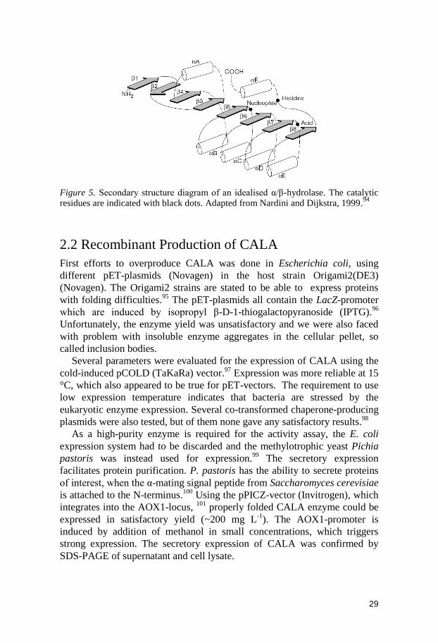
Figure 5. Secondary structure diagram of an idealised α/β-hydrolase. The catalytic residues are indicated with black dots. Adapted from Nardini and Dijkstra, 1999.
94
2.2 Recombinant Production of CALA
First efforts to overproduce CALA was done in Escherichia coli, using
different pET-plasmids (Novagen) in the host strain Origami2(DE3)
(Novagen). The Origami2 strains are stated to be able to express proteins
with folding difficulties.95
The pET-plasmids all contain the LacZ-promoter
which are induced by isopropyl β-D-1-thiogalactopyranoside (IPTG).96
Unfortunately, the enzyme yield was unsatisfactory and we were also faced
with problem with insoluble enzyme aggregates in the cellular pellet, so
called inclusion bodies.
Several parameters were evaluated for the expression of CALA using the
cold-induced pCOLD (TaKaRa) vector.97
Expression was more reliable at 15
°C, which also appeared to be true for pET-vectors. The requirement to use
low expression temperature indicates that bacteria are stressed by the
eukaryotic enzyme expression. Several co-transformed chaperone-producing
plasmids were also tested, but of them none gave any satisfactory results.98
As a high-purity enzyme is required for the activity assay, the E. coli
expression system had to be discarded and the methylotrophic yeast Pichia
pastoris was instead used for expression.99
The secretory expression
facilitates protein purification. P. pastoris has the ability to secrete proteins
of interest, when the α-mating signal peptide from Saccharomyces cerevisiae
is attached to the N-terminus.100
Using the pPICZ-vector (Invitrogen), which
integrates into the AOX1-locus, 101
properly folded CALA enzyme could be
expressed in satisfactory yield (~200 mg L-1
). The AOX1-promoter is
induced by addition of methanol in small concentrations, which triggers
strong expression. The secretory expression of CALA was confirmed by
SDS-PAGE of supernatant and cell lysate.
30
2.3 Manual Structure Modelling
2.3.1 Alanin-Scanning
An „alanin-scanning‟ (as described in chapter 1.6.3) was carried out by
SDM, where several CALA variants were produced with key residues
replaced by alanin. The enzyme variants produced were purified using
hydrophobic interaction chromatography (HIC). The enzyme variants were
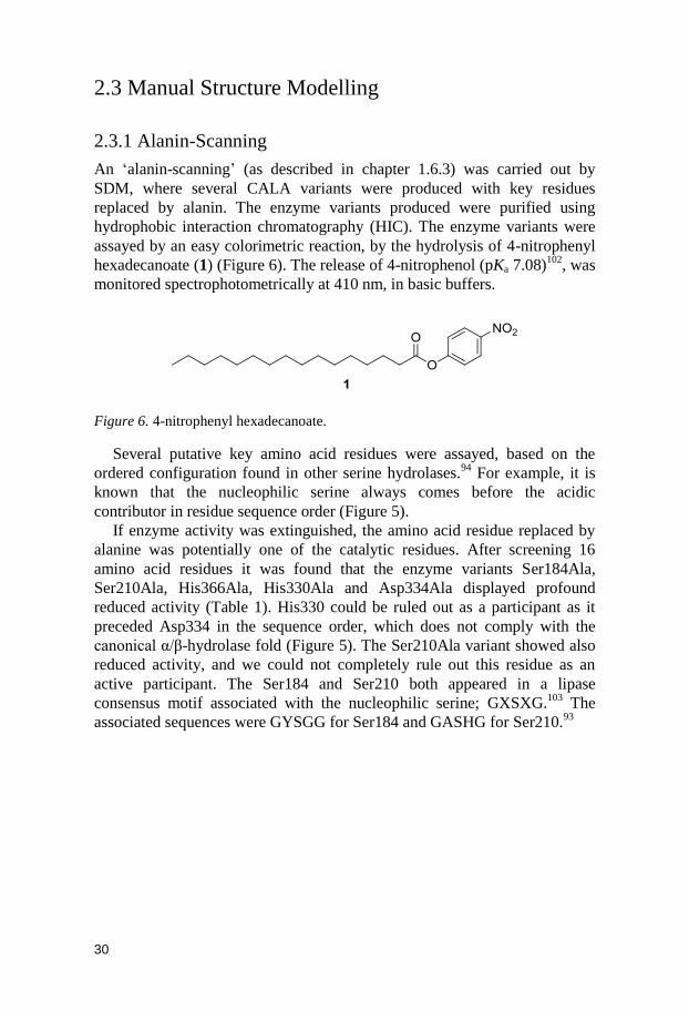
assayed by an easy colorimetric reaction, by the hydrolysis of 4-nitrophenyl
hexadecanoate (1) (Figure 6). The release of 4-nitrophenol (pKa 7.08)102
, was
monitored spectrophotometrically at 410 nm, in basic buffers.
Figure 6. 4-nitrophenyl hexadecanoate.
Several putative key amino acid residues were assayed, based on the
ordered configuration found in other serine hydrolases.94
For example, it is
known that the nucleophilic serine always comes before the acidic
contributor in residue sequence order (Figure 5).
If enzyme activity was extinguished, the amino acid residue replaced by
alanine was potentially one of the catalytic residues. After screening 16
amino acid residues it was found that the enzyme variants Ser184Ala,
Ser210Ala, His366Ala, His330Ala and Asp334Ala displayed profound
reduced activity (Table 1). His330 could be ruled out as a participant as it
preceded Asp334 in the sequence order, which does not comply with the
canonical α/β-hydrolase fold (Figure 5). The Ser210Ala variant showed also
reduced activity, and we could not completely rule out this residue as an
active participant. The Ser184 and Ser210 both appeared in a lipase
consensus motif associated with the nucleophilic serine; GXSXG.103
The
associated sequences were GYSGG for Ser184 and GASHG for Ser210.93

31
Table 1. Hydrolytic enzymatic activity of selected variants.
Entry Enzyme variant U mg-1[a]
1 Wild type 138 7.0
2 S184A 0.2 1.1
3 H366A 6.9 1.7
4 H330A 4.3 2.9
5 E298A 65 19
6 E314A 69 6.6
7 E308A 18.3 6.8
8 D334A 1.8 2.1
9 I301A 129 14
10 L367A 122 29
11 T118A 121 8.4
12 V120A 88 13
13 W129A 153 13
14 Y317A 39 12
15 Y183A 8.7 3.4
16 S210A 12 1.1
[a] One unit (U) of activity was defined as the amount of enzyme that released 1 µmol p-nitrophenol per minute under our assay conditions.
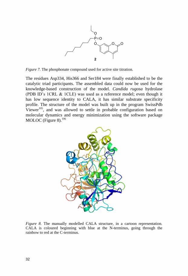
2.3.2 Active Site Titration
To distinguish between Ser184 or Ser210 as the nucleophilic residue, an
active site titration was carried out.104
Compound 2 was prepared by
coupling the fluorophore 4-methylumbelliferone with a phosphonate
compound (Figure 7). Compound 2 irreversible binds to the nucleophilic
serine, and can be used to determine whether an enzyme actually contains a
functional active site or not. The nucleophilic serine attacks the phosphorus
and the fluorescent moiety is released, resulting in an enzyme that is
irreversibly inhibited. The released fluorophore can be quantified by
fluorometry, and the fluorescence should display a linear correlation with the
amount of free active sites. The wild type CALA and the variants Ser184Ala
and Ser210Ala were subjected to active site titration. The wild type and
Ser210Ala both displayed correlation between fluorescence and amount of
enzyme. Fluorescence did not increase with increasing Ser184Ala enzyme
concentration, thus revealing that Ser184 was indeed the active site
nucleophile.
32
Figure 7. The phosphonate compound used for active site titration.
The residues Asp334, His366 and Ser184 were finally established to be the
catalytic triad participants. The assembled data could now be used for the
knowledge-based construction of the model. Candida rugosa hydrolase
(PDB ID‟s 1CRL & 1CLE) was used as a reference model; even though it
has low sequence identity to CALA, it has similar substrate specificity
profile. The structure of the model was built up in the program SwissPdb
Viewer105
, and was allowed to settle in probable configuration based on
molecular dynamics and energy minimization using the software package
MOLOC (Figure 8).106
Figure 8. The manually modelled CALA structure, in a cartoon representation. CALA is coloured beginning with blue at the N-terminus, going through the rainbow to red at the C-terminus.
33
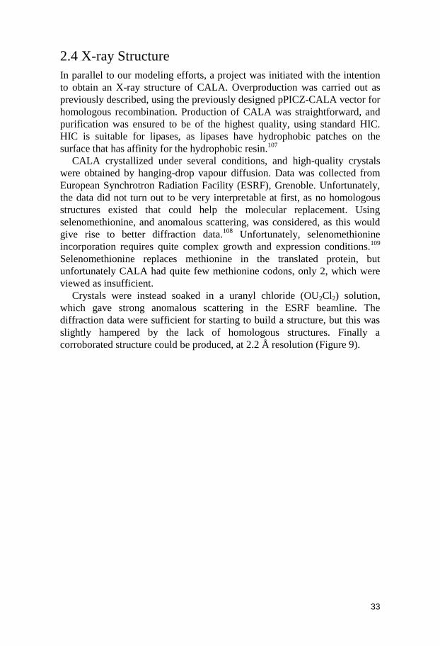
2.4 X-ray Structure
In parallel to our modeling efforts, a project was initiated with the intention
to obtain an X-ray structure of CALA. Overproduction was carried out as
previously described, using the previously designed pPICZ-CALA vector for
homologous recombination. Production of CALA was straightforward, and
purification was ensured to be of the highest quality, using standard HIC.
HIC is suitable for lipases, as lipases have hydrophobic patches on the
surface that has affinity for the hydrophobic resin.107
CALA crystallized under several conditions, and high-quality crystals
were obtained by hanging-drop vapour diffusion. Data was collected from
European Synchrotron Radiation Facility (ESRF), Grenoble. Unfortunately,
the data did not turn out to be very interpretable at first, as no homologous
structures existed that could help the molecular replacement. Using
selenomethionine, and anomalous scattering, was considered, as this would
give rise to better diffraction data.108
Unfortunately, selenomethionine
incorporation requires quite complex growth and expression conditions.109
Selenomethionine replaces methionine in the translated protein, but
unfortunately CALA had quite few methionine codons, only 2, which were
viewed as insufficient.
Crystals were instead soaked in a uranyl chloride (OU2Cl2) solution,
which gave strong anomalous scattering in the ESRF beamline. The
diffraction data were sufficient for starting to build a structure, but this was
slightly hampered by the lack of homologous structures. Finally a
corroborated structure could be produced, at 2.2 Å resolution (Figure 9).
34
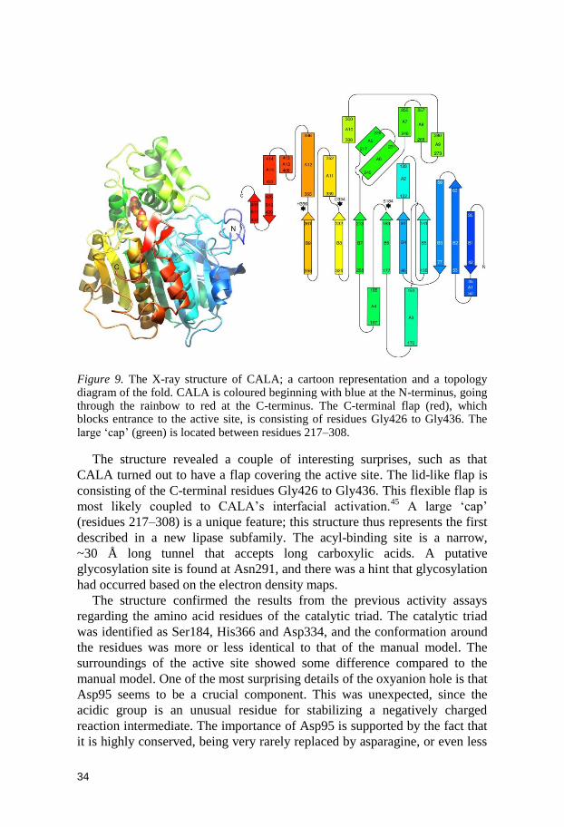
Figure 9. The X-ray structure of CALA; a cartoon representation and a topology diagram of the fold. CALA is coloured beginning with blue at the N-terminus, going through the rainbow to red at the C-terminus. The C-terminal flap (red), which blocks entrance to the active site, is consisting of residues Gly426 to Gly436. The large „cap‟ (green) is located between residues 217–308.
The structure revealed a couple of interesting surprises, such as that
CALA turned out to have a flap covering the active site. The lid-like flap is
consisting of the C-terminal residues Gly426 to Gly436. This flexible flap is
most likely coupled to CALA‟s interfacial activation.45
A large „cap‟
(residues 217–308) is a unique feature; this structure thus represents the first
described in a new lipase subfamily. The acyl-binding site is a narrow,
~30 Å long tunnel that accepts long carboxylic acids. A putative
glycosylation site is found at Asn291, and there was a hint that glycosylation
had occurred based on the electron density maps.
The structure confirmed the results from the previous activity assays
regarding the amino acid residues of the catalytic triad. The catalytic triad
was identified as Ser184, His366 and Asp334, and the conformation around
the residues was more or less identical to that of the manual model. The
surroundings of the active site showed some difference compared to the
manual model. One of the most surprising details of the oxyanion hole is that
Asp95 seems to be a crucial component. This was unexpected, since the
acidic group is an unusual residue for stabilizing a negatively charged
reaction intermediate. The importance of Asp95 is supported by the fact that
it is highly conserved, being very rarely replaced by asparagine, or even less

35
frequently, glutamine, in some distant relatives. A computational prediction
of pKa-values for ionisable protein residues, using PROPKA 2.0, suggested
that Asp95 has a high pKa of 7.9.110
This increases the plausibility that this
acidic residue can stabilize the oxyanion. The future will resolve whether
this claim is correct. An extensive review has described the different families
of oxyanion holes, and it is difficult to fit CALA into these defined
families.111
The uniqueness of the CALA-homologous sequences has resulted in the
designation of six homologous families and one new superfamily (called the
“Candida antarctica lipase A like” superfamily) in the Lipase Engineering
Database (LED).112
The X-ray structure is deposited at the RCSB Protein
Data Bank under the PDB ID: 2VEO.35
The manually constructed model and the X-ray structure are different in
some aspects. The manually constructed model was bound to have some
minor flaws, and one of these flaws originated from a misinterpretation of
the results from the study of CALA‟s interfacial activation.45
These data
were interpreted as there was no authentic interfacial activation in CALA
and the model was therefore not equipped with an active-site flap. The
modeled protein structure did also display an atypical Ramachandran plot.113
2.5 Conclusions
Protein structure determination is crucial for modern protein engineering, as
site-specific directed evolution techniques are becoming more powerful and
practical. The first part of this chapter demonstrates a novel knowledge-
based structure prediction approach. The latter part presents the resolved
structure of CALA; the first structure from an unexplored α/β-hydrolase
subfamily. The CALA fold will facilitate the generation of homology models
of potentially catalytically interesting enzymes.
Based on the X-ray structure of CALA it is concluded that the CALA
indeed have a C-terminal active-site flap, covering the active site. Molecular
modeling indicates that this flap is quite flexible, and that it is probably
responsible for the slight interfacial activation that has been observed.45
The
manual model proved correct in the assumptions regarding the active site
residues.
36
3. Directed Evolution of Candida antarctica Lipase A for Enhanced Enantioselectivity (Paper III and IV)
3.1 Introduction
The acquirement of the 3D-structure of CALA was a crucial key objective
for the planned structure-based directed evolution projects. One aim was to
achieve high enantioselectivity towards several chiral carboxylic acids. From
the start, the targeting of two interesting substrate families was intended. The
first target was the chiral allenic acids, and the second was the arylpropanoic
acids. The arylpropanoic acids are highly interesting as they form basis for
the „profen‟-group of pharmaceuticals. Early on, when the first draft of the
manually constructed CALA model was completed, the first attempt at
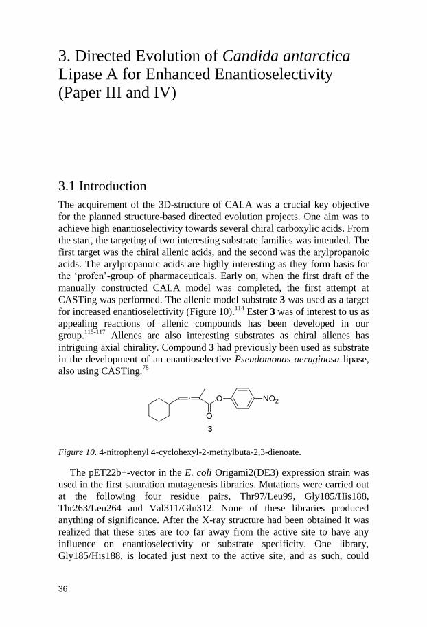
CASTing was performed. The allenic model substrate 3 was used as a target
for increased enantioselectivity (Figure 10).114
Ester 3 was of interest to us as
appealing reactions of allenic compounds has been developed in our
group.115-117
Allenes are also interesting substrates as chiral allenes has
intriguing axial chirality. Compound 3 had previously been used as substrate
in the development of an enantioselective Pseudomonas aeruginosa lipase,
also using CASTing.78
Figure 10. 4-nitrophenyl 4-cyclohexyl-2-methylbuta-2,3-dienoate.
The pET22b+-vector in the E. coli Origami2(DE3) expression strain was
used in the first saturation mutagenesis libraries. Mutations were carried out
at the following four residue pairs, Thr97/Leu99, Gly185/His188,
Thr263/Leu264 and Val311/Gln312. None of these libraries produced
anything of significance. After the X-ray structure had been obtained it was
realized that these sites are too far away from the active site to have any
influence on enantioselectivity or substrate specificity. One library,
Gly185/His188, is located just next to the active site, and as such, could
37
conceivably influence activity. However, this area is conserved due to
structural importance, for example backbone nitrogen of Gly185 is
responsible for oxyanion stabilization and therefore catalytic activity can be
severely reduced by even small perturbations.
After experimenting with temperature and IPTG concentration, and
bacterial host strains, some improvement could be achieved (as described in
chapter 2.2). The realization how detrimental the bacterial lysation procedure
was for activity, and the insufficient enzyme yields, forced us to look at
other options for enzyme expression.
A switch to a more efficient yeast expression system resulted in better
yields of protein and with higher purity. However, hydrolytic activity
towards the allenic substrate 3 was unsatisfactory. Even though the
expression levels were higher, the reaction was very slow, and appeared to
level off. Only marginal conversion of 3 occurred after several days, even
weeks of incubation. Strong product inhibition cannot be ruled out, and the
studies on 3 were abandoned for the time being.
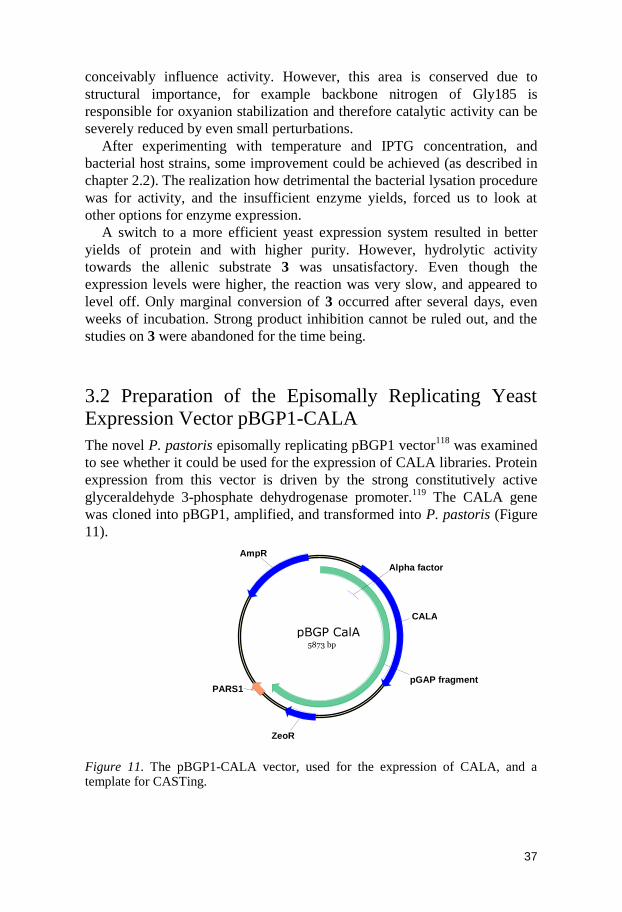
3.2 Preparation of the Episomally Replicating Yeast Expression Vector pBGP1-CALA
The novel P. pastoris episomally replicating pBGP1 vector118
was examined
to see whether it could be used for the expression of CALA libraries. Protein
expression from this vector is driven by the strong constitutively active
glyceraldehyde 3-phosphate dehydrogenase promoter.119
The CALA gene
was cloned into pBGP1, amplified, and transformed into P. pastoris (Figure
11).
Figure 11. The pBGP1-CALA vector, used for the expression of CALA, and a template for CASTing.
pBGP CalA 5873 bp
AmpR
ZeoR
CALA
pGAP fragment PARS1
Alpha factor
38
After examining expression levels of P. pastoris transformed with
pBGP1-CALA, it was concluded that the plasmid was highly useful for the
expression of CALA. In the supernatant, CALA reached a concentration of
approximately 100 mg L-1
.
3.3 Directed Evolution of CALA for Increased Enantioselective Towards 4-Nitrophenyl 2-Methylheptanoate
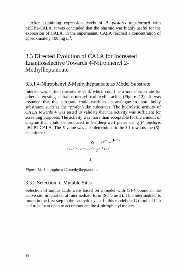
3.3.1 4-Nitrophenyl 2-Methylheptanoate as Model Substrate
Interest was shifted towards ester 4, which could be a model substrate for
other interesting chiral α-methyl carboxylic acids (Figure 12). It was
assumed that this substrate could work as an analogue to more bulky
substrates, such as the „profen‟-like substrates. The hydrolytic activity of
CALA towards 4 was tested to validate that the activity was sufficient for
screening purposes. The activity was more than acceptable for the amount of
enzyme that could be produced in 96 deep-well plates using P. pastoris
pBGP1-CALA. The E value was also determined to be 5.1 towards the (S)-
enantiomer.
Figure 12. 4-nitrophenyl 2-methylheptanoate.
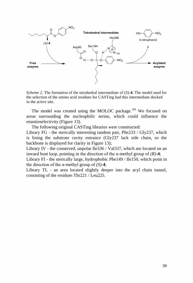
3.3.2 Selection of Mutable Sites
Selection of amino acids were based on a model with (S)-4 bound in the
active site in tetrahedral intermediate form (Scheme 2). This intermediate is
found in the first step in the catalytic cycle. In this model the C-terminal flap
had to be bent open to accommodate the 4-nitrophenyl moiety.
39
Scheme 2. The formation of the tetrahedral intermediate of (S)-4. The model used for the selection of the amino acid residues for CASTing had this intermediate docked in the active site.
The model was created using the MOLOC package.106
We focused on
areas surrounding the nucleophilic serine, which could influence the
enantioselectivity (Figure 13).
The following original CASTing libraries were constructed:
Library FG - the sterically interesting tandem pair, Phe233 / Gly237, which
is lining the substrate cavity entrance (Gly237 lack side chain, so the
backbone is displayed for clarity in Figure 13);
Library IV - the conserved, unpolar Ile336 / Val337, which are located on an
inward bent loop, pointing in the direction of the α-methyl group of (R)-4;
Library FI - the sterically large, hydrophobic Phe149 / Ile150, which point in
the direction of the α-methyl group of (S)-4;
Library TL - an area located slightly deeper into the acyl chain tunnel,
consisting of the residues Thr221 / Leu225.

40
Figure 13. The constructed CASTing libraries surrounding the active site. The active site with the catalytic residues His366 and Ser184 is displayed in each panel. The nucleophilic Ser184 is bound to the tetrahedral intermediate form of (S)-4.
3.3.3 Production of Libraries
The libraries were created by site-directed mutagenesis, using asymmetric
and degenerate primers. Asymmetric primers were used to lower the
potential primer duplex Tm (melting temperature), versus the primer-
template Tm. Libraries used in our CASTing had a reduced degeneracy, with
the NDT composition, to reduce the size of libraries.82
The PCR product was transformed into E. coli for amplification of the
library, to repair the nicked plasmid and to ensure that the plasmid yield was
sufficient. The total plasmid yield was of importance as the transformation
frequency of P. pastoris could be somewhat inadequate (approximately 500
cfu mg-1
). Diversity of the library had to be ensured by sequencing, as there
was a slight risk that a loss of diversity could occur in the first
transformation or in the PCR.
This amplified library plasmid preparation was transformed into
electrocompetent P. pastoris X33. The yeast libraries were grown for
approximately 96 h, and then the supernatant was harvested for screening of
the enzyme variants.
41
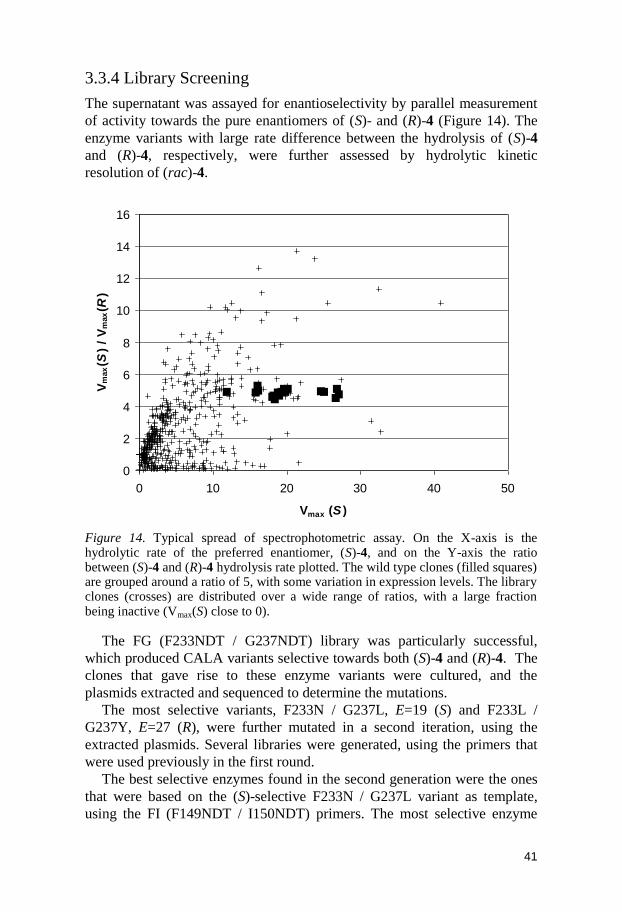
3.3.4 Library Screening
The supernatant was assayed for enantioselectivity by parallel measurement
of activity towards the pure enantiomers of (S)- and (R)-4 (Figure 14). The
enzyme variants with large rate difference between the hydrolysis of (S)-4
and (R)-4, respectively, were further assessed by hydrolytic kinetic
resolution of (rac)-4.
Figure 14. Typical spread of spectrophotometric assay. On the X-axis is the hydrolytic rate of the preferred enantiomer, (S)-4, and on the Y-axis the ratio between (S)-4 and (R)-4 hydrolysis rate plotted. The wild type clones (filled squares) are grouped around a ratio of 5, with some variation in expression levels. The library clones (crosses) are distributed over a wide range of ratios, with a large fraction being inactive (Vmax(S) close to 0).
The FG (F233NDT / G237NDT) library was particularly successful,
which produced CALA variants selective towards both (S)-4 and (R)-4. The
clones that gave rise to these enzyme variants were cultured, and the
plasmids extracted and sequenced to determine the mutations.
The most selective variants, F233N / G237L, E=19 (S) and F233L /
G237Y, E=27 (R), were further mutated in a second iteration, using the
extracted plasmids. Several libraries were generated, using the primers that
were used previously in the first round.
The best selective enzymes found in the second generation were the ones
that were based on the (S)-selective F233N / G237L variant as template,
using the FI (F149NDT / I150NDT) primers. The most selective enzyme
0
2
4
6
8
10
12
14
16
0 10 20 30 40 50
Vmax (S )
Vm
ax(S
) /
Vm
ax(R
)
42
found in the second round had the amino acid composition T64M / F149S /
I150D / F233N / G237L with an E value of 52 (S).
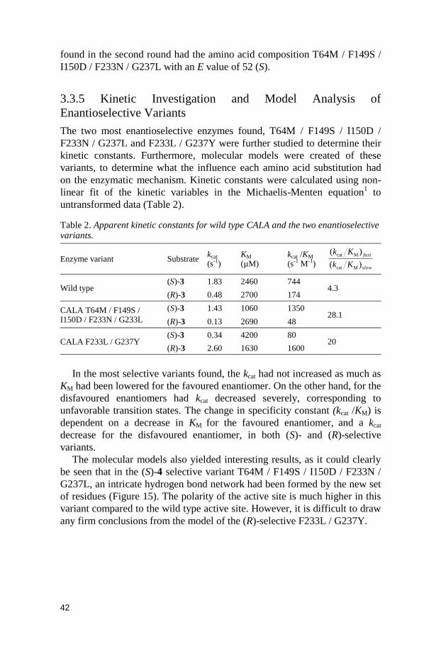
3.3.5 Kinetic Investigation and Model Analysis of
Enantioselective Variants
The two most enantioselective enzymes found, T64M / F149S / I150D /
F233N / G237L and F233L / G237Y were further studied to determine their
kinetic constants. Furthermore, molecular models were created of these
variants, to determine what the influence each amino acid substitution had
on the enzymatic mechanism. Kinetic constants were calculated using non-
linear fit of the kinetic variables in the Michaelis-Menten equation1 to
untransformed data (Table 2).
Table 2. Apparent kinetic constants for wild type CALA and the two enantioselective variants.
Enzyme variant Substrate kcat (s-1)
KM (µM)
kcat /KM (s-1 M-1)
slow
fast
Kk
Kk
)(
)(
Mcat
Mcat
Wild type (S)-3 1.83 2460 744
4.3 (R)-3 0.48 2700 174
CALA T64M / F149S / I150D / F233N / G233L
(S)-3 1.43 1060 1350 28.1
(R)-3 0.13 2690 48
CALA F233L / G237Y (S)-3 0.34 4200 80
20 (R)-3 2.60 1630 1600
In the most selective variants found, the kcat had not increased as much as
KM had been lowered for the favoured enantiomer. On the other hand, for the
disfavoured enantiomers had kcat decreased severely, corresponding to
unfavorable transition states. The change in specificity constant (kcat /KM) is
dependent on a decrease in KM for the favoured enantiomer, and a kcat
decrease for the disfavoured enantiomer, in both (S)- and (R)-selective
variants.
The molecular models also yielded interesting results, as it could clearly
be seen that in the (S)-4 selective variant T64M / F149S / I150D / F233N /
G237L, an intricate hydrogen bond network had been formed by the new set
of residues (Figure 15). The polarity of the active site is much higher in this
variant compared to the wild type active site. However, it is difficult to draw
any firm conclusions from the model of the (R)-selective F233L / G237Y.

43
Figure 15. Models displaying the active site of A) the (S)-4 selective T64M / F149S / I150D / F233N / G237L variant, and B) the (R)-4 selective F233L / G237Y variant. Note the intricate hydrogen bond network in the (S)-4 selective CALA, where Asn233, Asp150, and Ser149 connect.
3.4 Directed Evolution of CALA towards 4-Nitrophenyl 2-Phenylpropanoate
3.4.1 4-Nitrophenyl 2-Phenylpropanoate as Model Substrate
The main objective of the directed evolution project was to develop CALA
variants that displayed good enantioselectivity towards bulky „profen‟-like
substrates. The enantioselective variants that were derived using 4 as model
substrate did unfortunately not display activity nor enantioselectivity
towards the „profens‟. The evolved variants were most likely not able to
accommodate these bulky substrates, as they were adapted to the more
slender substrate 4. The libraries previously produced were therefore once
more screened, this time against the „profen‟-analogous substrate 4-
nitrophenyl 2-phenylpropanoate (5) (Figure 16).
Figure 16. 4-nitrophenyl 2-phenylpropanoate.
44
3.4.2 Library Screening
It was initially determined that wild type CALA displayed insufficient
activity towards ester 5 for screening and kinetic resolution, and
consequently it was decided that a directed evolution project should
commence with the prime objective to increase the activity of the enzyme.
The wild type exhibited some enantioselectivity (E = 20).
Library FG (Phe233NDT and Gly237NDT) had been previously observed
to have a large influence on the enantioselectivity towards ester 4. It seemed
reasonable that this library would also have an impact on the substrate
binding of the enzyme towards ester 5. Library FG was therefore used for a
spectrophotometric assay, using rac-5. The racemic substrate was used to
assay activity.
In library FG, several variants with improved activity were found. The
enantioselectivity of these variants were assayed by proper kinetic
resolution. A variant, with the single mutation Phe233Gly (designated
F233G), displayed strong enantioselectivity (E=259) and good activity
(Table 3). This variant displayed (R)-selectivity, which is contrasting to the
(S)-selective wild type.
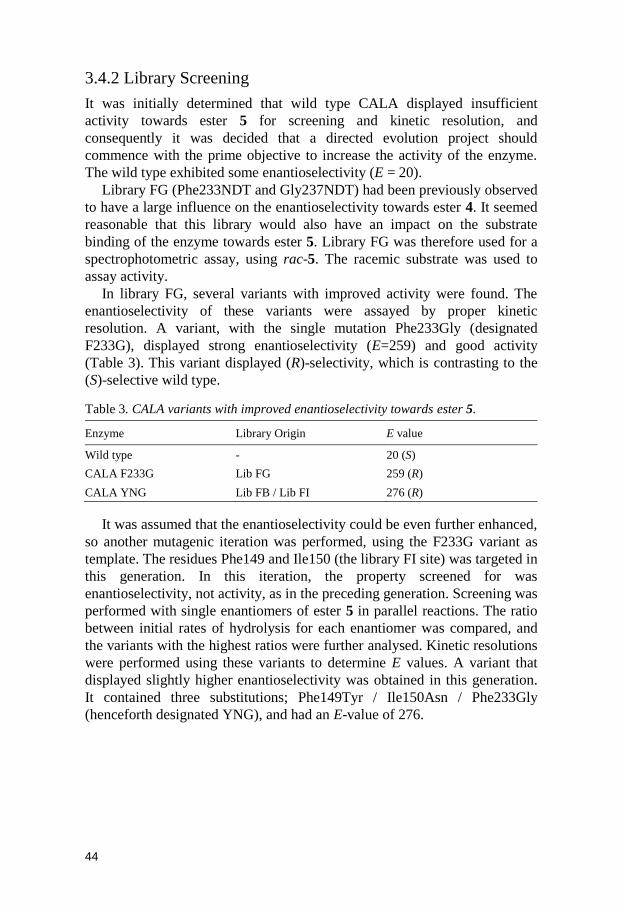
Table 3. CALA variants with improved enantioselectivity towards ester 5.
Enzyme Library Origin E value
Wild type - 20 (S)
CALA F233G Lib FG 259 (R)
CALA YNG Lib FB / Lib FI 276 (R)
It was assumed that the enantioselectivity could be even further enhanced,
so another mutagenic iteration was performed, using the F233G variant as
template. The residues Phe149 and Ile150 (the library FI site) was targeted in
this generation. In this iteration, the property screened for was
enantioselectivity, not activity, as in the preceding generation. Screening was
performed with single enantiomers of ester 5 in parallel reactions. The ratio
between initial rates of hydrolysis for each enantiomer was compared, and
the variants with the highest ratios were further analysed. Kinetic resolutions
were performed using these variants to determine E values. A variant that
displayed slightly higher enantioselectivity was obtained in this generation.
It contained three substitutions; Phe149Tyr / Ile150Asn / Phe233Gly
(henceforth designated YNG), and had an E-value of 276.

45
3.4.3 Substrate Scope
The notion that analogous substrates could be accommodated by these
enzyme variants had to be determined. The F233G and YNG variants were
therefore used for the kinetic resolution of several 4-nitrophenyl esters. Both
the YNG and F233G variants displayed high activity for an unexpectedly
broad substrate range. The YNG variant also displayed high to excellent E-
values for a broad range of substrates (Table 4). The F233G variant, on the
other hand, displayed more moderate E-values towards most substrates, and
was very poor towards the 2-benzylpropanoate 9 (Table 4, entry 14). The
F233G variant displayed slightly higher enantioselectivity towards ester 10
compared to YNG.
It is interesting to note that the enantioselective variants were (R)-
selective towards all these chiral esters, which is reversed compared to WT
for esters 5, 6, 9, 10 and 11.
The 4-methyl group on the phenyl (6) was accepted with an E value of 64
by the YNG variant (Table 4, entry 6). The ibuprofen ester, 4-nitrophenyl 2-
(4-isobutylphenyl)propanoate (11), was also accepted however with low
enantioselectivity and activity. YNG and F233G surprisingly tolerated large
substituent in the α-position, as can be seen in the activities towards 7 and 8.
It was interesting to note that the YNG has evolved higher enantioselectivity
towards (R)-4 (Table 4, entry 18) than the (R)-4 selective F233L / G237Y
variant (See chapter 3.3.4), even though that variant were obtained by
screening in particular towards 4. The WT E-value towards 4 was also more
reliable, compared to the values reported in chapter 3.3.1. The larger reaction
scale and improved work-up procedure increased reliability.

46
Table 4. Results from the kinetic resolution of different 4-nitrophenyl esters, using the wild type CALA (WT), the single mutant Phe233Gly (F233G) and triple mutant Phe149Tyr / Ile150Asn / Phe233 (YNG).
a
Entry Substrate Enzyme Timeb
(min)
Conversionb,c
(%) eep
b,d E
1
WT 150 38 84.7 20 (S)
2 F233G 3 25 98.9 259 (R)
3 YNG 3.5 31 98.9 276 (R)
4
WT 240 23 55.6 4 (S)
5 F233G 2.5 29 90.1 32 (R)
6 YNG 5 38 94.1 63 (R)
7
WT 24 11 17.0 2 (R)
8 F233G 0.5 20 95.4 57 (R)
9 YNG 1.7 17 97.0 79 (R)
10
WT 270 11 88.1 18 (R)
11 F233G 2.5 26 97.1 88 (R)
12 YNG 5.3 14 97.8 109 (R)
13
WT 240 7 80.3 10 (S)
14 F233G 5 7 44.7 3 (R)
15 YNG 15 27 96.7 84 (R)
16
WT 3.7 18 80.7 11 (S)
17 F233G 2.5 28 85.0 17 (R)
18 YNG 3.3 31 96.7 104 (R)
19
WT 60 14 75.3 19 (S)
20 F233G 3.3 9 96.0 54 (R)
21 YNG 3.5 6 95.4 45 (R)
a) Reaction conditions: 4-nitrophenylester (1.25 mL, 2 mg mL-1 in acetonitrile), enzyme solution (20 µL, 10 mg mL-1), potassium phosphate-buffer (8.5 mL, 100 mM, pH 8.0) b) Mean value of 2-4 reactions. c) Determined by 1H NMR. d) Determined by chiral GC.
3.4.3 Kinetic Resolution of 2-Phenylpropanoates with Different
Alcohol Moieties
CALA is known to be very slow reacting towards simple alkyl esters.120
Therefore, it was of interest to test the obtained variants towards esters that
did compose of less reactive alcohol moieties than 4-nitrophenol. The
comparatively high reactivity of 4-nitrophenyl esters are correlated to the
low pKa of 7.08 for 4-nitrophenol.102
Three analogues of ester 5, containing
ethyl (12), nonyl (13) and phenyl (14) moieties were used as substrates in the
kinetic resolution with YNG as catalyst. The result shows that enantio-
selectivity was maintained for all three esters (Table 5), and for the nonyl
and phenyl ester the E values were even higher. As expected, the hydrolysis
of the ethyl (pKa = 15.9) and nonyl esters was slower than that of the phenyl

47
(pKa = 9.55) ester but much faster than the corresponding hydrolysis of alkyl
esters by wild type CALA.120
Table 5. Kinetic resolution of esters with different alcohol side chains.a
Entry R Time
(h)
Conv.b
(%)
eepc
(%) E pKa
d
1 Et 3 14 98.9 >200 (211) 15.9
2 Nonyl 3 21 99.6 >200 (650) ~15.9
3 Ph 0.5 22 99.6 >200 (657) 9.55
a) Reaction conditions: Ester (1.25 mL, 2 mg mL-1 in acetonitrile), enzyme solution (100 µL (entries 1 and 2) or 20 µL (entry 3), 10 mg mL-1), potassium phosphate-buffer (8.5 mL, 100 mM, pH 8.0). b) Determined by 1H NMR. c) Determined by chiral GC. d) pKa of alcohol, from ref.102
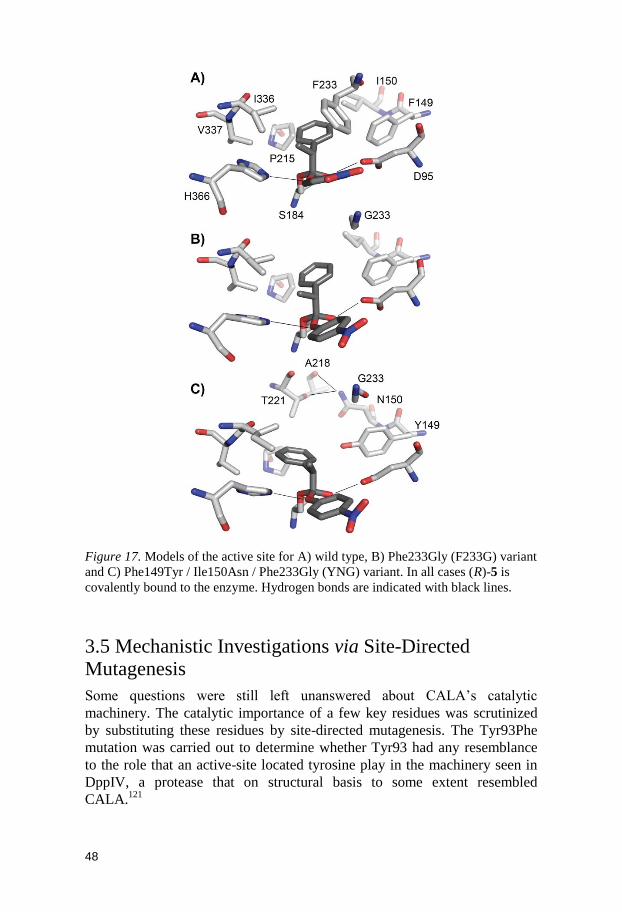
3.4.4 Models of Enantioselective Enzyme Variants
Active site models of the enzyme variants F233G and YNG were created
using docking and molecular dynamics, and were compared with a model of
the wild type enzyme (Figure 17). By examining the models, it is clear that
substitution of the large phenylalanine (Phe233) with a small glycine creates
much more space in the active site. The increased space in the active site can
be used to accommodate the substrates, and this would explain the observed
increase in activity. The results indicate that it is only (R)-enantiomer that
can benefit from the space created, which rationalizes the large effect on the
enantioselectivity. The increased enantioselectivity for the triple mutant
(YNG) could be explained by the increase in steric bulk introduced; the
addition of hydroxyl group to the Phe149 side chain (Phe149Tyr) creates
sterical clashes at the the α-methyl group, as well as an unfavorable polar
environment, which disfavors the (S) configuration. In addition, two new
hydrogen bonds are found in the YNG variant.
48
Figure 17. Models of the active site for A) wild type, B) Phe233Gly (F233G) variant
and C) Phe149Tyr / Ile150Asn / Phe233Gly (YNG) variant. In all cases (R)-5 is
covalently bound to the enzyme. Hydrogen bonds are indicated with black lines.
3.5 Mechanistic Investigations via Site-Directed Mutagenesis
Some questions were still left unanswered about CALA‟s catalytic
machinery. The catalytic importance of a few key residues was scrutinized
by substituting these residues by site-directed mutagenesis. The Tyr93Phe
mutation was carried out to determine whether Tyr93 had any resemblance
to the role that an active-site located tyrosine play in the machinery seen in
DppIV, a protease that on structural basis to some extent resembled
CALA.121

49
Hydrolytic activity was not diminished in the Tyr93Phe variant, thus it
could be concluded that Tyr93 did not participate in the catalytic machinery
(Figure 18).
Figure 18. Normalized initial hydrolytic rates of selected enzyme variants, using 1 as substrate.
Asp95 was also substituted by asparagine and valine to determine
whether this residue has the assumed importance as an oxyanion stabilizer.
The results show that the catalytic rate was severely reduced by these
substitutions. Asp95 is a highly interesting residue, as it is extraordinary to
find acidic residues as a component of the oxyanion hole. Further studies
should be carried out on CALA and related enzymes to bring more light on
this subject.
3.5 Conclusions
Variants of CALA, selective towards several types of α-substituted
carboxylic acids, were produced in the described directed evolution projects.
These variants also displayed medium to high increase in activity compared
to the wild type. The CASTing approach, combined with an episomally
replicating yeast plasmid, created a profitable synergy which could be highly
recommended for other directed evolution projects targeted at eukaryotic
enzymes. In chapter 3.3 the acquirement of CALA variants, selective
towards (R)- and (S)-4, is described. Kinetic constants were determined for
these variants.
0
100
WT Tyr93Phe Asp95Asn Asp95Val
No
rmalized
acti
vit
y

50
The obtainment of a variant with broad substrate scope towards α-
substituted esters is described in chapter 3.4. The initial library was only
screened for activity, and a racemic substrate was used. In this library the
F233G variant was discovered. This variant surprisingly displayed high
enantioselectivity, and at the same time displayed strong activity. In
contrast, the wild type enzyme had a very low activity towards the targeted
substrate. We conclude that in the process of increasing the activity of a non-
reactive enzyme, there is concurrently a high chance of developing strong
enantioselectivity, especially towards bulky substrates. The novel topology
of the active site of the developed enzyme has a higher chance of having
gained accommodation for just one of the enantiomers, than for both.
Removing the potential sterical clashes of a single enantiomer is more
probable. Therefore, if high enantioselectivity is desired, racemic substrates
can be used in the initial part of the screening process, if the initial enzyme
template has low activity. The use of racemic substrates in the screening
process would help speed up directed evolution projects aiming for enhanced
enantioselectivity, as pure enantiomers does not need to be prepared.

51
4. Combinatorial Reshaping of the Substrate Pocket (Paper V)
4.1 Introduction
Ibuprofen is presently one of the most consumed chiral NSAIDs, and as such
a highly interesting substrate.92
The (S)-enantiomer of ibuprofen shows the
strongest anti-inflammatory effect. Ibuprofen is marketed as a racemate, in
contrast to naproxen, which is sold as the pure (S)-enantiomer.
As mentioned in the previous chapter, low activity and enantioselectivity
was encountered when the YNG and Phe233Gly variants were used towards
an ester containing an ibuprofen moiety. It was assumed that we could obtain
an active variant towards ibuprofen with directed evolution using YNG as
template. This is a reasonable approach, as directed evolution has been used
for stepwise enzyme adaptation towards a slightly different substrate in each
evolutionary step, so called substrate walking.122-123
Several sites were
targeted and subjected to mutagenesis, using the YNG as well as the parent
F233G as templates. Using the ibuprofen ester 11 as model substrate (Figure
19), several libraries were revisited and assayed, yet none increased activity.
Thr221 / Leu225, that have interaction with the potential isobutyl group, and
Phe230 / Phe233, that should give rise to increased sterical relaxation, did
not yield any fruitful results. Also, a library based on Phe149 / Ile150 gave
the same negative results. Even the best variants isolated from these libraries
had minor improvement of activity, and unsatisfying enantioselectivity.
Using ISM, highly enantioselective variants of Pseudomonas aeruginosa
lipase has been derived towards similar substrates.124
This problem was faced as a challenging opportunity to develop a
different experimental protein engineering approach for obtaining higher
activity and enantioselectivity. It was considered that a more drastic protein
engineering approach would conceivably bring about changes that would
lead to accommodation of this sterically demanding substrate. It has
previously been shown that much reduced sets of amino acids can be used in
directed evolution of enzymes for obtaining enantioselectivity,125
and that
strong synergistic effects can be seen when combining many residue
substitutions.80
Simultaneous variation of 8-14 sites with binary variation of
amino acid residue has successfully been used for obtaining diverse GFP
proteins89
and thermostable proteins84,87
but it has not yet been tested for
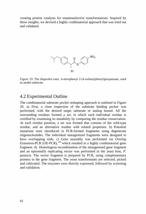
52
creating protein catalysts for enantioselective transformations. Inspired by
these insights, we devised a highly combinatorial approach that was tried out
and validated.
Figure 19. The ibuprofen ester, 4-nitrophenyl 2-(4-isobutylphenyl)propanoate, used as model substrate.
4.2 Experimental Outline
The combinatorial substrate pocket reshaping approach is outlined in Figure
20. a) First, a close inspection of the substrate binding pocket was
performed, with the desired target substrate or analog bound. All the
surrounding residues formed a set, in which each individual residue is
certified by examining its mutability by comparing the residue conservation.
At each residue position, a set was formed that consists of the wild-type
residue, and an alternative residue with related properties. b) Potential
mutations were introduced in PCR-formed fragments using degenerate
oligonucleotides. The individual mutagenized fragments were designed to
have overlapping ends. c) Gene assembly was performed via Overlap
Extension-PCR (OE-PCR),126
which resulted in a highly combinatorial gene
fragment. d) Homologous recombination of the mutagenized gene fragment
and an episomally replicating vector was performed in the yeast host, P.
pastoris. The vector fragment is prepared by PCR, using complementary
primers to the gene fragment. The yeast transformants are selected, picked
and cultivated. The enzymes were directly expressed, followed by screening
and validation.
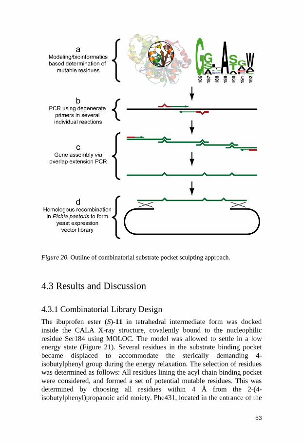
53
Figure 20. Outline of combinatorial substrate pocket sculpting approach.
4.3 Results and Discussion
4.3.1 Combinatorial Library Design
The ibuprofen ester (S)-11 in tetrahedral intermediate form was docked
inside the CALA X-ray structure, covalently bound to the nucleophilic
residue Ser184 using MOLOC. The model was allowed to settle in a low
energy state (Figure 21). Several residues in the substrate binding pocket
became displaced to accommodate the sterically demanding 4-
isobutylphenyl group during the energy relaxation. The selection of residues
was determined as follows: All residues lining the acyl chain binding pocket
were considered, and formed a set of potential mutable residues. This was
determined by choosing all residues within 4 Å from the 2-(4-
isobutylphenyl)propanoic acid moiety. Phe431, located in the entrance of the
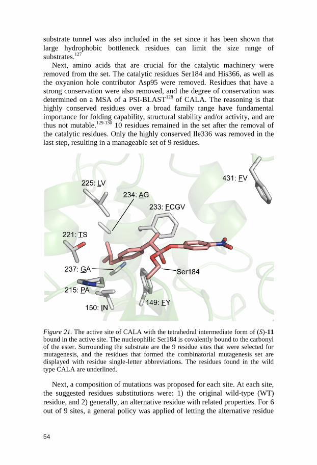
54
substrate tunnel was also included in the set since it has been shown that
large hydrophobic bottleneck residues can limit the size range of
substrates.127
Next, amino acids that are crucial for the catalytic machinery were
removed from the set. The catalytic residues Ser184 and His366, as well as
the oxyanion hole contributor Asp95 were removed. Residues that have a
strong conservation were also removed, and the degree of conservation was
determined on a MSA of a PSI-BLAST128
of CALA. The reasoning is that
highly conserved residues over a broad family range have fundamental
importance for folding capability, structural stability and/or activity, and are
thus not mutable.129-130
10 residues remained in the set after the removal of
the catalytic residues. Only the highly conserved Ile336 was removed in the
last step, resulting in a manageable set of 9 residues.
Figure 21. The active site of CALA with the tetrahedral intermediate form of (S)-11 bound in the active site. The nucleophilic Ser184 is covalently bound to the carbonyl of the ester. Surrounding the substrate are the 9 residue sites that were selected for mutagenesis, and the residues that formed the combinatorial mutagenesis set are displayed with residue single-letter abbreviations. The residues found in the wild type CALA are underlined.
Next, a composition of mutations was proposed for each site. At each site,
the suggested residues substitutions were: 1) the original wild-type (WT)
residue, and 2) generally, an alternative residue with related properties. For 6
out of 9 sites, a general policy was applied of letting the alternative residue
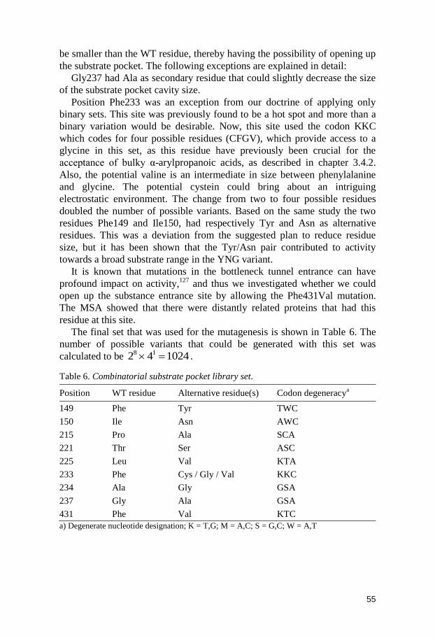
55
be smaller than the WT residue, thereby having the possibility of opening up
the substrate pocket. The following exceptions are explained in detail:
Gly237 had Ala as secondary residue that could slightly decrease the size
of the substrate pocket cavity size.
Position Phe233 was an exception from our doctrine of applying only
binary sets. This site was previously found to be a hot spot and more than a
binary variation would be desirable. Now, this site used the codon KKC
which codes for four possible residues (CFGV), which provide access to a
glycine in this set, as this residue have previously been crucial for the
acceptance of bulky α-arylpropanoic acids, as described in chapter 3.4.2.
Also, the potential valine is an intermediate in size between phenylalanine
and glycine. The potential cystein could bring about an intriguing
electrostatic environment. The change from two to four possible residues
doubled the number of possible variants. Based on the same study the two
residues Phe149 and Ile150, had respectively Tyr and Asn as alternative
residues. This was a deviation from the suggested plan to reduce residue
size, but it has been shown that the Tyr/Asn pair contributed to activity
towards a broad substrate range in the YNG variant.
It is known that mutations in the bottleneck tunnel entrance can have
profound impact on activity,127
and thus we investigated whether we could
open up the substance entrance site by allowing the Phe431Val mutation.
The MSA showed that there were distantly related proteins that had this
residue at this site.
The final set that was used for the mutagenesis is shown in Table 6. The
number of possible variants that could be generated with this set was
calculated to be 102442 18 .
Table 6. Combinatorial substrate pocket library set.
Position WT residue Alternative residue(s) Codon degeneracya
149 Phe Tyr TWC
150 Ile Asn AWC
215 Pro Ala SCA
221 Thr Ser ASC
225 Leu Val KTA
233 Phe Cys / Gly / Val KKC
234 Ala Gly GSA
237 Gly Ala GSA
431 Phe Val KTC
a) Degenerate nucleotide designation; K = T,G; M = A,C; S = G,C; W = A,T
56
4.3.2 Mutagenesis and Homologous Recombination
Some of the mutations could be grouped on one oligonucleotide pair, due to
sequence proximity. The 9 mutations could be clustered into 4 groups, and
with flanking sequences resulted in five fragments. The five fragments were
simultaneously assembled in a modified OE-PCR.131-132
The flanking
sequences of the final assembled gene did not include any mutations, as
these had to be mutation-free for the later homologous recombination
(Figure 20). PCR was used for forming the vector fragment, using
oligonucleotides that were complementary to the flanking sequences of the
gene fragment. The gene and vector fragments were mixed and
electroporated with P. pastoris cells. The yeasts‟ internal homologous
recombination system133
was used for the generation of the secretory
episomally replicating vector.118
The yeast was spread on agar plates, and
colonies were allowed to grow. Several transformants were sequenced to
validate that the diversity of the library was adequate. It was determined that
there was no observed bias towards wild-type residues. Colonies were
picked and grown in individual wells, and the enzyme variants were secreted
in the supernatant.
4.3.3 Functional Diversity of the Library
There is a strong demand that the efficacy of a protein engineering approach
can be validated, as it is very important for potential experimenters to
determine if the approach has a general applicability.76
A small subset of the
library was compared to different 4-nitrophenyl esters that placed dissimilar
sterical demands on the enzyme variants, to explore how the functional
diversity of the library appeared (Figure 22). Racemic esters 4, 5 and 11
were used as screening substrates for the spectrophotometric activity assay.
Several interesting aspects could be observed. 1) The library had large
functional diversity, i.e. both numerous more active and less active variants
than the wild type were found. This is an important criterion for a library of
good quality. 2) There seemed to be some correlation between activities
towards different substrates. 3) There were some enzyme variants that
deviated from this correlation. There were several enzyme variants that
showed distinguished activity towards one of the esters. One of these
variants was a „SVCAV‟ variant (vide infra), which displayed unique strong
activity towards 11. At the same time, the enzyme variants that displayed the
strongest activity towards 4 and 5, displayed no activity towards 11. This
implies that it would have been difficult to reach a high activity of 11 by
using 4 and 5 as model substrates. 4) Only a small minority of the library
had increased activity towards the ibuprofen ester 11. This should in no way
be held against the library design procedure, but rather as an evidence of the

57
small minority of enzyme variants with sufficiently large substrate pockets
to accept the bulky 11.
Figure 22. Comparison of activity for a small subset (89 colonies) of the combinatorial library towards different 4-nitrophenyl esters. The colonies are ordered after increasing activity towards substrate 5. The highest activity for each substrate in this subset was normalized to 100. P. pastoris X33 supernatants were used as blank. The highest activity towards 11 found in this subset is an „SVCAV‟ variant.
4.3.4 Library Screening towards Ibuprofen Ester
The enzyme variants were screened for increased hydrolytic activity, using
the ester (rac)-11. 2400 transformants were assayed, which corresponds to a
sequence coverage of 90%.134
Of all the screened transformants, only a small
group displayed exceptional activity towards 11. These transformants were
sequenced, and they were composed of only two unique enzyme variants.
One variant, designated „SVCAV‟ had incorporated the 5 following
alternative residues; Thr221Ser / Leu225Val / Phe233Cys / Gly237Ala /
Phe431Val. The other variant, designated „SVCV‟ had the same composition
of substitutions, except that it lacked the Gly237Ala substitution. Wild type
CALA and these two variants were purified using HIC. Kinetic resolution
using rac-11 as substrate was performed with the purified variants. The
specific activity was also calculated, based on a spectrophotometric assay
using the purified enzymes. The SVCAV variant displayed good
enantioselectivity and specific activity (Table 7). The SVCV variant
displayed more moderate activity and acceptable enantioselectivity.
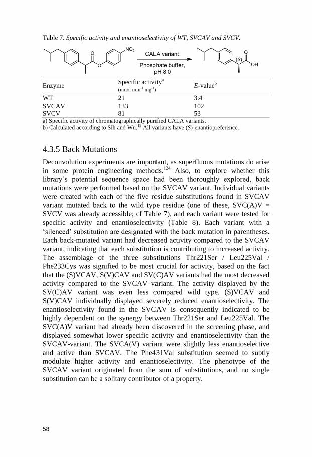
58
Table 7. Specific activity and enantioselectivity of WT, SVCAV and SVCV.
Enzyme Specific activity
a
(nmol min-1 mg-1) E-value
b
WT 21 3.4
SVCAV 133 102 SVCV 81 53 a) Specific activity of chromatographically purified CALA variants. b) Calculated according to Sih and Wu.19 All variants have (S)-enantiopreference.
4.3.5 Back Mutations
Deconvolution experiments are important, as superfluous mutations do arise
in some protein engineering methods.124
Also, to explore whether this
library‟s potential sequence space had been thoroughly explored, back
mutations were performed based on the SVCAV variant. Individual variants
were created with each of the five residue substitutions found in SVCAV
variant mutated back to the wild type residue (one of these, SVC(A)V =
SVCV was already accessible; cf Table 7), and each variant were tested for
specific activity and enantioselectivity (Table 8). Each variant with a
„silenced‟ substitution are designated with the back mutation in parentheses.
Each back-mutated variant had decreased activity compared to the SVCAV
variant, indicating that each substitution is contributing to increased activity.
The assemblage of the three substitutions Thr221Ser / Leu225Val /
Phe233Cys was signified to be most crucial for activity, based on the fact
that the (S)VCAV, S(V)CAV and SV(C)AV variants had the most decreased
activity compared to the SVCAV variant. The activity displayed by the
SV(C)AV variant was even less compared wild type. (S)VCAV and
S(V)CAV individually displayed severely reduced enantioselectivity. The
enantioselectivity found in the SVCAV is consequently indicated to be
highly dependent on the synergy between Thr221Ser and Leu225Val. The
SVC(A)V variant had already been discovered in the screening phase, and
displayed somewhat lower specific activity and enantioselectivity than the
SVCAV-variant. The SVCA(V) variant were slightly less enantioselective
and active than SVCAV. The Phe431Val substitution seemed to subtly
modulate higher activity and enantioselectivity. The phenotype of the
SVCAV variant originated from the sum of substitutions, and no single
substitution can be a solitary contributor of a property.
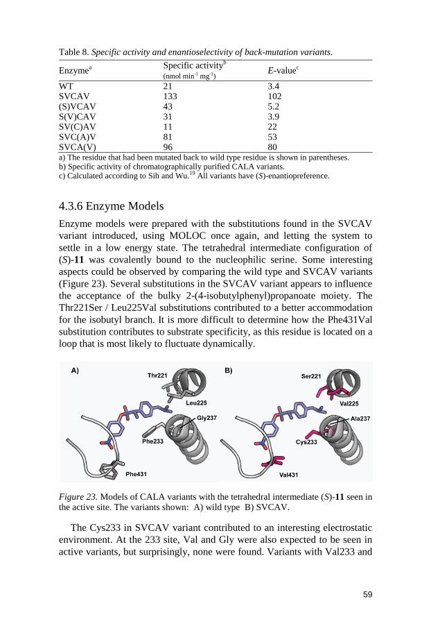
59
Table 8. Specific activity and enantioselectivity of back-mutation variants.
Enzymea
Specific activityb
(nmol min-1 mg-1) E-value
c
WT 21 3.4 SVCAV 133 102 (S)VCAV 43 5.2 S(V)CAV 31 3.9 SV(C)AV 11 22 SVC(A)V 81 53 SVCA(V) 96 80 a) The residue that had been mutated back to wild type residue is shown in parentheses. b) Specific activity of chromatographically purified CALA variants. c) Calculated according to Sih and Wu.19 All variants have (S)-enantiopreference.
4.3.6 Enzyme Models
Enzyme models were prepared with the substitutions found in the SVCAV
variant introduced, using MOLOC once again, and letting the system to
settle in a low energy state. The tetrahedral intermediate configuration of
(S)-11 was covalently bound to the nucleophilic serine. Some interesting
aspects could be observed by comparing the wild type and SVCAV variants
(Figure 23). Several substitutions in the SVCAV variant appears to influence
the acceptance of the bulky 2-(4-isobutylphenyl)propanoate moiety. The
Thr221Ser / Leu225Val substitutions contributed to a better accommodation
for the isobutyl branch. It is more difficult to determine how the Phe431Val
substitution contributes to substrate specificity, as this residue is located on a
loop that is most likely to fluctuate dynamically.
Figure 23. Models of CALA variants with the tetrahedral intermediate (S)-11 seen in the active site. The variants shown: A) wild type B) SVCAV.
The Cys233 in SVCAV variant contributed to an interesting electrostatic
environment. At the 233 site, Val and Gly were also expected to be seen in
active variants, but surprisingly, none were found. Variants with Val233 and

60
Gly233 were observed when validating the sequence diversity, so apparently
there was no bias against these two residues.
4.3.7 Protein Fitness Landscapes
A protein fitness landscape is a conceptual multidimensional sequence space
landscape with topology based on „fitness‟, a variable based on the protein
phenotype.135-136
The fitness is often, for enzymes, defined as the enzyme‟s
thermal stability, activity or enantioselectivity. Directed evolution is a highly
useful method to explore and climb in protein fitness landscapes. One of the
most interesting effects observed when combining several mutations in
iterative saturation mutagenesis has been the high degree of epistatic
effects.80
Epistatic interactions occur when one mutation influences the
effect of another mutation.135
This implies that it is possible to bypass valleys
in the fitness landscape by utilizing strong synergistic epistatic effects in
some protein engineering methods.137
Some protein fitness landscapes have
hard to reach fitness peaks.135
Established protein engineering methods may
be ineffectual, if the desired function is located far away in the fitness
landscape. If the sequence space surrounding the starting point (Point A in
Figure 24) in the fitness landscape has a topology that is neutral137
, it is not
possible to make an upward fitness climb by traditional evolutionary
methods, such as epPCR. If that is the case, then it is imperative to develop
methods that can scan the fitness landscape effectively to discover
topologies with higher fitness.
The method described in this chapter was an attempt to improve the
exploration of a fitness landscape that has a mostly neutral topology (Figure
24). To be able to reach a far-away patch on the fitness landscape, radical
protein engineering had to be utilized, where the potential to obtain
synergistic epistasis can be utilized for maximum efficiency. One such
example was the resculpting of an enzyme active site so it can accept a much
larger non-natural substrate, requiring the introduction of several mutations
in the substrate pocket. This was hard to reach by single site saturation
mutagenesis, and were difficult even using a proven strong method such as
ISM (as described in chapter 4.1). Combinatorial mutagenesis is the
mutagenesis of several sites simultaneously. This represents not only
exploring the multi-dimensionality of sequence space, but also probing many
potential epistatic effects. Highly combinatorial methods is in effect pole-
vaulting in the fitness landscape, which enables traversing fitness barriers,
and also probing distant patches in the landscape (Figure 24).
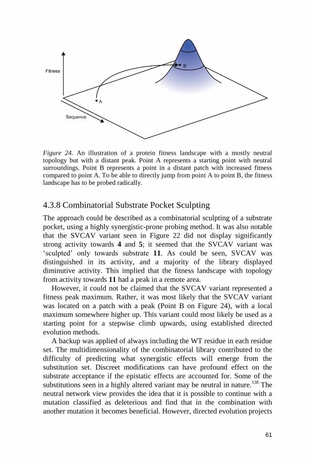
61
Figure 24. An illustration of a protein fitness landscape with a mostly neutral topology but with a distant peak. Point A represents a starting point with neutral surroundings. Point B represents a point in a distant patch with increased fitness compared to point A. To be able to directly jump from point A to point B, the fitness landscape has to be probed radically.
4.3.8 Combinatorial Substrate Pocket Sculpting
The approach could be described as a combinatorial sculpting of a substrate
pocket, using a highly synergistic-prone probing method. It was also notable
that the SVCAV variant seen in Figure 22 did not display significantly
strong activity towards 4 and 5; it seemed that the SVCAV variant was
„sculpted‟ only towards substrate 11. As could be seen, SVCAV was
distinguished in its activity, and a majority of the library displayed
diminutive activity. This implied that the fitness landscape with topology
from activity towards 11 had a peak in a remote area.
However, it could not be claimed that the SVCAV variant represented a
fitness peak maximum. Rather, it was most likely that the SVCAV variant
was located on a patch with a peak (Point B on Figure 24), with a local
maximum somewhere higher up. This variant could most likely be used as a
starting point for a stepwise climb upwards, using established directed
evolution methods.
A backup was applied of always including the WT residue in each residue
set. The multidimensionality of the combinatorial library contributed to the
difficulty of predicting what synergistic effects will emerge from the
substitution set. Discreet modifications can have profound effect on the
substrate acceptance if the epistatic effects are accounted for. Some of the
substitutions seen in a highly altered variant may be neutral in nature.138
The
neutral network view provides the idea that it is possible to continue with a
mutation classified as deleterious and find that in the combination with
another mutation it becomes beneficial. However, directed evolution projects

62
are often designed to disregard variants that display lower or neutral fitness
than the starting variant. Simultaneous multiple site-directed mutagenesis
could thus utilize neutral networks to discover new pathways in normally
untraversable dimensions in sequence space.
4.3.9 Other Considerations
Highly combinatorial methods suffer from „the numbers‟ problem77
. If we
consider that all 9 sites were chosen to be targets, the enzyme complexity
generated could be enormous. If NDT degeneracy, which codes for 12 amino
acid residues, would have been used at each site then 129 = 5.2 × 10
10
variants would have been generated. Also, if NNK degeneracy would have
been applied, unmanageable 209 = 5.1 × 10
12 enzyme variants would have
formed. On the other hand, if we would have stayed to our proclaimed
doctrine of keeping only to „binary‟ sets, then manageable 29 = 512 variants
would form. The use of this extremely condensed residue set was not
without problem. The choice of the alternative residue requires some
reasoning, as if an unacceptable residue were chosen; half the library is
„wasted‟. The selection of residue sets required an X-ray structure or at least
a good homology model. Mutational suggestions derived from
bioinformatics have been highly successful for deriving enantioselective
enzyme variants.125,139
In our approach, the information derived from
bioinformatics is generally only used for the detection of non-allowed
residue sites, not to give mutational suggestions.
The only a priori knowledge that we had was that 149 / 150 pair could
exist as a synergistic Tyr / Asn pair, and that Phe233 could be susbstituted to
a glycine, for acceptance of larger substrates. Interestingly, none of these
presumptions were implemented in the final enzyme variant. This is
promising as it points to that single site saturation mutagenesis were not
needed to find the beneficial substitutions. The suggested substitutions
Phe149Tyr and Ile150Asn were deviations from our suggested residue size-
reducing approach, but there were no a priori empirical evidence to suggest
that it would be unwise to implement these mutations. For example, this pair
contributed to activity towards broad substrate range in the YNG variant
(Chapter 3.4.3).
The computational determination of suggested residues has gone great
strides. Methods such IPRO140
can be used in combinatorial scrambling
methods, such as the one described in this paper. It shows promise as a
computational method for sculpting the pocket of ligands.141
Computational
de novo design of proteins and enzymes is still in its infancy,142
but has
already produced encouraging results.143
For the moment being, modulating
existing enzyme functions and scaffolds will probably be the main current in
the protein engineering field.123
63
4.4 Conclusions
The highly combinatorial method described can be used to obtain enzymes
that are active towards normally unacceptable molecules, such as very bulky
substrates. The substrate pocket can in this way be radically reshaped to
accommodate the substrate. There is higher possibility of obtaining positive
synergistic effects as multiple sites are altered simultaneously, yet at the
same time reducing possible variants to a minimum. The claim that there is a
high chance of obtaining enantioselective enzyme variants when unreactive
racemic substrates are used in the screening, (cf chapter 3.5) is once more
indicated.
This highly combinatorial protein engineering approach is useful for the
development of highly focused libraries, as well as reaching hard-to-reach
regions on the fitness landscape by its efficient probing capability. This
combinatorial substrate pocket sculpting approach should be validated
towards different enzyme classes, to determine whether it has general
applicability as a protein engineering tool. We believe that the demonstrated
method has a place in the protein engineering toolbox in the future.
64
5. Concluding Remarks
The main subject of this thesis has been the semi-rational design of custom-
made enzymes variants. The entire process is demonstrated, starting with a
lipase with unknown structure to the successful implementation of protein
engineering tools, ending up with highly adapted enzyme variants. The main
scientific goal has been the exploration of Candida antarctica lipase A‟s
structural mutability and biocatalytic potential for kinetic resolution of α–
methyl carboxylic acid esters. Also, this enzyme has served as an excellent
template for assessing different protein engineering approaches. Both the
now established directed evolution method ISM as well as our own
proprietary „combinatorial substrate pocket sculpting‟ approach was used.
Several highly enantioselective and active variants were successfully
obtained using these methods.

65
Acknowledgments
I would like to express my appreciation to:
My Supervisor, Prof. Jan-Erling Bäckvall for accepting me as a Ph.D.
candidate in his group, for his excellent guidance, enthusiasm and love of
science
I would like to thank Karin Engström and Dr. Lynn Kamerlin for
suggestions on the improvement of this thesis
I would like to acknowledge my great co-workers and collaborators;
Dr. Alex Kasrayan, for sharing the tools of the trade
Karin Engström, for everything she‟s done in our joint projects, and for
whom I feel immense gratitude
Jonas Nyhlén, for being a never-ending source of great ideas
Ylva Wikmark, for being creative, always working hard and never giving up
when we were facing difficulties
Dr. Mozaffar Shakeri, for being bold and highly interdisciplinary
Dr. Gaston Lavèn, for interesting discussions and wild ideas
Dr. Marco Bocola
Dr. Sarah Whitehead
Prof. Sherry Mowbray, her group, and Daniel Ericsson for their tenacity, and
for sharing passion and knowledge of crystallography and structural biology
Dr. Charles C. Lee
Former diploma students and project workers Michael Lindh, Kristin
Jacobsen, Roger Chang, Marie Gullstrand, Maria Cammenberg and Alessio
Ferrone. I wish you all best in the future!
The organizers and members of the Research School in Gene Expression,
Stockholm University
All my foreign friends and collaborators, for who I hope for a continuation
of joint projects, and wish the best of luck
66
The people at the KTH Royal Institute of Technology. Special thanks to
Prof. Karl Hult for advice
All the current and former members of the awesome JEB-group. You are
great friends, and thank you all for all those happy moments!
The always helpful TA-staff at the department
The unbreakable Gym Crew
All the nice people at the Department of Organic Chemistry
The AstraZeneca R&D, Local Discovery CNS&Pain Control, Södertälje
All of my awesome friends from Borås! Special greetings to the Grenzlos
Crew! 105%!
All my amazing friends from Stockholm! Tack speciellt till Shawon, Gustav,
Gustaf, Micke, Richard, Roger, Stefan och Thomas, för allt kul!
My mates from the Molecular Biology program and courses
The Sitzenbleiben Crew! Loska på världen!
The nice guys and gals involved in the Swedish transhumanist movement
All my friends that find politics exciting, important and a worthy pastime
All my other friends whom I haven‟t mentioned! You know who you are!
Alla mina släktingar för stöd och uppmuntran
Min familj: Mamma, Pappa, Evelina, Andreas, Max och Eva. Tack för all er
värme!
Min älskade Ellen, tack för allt du gjort för mig. Du har gjort mig till en
bättre man!
67
References
(1) Nelson, D. L.; Cox, M. M. Lehninger principles of biochemistry; 3rd ed.; Worth Publishers: New York, 2000.
(2) Koshland, D. E. Proc. Natl. Acad. Sci. U. S. A. 1958, 44, 98-104. (3) Khersonsky, O.; Tawfik, D. S. Annu. Rev. Biochem. 2010, 79, 471-505. (4) Kamerlin, S. C. L.; Sharma, P. K.; Chu, Z. T.; Warshel, A. Proc. Natl. Acad.
Sci. U. S. A. 2010, 107, 4075-4080. (5) Warshel, A. Proc. Natl. Acad. Sci. U. S. A. 1978, 75, 5250-5254. (6) Kamerlin, S. C. L.; Mavri, J.; Warshel, A. FEBS Lett. 2010, 584, 2759-2766. (7) Benkovic, S. J.; Hammes-Schiffer, S. Science 2003, 301, 1196-1202. (8) Kamerlin, S. C. L.; Warshel, A. Proteins: Struct., Funct., Bioinf. 2010, 78,
1339-1375. (9) Radzicka, A.; Wolfenden, R. Science 1995, 267, 90-93. (10) Cavalieri, D.; McGovern, P. E.; Hartl, D. L.; Mortimer, R.; Polsinelli, M. J.
Mol. Evol. 2003, 57, 226-232. (11) Iding, H.; Siegert, P.; Mesch, K.; Pohl, M. Biochim. Biophys. Acta, Protein
Struct. Mol. Enzymol. 1998, 1385, 307-322. (12) Faber, K. Biotransformations in Organic Chemistry; 4th ed.; Springer-
Verlag: Berlin, 2000. (13) Prather, K. L. J.; Martin, C. H. Curr. Opin. Biotechnol. 2008, 19, 468-474. (14) Lopez-Gallego, F.; Schmidt-Dannert, C. Curr. Opin. Chem. Biol. 2010, 14,
174-183. (15) Pasteur, L. Ann. Chim. Phys. 1848, 24, 442–459. (16) Podlech, J. Angew. Chem., Int. Ed. 1999, 38, 477-478. (17) Martín-Matute, B.; Bäckvall, J.-E. Curr. Opin. Chem. Biol. 2007, 11, 226-
232. (18) Janes, L. E.; Kazlauskas, R. J. Tetrahedron: Asymmetry 1997, 8, 3719-3733. (19) Sih, C. J.; Wu, S. H. Top. Stereochem. 1989, 19, 63-125. (20) Faber, K. Pure Appl. Chem. 1997, 69, 1613-1632. (21) Verger, R. Trends Biotechnol. 1997, 15, 32-38. (22) van Rantwijk, F.; Hacking, M.; Sheldon, R. A. Monatsh. Chem. 2000, 131,
549-569. (23) Klibanov, M. Acc. Chem. Res. 1990, 23, 114-120. (24) Pamies, O.; Bäckvall, J.-E. Trends Biotechnol. 2004, 22, 130-135. (25) Pellissier, H. Tetrahedron 2008, 64, 1563-1601. (26) Kim, M. J.; Ahn, Y.; Park, J. Curr. Opin. Biotechnol. 2002, 13, 578-587. (27) Pamies, O.; Bäckvall, J.-E. Chem. Rev. 2003, 103, 3247-3262. (28) Thalén, L. K.; Zhao, D. B.; Sortais, J. B.; Paetzold, J.; Hoben, C.; Bäckvall,
J.-E. Chem. Eur. J. 2009, 15, 3403-3410. (29) Leijondahl, K.; Borén, L.; Braun, R.; Bäckvall, J.-E. J. Org. Chem. 2009, 74,
1988-1993. (30) Träff, A.; Bogar, K.; Warner, M.; Bäckvall, J.-E. Org. Lett. 2008, 10, 4807-
4810.
68
(31) Gotor-Fernandez, V.; Brieva, R.; Gotor, V. J. Mol. Catal. B: Enzym. 2006, 40, 111-120.
(32) Schmid, R. D.; Verger, R. Angew. Chem., Int. Ed. 1998, 37, 1608. (33) Warshel, A.; Naray-Szabo, G.; Sussman, F.; Hwang, J. K. Biochemistry
1989, 28, 3629-3637. (34) Kamerlin, S. C. L.; Chu, Z. T.; Warshel, A. J. Org. Chem. 2010, 75, 6391-
6401. (35) Ericsson, D. J.; Kasrayan, A.; Johansson, P.; Bergfors, T.; Sandström, A. G.;
Bäckvall, J.-E.; Mowbray, S. L. J. Mol. Biol. 2008, 376, 109-119. (36) Goto, S.; Sugiyama, J.; Iizuka, H. Mycologia 1969, 61, 748-774. (37) Dominguez de Maria, P.; Carboni-Oerlemans, C.; Tuin, B.; Bargeman, G.;
van der Meer, A.; van Gemert, R. J. Mol. Catal. B: Enzym. 2005, 37, 36-46. (38) Høegh, I.; Patkar, S.; Halkier, T.; Hansen, M. T. Can. J. Bot. 1995, 73, 869-
875. (39) Wei, Y. H.; Lee, F. L.; Hsu, W. H.; Chen, S. R.; Chen, C. C.; Wen, C. Y.;
Lin, S. J.; Chu, W. S.; Yuan, G. F.; Liou, G. Y. Bot. Bull. Acad. Sin. 2005, 46, 223-229.
(40) Kakugawa, K.; Shobayashi, M.; Suzuki, O.; Miyakawa, T. Biosci. Biotechnol. Biochem. 2002, 66, 1328-1336.
(41) Kamper, J.; Kahmann, R.; Bolker, M. et. al. Nature 2006, 444, 97-101. (42) DSMZ - Deutsche Sammlung von Mikroorganismen und Zellkulturen
GmbH. (43) Liu, D.; Schmid, R. D.; Rusnak, M. Appl. Microbiol. Biotechnol. 2006, 72,
1024-1032. (44) Patkar, S. A.; Björking, F.; Zundel, M.; Schulein, M.; Svendsen, A.;
Heldthansen, H. P.; Gormsen, E. Indian J. Chem. 1993, 32, 76-80. (45) Martinelle, M.; Holmquist, M.; Hult, K. Biochim. Biophys. Acta 1995, 1258,
272-276. (46) Kirk, O., Christensen, M.W. Org. Process Res. Dev. 2002, 6, 446-451. (47) Rogalska, E.; Cudrey, C.; Ferrato, F.; Verger, R. Chirality 1993, 5, 24-30. (48) Li, X.-G.; Kanerva, L. T. Org. Lett. 2006, 8, 5593-5596. (49) Liljeblad, A.; Kanerva, L. T. Tetrahedron 2006, 62, 5831-5854. (50) Solymar, M.; Fülop, F.; Kanerva, L. T. Tetrahedron: Asymmetry 2002, 13,
2383-2388. (51) Barbayianni, E.; Fotakopoulou, I.; Höhne, M.; Constantinou-Kokotou, V.;
Bornscheuer, U. T.; Kokotos, G. J. Org. Chem. 2005, 70, 3737-3740. (52) Kourist, R.; Dominguez de Maria, P.; Bornscheuer, U. T. ChemBioChem
2008, 9, 491-498. (53) Svendsen, A.; Clausen, I. G.; Borch, K.; Egel-Mitani, M.; Patkar, S. A.;
Hansen, M. T. C. antarctica lipase and lipase variants. WO9401541. Novo Nordisk A/S., 1994.
(54) Svendsen, A.; Clausen, I. G.; Borch, K.; Egel-Mitani, M.; Patkar, S. A.; Hansen, M. T. C. antarctica lipase and lipase variants. U.S. Patent 6074863. Novo Nordisk A/S., 2000.
(55) Crick, F. Nature 1970, 227, 561-563. (56) Enzyme Functionality; Svendsen, A., Ed.; Marcel Dekker, Inc.: New York,
2004. (57) Darwin, C. On the Origin of Species by Means of Natural Selection, or the
Preservation of Favoured Races in the Struggle for Life; 1st ed.; John Murray: London, 1859.
(58) Haldane, J. B. S. Trans. Camb. Phil. Soc. 1924, 23, 19-41. (59) Alberts, B. Molecular Biology of the Cell; Garland Science: New York, 2002.
69
(60) Farinas, E. T.; Bulter, T.; Arnold, F. H. Curr. Opin. Biotechnol. 2001, 12, 545-551.
(61) Tobin, M. B.; Gustafsson, C.; Huisman, G. W. Curr. Opin. Struct. Biol. 2000, 10, 421-427.
(62) Directed molecular evolution of proteins: Or how to improve enzymes for biocatalysis; Brackmann, S.; Johnsson, K., Eds.; Wiley-VCH, 2002.
(63) Tao, H.; Cornish, V. W. Curr. Opin. Chem. Biol. 2002, 6, 858-864. (64) Lutz, S.; Patrick, W. M. Curr. Opin. Biotechnol. 2004, 15, 291-297. (65) Directed evolution library creation: Methods and protocols. Methods in
molecular biology; Arnold, F. H.; Georgiou, G., Eds.; Humana Press: Totowa, 2003; Vol. 231.
(66) Bloom, J. D.; Meyer, M. M.; Meinhold, P.; Otey, C. R.; MacMillan, D.; Arnold, F. H. Curr. Opin. Struct. Biol. 2005, 15, 447-452.
(67) Mullis, K.; Faloona, F.; Scharf, S.; Saiki, R.; Horn, G.; Erlich, H. Cold Spring Harbor Symp. Quant. Biol. 1986, 51, 263-273.
(68) Horsman, G. P.; Liu, A. M. F.; Henke, E.; Bornscheuer, U. T.; Kazlauskas, R. J. Chem. Eur. J. 2003, 9, 1933-1939.
(69) Morley, K. L.; Kazlauskas, R. J. Trends Biotechnol. 2005, 23, 231-237. (70) Stemmer, W. P. C. Proc. Natl. Acad. Sci. U. S. A. 1994, 91, 10747-10751. (71) Carbone, M. N.; Arnold, F. H. Curr. Opin. Struct. Biol. 2007, 17, 454-459. (72) Hiraga, K.; Arnold, F. H. J. Mol. Biol. 2003, 330, 287-296. (73) Zhao, H. M.; Arnold, F. H. Protein Eng. 1999, 12, 47-53. (74) Hutchison, C. A.; Phillips, S.; Edgell, M. H.; Gillam, S.; Jahnke, P.; Smith,
M. J. Biol. Chem. 1978, 253, 6551-6560. (75) Cunningham, B. C.; Wells, J. A. Science 1989, 244, 1081-1085. (76) Kazlauskas, R. J.; Bornscheuer, U. T. Nat. Chem. Biol. 2009, 5, 526-529. (77) Reetz, M. T.; Kahakeaw, D.; Lohmer, R. ChemBioChem 2008, 9, 1797-1804. (78) Carballeira, J. D.; Krumlinde, P.; Bocola, M.; Vogel, A.; Reetz, M. T.;
Bäckvall, J.-E. Chem. Commun. 2007, 1913-1915. (79) Reetz, M. T.; Bocola, M.; Carballeira, J. D.; Zha, D.; Vogel, A. Angew.
Chem., Int. Ed. 2005, 44, 4192-4196. (80) Reetz, M. T.; Sanchis, J. ChemBioChem 2008, 9, 2260-2267. (81) Reetz, M. T.; Carballeira, J. D. Nat. Protoc. 2007, 2, 891-903. (82) Clouthier, C. M.; Kayser, M. M.; Reetz, M. T. J. Org. Chem. 2006, 71, 8431-
8437. (83) Kato, R.; Nakano, H.; Konishi, H.; Kato, K.; Koga, Y.; Yamane, T.;
Kobayashi, T.; Honda, H. J. Mol. Biol. 2005, 351, 683-692. (84) Amin, N.; Liu, A. D.; Ramer, S.; Aehle, W.; Meijer, D.; Metin, M.; Wong,
S.; Gualfetti, P.; Schellenberger, V. Protein Eng. Des. Sel. 2004, 17, 787-793. (85) Poole, A. M.; Ranganathan, R. Curr. Opin. Struct. Biol. 2006, 16, 508-513. (86) Jäckel, C.; Bloom, J. D.; Kast, P.; Arnold, F. H.; Hilvert, D. J. Mol. Biol.
2010, 399, 541-546. (87) Hamamatsu, N.; Aita, T.; Nomiya, Y.; Uchiyama, H.; Nakajima, M.; Husimi,
Y.; Shibanaka, Y. Protein Eng. Des. Sel. 2005, 18, 265-271. (88) Kourist, R.; Jochens, H.; Bartsch, S.; Kuipers, R.; Padhi, S. K.; Gall, M.;
Böttcher, D.; Joosten, H. J.; Bornscheuer, U. T. ChemBioChem 2010, 11, 1635-1643.
(89) Treynor, T. P.; Vizcarra, C. L.; Nedelcu, D.; Mayo, S. L. Proc. Natl. Acad. Sci. U. S. A. 2007, 104, 48-53.
(90) Lippow, S. M.; Tidor, B. Curr. Opin. Biotechnol. 2007, 18, 305-311. (91) Fox, R. J.; Davis, S. C.; Mundorff, E. C. et. al. Nat. Biotechnol. 2007, 25,
338-344.
70
(92) Rao, P. N. P.; Knaus, E. E. J. Pharm. Pharm. Sci. 2008, 11, 81-110. (93) Kasrayan, A.; Bocola, M.; Sandström, A. G.; Lavén, G.; Bäckvall, J.-E.
ChemBioChem 2007, 8, 1409-1415. (94) Nardini, M.; Dijkstra, B. W. Curr. Opin. Struct. Biol. 1999, 9, 732-737. (95) Bessette, P. H.; Åslund, F.; Beckwith, J.; Georgiou, G. Proc. Natl. Acad. Sci.
U. S. A. 1999, 96, 13703-13708. (96) Dubendorff, J. W.; Studier, F. W. J. Mol. Biol. 1991, 219, 45-59. (97) Xia, B.; Etchegaray, J. P.; Inouye, M. J. Biol. Chem. 2001, 276, 35581-
35588. (98) Nishihara, K.; Kanemori, M.; Kitagawa, M.; Yanagi, H.; Yura, T. Appl.
Environ. Microbiol. 1998, 64, 1694-1699. (99) Daly, R.; Hearn, M. T. J. Mol. Recognit. 2005, 18, 119-138. (100) Cereghino, G. P.; Cereghino, J. L.; Ilgen, C.; Cregg, J. M. Curr. Opin.
Biotechnol. 2002, 13, 329-332. (101) Ellis, S. B.; Brust, P. F.; Koutz, P. J.; Waters, A. F.; Harpold, M. M.;
Gingeras, T. R. Mol. Cell. Biol. 1985, 5, 1111-1121. (102) Chemistry; 5th ed.; Silberberg, M. S., Ed.; McGraw-Hill: New York, 2009. (103) Wong, H.; Schotz, M. C. J. Lipid Res. 2002, 43, 993-999. (104) Fujii, R.; Utsunomiya, Y.; Hiratake, J.; Sogabe, A.; Sakata, K. Biochim.
Biophys. Acta, Mol. Cell Biol. Lipids 2003, 1631, 197-205. (105) Guex, N.; Peitsch, M. C. Electrophoresis 1997, 18, 2714-2723. (106) Gerber, P. R. J. Comput-Aided. Mol. Des. 1998, 12, 37-51. (107) Lienqueo, M. E.; Mahn, A.; Salgado, J. C.; Asenjo, J. A. J. Chromatogr., B:
Anal. Technol. Biomed. Life Sci. 2007, 849, 53-68. (108) Larsson, A. M.; Ståhlberg, J.; Jones, T. A. Acta Crystallogr., Sect. D: Biol.
Crystallogr. 2002, 58, 346-348. (109) Hendrickson, W. A.; Horton, J. R.; Lemaster, D. M. EMBO J. 1990, 9, 1665-
1672. (110) Li, H.; Robertson, A. D.; Jensen, J. H. Proteins: Struct., Funct., Bioinf. 2005,
61, 704-721. (111) Pleiss, J.; Fischer, M.; Peiker, M.; Thiele, C.; Schmid, R. D. J. Mol. Catal. B:
Enzym. 2000, 10, 491-508. (112) Widmann, M.; Juhl, P. B.; Pleiss, J. BMC Genomics 2010, 11, 123-123. (113) Hovmöller, S.; Zhou, T.; Ohlson, T. Acta Crystallogr., Sect. D: Biol.
Crystallogr. 2002, 58, 768-776. (114) Nyhlén, J.; Eriksson, L.; Bäckvall, J.-E. Chirality 2008, 20, 47-50. (115) Piera, J.; Persson, A.; Caldentey, X.; Bäckvall, J.-E. J. Am. Chem. Soc. 2007,
129, 14120-14121. (116) Piera, J.; Krumlinde, P.; Strübing, D.; Bäckvall, J.-E. Org. Lett. 2007, 9,
2235-2237. (117) Deska, J.; Ochoa, C. D.; Bäckvall, J.-E. Chem. Eur. J. 2010, 16, 4447-4451. (118) Lee, C. C.; Williams, T. G.; Wong, D. W.; Robertson, G. H. Plasmid 2005,
54, 80-85. (119) Waterham, H. R.; Digan, M. E.; Koutz, P. J.; Lair, S. V.; Cregg, J. M. Gene
1997, 186, 37-44. (120) Barbayianni, E.; Fotakopoulou, I.; Schmidt, M.; Constantinou-Kokotou, V.;
Bornscheuer, U. T.; Kokotos, G. J. Org. Chem. 2005, 70, 8730-8733. (121) Bjelke, J. R.; Christensen, J.; Branner, S.; Wagtmann, N.; Olsen, C.;
Kanstrup, A. B.; Rasmussen, H. B. J. Biol. Chem. 2004, 279, 34691-34697. (122) Chen, Z. L.; Zhao, H. M. J. Mol. Biol. 2005, 348, 1273-1282. (123) Savile, C. K.; Janey, J. M.; Mundorff, E. C. et. al. Science 2010, 329, 305-
309.
71
(124) Reetz, M. T.; Prasad, S.; Carballeira, J. D.; Gumulya, Y.; Bocola, M. J. Am. Chem. Soc. 2010, 132, 9144-9152.
(125) Reetz, M. T.; Wu, S. Chem. Commun. 2008, 5499-5501. (126) Horton, R. M.; Hunt, H. D.; Ho, S. N.; Pullen, J. K.; Pease, L. R. Gene 1989,
77, 61-68. (127) Schliessmann, A.; Hidalgo, A.; Berenguer, J.; Bornscheuer, U. T.
ChemBioChem 2009, 10, 2920-2923. (128) Altschul, S. F.; Madden, T. L.; Schaffer, A. A.; Zhang, J. H.; Zhang, Z.;
Miller, W.; Lipman, D. J. Nucleic Acids Res. 1997, 25, 3389-3402. (129) Mirny, L.; Shakhnovich, E. J. Mol. Biol. 2001, 308, 123-129. (130) Schueler-Furman, O.; Baker, D. Protein. Struct. Funct. Genet. 2003, 52, 225-
235. (131) An, Y. F.; Ji, J. F.; Wu, W. F.; Lv, A.; Huang, R. B.; Wei, Y. T. Appl.
Microbiol. Biotechnol. 2005, 68, 774-778. (132) Peng, R. H.; Xiong, A. S.; Yao, Q. H. Appl. Microbiol. Biotechnol. 2006, 73,
234-240. (133) Cregg, J. M.; Cereghino, J. L.; Shi, J.; Higgins, D. R. Mol. Biotechnol. 2000,
16, 23-52. (134) Bosley, A. D.; Ostermeier, M. Biomol. Eng. 2005, 22, 57-61. (135) Romero, P. A.; Arnold, F. H. Nat. Rev. Mol. Cell Biol. 2009, 10, 866-876. (136) Smith, J. M. Nature 1970, 225, 563-564. (137) Poelwijk, F. J.; Kiviet, D. J.; Weinreich, D. M.; Tans, S. J. Nature 2007, 445,
383-386. (138) Peisajovich, S. G.; Tawfik, D. S. Nat. Meth. 2007, 4, 991-994. (139) Jochens, H.; Bornscheuer, U. T. ChemBioChem 2010, 11, 1861-1866. (140) Saraf, M. C.; Moore, G. L.; Goodey, N. M.; Cao, V. Y.; Benkovic, S. J.;
Maranas, C. D. Biophys. J. 2006, 90, 4167-4180. (141) Fazelinia, H.; Cirino, P. C.; Maranas, C. D. Biophys. J. 2007, 92, 2120-2130. (142) Fischer, A.; Enkler, N.; Neudert, G.; Bocola, M.; Sterner, R.; Merkl, R. BMC
Bioinformatics 2009, 10, 16. (143) Röthlisberger, D.; Khersonsky, O.; Wollacott, A. M. et. al. Nature 2008, 453,
190-194.