protein phosphorylation and cellular regulation, i
TRANSCRIPT

72
PROTEIN PHOSPHORYLATION AND CELLULARREGULATION, I
Nobel Lecture, December 8, 1992
byEDWIN G. K R E B S
Departments of Pharmacology and Biochemistry, School of Medicine, SL-15,University of Washington, Seattle, WA 98195, USA.
INTRODUCTION
This presentation and the one to be given immediately thereafter will beconcerned with the reversible phosphorylation of proteins and the role ofthis process in biological regulation. In addition to discussing our own earlycontributions to this field, Ed Fischer and I will describe some of thedevelopments that have occurred subsequently. These developments haveled to the realization that protein phosphorylation constitutes a majormechanism by which cellular processes are controlled. My own remarks willreview the historical background that provided the setting in which ourjoint work was carried out in the 1950s and early 1960s. Then I’ll turn to adiscussion of this work itself, to be followed by comments on the cyclicAMP-dependent protein kinase. Finally I will talk about the intracellulartransmission of hormone and growth factor signals through protein kinasecascades.
BACKGROUND
By 1940, which is the year in which I entered medical school at WashingtonUniversity in St Louis, it was well established that the breakdown of glyco- gen in skeletal muscle and other types of cells occurs by the process ofphosphorolysis, catalyzed by the enzyme phosphorylase (for review, see ref.1). Carl Cori was Professor and Chairman of the Department of Pharmacol-ogy at Washington University, when I started my training there, but he wassoon to take over the Department of Biological Chemistry. I gradually cameto know him, at first distantly, the way medical students usually know theirprofessors, but later on somewhat better after I had become a teachingassistant in biochemistry. In this latter role I also became acquainted withArda Green, who, together with Carl and Gerty Cori, was purifying rabbitmuscle phosphorylase . It was not long until I began to hear about theunusual properties of this remarkable enzyme, which they had foun d exist-
Edwin G. Krebs 73
ed in skeletal muscle in two different forms that they designated a s phos-phorylase b and phosphorylase a (2,3). The a form was purified and ob-tained as a crystalline enzyme. Kinetically, it was shown that phosphorylas e brequired high concentrations of 5’-AMP for activity whereas phosphorylasea was active in the absence of this nucleotide. Since the concentration of5’-AMP required for the activity of phosphorylase b was considerably higher than that found in muscle, this form was considered to be physiologicallyinactive. Phosphorylase a was thought of as the physiologically active spe-cies. Evidence was obtained that the two forms are interconvertible withinthe cell, and it was postulated by the Coris that interconversion of the formsof phosphorylase constitutes a physiologically significant regulatory mecha-nism. Resting muscle was reported to contain phosphorylase predominantlyin the a form, whereas electrically stimulated muscle contained th e b form(4). As will be discussed later (see below) the reverse is actually true.Regardless of this latter point, however, the finding that an enzyme mightshuttle back and forth between two forms within the cell was a remarkableadvance, which set the stage for important later developments.
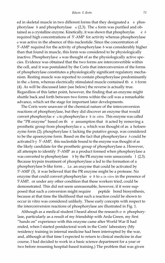
The Coris were unaware of the chemical nature of the interconversionreactions of phosphorylase, but they did discover an enzyme that wouldconvert phosphorylas e a to phosphorylas e b in vitro. This enzyme was calledthe “PR enzyme” based on th e assumption that it acted by removing aprosthetic group from phosphorylas e a, which was thought of as a holoen-zyme form (2); phosphorylase 6, lacking the putative group, was consideredto be the apoenzyme form. Based on the fact that phosphorylas e b could beactivated b y 5’-AMP, this nucleotide bound to the enzyme was thought of asthe likely candidate for the prosthetic group of phosphorylase a. However,all attempts to identify 5’-AMP as a product formed when phosphorylase awas converted to phosphorylase b by the PR enzyme were unsuccessfu l (2,3).Because trypsin treatment of phosphorylase a led to the formation of aphosphorylase b-like form , i.e. an enzyme that could be activated by5’-AMP (3), it was believed that the PR enzyme might be a protease. Noenzyme that could convert phosphorylas e b to a in vitro in the presence of5’AMP, or under any other condition that these workers tried, could bedemonstrated. This did not seem unreasonable, however, if it were sup-posed that such a conversion might require peptide bond biosynthesis,because at that time the likelihood that such a reaction could be shown tooccur in vitro was considered unlikely. These early concepts with respect tothe interconversion reactions of phosphorylase are illustrated in Fig. 1.
Although as a medical student I heard about the research o n phosphory-lase, particularly as a result of my friendship with Arda Green, my first“hands on” experience with this enzyme came after World War II hadended, when I started postdoctoral work in the Coris’ laboratory (Myresidency training in internal medicine had been interrupted by the war,and, although at that time I expected to return to clinical medicine in duecourse, I had decided to work in a basic science department for a year ortwo before resuming hospital-based training.) The problem that was given

74 Physiology or Medicine 1992
Phosphorylase( inactive)
+ A M P Active Enzyme
Phosphorylase( active ))
Phosphorylase(requires AMP
for activity)
A M P Prosthetic Group”Removing enzyme
+ AMP
Phosphorylase Phosphorylase
Fig. I. Early concepts of the interconversion reactions of muscle phosphorylase . Inactive phosphorylaseb becomes an active enzym e in the presence of 5’-AMP. Phosphorytase a, the physiologicallyactive form of the enzyme, is inactivated by the prosthetic group removing (PR) enzyme. It wasthought likely that 5’-AMP would be product of this reaction, but this could not be demonstrat-cd.
to me by the Coris involved solubility measurements on phosphorylase, andI also studied the effect of protamine and polylysine on the two forms of theenzyme. It was of interest that in the presence of these polyanionic sub-stances, inosinic acid, as well o n 5’-AMP, was very effective as an activator ofphosphorylase b. All of these studies on the effects of nucleotides onphosphorylase were carried out before the revelations of Jacob and Monodconcerning allosterism and protein conformation and at that time wereviewed simply as unexplained phenomenology. Indeed for several yearsCarl Cori advised against my publishing these findings unless I couldexplain them. Eventually, after Neil Madsen found that protamine couldphysically bind to phosphorylase, which was at least a step in the direction ofhow it might affect the activity of the enzyme, Cori let me send in my paper.
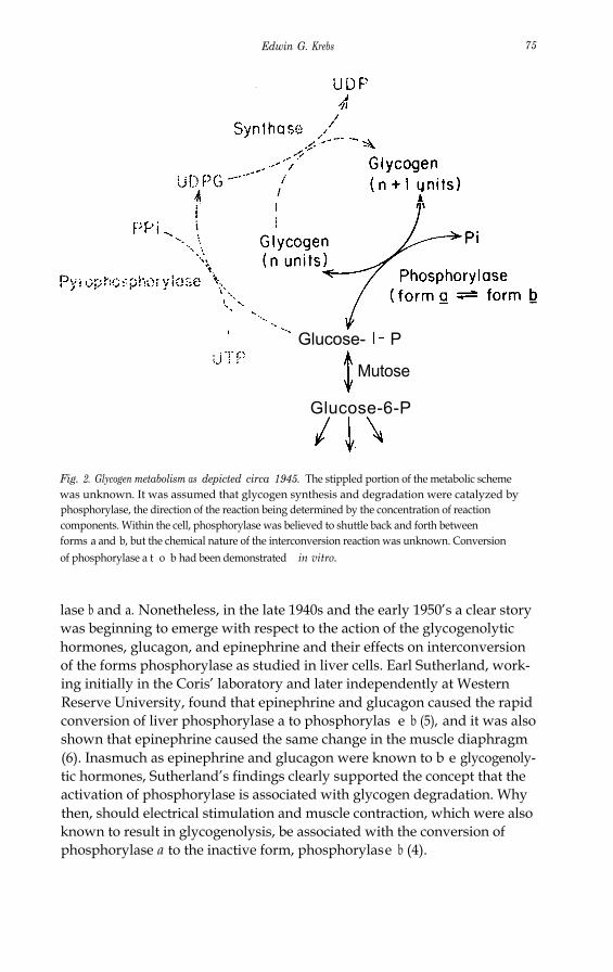
Although, as I have pointed out, knowledge with respect to the propertiesof phosphorylase as an enzyme increased significantly during th e 1940s, itwas often difficult to explain their physiological importance. This can beunderstood if one considers what was known about glycogen metabolism atthat time. The epochal work of Leloir on the mechanism of glycogensynthesis was still to come, i.e. the role of UDG in polysaccharide synthesisand the existence of the enzyme, glycogen synthase, were completely un-known. Hence, it was believed that phosphorylase was involved in glycogensynthesis as well as in glycogenolysis (Fig. 2). It was assumed that thedirection of the phosphorylase reaction would be determined by the con-centrations of the components of the reaction, particularly by the concen-tration of inorganic phosphate. This situation obviously made it difficult tointerpret findings relating to the interconversion reactions o f phosphory-
Edwin G. Krebs 75
Glucose- P
Mutose
Glucose-6-P
Fig. 2. Glycogen metabolism as depicted circa 1945. The stippled portion of the metabolic schemewas unknown. It was assumed that glycogen synthesis and degradation were catalyzed byphosphorylase, the direction of the reaction being determined by the concentration of reactioncomponents. Within the cell, phosphorylase was believed to shuttle back and forth betweenforms a and b, but the chemical nature of the interconversion reaction was unknown. Conversionof phosphorylase a t o b had been demonstrated in vitro.
lase b and a. Nonetheless, in the late 1940s and the early 1950’s a clear storywas beginning to emerge with respect to the action of the glycogenolytichormones, glucagon, and epinephrine and their effects on interconversionof the forms phosphorylase as studied in liver cells. Earl Sutherland, work-ing initially in the Coris’ laboratory and later independently at WesternReserve University, found that epinephrine and glucagon caused the rapidconversion of liver phosphorylase a to phosphorylas e b (5), and it was alsoshown that epinephrine caused the same change in the muscle diaphragm(6). Inasmuch as epinephrine and glucagon were known to b e glycogenoly-tic hormones, Sutherland’s findings clearly supported the concept that theactivation of phosphorylase is associated with glycogen degradation. Whythen, should electrical stimulation and muscle contraction, which were alsoknown to result in glycogenolysis, be associated with the conversion ofphosphorylase a to the inactive form, phosphorylase b (4).

76 Physiology or Medicine 1992
CONVERSION OF RABBIT SKELETAL MUSCLE PHOSPHORYLAS E bTO PHOSPHORYLASE a IN VITRO
In Fall 1953, Ed Fischer joined the Department of Biochemistry at theUniversity of Washington , where I was already a faculty member. As agraduate student in Geneva, Ed had worked on potato phosphorylase, andwe thus shared a common interest in this particular enzyme. We discussedsome of the puzzling features of phosphorylase and were particularly intri-gued by the still unsolved nature of th e 5’-AMP effect, i.e. how 5’AMPactivates phosphorylas e b but is seemingly unnecessary for phosphorylase a.It seemed worthwhile, purely as a secondary undertaking for each of us,since we both had problems that we considered as our major areas ofconcentration, to pool our efforts to see whether or not we could obtaininformation on this point. The initial experiment that we undertook togeth-er was simply to prepare pure crystalline phosphorylase a by the Coris’procedure (7). However , our initial attempts to do this were woefullyunsuccessful. The enzyme would not crystallize, a step that was essential inorder to obtain pure protein. More puzzling was the fact that the partiallypure enzyme, which we did obtain, was always in th e b form rather than thea form. Insofar as we could determine we had followed the Coris’ proce-dure exactly except that we had clarified the original muscle extract bycentrifugation rather than by filtration through paper. Although thisseemed like a trivial change, we nonetheless included the filtration step inour next preparation. To our surprise, this time we obtained phosphorylasea, which crystallized readily as it was supposed to do. Two conclusions couldbe drawn that would explain these results. First, “resting” rabbit muscleextracts must contain phosphorylase predominantly in th e b form ratherthan the a form as had been postulated by the Cori s (4,8), and second,filtration of muscle extracts through paper must trigger conversion ofphosphorylase b to a in vitro (9). Subsequently we found that thoroughwashing of the filter paper before use eliminated conversion of b to a,implying that some component in the paper must have been extracted toaccount for the effect. It was also found that this conversion failed to occurif the muscle extract were aged prior to filtration. The critical componentfrom the filter paper that was required in th e b to a step was shown to becalcium, and the essential component in the extract that was lost on agingwas found to be ATP.
Armed with the knowledge that there is an ATP requirement for conver-sion of phosphorylas e b to phosphorylase a, we thought it likely that we weredealing with a phosphotransferase reaction in which the termina l phos-phoryl group of ATP was being transferred either to the protein itself orpossibly to a nonprotein component bound to the enzyme. It soon becameapparent that a “converting enzyme” , which could be separated fromphosphorylase b, was required in the reaction (10). After it had beendetermined that ADP, as well as phosphorylase a were products of thereaction (1 l), we spoke of this enzyme as phosphorylase b kinase, or simply

Edwin G. Krebs 77
phosphorylase kinase, rather than converting enzyme (11). The nature ofthe Ca2+ requirement for the b to a reaction was not apparent immediately,but eventually it was determined by one of our graduate students, WilliamL. Meyer, that the effect of this metal was two-fold. On the one han d Ca2+
was needed for the activation of a kinase activating factor, (RAF), but inaddition it was also required for the activity of phosphorylase kinase itself(12). RAF was later shown to be a Ca2+-dependent protease by anotherstudent, R. Bruce Huston (13). (The current name for thi s protease iscalpain.) We determined the stoichiometry of the phosphoryla se b to phos-phorylase a reaction (11) and also obtained the amino acid sequence sur-rounding the phosphorylated serine in phosphorylase a (14). It was demon-strated that inorganic phosphate is released in the muscle phosphorylase ato b reaction catalyzed by the PR enzyme, which could now be termedphosphorylase phosphatase. Thus, by the lat e 1950’s, it was possible to writespecific equations for the interconversion reactions of muscl e phosphory-lase as follows:
phosphorylase b + 2ATP(dimer)
phosphorylase a + 2H2O(dimer)
phosphorylasekinase
phosphorylase a + 2ADP(dimer)
phosphorylasephosphatase
phosphorylase b + 2Pi(dimer)
During the same period that our work on the muscle phosphorylasesystem was being carried out, very similar studies relating to live r phosphor-ylase were being conducted independently in the laboratory of Earl Suther-land at Western Reserve University. Sutherland’s major coworkers in thiseffort were Walter D. Wosilait and Theodore W. Rall. In an early study (15)these investigators obtained evidence that inorganic phosphate was releasedwhen partially purified active liver phosphorylase was incubated with whatthey referred to as inactivating enzyme, and they also determined thatphosphate was incorporated into phosphorylase when liver slices wereincubated in the presence o f 32P-phosphate. Later they showed that both ofthe interconversion reactions of liver phosphorylase could be demonstratedin cell free system s (16,17). A monumental “ancillary finding” that grew outof the work on liver phosphorylase by the Sutherland laboratory was, ofcourse, the discovery of cyclic AMP, the first identified “second messenger”of hormone action (18).
It is of interest to consider whether there had been any indications thatthe phosphorylation and dephosphorylation of proteins might be of regula-tory significance prior to elucidation of the muscle and liver phosphorylase

78 Physioloa or Medicine 1992
systems. The existence of phosphoproteins had, of course, been known formany years before 1950. Phosphoproteins were classified as “conjugatedproteins” and for the most part consisted of proteins of nutritional signifi-cance associated with the feeding of the young, e.g . casein of milk andseveral phosphoproteins found in egg yolk. Interestingly, it was also appre-ciated that pepsinogen contained one mole of firmly bound phosphate permole of enzyme, but the significance of this wasn’t (and still isn’t) known. Itwas also known that nonspecific phosphatases could catalyze the release ofphosphate from phosphoproteins. In an important study that was carriedout at about the same time as the work on phosphorylase was underway,Burnett and Kennedy described an enzyme that catalyzed th e phosphoryla-tion of casein (19). These workers were aware of the high rate of turnover ofphosphate in proteins and were the first to describe a protein kinase. Ingeneral, however, a realization that protein phosphorylation- dephosphory-lation is a dynamic process affecting enzymes and important in the controlof metabolism had not bee n forseen.
PROTEIN PHOSPHORYLATION AND THE TRANSMISSIONOF EXTRACELLULAR SIGNALS
As we have seen, an interest in the mechanisms of action of epinephrine andglucagon had an important part in work on the interconversion reactions ofphosphorylase (5,6) which in turn led to discovery of the dynamic phos-phorylation and dephosphorylation of proteins. Going back even further,however, it can be noted that the original studies of the Coris, which led tothe finding of phosphorylase itself, grew out of the longstanding interest ofthese investigators on the role of epinephrine in the regulation of glycogenmetabolism (20). That an interest in hormone action would contributeimportantly to the development of the field of protein phosphorylation-dephosphorylation was no accident, since we now know that one of themajor functions of protein phosphorylation as a regulatory process is in thetransmission of signals that impinge on cells. This is true not only withrespect to the transduction of hormone and growth factor signals but forother types of stimuli as well. For example, as mentioned earlier, electricalstimulation of muscle leads to changes in protein phosphorylation withinthe cell. It is probably more than coincidental that the extent of proteinphosphorylation is much greater in eukaryotic cells than it is prokaryotes,particularly in eukaryotic cells of higher animals that are subject to complexforms of external regulation.
It was apparent very early that the relative amounts of phosphorylase band a that would be present within the cell at any particularly time woulddepend on the relative rates of the phosphorylase kinase and phosphatasereactions, and it was anticipated that one or both of these enzymes must besubject to regulation. Furthermore, two factors capable of influencing thebalance between the two forms of phosphorylase had been found. As noted,these were calcium ions, identified as the metal ion that caused phosphory-

Edwin G. Krebs 79
lase a formation in muscle extracts (9) and cyclic AMP, which promotedphosphorylase a formation in liver cells and homogenates (reviewed in ref.21). We subsequently showed that cyclic AMP would also cause phosphory-lase a formation in muscle extracts (22). In the muscle system it wasdetermined that the effects of Ca2+ and cyclic AMP in stimulating phos-phorylase activation in vitro were a result of phosphorylase kinase activationrather than phosphorylase phosphatase inhibition (11).
The coupling of muscle contraction to glycogenolysis: The finding that themajor form of phosphorylase present in resting muscle is phosphorylas e b(8) rather than phosphorylase a (4,7), together with a realization thatphosphorylase b can be converted to phosphorylas e a if Ca2+ is introducedinto muscle extracts containing AT P (9), made it possible to arrive at arational position with respect to the effect of electrical stimulation onphosphorylase. In 1956 (23) Cori was able to demonstrate that musclecontraction causes conversion of phosphorylas e b to a, rather than the otherway around; this was in keeping with the known effect of contraction onglycogen breakdown. The effect of Ca2+ on the activation of phosphorylasekinase now fits well into a scheme (Fig. 3) whereby this metal, acting as amessenger substance associated with muscle contraction, could be responsi-ble for the coupling of glycogenolysis (an energy-yielding process) to con-traction (an energy-utilizing process). Of the two different mechanisms bywhich Ca2+ could regulate phosphorylase kinase, i.e. through limited pro-teolysis involving KAF, or as a result of the requirement of phosphorylasekinase for Ca2+ per se (12), only the latter was considered to be physiologi-cally significant. The work of Ozaw a et al. (24), who quantified the effect ofCa2+ on the phosphorylase kinase reaction, was critical with respect to ourunderstanding of this process. Many years later the actual mechanism by
Nerve St imulat ion+
Releose of Ca2+ f romSarcoplasmic reticulum
PhosphorylaseKinase
Phosphorylase P hosphorylase
Glycogen Glucose- l - P
Fig. 3 The role of calcium ions in regulating glycogenolysis (circa 1962).

8 0 P h y si ol o g y or M e di ci n e 1 9 9 2
w hi c h C a 2 + sti m ul at e s p h o s p h o r yl a s e ki n a s e s b e c a m e a p p a r e nt w h e n it w a s
f o u n d t h at c al m o d uli n i s a s u b u nit of p h o s p h o r yl a s e ki n a s e ( 2 5).
T he me c h a nis m of a cti o n of c y cli c A M P a n d t he c y cli c A M P- de pe n de nt pr otei n
ki n ase : T h e m e c h a ni s m b y w hi c h c y cli c A M P c a u s e s a n i n c r e a s e i n p h o s-
p h o r yl a s e ki n a s e a cti vit y t u r n e d o ut t o b e m o r e c o m pl e x t h a n t h at o f C a 2 +.
W h e r e a s C a 2 + i s a n e s s e nti al a cti v ati n g c o m p o n e nt i n t h e p h o s p h o r yl a s e b t o
a r e a cti o n it s elf, c y cli c A M P c a u s e s a cti v ati o n of p h o s p h o r yl a s e ki n a s e b y
p r o m oti n g it s p h o s p h o r yl ati o n. T h e fi r st cl u e t h at p h o s p h o r yl ati o n mi g ht
b e i n v ol v e d i n t h e a cti v ati o n of t h e ki n a s e c a m e wit h t h e fi n di n g t h at A T P i s
r e q ui r e d i n o r d e r t o o b s e r v e t h e a cti v ati o n r e a cti o n a n d t h e c y cli c A M P
eff e ct i n r a b bit s k el et al m u s cl e e xt r a ct s ( 2 2). A k e y c o w o r k e r w h o c a r ri e d
o ut t h e i niti al e x p e ri m e nt t h at r e v e al e d t hi s r e q ui r e m e nt w a s a g r a d u at e
st u d e nt, D o n al d A. G r a v e s. A cti v ati o n of m u s cl e p h o s p h o r yl a s e ki n a s e w a s
c h a r a ct e ri z e d b y m a r k e d e n h a n c e m e nt of it s a cti vit y a s m e a s u r e d at p H 7 o r
b el o w, a n d o nl y m o d e r at e i n c r e a s e s i n a cti vit y w e r e s e e n w h e n t h e b t o a
r e a cti o n w a s c a r ri e d o ut at hi g h e r p H v al u e s. T h e r ati o of a cti vit y a t p H 6. 8
t o a cti vit y at p H 8. 2 w a s f o u n d t o s e r v e a s a u s ef ul i n d e x of t h e st at e of
p h o s p h o r yl a s e ki n a s e a cti v ati o n ( 2 6). Si g nifi c a ntl y, o n c e p h o s p h o r yl a s e
ki n a s e h a d b e e n a cti v at e d b y p r ei n c u b ati o n wit h A T P, t h e p r e s e n c e of c y cli c
A M P w a s n o l o n g e r e s s e nti al- a s mi g ht b e a nti ci p at e d if t h e ki n a s e w e r e
b ei n g m o difi e d c o v al e ntl y i n a n a cti v ati o n pr o c e s s sti m ul at e d b y c y cli c A M P
( 2 2).P h o s p h o r yl a s e ki n a s e w a s p u rifi e d e xt e n si v el y a n d o bt ai n e d a s a n e a rl y
h o m o g e n e o u s hi g h m ol e c ul a r w ei g ht p r ot ei n, ( M r = 1. 2 X 1 0 6 ), w hi c h still
r et ai n e d t h e a bilit y t o b e a cti v at e d b y p r ei n c u b ati o n wit h M g A T P i n a
r e a cti o n t h at w a s st r o n gl y sti m ul at e d b y c y cli c A M P. Alt h o u g h t h e r e w e r e
i n di c ati o n s t h at a s e c o n d p r ot ei n ki n a s e mi g ht b e i n v ol v e d i n t h e a cti v ati o n
p r o c e s s ( 2 7), t h e ki n eti c s of t h e r e a cti o n w e r e c o m pl e x a n d t h e w o r k of
D e L a n g e et al. ( 2 8) s u g g e st e d i n st e a d t h at a n a ut o c at al yti c r e a cti o n w a s
o c c u r ri n g. H o w e v e r, t h e e xi st e n c e of a s e p a r at e “ c y cli c A M P- d e p e n d e nt
p h o s p h o r yl a s e ki n a s e ki n a s e ” , w hi c h a c c o m p a ni e d p h o s p h o r yl a s e ki n a s e
ki n a s e d uri n g it s p urifi c ati o n, w a s e v e nt u all y e st a bli s h e d b y D o n al A. W al s h
a n d J o h n P. P e r ki n s, p o st d o ct o r al f ell o w s i n m y l a b o r at o r y ( 2 9). T h e y
s u c c e e d e d i n p u rif yi n g t h e n e w ki n a s e a b o ut 2 0 0-f ol d f r o m r a b bit m u s cl e
e xt r a ct a n d s e p a r at e d it c o m pl et el y f r o m p h o s p h o r yl a s e ki n a s e. T h e ki n a s e
w a s r ef e r r e d t o a s t h e “ c y cli c A M P- d e p e n d e nt p r ot ei n ki n a s e ” r at h e r t h a n
p h o s p h o r yl a s e ki n a s e ki n a s e, b e c a u s e it w a s f o u n d ( 2 9) t o h a v e a b r o a d e r
s p e cifi cit y t h a n w o ul d h a v e b e e n i m pli e d b y u s e of t h e m o r e r e st ri cti v e
n a m e. I n r et r o s p e ct, t h e c y cli c A M P- d e p e n d e nt p r ot ei n ki n a s e p r o b a bl y
r e p r e s e nt s t h e s a m e a cti vit y r e p o rt e d b y H uiji n g a n d L a r n e r ( 3 0) t o b e
i n v ol v e d i n c y cli c A M P- sti m ul at e d gl y c o g e n s y nt h a s e p h o s p h o r yl ati o n. T h e
fi n di n g of a c y cli c A M P- d e p e n d e nt p r ot ei n ki n a s e s e p a r a bl e f r o m p h o s-
p h o r yl a s e ki n a s e m a d e it p o s si bl e t o c o n st r u ct a c o m pl et e c a s c a d e m e c h a-
ni s m s h o wi n g t h e eff e ct of e pi n e p h ri n e o n gl y c o g e n ol y si s ( Fi g. 4). T hi s
r e p r e s e nt e d t h e fi r st e x a m pl e of a p r ot ei n ki n a s e c a s c a d e i n w hi c h o n e
p r ot ei n ki n a s e i s p h o s p h o r yl at e d a n d a cti v at e d b y a n ot h e r. D e s pit e t h e

Edwin G. Krebs 81
Adrenaline
i
ATP cyclic AMP
Cyclic AMP-dep PK
Nonactivatedphosphorylasekinase
Activatedphosphorylasekinase
Phosphorylase Phosphorylase
Glycogen Glucose -I- P
Fig. 4 The regulation of glycogenolysis by epinephrine (adrenaline)
potential usefulness of a cascade device (amplification of the signal, provi-sion of branch points etc.) very few other examples have been reported.Recently, however, a complicated multi-step protein kinase cascade relatedto the action of numerous growth factors has been detected; this will bediscussed at the end of this lecture.
The cyclic AMP-dependent protein kinase has served as a prototype forstudies on protein kinases in general. This kinase is made up of regulatory(R) and catalytic (C) subunits and has the general structure , R2C2. In thenext lecture Ed Fischer will speak about the mechanism by which cyclicAMP regulates the activity of the kinase. The catalytic subunit of thisenzyme was the first protein kinase for which the complete amino acidsequence was determine d (31), and it was also the first protein kinase tohave had its X-ray crystallographic structure elucidated (32). The cyclicAMP-dependent protein kinase also played a very significant part in ourunderstanding of protein kinase specificity, which has become increasinglyimportant as the number of known kinases has sky rocketed. Although it willnot be possible to do more than touch on this latter topic here, it can benoted that the cyclic AMP-dependent protein kinase was the first kinase forwhich a “consensus” phosphorylation site sequence was clearly recognized(33-37). This sequence, Arg-Arg-X-Ser-X, is found in many, but not all,

82 Physiology or Medicine I992
substrates for the cyclic AMP-dependent protein kinase. In connection withthe specificty studies that were carried out within my own laboratory, Iwould like to call particular attention to the work of David B . Bylund andBruce E. Kemp.
A protein serine/threonine kinase cascade activated in response to insulin andrelated growth factors: One of the enduring problems in endocrinology hasbeen the question of the mechanism of action of insulin. Indeed, investiga-tors have been interested in this area for decades, and over the yearsinformation on this subject gradually accumulated through the applicationof whatever tools became available. At first, experiments were carried outwith intact animals, later with perfused organs, eventually with isolatedtissues such as the muscle diaphragm, and finally with cells in culture. Untilcomparatively recently it had not been possible, however, to demonstrate ameaningful insulin response in homogenates or other types of cell-freesystem. Approximately ten years ago, however, it was found that the EGFreceptor is a protein tyrosine kinase, and shortly after that it was deter-mined that this is also true for the insulin recepto r (38,39). Now it is knownthat eight or nine different growth factor receptors are protein tyrosinekinases. These findings ushered in a new era of research with respect toinsulin and related hormones or growth factors. However, although mostworkers in the field felt that it would now be a simple matter to identifymeaningful substrates for thes e receptor/kinases, and to elucidate thecomplete intracellular signal transduction pathway involved, this did notoccur. Several tyrosine-phosphorylated proteins were found in cells treatedwith insulin or the other growth factors, but none of these proteins could atfirst be readily connected with the known cellular actions of the growthfactors involved. Nonetheless, it was clearly demonstrated that the proteintyrosine kinase activity of these receptors was essential for their action, andthere was little if any doubt that initiation of their signals must involveligand-stimulated tyrosine phosphorylation.
An investigator who is interested in determining the steps of a signaltransduction pathway can either wor k “downstream” from a receptor inorder to identify components of the pathway, or he can work “upstream”toward the receptor starting with a well established cellular effect of thegrowth factor in question. A number of laboratories, including my own,which are interested in signaling from the insulin or related receptors, haveused the latter strategy in their approach. The particular cellular effect thatthese groups have chosen as their starting point has been the activation ofprotein serine and threonine kinases, which results from the simulation oftyrosine kinases. It has long been known that the stimulation of proteintyrosine kinases in cells results in changes in serine/threonine phosphoryla-tion as well as in changes in tyrosine phosphorylation, but the mechanisminvolved in the coupling of the two types of protein phosphorylation hasbeen unknown (40).
One of the cellular proteins that is readily phosphorylated on serineresidues in response to the stimulation of receptors having protein tyrosine
Edwin G. Krebs 83
kinase activity is ribosomal protein S6, although the role of this phosphory-lation insofar as the regulation of protein synthesis is not clear. Thisphenomenon had been discovered a number of years ago, prior to theinvolvement of my own laboratory in the problem, and a good start hadalready been made in identifying components involved in regulating S6phosphorylation. For example, it had been shown that in adipocytes orSwiss 3T3 cells stimulated by insulin an S6 kinase becomes activated (41,42)and there was good evidence that the activation was due to covalent modifi-cation, i.e. phosphorylation. It was determined that the protei n serine/threonine phosphatases would inactivate the activate d (43,44). Maller andcoworkers had shown that an S6 Kinase (S6 Kinase II), purified to homo-geneity from Xenopus laevis eggs, could also be inactivated by proteinphosphatases (45) . Importantly, this latter S6 kinase could be reactivated bya different protein serine/threonine kinase, microtubule-associated protein2 kinase (MAP kinase), which was known to be stimulated by insulin treat-ment of 3T3 L1 cells (46). These results strongly suggested the existence of aprotein serine/threonine kinase cascade in which one growth factor-activat-ed protein kinase phosphorylated and activated a second protein kinase.Gregory el al. (47) confirmed these findings using a phosphatase-treated S6kinase from rabbit liver and MAP kinase from insulin-stimulated Rat 1HIRc B cells. Then in this laboratory we showed that an EGF or insulin-stimulated MAP kinase from Swis s 3T3 cells could phosphorylate andactivate an S6 kinase obtained from this same cell type (48). MAP kinase, inits active form, appeared to have been activated as a result o f serine/threo-nine phosphotylation, since it could be inactivated by protein phosphatase2A. This suggested the existence of even a third protein serine/threoninekinase in this growth factor-stimulated process.
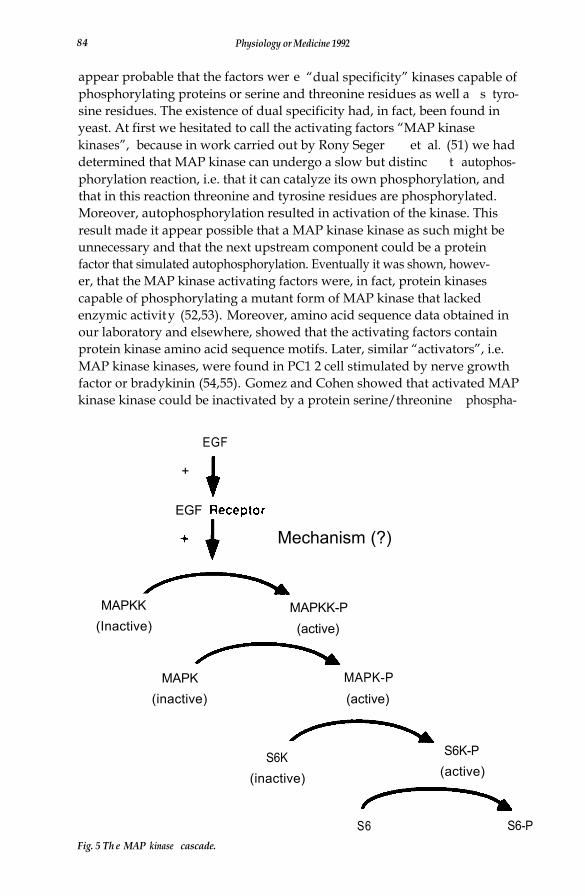
A finding of major significance from Sturgill’s laboratory was the fact thatMAP kinase in its active form contains phosphotyrosine as well a s phos-phothreonine, and that in addition to being inactivated by protein phospha-tase 2A, it can also be inactivated by CD45, a protein tyrosine phosphatase(49). These investigators went on to show that phosphorylation of MAPkinase on tyrosine and threonine is essential for its activity, and it seemedlikely that two different types of protein kinase, i.e. a protei n serine/threo-nine kinase and a protein tyrosine kinase, would be involved as upstreamcomponents involved in its activation. There was no evidence, however, thata receptor-type protein tyrosine kinase could serve as either one of thesekinases. It was thus of great interest to search for an enzyme or enzymes,MAP kinase kinases, that would catalyze MAP kinase phosphorylation andactivation.
In this laboratory Natalie Ahn et al. (50) discovered two separable activa-ting factors from Swiss 3T3 cells that catalyzed the activation of an inactiveform of MAP kinase in vitro. Each of these factors required the presence ofMgATP in order to bring about activation of MAP kinase, and it did notmatter whether the latter had first been inactivated by a protei n serine/threonine phosphatase or by a protein tyrosine phosphatase. This made it
84 Physiology or Medicine 1992
appear probable that the factors wer e “dual specificity” kinases capable ofphosphorylating proteins or serine and threonine residues as well a s tyro-sine residues. The existence of dual specificity had, in fact, been found inyeast. At first we hesitated to call the activating factors “MAP kinasekinases”, because in work carried out by Rony Seger et al. (51) we haddetermined that MAP kinase can undergo a slow but distinc t autophos-phorylation reaction, i.e. that it can catalyze its own phosphorylation, andthat in this reaction threonine and tyrosine residues are phosphorylated.Moreover, autophosphorylation resulted in activation of the kinase. Thisresult made it appear possible that a MAP kinase kinase as such might beunnecessary and that the next upstream component could be a proteinfactor that simulated autophosphorylation. Eventually it was shown, howev-er, that the MAP kinase activating factors were, in fact, protein kinasescapable of phosphorylating a mutant form of MAP kinase that lackedenzymic activity (52,53). Moreover, amino acid sequence data obtained inour laboratory and elsewhere, showed that the activating factors containprotein kinase amino acid sequence motifs. Later, similar “activators”, i.e.MAP kinase kinases, were found in PC1 2 cell stimulated by nerve growthfactor or bradykinin (54,55). Gomez and Cohen showed that activated MAPkinase kinase could be inactivated by a protein serine/threonine phospha-
EGF
+
EGF
Mechanism (?)
MAPKK(Inactive)
MAPKK-P(active)
MAPK(inactive)
MAPK-P(active)
S6K(inactive)
S6K-P(active)
S6 S6-PFig. 5 Th e MAP kinase cascade.

Edwin G. Krebs 85
tase but was not affected by protein tyrosine phosphatases (54). This latterresult indicated that an additional upstream protein serine/threonin e kin-ase, i.e. a MAP kinase kinase kinase, was probably a component of thepathway. The present status of the MAP kinase cascade, without the puta-tive MAP kinase kinase kinase, is shown in Fig. 5.
Intense effort on the part of many different laboratories is currentlycentered on the question of the identity of MAP kinase kinase kinase andthe mechanism by which such an enzyme might be regulated. It was report-ed by Kyriakis et al. that Raf-1 activates MAP kinase kinase and thus mightitself be a candidate for the kinase kinase kinase (56). This result wasconfirmed by Dent et al. (57). Other studies have implicated the possibilitythat cdc2 might be an upstream component of the MAP kinase cascade butnot an immediate kinase kinase kinase itself (58). Finally, there is consider-able evidence that p21 ras is a component acting at some site between thereceptor and the components of the cascade as currently identified (59-62). A somewhat neglected area of study with respect to the MAP kinasecascade relates to the question of its precise function and the reasons forthe existence of such a complicated scheme. It is probable that numeroussubstrates may exist for each of the protein kinases in the cascade, and inthis connection it is noteworthy that a number of transcription factorsappear to be targeted by MAP kinase and by the S6 kinase. The differentkinases may also have specific metabolic enzyme targets that have not as yetbeen identified. Finally, some of the kinases may branch off and regulatestill other kinases (63).
CONCLUSION
In this talk I have reviewed the early work on the interconversion reactionsof the two forms of glycogen phosphorylase and phosphorylase kinase. Athird enzyme that played an important part in early work on proteinphosphorylation-dephosphorylation was glycogen synthase. Much of theoriginal research on that enzyme was carried out by Joseph Larner and hisassociates during the early 1960s. These investigators found that, in con-trast to the effect of phosphorylation on phosphorylase and phosphorylasekinase, phosphorylation caused a decrease in the activity of the synthase.Because the “field” of protein phosphorylation during the first ten yearswas dominated by those interested in glycogen metabolism, some evenexpressed the idea that perhaps regulation of enzymes by phosphorylation-dephosphorylation might be restricted to this area. However, the finding ofa multifunctional cyclic AMP-dependent protein kinase, which was almostimmediately shown by Kuo and Greengard (64) to be very widespread innature, served to change this concept. Another important finding thatdispelled the idea that protein phosphorylation was very limited in scopewas the report that pyruvate dehydrogenase is regulated by phosphorylation(65). As can be noted in Fig. 6, the number of enzymes reported to undergoregulation as a result of this being phosphorylated started growin g precipi-


Edwin G. Krebs 87
very important contributions made by numerous talented unnamed stu-dents and postdoctoral fellows,.some but not all of whom I have mentionedin this article, who carried out the actual experiments reported here and inaddition helped to provide direction for many of the studies that arereported.
REFERENCES
1.
2.3.4.5.6.
7.8.9.
10.11.12.
13.14.
15.16.17.18.19.20.21.
22.
23.
24.25.
26.27.
28.
29.
30.
Cori, C.(1939) Cold Spring Harbor Symposia on Quantitative Biologyol. VII, p.260 - 268.Cori, G. T. and Green, A. A. (1945) J. Biol. Chem. 158, 32 1 - 332.Cori, G. T. and Cori, C F. (1943) J. Biol. Chem., 151, 31-38.Cori, G. T. (1945)J. Biol. Chem., 158, 333-339.Sutherland, E. W. and Cori, C. F. (1951) J. Biol. Chem., 188, 531-543.Sutherland, E. W. (1950) Recent Progress in Hormone Research, Proceedings ofthe I.aurentian Hormone Conference, Vol. 5, Academic Press, New York, p.441463.Green, A. A. and Cori, G. T. (1943) J. Biol. Chem., 151, 21-29.Krebs, E. G. and Fischer, E. H. (1955) J. Biol. Chem., 216 , 113 -120.Fischer, E. H . and Krebs, E . G. (1955) J. Biol. Chem., 216, 121 -132.Krebs, E. G. and Fischer, E. H. (1956) J. Biochim. Biophys. Acta, 20, 150 -157.Krebs, E. G., Kent, A. B. and Fischer, E. H. (1958) J. Biol. Chem., 231, 78-83.Meyer, W. L., Fischer, E. H. and Krebs, E. G: (1964 ) Biochemistry, 3, 1033 -1039.Huston, R. B. and Krebs, E. G. (1968) Biochemistry, 7, 2116-2122.Fischer, E. H., Graves , D. J., Crittenden, E. R. S. and Krebs, E. G. (1959) J. Biol.Chem., 234 , I698 - 1704.Sutherland, E. W. and Wosilait, W. D. (1955) Nature, 175, 169.Rall, T. W., Sutherland, E. W. and Wosilait, W. D. (1956) J. Biol. Chem., 218,Wosilait, W . D. (1958) J. Biol. Chem. 233, 597.Sutherland, E. W. and Rall, T. W. (1958) J. Biol. Chem., 233, 1077-1091.Burnett, G. and Kennedy, E. P. (1954) J. Biol. Chem., 211, 969 -988.Cori, C. F. and Cori, G. T. (1928) J. Biol. Chem., 79, 309 -355.Sutherland, E. W. (1962) The Harvey Lectures, Series 57, Academic Press, NewYork, p. 17-33.Krebs, E. G., Graves, D. J. and Fischer, E. H. (1959) J. Biol. Chem. 234:2867-2873.Cori, C. F. in O. H. Gaebler, ed., Enzymes: Units of Biological Structure andFunction, Academic Press, New York, 1956, p. 573.Ozawa, E., Hosoi, K., and Ebashi, S. (1967) J. Biochem. 61, 531-533.Grand, R. J. A., Shenolikar, S. and Cohen, P. (1981) Eur. J. Biochem. 113, 359 -367.Posner, J. B., Stern, R. and Krebs, E. G. (1965) J. Biol. Chem., 240, 982 - 985.Krebs, E. G., DeLange, R. J., Kemp, R. G. and Riley, W. D. (1966) Pharmacol.Rev., 18, 163-171.DeLange, R. J., Kemp, R. G., Riley, W. D., Cooper, R. A. and Krebs, E. G.(1968) J. Biol. Chem., 243, 2200 -2208.Walsh, D. A., Perkins, J. P. and Krebs, E. G. (1968) J. Biol. Chem., 243, 376 3 -3765.Huijing, F. and Larner, J. (1966) Biochem. and Riophs Res. Commun., 23, 259-263.

88 Physiology or Medicine 1992
31. Shoji, S., Parmelee, D. C., Wade, R. D., Kumar, S., Ericsson, L H., Walsh, K.A., Neurath, H., Long , G. L., DeMaille, J. G., Fischer, E. H. and Titani, K.(1981) Proc. Natl. Acad. Sci. USA, 78 , 848-851.
32. Knighton, D. R., Zheng, J., Eyck, L. F. T. , Ashford, V. A., Xuong, N.-H.,Taylor, S. S. and Sowadski, J. M. (1991) Science, 253, 407-414.
33. Kemp, B. E., Bylund, D. B., Huang, T. S. and Krebs, E. G. (1975 ) Proc. Natl.Acad. Sci. USA, 72, 344 8 -3452.
34. Humble, E., Berglund, L., Titanji, V., Ljungstrom, O. Edlund, B., Zetterquist , 0.and Engstrom, L. (1975) Biochem. Biophys. Res. Commun. 66, 614 - 62 1.
35. Daile, P., Carnegie, P. R. and Young, J. D. (1975) Nature, 257 , 416-418.36. Zetterquist, O., Ragnarsson, U., Humble, E., Berglund, L. and Engstrom, L.
(1976) Biochem. Biophys. Res. Commun. 70, 696-- 703.37. Kemp, B. E., Graves, D. J., Benjamini, E. and Krebs, E. G. (1977) J. Biol. Chem.,
252, 4888- 4894.38. Cohen, Carpenter, G. and King, L. E., Jr. (1980) J. iol. Chem., 255, 4834-
4842.39. Kasuga, M., Zick, Y., Blithe, D. L., Karlsson, F. A., Haring, H. U., and Kahn, C.
R. (1982) J. Biol . Chem., 257, 9891-9894.40. Denton, R. M. (1986 ) Advances in Cyclic Nucleotide and Protein Phosphorylation
Research (P. Crecngard and G. A. Robinson , Eds.) Raven Press, New York, p.293-341.
41. Cobb, M. H. and Rosen, 0. M. (1982) J. Biol. Chem., 258 , 12472-12481.42. Novak-Hofer, I., and Thomas, G. (1984) J. io . Chem., 259, 5995-6000.43. Ballou, L. M., Jeno, P. and Thomas, G. (1988) J. Biol. Chem., 263, 1188- 1194.44. Ballou , L. M., Siegmann, M. and Thomas, G. (1988) Prac. Natl. Acad. Sci. USA,
85,7154-7158.45. Andres, J. L. and Mallet-, J. L. (1989) J. io. Chem., 264, 151 - 156.46. Sturgill, T. W., Ray, I,. B., Erickson, E. and Mallet-, J. I.. (1988) Nature, 334 ,
715-718.47. Gregory, J. S., Boulton, T. G., Sang, B.-C., and Cobb, M. H. (1989) J. Biol.
Chem., 264 , 18, 397- 18, 401.48. Ahn, N . G. and Krebs, E. G. (1990) J. io. Chem., 265, 11495- 11501.49. Anderson, N. G., Maller, J. L., Tonks, N. K., and Sturgill, T. W. (1990) Nature,
343, 651-653.50. Ahn, N . G., Seger, R., Bratlien, R. L., Diltz, C. D., Tonks, N.K. and Krebs, E. G.
(1991) J. Biol. Chem., 266, 4220 - 4227.51. Seger, R., Ahn, N. G., Boulton, T. G., Yancopoulos, G. D., Panayotatos, N.,
Radziqjewska, E., Ericsson, I,., Bratlien, R. L., Cobb, M. H. and Krebs, E. G.(1991) Proc. Natl. Acad. Sci. USA, 88, 6142-6146,
52. Posada, J. and Cooper, J. A. (1992), Science, 255, 212-215.53.. Seger, R., Ahn, N. G., Posada, J., Munar, E. S., Jensen, A. M., Cooper, J. A.,
Cobb, M. H., and Krebs, E. G. (1992 ) J. Biol. Chem., 267, 14373 - 14381.54. Gomez, N. and Cohen, P. (1991) Nature, 351, 69-72.55. Ahn, N. C. , Robbins, D. J., Haycock, J. W., Seger, R., Cobb, M. H., and Krebs,
E. G. (1992 ) J. Neurochemistry, 147- 156.56. Kyriakis, J. M., App, H., Zhang, X., Banerjee, P., Brautigan, D. L., Rapp, U. R.,
Avruch, J. (1992) Nature, 358,417-421.57. Dent, P., Haser, W., Haystead, T. A. J., Vincent, L. A., Roberts, T. M., Sturgill,
T. W. (1992) Science, 257, 1404- 1406.58. Matsuda, S., Kosako, H., Takenaka, K., Moriyama, K., Sakai, H., Akiyama, T.,
Cotoh, Y., Nishida, E. (1992) EMBO J., 11, 973- 982 .59. Thomas, S. M., DeMarco, M., D’Arcangelo, G., Halegoua, S., Brugge, J. S.
(1992) Cell, 68, 1031-1040.60. Wood, K. W., Sarnecki, C., Roberts, T. M., Blenis, J. (1992) Cell, 68, 1041-
1050.

Edwin G. Krebs 89
61. deVries-Smits, A. M. M., Burgering, B. M. T., Leevers, S. J., Marshall, C. J.,Bos, J. L. (1992) Nature, 357, 602-604.
62. Robbins, D. J., Cheng, M., Zhen, E., Vanderbilt, C. A., Feig, L. A., Cobb, M. H.(1992) Proc. Natl. Acad. Sci. USA, 89, 6924-6928.
63. Stokoe, D., Campbell, D. G., Nakielny, S., Hidaka, H., Leevers, S. J., Marshall,C. and Cohen, P. (1992) EMBOJ., 11,3985-3994.
64. Kuo, J. F. and Greengard, P. (1969) J. Biol. Chem., 244, 3417-3419.65. Linn, T. C., Pettit, F. H., Hucho, F. and Reed, L. J. (1969a) Proc. Natl. Acad. Sci.
USA, 64,227 - 234.