protein phylogenies and signature sequences: a reappraisal ... · archaebacteria and eubacteria...
TRANSCRIPT
MICROBIOLOGY AND MOLECULAR BIOLOGY REVIEWS,1092-2172/98/$04.0010
Dec. 1998, p. 1435–1491 Vol. 62, No. 4
Copyright © 1998, American Society for Microbiology. All Rights Reserved.
Protein Phylogenies and Signature Sequences: A Reappraisalof Evolutionary Relationships among Archaebacteria,
Eubacteria, and EukaryotesRADHEY S. GUPTA*
Department of Biochemistry, McMaster University, Hamilton, Ontario L8N 3Z5, Canada
PREFACE ..................................................................................................................................................................1435CURRENT EVOLUTIONARY PERSPECTIVE ...................................................................................................1436MOLECULAR PHYLOGENIES: ASSUMPTIONS, LIMITATIONS, AND PITFALLS .................................1438SEQUENCE SIGNATURES AND THEIR IMPORTANCE IN EVOLUTIONARY STUDIES......................1442ROOT OF THE PROKARYOTIC TREE: ANCESTRAL NATURE OF ARCHAEBACTERIA AND
GRAM-POSITIVE BACTERIA .......................................................................................................................1444EVOLUTIONARY RELATIONSHIPS AMONG PROKARYOTES...................................................................1446
Signature Sequences Showing the Distinctness of Archaebacteria ...............................................................1447Signature Sequences Distinguishing Archaebacteria and Gram-Positive Bacteria from Gram-Negative
Bacteria ..............................................................................................................................................................1449A Specific Relationship between Archaebacteria and Gram-Positive Bacteria and the Distinctness of
Gram-Negative Bacteria Is Consistent with Prokaryotic Cell Structures and Other GenePhylogenies ........................................................................................................................................................1450
Signature Sequence Distinguishing between Low-G1C and High-G1C Gram-Positive Bacteria andPointing to a Specific Relationship of the Latter Group to the Gram-Negative Bacteria .....................1455
Signature Sequences Indicating that Deinococcus and Thermus Are Intermediates in the Transitionfrom Gram-Positive to Gram-Negative Bacteria ..........................................................................................1456
Phylogenetic Placement of Cyanobacteria and Their Close Evolutionary Relationship to theDeinococcus-Thermus Group ............................................................................................................................1458
Signature Sequences Defining Proteobacteria and Some of Their Subdivisions.........................................1461Nature of the Archaebacterial Group and Its Relationship to Gram-Positive Bacteria ............................1461Possible Selective Forces Leading to Horizontal Gene Transfers .................................................................1465Evolutionary Relationships within Prokaryotes: an Integrated View Based on Molecular and
Phenotypic Characteristics ..............................................................................................................................1470EVOLUTIONARY RELATIONSHIP BETWEEN EUKARYOTES AND PROKARYOTES...........................1473
Some Critical Assumptions in Studying Prokaryote-Eukaryote Relationships ...........................................1473Most Genes for the Information Transfer Processes Are Derived from Archaebacteria ...........................1473Hsp70 Provides the Clearest Example of the Contribution of Eubacteria to the Nuclear-Cytosolic
Genome...............................................................................................................................................................1474The Eukaryotic Nuclear Genome Is a Chimera of Genes Derived from Archaebacteria and
Gram-Negative Bacteria...................................................................................................................................1475Origin of the Nucleus and Endoplasmic Reticulum ........................................................................................1477Did Mitochondria and the First Eukaryotic Cell Originate from the Same Fusion Event? .....................1481
CONCLUDING REMARKS....................................................................................................................................1485ACKNOWLEDGMENTS .........................................................................................................................................1487REFERENCES ..........................................................................................................................................................1487
“The credible is, by definition, what is believed already,and there is no adventure of the mind there.”
Northrop Frye (74)
PREFACE
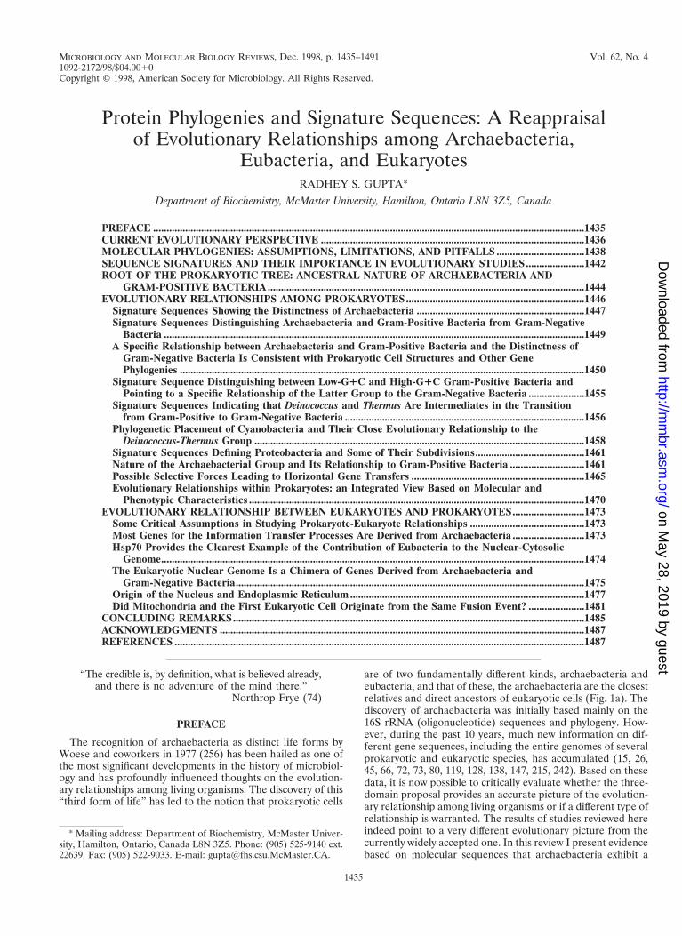
The recognition of archaebacteria as distinct life forms byWoese and coworkers in 1977 (256) has been hailed as one ofthe most significant developments in the history of microbiol-ogy and has profoundly influenced thoughts on the evolution-ary relationships among living organisms. The discovery of this“third form of life” has led to the notion that prokaryotic cells
are of two fundamentally different kinds, archaebacteria andeubacteria, and that of these, the archaebacteria are the closestrelatives and direct ancestors of eukaryotic cells (Fig. 1a). Thediscovery of archaebacteria was initially based mainly on the16S rRNA (oligonucleotide) sequences and phylogeny. How-ever, during the past 10 years, much new information on dif-ferent gene sequences, including the entire genomes of severalprokaryotic and eukaryotic species, has accumulated (15, 26,45, 66, 72, 73, 80, 119, 128, 138, 147, 215, 242). Based on thesedata, it is now possible to critically evaluate whether the three-domain proposal provides an accurate picture of the evolution-ary relationship among living organisms or if a different type ofrelationship is warranted. The results of studies reviewed hereindeed point to a very different evolutionary picture from thecurrently widely accepted one. In this review I present evidencebased on molecular sequences that archaebacteria exhibit a
* Mailing address: Department of Biochemistry, McMaster Univer-sity, Hamilton, Ontario, Canada L8N 3Z5. Phone: (905) 525-9140 ext.22639. Fax: (905) 522-9033. E-mail: [email protected].
1435
on May 28, 2019 by guest
http://mm
br.asm.org/
Dow
nloaded from

close and specific relationship to gram-positive bacteria andthat the primary division within prokaryotes is not betweenarchaebacteria and eubacteria but, rather, between organismsthat have either a monoderm cell structure (i.e., prokaryoticcells surrounded by a single membrane, which includes allarchaebacteria and gram-positive bacteria) or a diderm cellstructure (i.e., prokaryotic cells surrounded by an inner cyto-plasmic membrane and an outer membrane, which includes alltrue gram-negative bacteria) (Fig. 1b) (100). The sequencedata also strongly indicate that the ancestral eukaryotic cell isnot a direct descendant of the archaebacterial lineage but is achimera that resulted from a unique fusion event involving twovery different groups of prokaryotes—a thermoacidophillic ar-chaebacterium (monoderm) and a gram-negative eubacterium(diderm), followed by integration of their genomes. Thus, alleukaryotic organisms, including the amitochondriate andaplastidic cells, received and retained gene contributions fromboth lineages.
CURRENT EVOLUTIONARY PERSPECTIVE
The quest for an understanding of the evolutionary relation-ships between extant organisms has posed a major challenge tobiologists for centuries (23, 43, 159, 167). Since all living or-ganisms are specifically related to each other by the presenceof numerous common (or related) biomolecules and follow asimilar complex strategy for growth and propagation, there isnow little doubt that they all evolved from a common (univer-sal) ancestor (3, 228). However, discerning how different majorgroups of organisms are related to each other and tracing theirevolution from the common ancestor remains controversialand unresolved. After the invention of the microscope in the17th century, studies on the morphological characteristics ofcells from extant organisms led to the identification of two
distinct types of cells (3), later termed prokaryotes and eu-karyotes (34, 173), which could be readily distinguished. Theeukaryotic cells are distinguished from prokaryotes by a num-ber of different characteristics including the presence of acytoskeleton, endomembrane system, etc. (3, 159). However,the hallmark feature of all eukaryotic cells is the presence of amembrane-bounded nucleus, and any organism lacking a nu-clear membrane is considered a prokaryote (4, 34, 173). Eu-karyotic organisms were classified into a number of differentgroups or kingdoms, namely, Animalia, Plantae, Fungi, andProtoctista, based on their detailed and complex morphologiesand with the aid of fossil records (164, 248). However, a similarLinnaean approach to classification based on cell shape, phys-iology, and other characteristics was unsuccessful in detectingthe phylogeny of prokaryotic organisms (23, 24, 121, 140, 194,195, 227, 228, 230, 245, 250, 252, 254). The problem was partlydue to their very simple morphologies but was also due in largepart to the difficulty in determining which of the cellular fea-tures and characteristics of prokaryotes is most meaningful fortaxonomic purposes.
Despite the ill-defined state of bacterial taxonomy, one em-pirical criterion that has proven of much practical value in theclassification/identification of prokaryotes is their response tothe Gram stain (121), discovered by Christian Gram in 1884(88). As has been noted by Murray, “Gram-positiveness andGram-negativeness are still unassailable characters except inArchaebacteria, the radiation-resistant cocci and . . . the wall-less mollicutes” (175). Gram staining involves successive treat-ment of cells with the basic dye crystal violet followed bytreatment with iodine solution and then extraction with a polarorganic solvent such as alcohol or acetone. The cells whichresist decolorization and retain the blue-black dye complex arereferred to as gram positive, whereas those which do not retainthe stain are classified as Gram negative (12, 13, 88, 121). The
FIG. 1. Evolutionary relationships among living organisms in the three-domain model of Woese et al. (258) (a) and as suggested here based on protein sequencedata and structural characteristics of organisms (b). In panel b, the solid arrows identify taxa that evolved from each other in the directions shown by accumulation ofmutations and the dotted lines denote symbiotic events that led to the acquisition of mitochondria and plastids. These latter events, which are common in both models,are not shown in panel a. In panel b, the double-headed arrow between archaebacteria and gram-positive bacteria indicates the polyphyletic relationship between thesegroups for several genes. The terms “monoderm” and “diderm” refer to prokaryotic cells that are bounded by only one membrane or two different (cytoplasmic andouter) membranes, respectively. The dashed lines indicate the first fusion between an archaebacterium and a gram-negative bacterium that is postulated to have givenrise to the ancestral eukaryotic cell (102, 105). Abbreviations: CM, cytoplasmic membrane; CW, cell wall; OM, outer membrane, PE, periplasm.
1436 GUPTA MICROBIOL. MOL. BIOL. REV.
on May 28, 2019 by guest
http://mm
br.asm.org/
Dow
nloaded from
Gram-staining response, although not always reliable due to itsdependence on cell physiology and cell integrity (11, 228), thusdivides prokaryotes into two main groups, the gram-positiveand the gram-negative (121, 228). Although the Gram reactionis an empirical criterion, its basis lies in the marked differencesin the ultrastructure and chemical composition of the cell wall(14, 192, 228, 229, 235). The Gram-positive bacteria in generalcontain a thick cell wall (20 to 80 nm) that is very rich incross-linked peptidoglycan (accounting for between 40 and90% of the dry weight) and also containing teichoic acids,teichuronic acid, and polysaccharides (6, 14, 192, 229). Becauseof their rigid cell walls, these bacteria have been named Fir-micutes in Bergey’s Manual of Systematic Bacteriology (174); anumber of other bacteria which possess the above structuralcharacteristics but may show gram-variable (or gram-negative)staining are also placed in the same group. In contrast, all“true” gram-negative bacteria, named Gracilicutes in Bergey’sManual (174), have only a thin layer of peptidoglycan (2 to 3nm) and have, in addition to the cytoplasmic membrane, anouter membrane containing lipopolysaccharides, which liesoutside of the peptidoglycan layer. As noted by Truper andSchleifer (244) “A clear separation of the Gram-positive andGram-negative bacteria can be obtained by the differences inthe ultrastructure and chemical composition of the cell wall”.In the present work, I have used the term “gram negativebacteria” to describe prokaryotes whose envelopes contain acytoplasmic membrane, a murine cell wall, and an outer mem-brane rather than by their Gram-staining response.
Based on the nature of the bounding layer of the cells, whichis reflected in the Gram-staining reaction, a major microbiol-ogy textbook (228) suggested the division of prokaryotes intothree main groups: “The Mycoplasma which do not synthesizea cell wall, the membrane serving as the outer bounding layer;the Gram-positive bacteria, which synthesize a monolayeredcell wall; and the Gram-negative bacteria, which synthesize acell wall composed of at least two structurally distinct layers.”Although they could not know the extent of the problem, manyearlier bacteriologists recognized the importance of cell struc-ture and the bounding layer in the classification of prokaryotes:“It is self evident that the shape of the cell is of outstandingimportance for determining the place of bacterium in anyphylogenetic system” (140). However, as noted in a leadingtextbook, distinguishing between cells containing differenttypes of envelopes was not an easy task (228): “The Gram-staining procedure is not always a wholly reliable method (and)the differentiation of these two subgroups (i.e., Gram-positiveand Gram-negative) by other and more reliable methods is noteasy; it requires either electron microscopic examination ofwall structure in thin sections of the cells or chemical detectionof the group specific polymers.” In view of these difficulties, theresults obtained were often difficult to integrate into a coherentscheme (24, 121, 174, 194, 195, 227, 228, 230, 245).
By the late 1950s and early 1960s, when microbiologists werefeeling increasingly frustrated in their attempts to understandthe natural relationship among prokaryotes, the era of molec-ular biology dawned. With this came the important realization,spelled out clearly by Zuckerkandl and Pauling (264), that thelinear sequences of bases and amino acids in nucleic acids andproteins are informative documents containing a record oforganismal evolutionary history from the very beginning andthat in this regard the prokaryotic organisms are just as com-plex and informative as any eukaryote (65, 264). Thus, a com-parison of sequences of the same gene or protein from variousspecies could be used to deduce and reconstruct the evolution-ary history of organisms. This marked the beginning of the fieldof molecular evolution. The rationale for using molecular se-
quence data to deduce the evolutionary relationship betweenorganisms is described in a number of excellent reviews (58, 60,61, 64, 65, 178, 236) and is not covered here except for certainrelevant points.
The initial molecular approaches based on DNA base com-position, nucleic acid hybridization, and immunological cross-reactivities were of limited use and were generally successful inestablishing or rejecting relationships only among bacteria thatwere thought to be closely related species (224, 226, 228). Thefull impact of the molecular approach on evolutionary biologydid not become evident until Woese and coworkers (71, 250,256) had completed systematic studies of a significant numberof living organisms based on the small-subunit rRNA se-quences (SSU or 16S rRNA). The earlier studies in this regardwere based on comparison of the oligonucleotide catalogs ofthe 16S rRNA, but these were later supplanted by phylogeneticanalysis based on complete sequences of the molecules. Thesestudies revealed that, based on genetic distances and signaturesequences in the 16S rRNA, various prokaryotic and eukary-otic organisms fell into three distinct groups (71, 250, 256).One group consisted of all eukaryotic organisms, the secondconsisted of all commonly known bacteria (the term “eubac-teria” was suggested for this group) including various genera ofgram-positive and gram-negative bacteria and cyanobacteria,and the third group consisted of a number of previously little-studied prokaryotes (methanogens, extreme thermoacidophiles,and extreme halophiles) which grow in unusual habitats. Becauseof their assumed antiquity, this last group of prokaryotes wasnamed “archaebacteria” (256).
In terms of their genetic distances (or similarity coefficientsfrom oligonucleotide catalogs) based on rRNA, the archaebac-teria were no more closely related to the eubacteria than to theeukaryotes. This observation, in conjunction with a number ofunique characteristics of archaebacteria (e.g., lack of muramicacid in cell walls [127)] membrane lipids that contain ether-linked isoprenoid side chains [127, 133)], distinctive RNA poly-merase subunits structures [263], and lack of ribothymine inthe TCC loop of tRNA), led Woese and collaborators topropose that the archaebacteria were totally distinct fromother bacteria and constituted one of the three aboriginal linesof descent from the universal ancestor (71, 250, 256). Theprokaryotes thus consisted of two distinct and non-overlapping(i.e., monophyletic) groups: eubacteria and archaebacteria,which were no more specifically related to each other thaneither was to the eukaryotes (250, 256). Since microbiology atthe time was lacking any formal basis for phylogeny, this pro-posal, based on more defined and quantitative molecular char-acteristics, was generally favorably received, and within a de-cade most microbiology textbooks took notice of or wererevised in the light of these new findings (6, 8, 14, 121, 192,229).
The archaebacterial proposal received a major boost in 1989when the phylogenies based on a number of protein sequenceswere added to the analysis, including those for the proteinsynthesis elongation factors EF-1a/Tu and EF-2/G, RNA poly-merase subunits II and III, and F- and V-type ATPases (82,126, 196). These studies again supported the distinctness ofarchaebacteria from eubacteria. Further, in contrast to therRNA phylogeny, where only an unrooted tree was possible forarchaebacteria, eubacteria, and eukaryotes, for the paralogouspairs of protein sequences (namely, EF-Tu and EF-G; and F-and V-ATPases) which appeared to be the results of ancientgene duplication events in the common ancestor of all extantlife, it was possible to root the universal tree by using one setof genes as an outgroup for the other (82, 126). These studiesindicated that the root of the universal tree lay between ar-
VOL. 62, 1998 PHYLOGENY OF PROKARYOTES AND EUKARYOTES 1437
on May 28, 2019 by guest
http://mm
br.asm.org/
Dow
nloaded from
chaebacteria and eubacteria, and in both cases the eukaryoteswere indicated as specific relatives of archaebacteria (82, 126).In 1990, Woese et al. (258) adopted this rooting, and a formalthree-domain proposal for the classification of organisms wasput forward. The proposal assigned each of the three groups,archaebacteria, eubacteria, and eukaryotes, a Domain status (anew highest taxonomic level) and renamed them Archaea, Bac-teria, and Eucarya. The name Archaea was specifically pro-posed to indicate that this group of prokaryotes bear no spe-cific relationship to the other prokaryotes (i.e., Bacteria oreubacteria) (258). This rooted version of the universal tree(Fig. 1a), commonly referred to as the archaebacterial orthree-domain tree, is now widely accepted as the current par-adigm in the field (54, 91, 171, 187, 258).
But does this tree or view represent the true relationshipbetween the organisms? In recent years, much new informa-tion based on a large number of gene and protein sequences,including the complete genomes of several prokaryotic andeukaryotic organisms, has become available (26, 45, 66, 72, 73,80, 119, 128, 138, 147, 215, 242). Based on this information, itis now possible to critically evaluate the three-domain proposaland its various predictions and to determine if this view issupported by all data or is true only for a subset of gene andprotein sequences. These studies should also indicate whethera different sort of relationship between the organisms is moreconsistent with most of the available data. Since most biologistsare not familiar with the assumptions and pitfalls of phyloge-netic analyses, I will try to point out the strengths as well thesubjective and weak aspects of such analyses so that the read-ers can understand and evaluate the results which form thebases for any classification.
MOLECULAR PHYLOGENIES: ASSUMPTIONS,LIMITATIONS, AND PITFALLS
The use of molecular sequences for phylogenetic studies isbased on the assumption that changes in gene sequences occurrandomly and in a time-dependent manner and that a certainproportion of these become fixed in the molecules (58, 65, 136,178, 236). The accumulation of changes in gene sequences in aquasi clock-like manner has given rise to the concept of “evo-lutionary clock” or molecular chronometer (136). Followingthe clock analogy (252), just as different hands or features (e.g.,the month, day, minute, and second) in a clock move at verydifferent rates, the changes in different gene sequences (orsometimes within different parts of the same gene) also occurat vastly different rates. Thus, some sequences which changevery slowly (like the year, month, or day) are well suited formonitoring ancient events, while others, with a higher rate ofchange (like the hour, minute, or second), provide the sensi-tivity and resolution to measure relatively recent occurrences.Since the evolutionary history of life on this planet spans a vastperiod (approximately 3.8 Ga, 109 years), different sequenceshave different utilities in evolutionary studies. In the presentcontext, where our main focus is on examining very ancientevolutionary events (e.g., relationships within the higher pro-
karyotic taxa and the origin of eukaryotic cells), the sequenceswhich change very slowly and hence show a high degree ofconservation in all extant organisms (i.e., the best-preservedmolecular fossils) are most useful.
Phylogenetic analysis can be carried out based on eithernucleic acid or protein sequences. For noncoding sequencessuch as various rRNAs, tRNAs, and introns, phylogenetic anal-ysis can be carried out based on only the nucleotide sequencedata. However, for gene sequences that encode proteins, anal-yses can be performed based on either the nucleic acid or theamino acid sequence data. For proteins, the two kinds of anal-yses appear analogous at first. In fact, the analysis based onnucleic acid sequences, with three times as many characters,would seem to be more informative (181, 250). While this istrue in principle, for phylogenetic analyses involving distantlyrelated taxa the increased information content in nucleic acidsequences as opposed to protein sequences is merely an illu-sion and in most cases is a major liability. The main reason forthis lies in the degeneracy of the genetic code. All but twoamino acids (Met and Trp) are encoded by at least two codonswhich differ in the third position. In view of this degeneracy,most changes in the third codon positions are selectively neu-tral (i.e., they do not result in any change in the protein se-quence) and, as a consequence, change frequently even inclosely related species (58, 60, 136). In distantly related taxa,which diverged from each other a long time ago, the bases atthe third codon positions may have changed so many times thatthe actual bases found at these positions are random in natureand their information content is virtually nil. The inclusion ofsuch bases in the analyses, therefore, would lead to uncertaintyat every third position, thereby reducing the signal (i.e., posi-tions which are evolutionary important)-to-noise (i.e., posi-tions or changes which provide no evolutionary information)ratio in the data set.
Another important factor affecting the usefulness of nucleicacid sequences compared to protein sequences relates to thedifferences in the genomic G1C content of species (113, 231).The G1C content of different species is known to differ greatly(this is often true for two species within the same genus aswell), and it is generally homogenized over the entire genome.In the protein-coding sequences, these differences in the G1Ccontents are accommodated by selective changes (i.e., codonpreferences) in the third codon positions. The species whichare rich in G1C show a strong preference for codons that haveG or C in the third position (often .90%), whereas specieswith low G1C content predominantly utilize the codons withA or T in these positions. Thus, two unrelated species withsimilar G1C contents (e.g., either very high or very low) mayhave very similar bases in the third codon positions. If phylo-genetic analysis is carried out based on nucleic acid sequences,these species may show a strong affinity for each other but forthe wrong reason (113, 231). Thus, the third codon positions,rather than being informative, can introduce major bias intothe analyses. For a similar reason but to a lesser extent, thebases in the first codon positions are also evolutionarily lessinformative and can cause reduced signal-to-noise ratio. Thus,
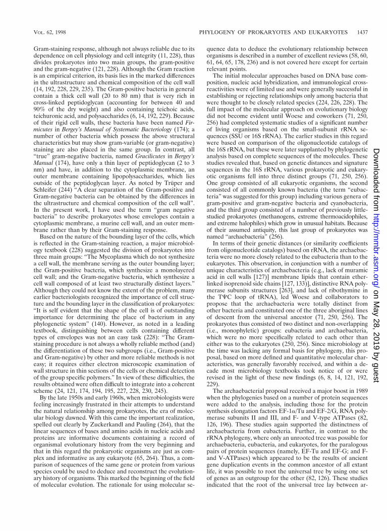
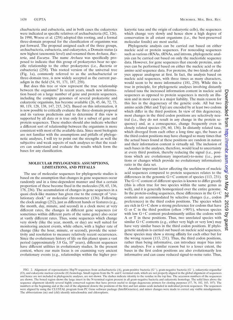
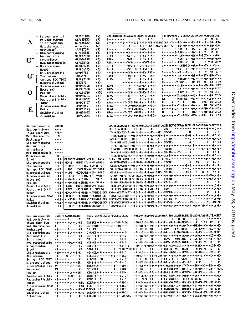
FIG. 2. Alignment of representative Hsp70 sequences from archaebacteria (A), gram-positive bacteria (G1), gram-negative bacteria (G2), eukaryotic-organellar(O), and eukaryotic nuclear-cytosolic (E) homologs. Small regions from the N- and C-terminal ends, which are not properly aligned in the global alignment of sequencesand hence are not included in phylogenetic analyses, are not shown. The dashes indicate identity to the residue in the top line. The accession numbers of the sequencesare shown. The boxed region shows the large insert in the N-terminal region present in all gram-negative bacteria and eukaryotic homologs. The solid lines above thesequence alignment identify several highly conserved regions that have proven useful to design degenerate primers for cloning purposes (57, 76, 102, 103, 107). Thenumbers at the beginning and at the end of the alignment denote the positions of the first and last amino acids included in individual protein sequences. The sequenceswere aligned by using the CLUSTAL program from PC Gene software package (IntelliGenetics), and minor changes were made to correct any visible misalignments.The abbreviations (m) and (chl) identify mitochondria and chloroplasts.
1438 GUPTA MICROBIOL. MOL. BIOL. REV.
on May 28, 2019 by guest
http://mm
br.asm.org/
Dow
nloaded from
VOL. 62, 1998 PHYLOGENY OF PROKARYOTES AND EUKARYOTES 1439
on May 28, 2019 by guest
http://mm
br.asm.org/
Dow
nloaded from
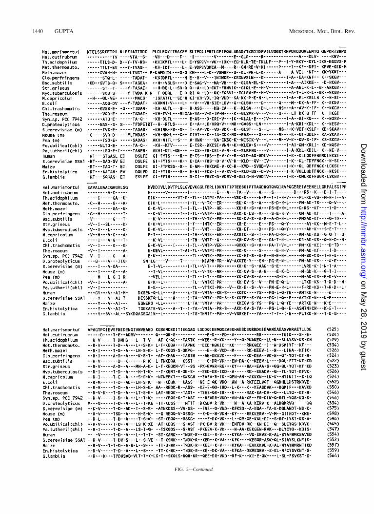
FIG. 2—Continued.
1440 GUPTA MICROBIOL. MOL. BIOL. REV.
on May 28, 2019 by guest
http://mm
br.asm.org/
Dow
nloaded from
in the phylogenetic analyses of distantly related taxa with vary-ing G1C contents, the larger number of characters in thenucleic acid sequences does not offer any real advantage, andif the bases at the third codon positions (and often those at thefirst positions as well) are not excluded from the analyses,misleading results could be obtained. In view of these consid-erations, for the protein-coding regions, the amino acid se-quences, which are minimally affected by the differences in theG1C contents of the species, have proven more reliable andare the preferred choice for phylogenetic analyses (111, 113,231).
In contrast to the protein-coding regions, where the codondegeneracy provides a natural mechanism for accommodatingchanges caused by G1C drifts, the effect of varying G1Ccompositions on structural nucleic acid sequences such asrRNA or tRNA remains largely undetermined. Thus, whencomparing sequences from different species with varying G1Ccompositions, it is difficult to distinguish between the changesthat are due to G1C drift (evolutionarily not significant) fromthose that are evolutionarily important. Thus, in any analysesbased on structural nucleic acid sequences, the signal-to-noiseratio is inherently low. The effect that this will have on phylo-genetic reconstruction cannot be easily determined or cor-rected, but this is a major and continuing source of concern inphylogenetic studies based on structural nucleic acids such asthe 16S rRNA. As pointed out by Woese (251), “The problem(of) disparity in base composition is far more troublesome thanis generally recognized and has almost received no attention todate. . . . It is important to understand the extent to which thegeneral pattern reflects rRNA compositional disparity ratherthan the true phylogeny.”
Another major problem in phylogenetic analyses is the reli-ability of the sequence alignment. The alignment of homolo-gous positions in a set of sequences is the starting point inphylogenetic analyses from which all inferences are derived.Hence, the importance of having a reliable alignment for phy-logenetic studies cannot be overemphasized. Most sequencealignment programs work by recognizing local similarity indifferent parts of molecules and then creating an alignment ofall positions which maximizes the number of matches betweenthe sequences, keeping the number of gaps introduced to aminimum (117). Although the alignment programs work sim-ilarly for both nucleic acid and protein sequences, there areimportant differences. In nucleic acid sequences there are onlyfour characters, and hence the number of matches between anytwo sequences (unrelated) is expected to be a minimum of25%; with the introduction of a small number of gaps, it iscommonly in the range of 40 to 50%. In view of this, theprobability of chance alignment of nonhomologous regions intwo sequences is quite high, particularly if the sequences beingcompared are of different lengths and have either unusuallyhigh or low G1C contents. In contrast, in proteins each char-acter has 20 states, which greatly reduces the probability ofchance alignment between nonhomologous regions. There areno standard criteria for a good alignment, but it is generallyassessed empirically by means of visual inspection. If the set ofsequences contains highly conserved regions dispersedthroughout the alignment, the proper alignment of such re-gions in all sequences is indicative of a good alignment. How-ever, for sequences which do not contain many such regions, itis often difficult to get a reliable alignment for phylogeneticstudies. Very often, differences in sequence alignment, theregions included in the phylogenetic analyses, or even theorder in which the sequences are added in an alignment (151)could lead to important differences in the inferences drawn(42, 112).
Most extensive phylogenetic studies of living organisms havebeen carried out based on the SSU rRNA sequences (8, 77, 86,149, 152, 224), which have been called the “ultimate molecularchronometers” by Woese (250). However, the alignment ofrRNA sequences from various prokaryotic and eukaryotic spe-cies presents unique problems. In view of the large differencesin the lengths of prokaryotic ('1,500 nucleotides) and eukary-otic ('2,000 nt) SSU rRNAs (mitochondrial SSU rRNA fromsome species is only 612 bp long [89]) and the wide variationsin the G1C contents of species, a reliable alignment of rRNAsequences from distantly related taxa cannot easily be obtainedbased on the primary sequence data alone. The approachtaken to get around this problem is to rely on the secondary-structure models of rRNA, based on the assumption that thesecondary structure of the rRNA is highly conserved and pro-vides a reliable guide for identification of homologous posi-tions (252, 257, 259). Based on this, portions of the foldedmolecules (i.e., particular loops or stems) that are postulatedto be similar in different sequences are aligned and used forphylogenetic studies.
The use of secondary-structure models for identification andalignment of homologous positions in the SSU rRNA is a veryserious and far-reaching assumption. From an energetic pointof view, the SSU rRNA can assume many different but equallylikely secondary structures (259). While the proposed struc-tures of rRNAs are supported by enzymatic digestion andchemical modification studies of some species (257, 259), theirvalidity in distantly related prokaryotic and eukaryotic taxa isfar from established. The effect that these far-reaching as-sumptions, on which all rRNA alignments are based (8, 33,181, 184, 189, 224, 251), will have on the deduced phylogeneticrelationships remains to be determined. However, it is clearthat these assumptions have the potential to profoundly influ-ence the outcome of any analyses (111).
In contrast to the rRNA sequence alignment, alignment ofamino acid sequences of a highly conserved protein such as the70-kDa heat shock chaperone protein (Hsp70) requires mini-mal or no assumptions. Because of the similar size of thisprotein in various prokaryotic and eukaryotic species (includ-ing organellar homologs) and its high degree of sequence con-servation, a good alignment of the sequences from variousspecies is readily obtained by using any common sequencealignment program (117) or even manually by placing the se-quences next to each other. Figure 2 shows an alignment of 25Hsp70 sequences covering the prokaryotic and eukaryoticspectrum as well as organellar homologs. The alignment shownwas obtained with the CLUSTAL program from the PCGENEsoftware, and only minor corrections to it have been mademanually. The large number of identical and conserved resi-dues present throughout the length of this alignment givesconfidence that the observed alignment is reliable. The globalalignment of Hsp70 sequences shows many regions that arenearly completely conserved in all species. Degenerate primersbased on these sequences have been successfully used to clonethe gene encoding Hsp70 from a wide range of prokaryotic andeukaryotic organisms (56, 57, 76, 102, 103, 107, 108).
Once a (reliable!) sequence alignment has been obtained,three main types of methods are used for phylogenetic recon-struction: those based on maximum parsimony (58, 64), thosebased on pairwise genetic distances between the species (65,207), and the maximum-likelihood method (58, 137). Thesemethods interpret the sequence alignment in different ways,and therefore the results obtained from them often differ (110,238). All these methods, as well as the others (e.g., evolution-ary parsimony [152]), can give rise to incorrect relationshipsunder different conditions. Five main factors affecting the out-
VOL. 62, 1998 PHYLOGENY OF PROKARYOTES AND EUKARYOTES 1441
on May 28, 2019 by guest
http://mm
br.asm.org/
Dow
nloaded from
come of these analyses are (i) an underestimation of the num-ber of genetic changes between the species (often multiplechanges in a position are counted as either one or no change);(ii) the long-branch-length effect, where two distantly relatedtaxa may appear more closely related than they truly are ifthere are no intermediate taxa to break the long branches (62);(iii) large differences in the evolutionary rates among differentspecies in the data set; (iv) horizontal or lateral gene transfersbetween the species (236a); and (v) comparison of paralogoussequences which are the results of unidentified ancient geneduplication events (62, 110, 152, 233, 238). In most cases, it isdifficult to ascertain the effects of different factors and to de-termine which phylogenetic method is more suitable or reli-able. Hence, phylogenetic analyses are generally carried out bydifferent methods to see if all the methods give similar results.
The reliability of phylogenetic relationships inferred fromthe above methods is commonly assessed by performing abootstrap test (59). In this test, the aligned sequences aresampled randomly and certain numbers of columns in theoriginal alignment are replaced with columns from elsewherein the sequences to obtain 100 or more different alignments,each containing the same number of columns. Thus, in a givenbootstrap set, some columns will not be included at all, otherswill be included once, and still others will be repeated two ormore times. Phylogenetic analysis is then performed on each ofthe bootstrap replicates, and a consensus tree from this data isdrawn. The main purpose that bootstrap analyses serve is toprovide a measure of the variability of the phylogenetic esti-mate or confidence levels in the observed evolutionary rela-tionships. If the sample data throughout the sequence lengthsupport a particular relationship, this will be reflected in thegrouping of the species in all (or a vast majority) of the boot-straps. The results of these analyses are presented by placingbootstrap scores (indicated by the percentages or the numberof times that different species group together in bootstraptrees) on different nodes in the tree. Bootstrap values of .80to 85% are generally considered to provide good support for aspecific phylogenetic relationship.
Despite due care in the alignment and analyses of the se-quence data, interpretation of the phylogenetic trees that areobtained is not straightforward. The most common problem inthis regard is that phylogenetic trees based on different genesor proteins may differ from each other in terms of the evolu-tionary information that they provide. Based on the clock anal-ogy discussed above, some genes are better suited to resolvecertain relationships than are others. Thus, while a particularrelationship may be clearly resolved and strongly supported byone gene phylogeny, the same relationship may not be obviousfrom a different gene phylogeny. Such results are generallyregarded as controversial by many scientists, including evolu-tionary biologists (49, 53, 69, 70), but it is important to realizethat they are not. Part of the problem in the interpretation ofnew data stems from the commonly held perception that phy-logenetic trees based on just one or two molecules (e.g., 16SrRNA) can clearly establish the evolutionary relationships be-tween all extant species (181, 184, 188, 202, 224, 250–252). Thismeans that any results that do not concur with the 16S rRNAphylogenies are generally considered deviant and suspicious(69). However, such a notion is clearly erroneous, in view ofthe limitations of the rRNA-based phylogenies noted aboveand the inability of the 16S rRNA trees to resolve the branch-ing orders of the deeply lying taxa within eubacteria: “(In the16S rRNA phylogeny) the majority of the bacterial phyla arisein such a tight radiation that their exact order of branching hasyet to be resolved” (252).
Cognizant of these problems, many scientists working in this
area have urged caution in the interpretation of phylogeneticdata. Woese wrote (252): “The scientifically proper stance forthe microbiologists to take at this juncture will be to treat thesephylogenies (bacterial) as hypotheses, and test them usingother molecules, phenotypic characteristics of the organisms,and so on. When the same or very similar relationships aregiven by different molecular systems or when new phenotypicsimilarities consistent with the projected phylogenies turn up,then that phylogeny can be confidently accepted”; Rothschildet al. wrote (202): “We encourage phylogenetic analyses wheremolecular approaches are evaluated in the light of other avail-able data, and where the strengths as well as subjective andweak aspects of the analyses are made explicit”; and Murray etal. stated (177): “The integrated use of phylogenetic and phe-notypic characteristics, called polyphasic taxonomy (38), is nec-essary for the delineation of taxa at all levels from Kingdom togenus”. I do not think any evolutionary scientist will disagreewith the above statements or suggested approaches.
It is clear from the above discussion that the results ofphylogenetic analyses should not be uncritically accepted butinstead should be evaluated in the light of other available data,including data from morphological, geological, and fossilsources. There is also a pressing need to develop additionalsequence-based criteria for determining the evolutionary rela-tionships among species, which are based on minimal assump-tions and which could be readily understood and interpreted byboth specialists and nonspecialists. In the next few sections, Ipresent evidence that conserved inserts or deletions restrictedto specific taxa (170), which are referred to as signature se-quences in the present work, provide such criteria.
SEQUENCE SIGNATURES AND THEIR IMPORTANCEIN EVOLUTIONARY STUDIES
Signature sequences in proteins could be defined as regionsin the alignments where a specific change is observed in theprimary structure of a protein in all members of one or moretaxa but not in the other taxa (99, 107, 198). The changes in thesequence could be either the presence of particular amino acidsubstitutions or specific deletions or insertions (i.e., indels). Inall cases, the signatures must be flanked by regions that areconserved in all the sequences under consideration. Theseconserved regions serve as anchors to ensure that the observedsignature is not an artifact resulting from improper alignmentor from sequencing errors. Although changes of various kindscan serve as sequence signatures (56, 99), in the analyses pre-sented here I have mainly considered only signatures involvingindels. My reason for focusing on indels is that I think they areless likely to result from independent mutational events occur-ring over a long period (see below), compared with change innucleotides and hence amino acids. Since this review is the firstdetailed attempt to use conserved indels as phylogenetic mark-ers to discern the course of evolutionary history, a discussion ofthe rationale for such studies as well as their limitations andpitfalls is provided.
The rationale of using conserved indels in evolutionary stud-ies could briefly be described as follows. When a conservedindel of defined length and sequence, and flanked by conservedregions (which ensure that the observed changes are not due toimproper alignment or sequencing errors), is found at preciselythe same position in homologs from different species, the sim-plest and most parsimonious explanation for this observation isthat the indel was introduced only once during the course ofevolution and then passed on to all descendants. This is aminimal assumption implicit in most evolutionary analyses.
1442 GUPTA MICROBIOL. MOL. BIOL. REV.
on May 28, 2019 by guest
http://mm
br.asm.org/
Dow
nloaded from

Thus, based on the presence or absence of a signature se-quence, the species containing or lacking the signature can bedivided into two distinct groups, which bear a specific evolu-tionary relationship to each other. A well-defined indel in agene or protein also provides a very useful milestone for evo-lutionary events, since all species emerging from the ancestralcell in which the indel was first introduced are expected tocontain the indel whereas all species that existed before thisevent or which did not evolve from this ancestor will lack theindel. Further, if specific indels could be identified in proteinsthat coincide with or were introduced at critical branch pointsduring the course of evolution, such signatures could serve asimportant phylogenetic markers for distinguishing among ma-jor groups of organisms.
In using conserved indels as phylogenetic markers, two po-tentially serious problems that could affect the interpretationof any data should be kept in mind. First, there is the possibilitythat the observed indel was introduced on multiple occasionsin different species due to similar functional constraints andselection pressure rather than being derived from a commonancestor. Second, lateral gene transfer between species couldalso readily account for the presence of shared sequence fea-tures in particular groups of organisms. While a definitiveresolution of the question whether a given sequence signatureis due to common ancestry or results from these two causes isdifficult in most cases, important insights concerning the sig-nificance of such data are often provided by consideration ofinformation from other sources.
The most important and relevant information bearing onthis issue is provided by consideration of cell structure andphysiology. In this context, it should be emphasized that theaim of phylogenetic analysis is to explain and reconstruct theevolutionary history of organisms. Hence, the structural andphysiological characteristics of organisms are of central impor-tance, and they should be the ultimate arbiter in determiningthe significance of such data. Without this context, phyloge-netic analysis of sequence data could become an end in itself,bearing little relation to the organisms. Therefore, if the infer-ence derived from a given signature sequence or phylogeneticanalysis is consistent with an important structural (e.g., cellenvelope structure) or physiological attribute of the organisms,it is likely that we are on the right track, and it gives confidencein the correctness of the inference. On the other hand, if theinferences based on signature sequences and phylogeneticanalyses are at a variance with important structural andphysiological characteristics, one should ask questions aboutwhy it is so rather than distrusting or ignoring these char-acteristics.
Another useful criterion in assessing whether a given signa-ture is of evolutionary significance is provided by its speciesdistribution. If a given sequence signature is present in allknown members of a given taxa, it is more probable that it wasintroduced only once in a common ancestor of the group andthen passed on to all descendants. In such cases, phylogeniesbased on other gene sequences are also expected to be gener-ally consistent with and support the inference drawn from thesignature. In contrast, when a shared indel is present either inonly certain members of particular taxa or when species con-taining the signature show no obvious structural or physiolog-ical relationship, the possibility that the observed signature is aresult of independent evolutionary events or horizontal genetransfers becomes more likely. In our analysis, we have comeacross several examples of signature sequences which provideevidence of lateral gene transfers between species (unpub-
lished results). Such signatures are of limited use in deducingphylogenetic analysis and, except for a few, will not be de-scribed here.
The presence of well-defined signature sequences in pro-teins should allow one to establish evolutionary relationshipsamong species by means of molecular cladistic analysis. Thisapproach, although not generally applicable to all proteins(because most proteins do not contain useful sequence signa-tures), has certain advantages over traditional phylogeneticanalyses based on the gene or protein sequences. First, intraditional phylogenetic analysis, the evolutionary relation-ships among different species are determined based upon theassumption of a constancy of evolutionary rate in all species(58, 60, 65, 136). Since this assumption is rarely correct overlong periods (84), the differences in evolutionary rates couldlead to incorrect species relationships. However, the signaturesequences, such as conserved indels of defined sizes, shouldnot be greatly affected by the differences in evolutionary rates.The proteins which are greatly affected by the differences inevolutionary rates are unlikely to contain well-defined indels inconserved regions and hence will be excluded from consider-ation. A second common and serious source of problems inphylogenetic analysis involves sequencing errors, and anyoneinvolved in DNA sequencing should be familiar with this. Forexample, sequence compressions which are not satisfactorilyresolved are a common occurrence, particularly in G1C-richsequences. The errors introduced in reading such regionscould lead to either localized (from base and amino acid sub-stitutions) or extended (from frameshifts) changes in the geneor protein sequences. In one study, the error frequency inDNA sequences in the databases has been estimated at 3.55%(146), although other estimates indicate it to be much lower(145). An additional but related problem involves the increas-ing number of sequences in the databases which have beenobtained by PCR amplification and sequenced by automatedmeans. The higher rates of sequence errors and contaminationin such sequences should be a cause of concern. These factorscould affect the branching orders of species in phylogenetictrees. However, it is highly unlikely that a sequencing errorcould give rise to an indel of a defined length and sequence ata precise position within a conserved region. A signature ofeven one amino acid involves the addition or deletion of threenucleotides in the DNA sequence at a precise position andhence is highly significant. Third, a very common problem inevolutionary analyses (discussed in the previous section) is thatthe phylogenetic trees based on certain genes (or proteins)may fail to resolve the branching orders (e.g., low bootstrapscores for the nodes) for particular groups of species and hencethe results of these studies will be indeterminate; i.e., theyneither support nor refute a particular relationship (21, 85).However, this is not a problem in the case of signature se-quences, where the relationship is assessed based on the pres-ence or absence of a given signature and thus its interpretationis unambiguous. One expects that the relationship indicated bysignature sequences should generally be consistent with andsupported by the phylogenetic analysis based on other gene orprotein sequences. However, the analyses based on signaturesequences are limited in one sense: whereas a phylogenetictree provides information about evolutionary interrelation-ships among all species in a tree, a given signature sequence islimited to distinguishing and establishing the evolutionary re-lationship between the two groups of species, i.e., those con-taining and those lacking the signature.
VOL. 62, 1998 PHYLOGENY OF PROKARYOTES AND EUKARYOTES 1443
on May 28, 2019 by guest
http://mm
br.asm.org/
Dow
nloaded from
ROOT OF THE PROKARYOTIC TREE: ANCESTRALNATURE OF ARCHAEBACTERIA AND
GRAM-POSITIVE BACTERIA
To fully understand and correctly interpret the implicationsof a given sequence signature, a reference point is required.When an indel is present in one group of species and absentfrom others, it is difficult to say a priori which of these groupsis ancestral and which is derived. While this problem cannot beresolved in most cases, one instance where valuable additionalinformation helpful in resolving this question is available cor-responds to a signature identified in the Hsp70 family of pro-teins. Hsp70 homologs from different gram-negative bacteriacontain a conserved insert of 21 to 23 amino acids which is notpresent in any homolog from gram-positive bacteria or archae-bacteria (Fig. 3) (103, 107, 108). This sequence signature couldresult either from a deletion in the common ancestor of allarchaebacteria and gram-positive bacteria or from an insertionin the common ancestor of all gram-negative bacteria. De-pending upon which of these scenarios is correct, one of thesegroups of prokaryotes becomes ancestral and the other be-comes derived. Resolution of this question is provided by anumber of different observations.
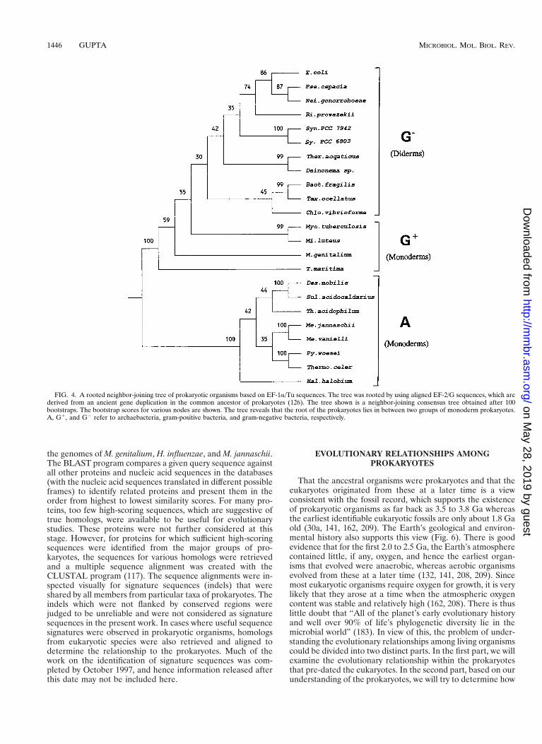
First, based on the duplicated gene sequences for EF-1a/Tuand EF-2/G proteins, where one set of sequences could beused to root the other tree, the roots of both EF-1a/Tu andEF-2/G trees have been shown to lie between the archaebac-terial lineage and the eubacterial species Thermotoga maritima(7, 21, 112). A tree for EF-1a/Tu sequences, which was rootedby using EF-2/G, is shown in Fig. 4. As seen in this figure, theroot of the tree lies in between archaebacteria and eubacteriaand the deepest branches within eubacteria consist of T. ma-ritima and other gram-positive bacteria. A similar rooting ofthe universal tree in between archaebacteria and T. maritimahas been independently made based on trees constructed fromhomologous isoleucine-, leucine-, and valine-tRNA synthetasesequences (20). Although the species T. maritima has beenassumed to be a gram-negative bacterium in the past (184, 250,251, 258), recent studies based on several proteins provideevidence that it should in fact be grouped with gram-positivebacteria (22). This inference is supported by signature se-quences in Hsp70 (Fig. 3) and a number of other proteins (see“Evolutionary relationships among prokaryotes”), where T.maritima behaves similarly to various gram-positive bacteriaand differently from different gram-negative bacteria. Phyloge-netic analyses based on a number of proteins, i.e., Rec A (55,131, 247) and sigma factor 70 (39), also provide evidence of agrouping of T. maritima with gram-positive bacteria. Most im-portantly, Cavalier-Smith (31) has pointed out that T. mari-tima, similar to other gram-positive bacteria, is bounded byonly a single unit lipid membrane, which I consider to be themain defining characteristic of gram-positive bacteria. In viewof these observations, the results of the above rootings indicatethat the root of the prokaryotes lies between archaebacteriaand gram-positive bacteria.
A second independent line of evidence supporting the an-cestral nature of the clade consisting of archaebacteria andgram-positive bacteria is provided by a comparison of se-quences for the Hsp70 and the MreB families of proteins. Wehave previously shown that MreB protein, which is about halfthe length of Hsp70 (about 340 amino acids [aa], with respectto 600 to 650 a.a. for Hsp70) and is present in all major groupsof prokaryotes (archaebacteria, gram-positive bacteria, andgram-negative bacteria), shows significant similarity to the N-terminal half of Hsp70 sequences (107), where the large indelin the Hsp70 homologs is present. The three-dimensional
structures of the MreB protein and the N-terminal half ofHsp70 are also very similar (18, 65a), supporting the view thatthese proteins have evolved from a common ancestor (18).Since both Hsp70 and MreB proteins are found in all maingroups of prokaryotes, they very probably evolved by an an-cient gene duplication in the universal ancestor, before Hsp70acquired the C-terminal domain (104, 107). In view of this, weexpect that if the above indel in Hsp70 is an insert in gram-negative bacteria, the MreB protein sequences should not pos-sess it. On the other hand, if the homologs containing theinsert are ancestral, this insert should also be found in theMreB sequences. A comparison of MreB and Hsp70 sequencesfrom the major group of prokaryotes (Fig. 5) shows that, sim-ilar to the Hsp70 from archaebacteria and gram-positive bac-teria, this insert is not present in any of the MreB sequences,including those from gram-negative bacteria. (It should bementioned that since MreB and Hsp70 are very distant ho-mologs, the sequence similarity between these proteins is lim-ited. However, despite this fact, the inference that MreB pro-tein does not contain the insert is quite apparent.) Thisobservation provides strong independent evidence that theprokaryotic organisms lacking the insert (i.e., archaebacteriaand gram-positive bacteria) are ancestral and that this insertwas introduced into Hsp70 in a common ancestor of the gram-negative bacteria (104, 107). I will refer to this insert, which isa distinguishing feature of gram-negative bacteria and eu-karyotes, as the diderm insert, signifying its point of evolution-ary origin.
Lastly, the view that archaebacteria and gram-positive bac-teria are ancestral lineages is also consistent with the availableevidence concerning the planet’s early environment. Based onEarth’s geological history, the conditions under which the ear-liest organisms evolved were hot and anaerobic (Fig. 6). Thewidespread prevalence of the ability to exist under these con-ditions in various archaebacteria and gram-positive bacteria(67, 186, 232, 250) is consistent with the view that these groupsare ancestral. Based on the above pieces of evidence, all ofwhich lead to a similar inference, I am going to assume that therooting of the prokaryotic tree between (or within) archaebac-teria and gram-positive bacteria is correct, and I will examinewhether this rooting can explain other observations and phy-logenies.
The root provides an important reference point for evolu-tionary studies. By using this reference point, it should now bepossible to understand and interpret signature sequences indifferent proteins to piece together the evolutionary relation-ship and history of the other groups of prokaryotes. In thefollowing sections, I describe signature sequences in differentgroups of species and my interpretation of them based on theabove rooting. Since a great deal of work that follows is basedon signature sequences that are reported for the first time, it isappropriate to describe the approach taken to identify thesignature sequences. The signature sequences in a number ofproteins such as Hsp70 and Hsp60 were empirically discovered(96, 104, 107, 108). However, the complete genomes of severalgram-positive bacteria (Mycoplasma genitalium [73], Myco-plasma pneumoniae [119], and Bacillus subtilis [147], gram-negative bacteria (Haemophilus influenzae [66], Escherichia coli[15], Synechococcus sp. strain PCC 6803 [128], Helicobacterpylori [242], Borrelia burgdorferi [72], and Aquifex aeolicus [45]),and archaebacteria (Methanococcus jannaschii [26], Meth-anobacterium thermoautotrophicum [215], and Archaeoglobusfulgidus [138]) have recently been reported. In view of this, tosearch for signature sequences in different proteins, a system-atic approach was used. For these purposes, we performed aBLAST search (5) on each of the unique proteins identified in
1444 GUPTA MICROBIOL. MOL. BIOL. REV.
on May 28, 2019 by guest
http://mm
br.asm.org/
Dow
nloaded from
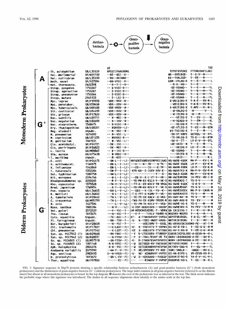
FIG. 3. Signature sequence in Hsp70 proteins showing a specific relationship between archaebacteria (A) and gram-positive bacteria (G1) (both monodermprokaryotes) and the distinctness of gram-negative bacteria (G2) (diderm prokaryotes). The large indel common in all gram-negative bacteria (referred to as the diderminsert) but absent in all monoderm prokaryotes is boxed. In the top diagram, W denotes the root of the prokaryotic tree as inferred in the text. The thick arrow indicatesthe probable stage where this signature was introduced. The dashes in all sequence alignments show identity to the amino acids in the top line.
VOL. 62, 1998 PHYLOGENY OF PROKARYOTES AND EUKARYOTES 1445
on May 28, 2019 by guest
http://mm
br.asm.org/
Dow
nloaded from
the genomes of M. genitalium, H. influenzae, and M. jannaschii.The BLAST program compares a given query sequence againstall other proteins and nucleic acid sequences in the databases(with the nucleic acid sequences translated in different possibleframes) to identify related proteins and present them in theorder from highest to lowest similarity scores. For many pro-teins, too few high-scoring sequences, which are suggestive oftrue homologs, were available to be useful for evolutionarystudies. These proteins were not further considered at thisstage. However, for proteins for which sufficient high-scoringsequences were identified from the major groups of pro-karyotes, the sequences for various homologs were retrievedand a multiple sequence alignment was created with theCLUSTAL program (117). The sequence alignments were in-spected visually for signature sequences (indels) that wereshared by all members from particular taxa of prokaryotes. Theindels which were not flanked by conserved regions werejudged to be unreliable and were not considered as signaturesequences in the present work. In cases where useful sequencesignatures were observed in prokaryotic organisms, homologsfrom eukaryotic species were also retrieved and aligned todetermine the relationship to the prokaryotes. Much of thework on the identification of signature sequences was com-pleted by October 1997, and hence information released afterthis date may not be included here.
EVOLUTIONARY RELATIONSHIPS AMONGPROKARYOTES
That the ancestral organisms were prokaryotes and that theeukaryotes originated from these at a later time is a viewconsistent with the fossil record, which supports the existenceof prokaryotic organisms as far back as 3.5 to 3.8 Ga whereasthe earliest identifiable eukaryotic fossils are only about 1.8 Gaold (30a, 141, 162, 209). The Earth’s geological and environ-mental history also supports this view (Fig. 6). There is goodevidence that for the first 2.0 to 2.5 Ga, the Earth’s atmospherecontained little, if any, oxygen, and hence the earliest organ-isms that evolved were anaerobic, whereas aerobic organismsevolved from these at a later time (132, 141, 208, 209). Sincemost eukaryotic organisms require oxygen for growth, it is verylikely that they arose at a time when the atmospheric oxygencontent was stable and relatively high (162, 208). There is thuslittle doubt that “All of the planet’s early evolutionary historyand well over 90% of life’s phylogenetic diversity lie in themicrobial world” (183). In view of this, the problem of under-standing the evolutionary relationships among living organismscould be divided into two distinct parts. In the first part, we willexamine the evolutionary relationship within the prokaryotesthat pre-dated the eukaryotes. In the second part, based on ourunderstanding of the prokaryotes, we will try to determine how
FIG. 4. A rooted neighbor-joining tree of prokaryotic organisms based on EF-1a/Tu sequences. The tree was rooted by using aligned EF-2/G sequences, which arederived from an ancient gene duplication in the common ancestor of prokaryotes (126). The tree shown is a neighbor-joining consensus tree obtained after 100bootstraps. The bootstrap scores for various nodes are shown. The tree reveals that the root of the prokaryotes lies in between two groups of monoderm prokaryotes.A, G1, and G2 refer to archaebacteria, gram-positive bacteria, and gram-negative bacteria, respectively.
1446 GUPTA MICROBIOL. MOL. BIOL. REV.
on May 28, 2019 by guest
http://mm
br.asm.org/
Dow
nloaded from

eukaryotic organisms are related to the prokaryotes. It shouldbe emphasized that these two questions are completely inde-pendent. Therefore, while considering the evolutionary rela-tionships within prokaryotes, there is no need to confound orbias the evolutionary relationships by considering sequencesfrom various prokaryotes and eukaryotes at the same time, ashas been commonly done in most earlier studies (7, 21, 49, 53,69, 70, 81, 112, 126, 196, 198, 258, 262).
Signature Sequences Showing the Distinctnessof Archaebacteria
Signature sequences consisting of distinct nucleotides thatare present at particular positions in the SSU rRNA and that
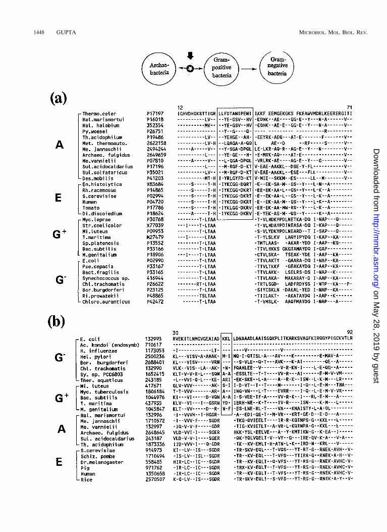
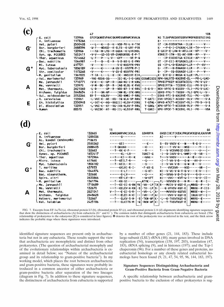
distinguish archaebacteria from other prokaryotes have beendescribed by Woese (251, 253). The view that archaebacteriaare distinct from other prokaryotes is also supported by signa-ture sequences in many proteins. The elongation factor EF-1/Tu provides a well-studied example (Fig. 7a), where a 12-aaindel is present in various archaebacteria but not in any of theeubacteria including different genera of gram-positive bacteria(99, 112). Some other proteins where signature sequencesunique to archaebacteria are found include ribosomal proteinsL5 (Fig. 7b), S5 (Fig. 7c), and L14 (Fig. 7d). As expected fromthese signatures, the inference that archaebacteria are distinctfrom other prokaryotes is strongly supported by phylogeneticanalyses based on rRNA, EF-1/Tu, and these other proteins (7,21, 71, 87, 112, 126, 250). For all the above proteins, the
FIG. 5. Alignment of Hsp70 and MreB sequences from different groups of species showing the absence of the diderm insert in the MreB sequences. The absenceof the insert in all MreB proteins, as well as Hsp70 homologs from archaebacteria and gram-positive bacteria (boxed region), provides evidence that the homologslacking the insert are ancestral. (104, 107). The numbers at the beginning and at the end indicate the position of the sequence in individual proteins.
FIG. 6. Time line showing some of the main events in the history of this planet based on geological and fossil evidence (132, 141, 208, 209).
VOL. 62, 1998 PHYLOGENY OF PROKARYOTES AND EUKARYOTES 1447
on May 28, 2019 by guest
http://mm
br.asm.org/
Dow
nloaded from
1448 GUPTA MICROBIOL. MOL. BIOL. REV.
on May 28, 2019 by guest
http://mm
br.asm.org/
Dow
nloaded from
identified signature sequences are present only in archaebac-teria but not in any eubacteria. These results support the viewthat archaebacteria are monophyletic and distinct from otherprokaryotes. (The question of archaebacterial monophyly andof the evolutionary relationships within archaebacteria is ex-amined in detail below; see “Nature of the archaebacterialgroup and its relationship to gram-positive bacteria”). In myworking model, which places the root between archaebacteriaand gram-positive bacteria, these signatures were probably in-troduced in a common ancestor of either archaebacteria orgram-positive bacteria after separation of the two lineages(diagram in Fig. 7). In addition to these signature sequences,the distinctness of archaebacteria from eubacteria is supported
by a number of other genes (21, 144, 183). These includelarge-subunit (LSU) rRNA (48); many genes involved in DNAreplication (54), transcription (158, 197, 203), translation (47,183), tRNA splicing (9), and in histones (197); and the Tcp-1chaperonin (96). For a number of these genes and proteins, noeubacterial homologs or any closely related eubacterial ho-mologs have been found (9, 21, 47, 54, 95, 96, 144, 183, 197).
Signature Sequences Distinguishing Archaebacteria andGram-Positive Bacteria from Gram-Negative Bacteria
A specific relationship between archaebacteria and gram-positive bacteria to the exclusion of other prokaryotes is sug-
FIG. 7. Excerpts from EF-1a/Tu (a), ribosomal protein L5 (b), ribosomal protein S5 (c), and ribosomal protein L14 (d) alignments identifying signature sequencesthat show the distinctness of archaebacteria (A) from eubacteria (G1 and G2). The common indels that distinguish archaebacteria from eubacteria are boxed. (Therelationship of prokaryotes to the eukaryotes [E] is considered in later figures). W denotes the root of the prokaryotic tree as inferred in the text, and the thick arrowindicates the probable stage where these signatures were introduced.
VOL. 62, 1998 PHYLOGENY OF PROKARYOTES AND EUKARYOTES 1449
on May 28, 2019 by guest
http://mm
br.asm.org/
Dow
nloaded from
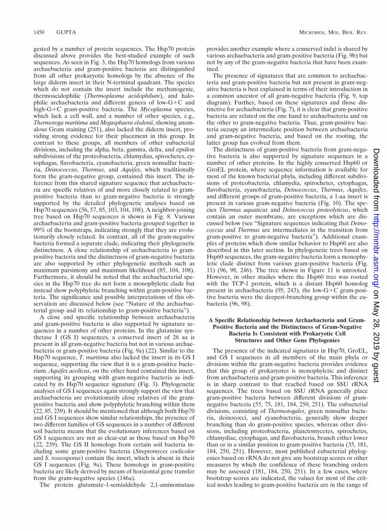
gested by a number of protein sequences. The Hsp70 proteindiscussed above provides the best-studied example of suchsequences. As seen in Fig. 3, the Hsp70 homologs from variousarchaebacteria and gram-positive bacteria are distinguishedfrom all other prokaryotic homologs by the absence of thelarge diderm insert in their N-terminal quadrant. The specieswhich do not contain the insert include the methanogenic,thermoacidophilic (Thermoplasma acidophilum), and halo-philic archaebacteria and different genera of low-G1C andhigh-G1C gram-positive bacteria. The Mycoplasma species,which lack a cell wall, and a number of other species, e.g.,Thermotoga maritima and Megasphaera elsdenii, showing anom-alous Gram staining (251), also lacked the diderm insert, pro-viding strong evidence for their placement in this group. Incontrast to these groups, all members of other eubacterialdivisions, including the alpha, beta, gamma, delta, and epsilonsubdivisions of the proteobacteria, chlamydias, spirochetes, cy-tophagas, flavobacteria, cyanobacteria, green nonsulfur bacte-ria, Deinococcus, Thermus, and Aquifex, which traditionallyform the gram-negative group, contained this insert. The in-ference from this shared signature sequence that archaebacte-ria are specific relatives of and more closely related to gram-positive bacteria than to gram-negative bacteria is stronglysupported by the detailed phylogenetic analyses based onHsp70 sequences (56, 57, 85, 103, 104, 108). A neighbor-joiningtree based on Hsp70 sequences is shown in Fig. 8. Variousarchaebacteria and gram-positive bacteria grouped together in99% of the bootstraps, indicating strongly that they are evolu-tionarily closely related. In contrast, all of the gram-negativebacteria formed a separate clade, indicating their phylogeneticdistinctness. A close relationship of archaebacteria to gram-positive bacteria and the distinctness of gram-negative bacteriaare also supported by other phylogenetic methods such asmaximum parsimony and maximum likelihood (85, 104, 108).Furthermore, it should be noted that the archaebacterial spe-cies in the Hsp70 tree do not form a monophyletic clade butinstead show polyphyletic branching within gram-positive bac-teria. The significance and possible interpretations of this ob-servation are discussed below (see “Nature of the archaebac-terial group and its relationship to gram-positive bacteria”).
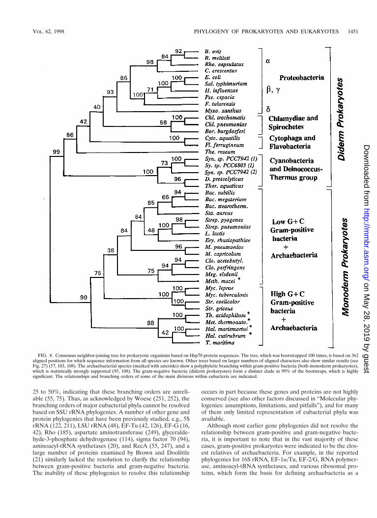
A close and specific relationship between archaebacteriaand gram-positive bacteria is also supported by signature se-quences in a number of other proteins. In the glutamine syn-thetase I (GS I) sequences, a conserved insert of 26 aa ispresent in all gram-negative bacteria but not in various archae-bacteria or gram-positive bacteria (Fig. 9a) (22). Similar to theHsp70 sequence, T. maritima also lacked the insert in its GS Isequence, supporting the view that it is a gram-positive bacte-rium. Aquifex aeolicus, on the other hand contained this insert,supporting its grouping with gram-negative bacteria as indi-cated by its Hsp70 sequence signature (Fig. 3). Phylogeneticanalyses of GS I sequences again strongly support the view thatarchaebacteria are evolutionarily close relatives of the gram-positive bacteria and show polyphyletic branching within them(22, 85, 239). It should be mentioned that although both Hsp70and GS I sequences show similar relationships, the presence oftwo different families of GS sequences in a number of differentsoil bacteria means that the evolutionary inferences based onGS I sequences are not as clear-cut as those based on Hsp70(22, 239). The GS II homologs from certain soil bacteria in-cluding some gram-positive bacteria (Streptomyces coelicolorand S. roseosporus) contain the insert, which is absent in theirGS I sequences (Fig. 9a). These homologs in gram-positivebacteria are likely derived by means of horizontal gene transferfrom the gram-negative species (146a).
The protein glutamate-1-semialdehyde 2,1-aminomutase
provides another example where a conserved indel is shared byvarious archaebacteria and gram-positive bacteria (Fig. 9b) butnot by any of the gram-negative bacteria that have been exam-ined.
The presence of signatures that are common to archaebac-teria and gram-positive bacteria but not present in gram-neg-ative bacteria is best explained in terms of their introduction ina common ancestor of all gram-negative bacteria (Fig. 9, topdiagram). Further, based on these signatures and those dis-tinctive for archaebacteria (Fig. 7), it is clear that gram-positivebacteria are related on the one hand to archaebacteria and onthe other to gram-negative bacteria. Thus, gram-positive bac-teria occupy an intermediate position between archaebacteriaand gram-negative bacteria, and based on the rooting, thelatter group has evolved from them.
The distinctness of gram-positive bacteria from gram-nega-tive bacteria is also supported by signature sequences in anumber of other proteins. In the highly conserved Hsp60 orGroEL protein, where sequence information is available formost of the known bacterial phyla, including different subdivi-sions of proteobacteria, chlamydia, spirochetes, cytophagas,flavobacteria, cyanobacteria, Deinococcus, Thermus, Aquifex,and different groups of gram-positive bacteria, a 1-aa insert ispresent in various gram-negative bacteria (Fig. 10). The spe-cies Thermus aquaticus and Deinococcus proteolyticus, whichcontain an outer membrane, are exceptions which are dis-cussed below (see “Signature sequences indicating that Deino-coccus and Thermus are intermediates in the transition fromgram-positive to gram-negative bacteria”). Additional exam-ples of proteins which show similar behavior to Hsp60 are alsodescribed in this later section. In phylogenetic trees based onHsp60 sequences, the gram-negative bacteria form a monophy-letic clade distinct from various gram-positive bacteria (Fig.11) (96, 98, 246). The tree shown in Figure 11 is unrooted.However, in other studies where the Hsp60 tree was rootedwith the TCP-1 protein, which is a distant Hsp60 homologpresent in archaebacteria (95, 243), the low-G1C gram-posi-tive bacteria were the deepest-branching group within the eu-bacteria (96, 98).
A Specific Relationship between Archaebacteria and Gram-Positive Bacteria and the Distinctness of Gram-Negative
Bacteria Is Consistent with Prokaryotic CellStructures and Other Gene Phylogenies
The presence of the indicated signatures in Hsp70, GroEL,and GS I sequences in all members of the main phyla ordivisions within the gram-negative bacteria provides evidencethat this group of prokaryotes is monophyletic and distinctfrom archaebacteria and gram-positive bacteria. This inferenceis in sharp contrast to that reached based on SSU rRNAsequences. The trees based on SSU rRNA generally placegram-positive bacteria between different divisions of gram-negative bacteria (55, 75, 181, 184, 250, 251). The eubacterialdivisions, consisting of Thermotogales, green nonsulfur bacte-ria, deinococci, and cyanobacteria, generally show deeperbranching than do gram-positive species, whereas other divi-sions, including proteobacteria, planctomycetes, spirochetes,chlamydiae, cytophagas, and flavobacteria, branch either lowerthan or in a similar position to gram-positive bacteria (35, 181,184, 250, 251). However, most published eubacterial phylog-enies based on rRNA do not give any bootstrap scores or othermeasures by which the confidence of these branching ordersmay be assessed (181, 184, 250, 251). In a few cases, wherebootstrap scores are indicated, the values for most of the crit-ical nodes leading to gram-positive bacteria are in the range of
1450 GUPTA MICROBIOL. MOL. BIOL. REV.
on May 28, 2019 by guest
http://mm
br.asm.org/
Dow
nloaded from
25 to 50%, indicating that these branching orders are unreli-able (55, 75). Thus, as acknowledged by Woese (251, 252), thebranching orders of major eubacterial phyla cannot be resolvedbased on SSU rRNA phylogenies. A number of other gene andprotein phylogenies that have been previously studied, e.g., 5SrRNA (122, 211), LSU rRNA (48), EF-Tu (42, 126), EF-G (16,42), Rho (185), aspartate aminotransferase (249), glyceralde-hyde-3-phosphate dehydrogenase (114), sigma factor 70 (94),aminoacyl-tRNA synthetases (20), and RecA (55, 247), and alarge number of proteins examined by Brown and Doolittle(21) similarly lacked the resolution to clarify the relationshipbetween gram-positive bacteria and gram-negative bacteria.The inability of these phylogenies to resolve this relationship
occurs in part because these genes and proteins are not highlyconserved (see also other factors discussed in “Molecular phy-logenies: assumptions, limitations, and pitfalls”), and for manyof them only limited representation of eubacterial phyla wasavailable.
Although most earlier gene phylogenies did not resolve therelationship between gram-positive and gram-negative bacte-ria, it is important to note that in the vast majority of thesecases, gram-positive prokaryotes were indicated to be the clos-est relatives of archaebacteria. For example, in the reportedphylogenies for 16S rRNA, EF-1a/Tu, EF-2/G, RNA polymer-ase, aminoacyl-tRNA synthetases, and various ribosomal pro-teins, which form the basis for defining archaebacteria as a
FIG. 8. Consensus neighbor-joining tree for prokaryotic organisms based on Hsp70 protein sequences. The tree, which was bootstrapped 100 times, is based on 362aligned positions for which sequence information from all species are known. Other trees based on larger numbers of aligned characters also show similar results (seeFig. 27) (57, 103, 108). The archaebacterial species (marked with asterisks) show a polyphyletic branching within gram-positive bacteria (both monoderm prokaryotes),which is statistically strongly supported (95, 108). The gram-negative bacteria (diderm prokaryotes) form a distinct clade in 99% of the bootstraps, which is highlysignificant. The relationships and branching orders of some of the main divisions within eubacteria are indicated.
VOL. 62, 1998 PHYLOGENY OF PROKARYOTES AND EUKARYOTES 1451
on May 28, 2019 by guest
http://mm
br.asm.org/
Dow
nloaded from
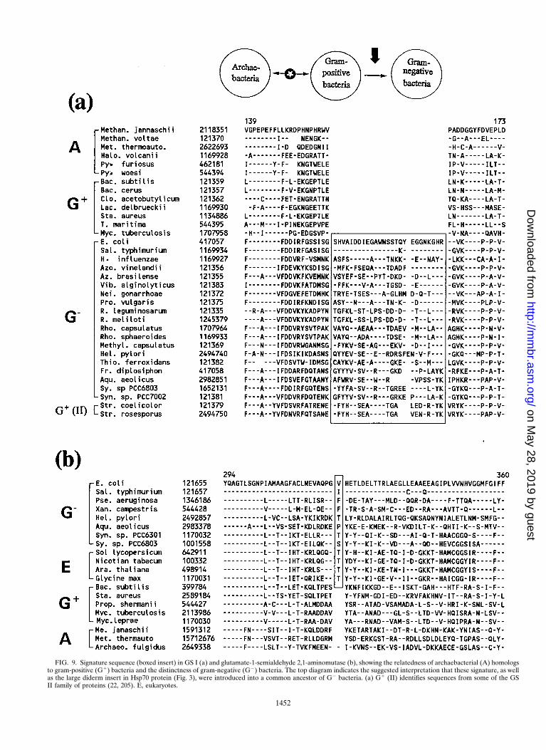
FIG. 9. Signature sequence (boxed insert) in GS I (a) and glutamate-1-semialdehyde 2,1-aminomutase (b), showing the relatedness of archaebacterial (A) homologsto gram-positive (G1) bacteria and the distinctness of gram-negative (G2) bacteria. The top diagram indicates the suggested interpretation that these signature, as wellas the large diderm insert in Hsp70 protein (Fig. 3), were introduced into a common ancestor of G2 bacteria. (a) G1 (II) identifies sequences from some of the GSII family of proteins (22, 205). E, eukaryotes.
1452
on May 28, 2019 by guest
http://mm
br.asm.org/
Dow
nloaded from
unique domain, the species Thermotoga maritima, which is nowknown to be gram positive, shows the closest relationship toarchaebacteria (7, 20, 21, 112, 250). Brown and Doolittle (21)recently reported phylogenies based on 66 protein sequences
for which sequence information was available from archaebac-teria, eubacteria, and eukaryotes. They tried to determinewhich of the three possible relationships among these groups(i.e., an archaebacterial-bacterial clade, an archaebacterial-eu-
FIG. 10. Excerpt from the GroEL (or Hsp60) protein sequence alignment showing a 1-aa insertion (boxed) that is shared by most divisions of G2 bacteria but absentfrom all G1 bacteria. The absence of this insert in Thermus aquaticus and Deinococcus proteolyticus, which are diderm prokaryotes that contain thick cell walls, indicatesthat this insert was introduced into an ancestral gram-negative lineage after the branching of the Deinococcus-Thermus group (thick arrow in the top diagram).
VOL. 62, 1998 PHYLOGENY OF PROKARYOTES AND EUKARYOTES 1453
on May 28, 2019 by guest
http://mm
br.asm.org/
Dow
nloaded from
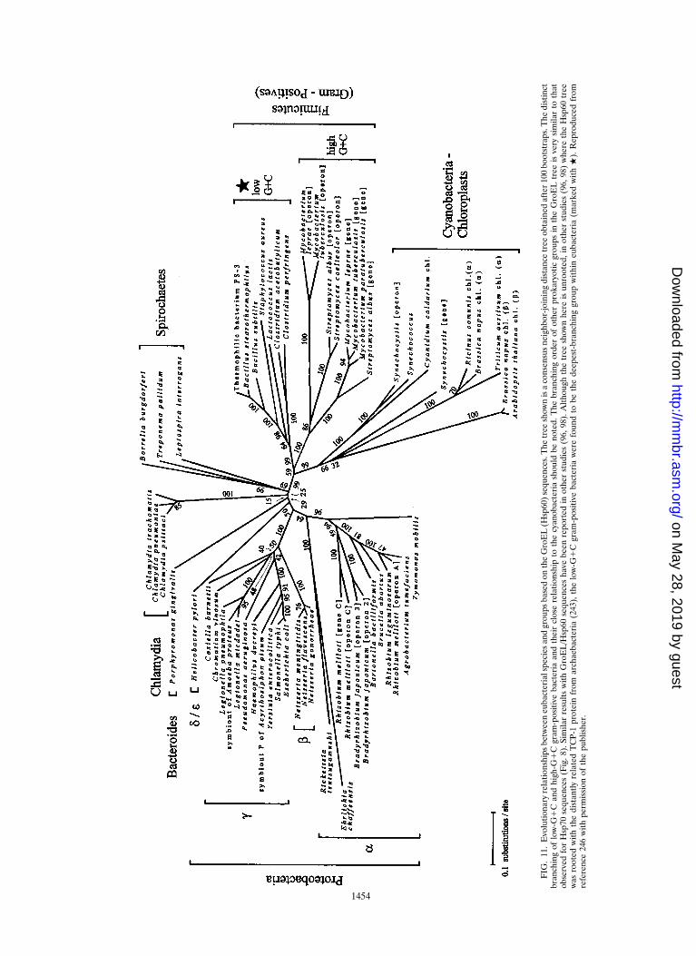
FIG
.11
.E
volu
tiona
ryre
latio
nshi
psbe
twee
neu
bact
eria
lspe
cies
and
grou
psba
sed
onth
eG
roE
L(H
sp60
)se
quen
ces.
The
tree
show
nis
aco
nsen
sus
neig
hbor
-join
ing
dist
ance
tree
obta
ined
afte
r10
0bo
otst
raps
.The
dist
inct
bran
chin
gof
low
-G1
Can
dhi
gh-G
1C
gram
-pos
itive
bact
eria
and
thei
rcl
ose
rela
tions
hip
toth
ecy
anob
acte
ria
shou
ldbe
note
d.T
hebr
anch
ing
orde
rof
othe
rpr
okar
yotic
grou
psin
the
Gro
EL
tree
isve
rysi
mila
rto
that
obse
rved
for
Hsp
70se
quen
ces
(Fig
.8).
Sim
ilar
resu
ltsw
ithG
roE
L/H
sp60
sequ
ence
sha
vebe
enre
port
edin
othe
rst
udie
s(9
6,98
).A
lthou
ghth
etr
eesh
own
here
isun
root
ed,i
not
her
stud
ies
(96,
98)
whe
reth
eH
sp60
tree
was
root
edw
ithth
edi
stan
tlyre
late
dT
CP-
1pr
otei
nfr
omar
chae
bact
eria
(243
),th
elo
w-G
1C
gram
-pos
itive
bact
eria
wer
efo
und
tobe
the
deep
est-
bran
chin
ggr
oup
with
ineu
bact
eria
(mar
ked
with
.).
Rep
rodu
ced
from
refe
renc
e24
6w
ithpe
rmis
sion
ofth
epu
blis
her.
1454
on May 28, 2019 by guest
http://mm
br.asm.org/
Dow
nloaded from
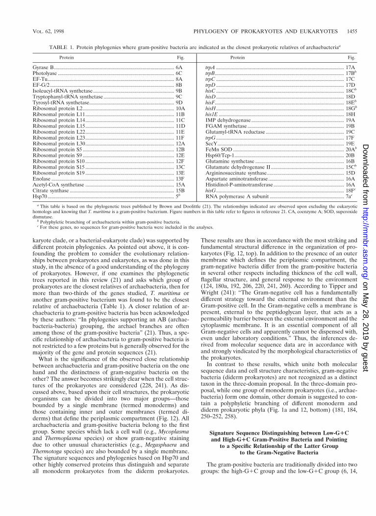
karyote clade, or a bacterial-eukaryote clade) was supported bydifferent protein phylogenies. As pointed out above, it is con-founding the problem to consider the evolutionary relation-ships between prokaryotes and eukaryotes, as was done in thisstudy, in the absence of a good understanding of the phylogenyof prokaryotes. However, if one examines the phylogenetictrees reported in this review (21) and asks which group ofprokaryotes are the closest relatives of archaebacteria, then formore than two-thirds of the genes studied, T. maritima oranother gram-positive bacterium was found to be the closestrelative of archaebacteria (Table 1). A closer relation of ar-chaebacteria to gram-positive bacteria has been acknowledgedby these authors: “In phylogenies supporting an AB (archae-bacteria-bacteria) grouping, the archael branches are oftenamong those of the gram-positive bacteria” (21). Thus, a spe-cific relationship of archaebacteria to gram-positive bacteria isnot restricted to a few proteins but is generally observed for themajority of the gene and protein sequences (21).
What is the significance of the observed close relationshipbetween archaebacteria and gram-positive bacteria on the onehand and the distinctness of gram-negative bacteria on theother? The answer becomes strikingly clear when the cell struc-tures of the prokaryotes are considered (228, 241). As dis-cussed above, based upon their cell structures, the prokaryoticorganisms can be divided into two major groups—thosebounded by a single membrane (termed monoderms) andthose containing inner and outer membranes (termed di-derms) that define the periplasmic compartment (Fig. 12). Allarchaebacteria and gram-positive bacteria belong to the firstgroup. Some species which lack a cell wall (e.g., Mycoplasmaand Thermoplasma species) or show gram-negative stainingdue to other unusual characteristics (e.g., Megasphaera andThermotoga species) are also bounded by a single membrane.The signature sequences and phylogenies based on Hsp70 andother highly conserved proteins thus distinguish and separateall monoderm prokaryotes from the diderm prokaryotes.
These results are thus in accordance with the most striking andfundamental structural difference in the organization of pro-karyotes (Fig. 12, top). In addition to the presence of an outermembrane which defines the periplasmic compartment, thegram-negative bacteria differ from the gram-positive bacteriain several other respects including thickness of the cell wall,flagellar structure, and general response to the environment(124, 180a, 192, 206, 220, 241, 260). According to Tipper andWright (241): “The Gram-negative cell has a fundamentallydifferent strategy toward the external environment than theGram-positive cell. In the Gram-negative cells a membrane ispresent, external to the peptidoglycan layer, that acts as apermeability barrier between the external environment and thecytoplasmic membrane. It is an essential component of allGram-negative cells and apparently cannot be dispensed with,even under laboratory conditions.” Thus, the inferences de-rived from molecular sequence data are in accordance withand strongly vindicated by the morphological characteristics ofthe prokaryotes.
In contrast to these results, which unite both molecularsequence data and cell structure characteristics, gram-negativebacteria (diderm prokaryotes) are not recognized as a distincttaxon in the three-domain proposal. In the three-domain pro-posal, while one group of monoderm prokaryotes (i.e., archae-bacteria) form one domain, other domain is suggested to con-tain a polyphyletic branching of different monoderm anddiderm prokaryotic phyla (Fig. 1a and 12, bottom) (181, 184,250–252, 258).
Signature Sequence Distinguishing between Low-G1Cand High-G1C Gram-Positive Bacteria and Pointing
to a Specific Relationship of the Latter Groupto the Gram-Negative Bacteria
The gram-positive bacteria are traditionally divided into twogroups: the high-G1C group and the low-G1C group (6, 14,
TABLE 1. Protein phylogenies where gram-positive bacteria are indicated as the closest prokaryotic relatives of archaebacteriaa
Protein Fig. Protein Fig.
Gyrase B............................................................................................ 6A trpA .................................................................................................. 17APhotolyase ......................................................................................... 6C trpB................................................................................................... 17Bb
EF-Tu................................................................................................. 8A trpC .................................................................................................. 17CEF-G/2 ............................................................................................... 8B trpD .................................................................................................. 17DIsoleucyl-tRNA synthetase.............................................................. 9B hisC .................................................................................................. 18Cb
Tryptophanyl-tRNA synthetase ...................................................... 9C hisD .................................................................................................. 18DTyrosyl-tRNA synthetase................................................................. 9D hisF................................................................................................... 18Eb
Ribosomal protein L2...................................................................... 10A hisH .................................................................................................. 18Gb
Ribosomal protein L11.................................................................... 11B his1E ................................................................................................ 18HRibosomal protein L14.................................................................... 11C IMP dehydrogenase ....................................................................... 19ARibosomal protein L15.................................................................... 11D FGAM synthetase .......................................................................... 19BRibosomal protein L22.................................................................... 11E Glutamyl-tRNA reductase ............................................................ 19CRibosomal protein L23.................................................................... 11F trpG .................................................................................................. 17FRibosomal protein L30.................................................................... 12A SecY................................................................................................. 19ERibosomal protein S5 ...................................................................... 12B FeMn SOD ..................................................................................... 20Ab
Ribosomal protein S9 ...................................................................... 12E Hsp60/Tcp-1.................................................................................... 20BRibosomal protein S10 .................................................................... 12F Glutamine synthetase .................................................................... 16BRibosomal protein S15 .................................................................... 13C Glutamate dehydrogenase II ........................................................ 15Cb
Ribosomal protein S19 .................................................................... 13E Argininosuccinate synthase........................................................... 15DEnolase .............................................................................................. 13F Aspartate aminotransferase.......................................................... 16AAcetyl-CoA synthetase .................................................................... 15A Histidinol-P-aminotransferase ...................................................... 16ACitrate synthase ................................................................................ 15B hisG.................................................................................................. 18Fc
Hsp70 ................................................................................................. 5b RNA polymerase A subunit ......................................................... 7ac
a This table is based on the phylogenetic trees published by Brown and Doolittle (21). The relationships indicated are observed upon excluding the eukaryotichomologs and knowing that T. maritima is a gram-positive bacterium. Figure numbers in this table refer to figures in reference 21. CA, coenzyme A; SOD, superoxidedismutase.
b Polyphyletic branching of archaebacteria within gram-positive bacteria.c For these genes, no sequences for gram-positive bacteria were included in the analyses.
VOL. 62, 1998 PHYLOGENY OF PROKARYOTES AND EUKARYOTES 1455
on May 28, 2019 by guest
http://mm
br.asm.org/
Dow
nloaded from
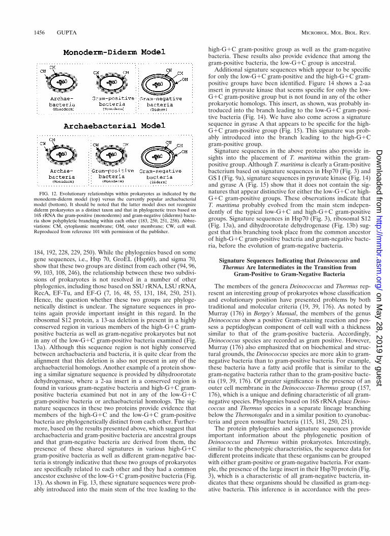
184, 192, 228, 229, 250). While the phylogenies based on somegene sequences, i.e., Hsp 70, GroEL (Hsp60), and sigma 70,show that these two groups are distinct from each other (94, 96,99, 103, 108, 246), the relationship between these two subdivi-sions of prokaryotes is not resolved in a number of otherphylogenies, including those based on SSU rRNA, LSU rRNA,RecA, EF-Tu, and EF-G (7, 16, 48, 55, 131, 184, 250, 251).Hence, the question whether these two groups are phyloge-netically distinct is unclear. The signature sequences in pro-teins again provide important insight in this regard. In theribosomal S12 protein, a 13-aa deletion is present in a highlyconserved region in various members of the high-G1C gram-positive bacteria as well as gram-negative prokaryotes but notin any of the low-G1C gram-positive bacteria examined (Fig.13a). Although this sequence region is not highly conservedbetween archaebacteria and bacteria, it is quite clear from thealignment that this deletion is also not present in any of thearchaebacterial homologs. Another example of a protein show-ing a similar signature sequence is provided by dihydroorotatedehydrogenase, where a 2-aa insert in a conserved region isfound in various gram-negative bacteria and high-G1C gram-positive bacteria examined but not in any of the low-G1Cgram-positive bacteria or archaebacterial homologs. The sig-nature sequences in these two proteins provide evidence thatmembers of the high-G1C and the low-G1C gram-positivebacteria are phylogenetically distinct from each other. Further-more, based on the results presented above, which suggest thatarchaebacteria and gram-positive bacteria are ancestral groupsand that gram-negative bacteria are derived from them, thepresence of these shared signatures in various high-G1Cgram-positive bacteria as well as different gram-negative bac-teria is strongly indicative that these two groups of prokaryotesare specifically related to each other and they had a commonancestor exclusive of the low-G1C gram-positive bacteria (Fig.13). As shown in Fig. 13, these signature sequences were prob-ably introduced into the main stem of the tree leading to the
high-G1C gram-positive group as well as the gram-negativebacteria. These results also provide evidence that among thegram-positive bacteria, the low-G1C group is ancestral.
Additional signature sequences which appear to be specificfor only the low-G1C gram-positive and the high-G1C gram-positive groups have been identified. Figure 14 shows a 2-aainsert in pyruvate kinase that seems specific for only the low-G1C gram-positive group but is not found in any of the otherprokaryotic homologs. This insert, as shown, was probably in-troduced into the branch leading to the low-G1C gram-posi-tive bacteria (Fig. 14). We have also come across a signaturesequence in gyrase A that appears to be specific for the high-G1C gram-positive group (Fig. 15). This signature was prob-ably introduced into the branch leading to the high-G1Cgram-positive group.
Signature sequences in the above proteins also provide in-sights into the placement of T. maritima within the gram-positive group. Although T. maritima is clearly a Gram-positivebacterium based on signature sequences in Hsp70 (Fig. 3) andGS I (Fig. 9a), signature sequences in pyruvate kinase (Fig. 14)and gyrase A (Fig. 15) show that it does not contain the sig-natures that appear distinctive for either the low-G1C or high-G1C gram-positive groups. These observations indicate thatT. maritima probably evolved from the main stem indepen-dently of the typical low-G1C and high-G1C gram-positivegroups. Signature sequences in Hsp70 (Fig. 3), ribosomal S12(Fig. 13a), and dihydroorotate dehydrogenase (Fig. 13b) sug-gest that this branching took place from the common ancestorof high-G1C gram-positive bacteria and gram-negative bacte-ria, before the evolution of gram-negative bacteria.
Signature Sequences Indicating that Deinococcus andThermus Are Intermediates in the Transition from
Gram-Positive to Gram-Negative Bacteria
The members of the genera Deinococcus and Thermus rep-resent an interesting group of prokaryotes whose classificationand evolutionary position have presented problems by bothtraditional and molecular criteria (19, 39, 176). As noted byMurray (176) in Bergey’s Manual, the members of the genusDeinococcus show a positive Gram-staining reaction and pos-sess a peptidoglycan component of cell wall with a thicknesssimilar to that of the gram-positive bacteria. Accordingly,Deinococcus species are recorded as gram positive. However,Murray (176) also emphasized that on biochemical and struc-tural grounds, the Deinococcus species are more akin to gram-negative bacteria than to gram-positive bacteria. For example,these bacteria have a fatty acid profile that is similar to thegram-negative bacteria rather than to the gram-positive bacte-ria (19, 39, 176). Of greater significance is the presence of anouter cell membrane in the Deinococcus-Thermus group (157,176), which is a unique and defining characteristic of all gram-negative species. Phylogenies based on 16S rRNA place Deino-coccus and Thermus species in a separate lineage branchingbelow the Thermotogales and in a similar position to cyanobac-teria and green nonsulfur bacteria (115, 181, 250, 251).
The protein phylogenies and signature sequences provideimportant information about the phylogenetic position ofDeinococcus and Thermus within prokaryotes. Interestingly,similar to the phenotypic characteristics, the sequence data fordifferent proteins indicate that these organisms can be groupedwith either gram-positive or gram-negative bacteria. For exam-ple, the presence of the large insert in their Hsp70 protein (Fig.3), which is a characteristic of all gram-negative bacteria, in-dicates that these organisms should be classified as gram-neg-ative bacteria. This inference is in accordance with the pres-
FIG. 12. Evolutionary relationships within prokaryotes as indicated by themonoderm-diderm model (top) versus the currently popular archaebacterialmodel (bottom). It should be noted that the latter model does not recognizediderm prokaryotes as a distinct taxon and that in phylogenetic trees based on16S rRNA the gram-positive (monoderms) and gram-negative (diderms) bacte-ria show polyphyletic branching within each other (183, 250, 251, 258). Abbre-viations: CM, cytoplasmic membrane; OM, outer membrane; CW, cell wall.Reproduced from reference 101 with permission of the publisher.
1456 GUPTA MICROBIOL. MOL. BIOL. REV.
on May 28, 2019 by guest
http://mm
br.asm.org/
Dow
nloaded from
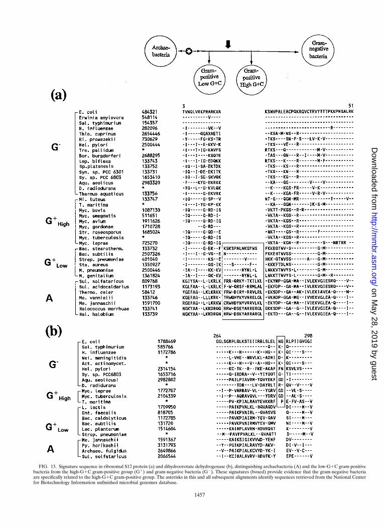
FIG. 13. Signature sequence in ribosomal S12 protein (a) and dihydroorotate dehydrogenase (b), distinguishing archaebacteria (A) and the low-G1C gram-positivebacteria from the high-G1C gram-positive group (G1) and gram-negative bacteria (G2). These signatures (boxed) provide evidence that the gram-negative bacteriaare specifically related to the high-G1C gram-positive group. The asterisks in this and all subsequent alignments identify sequences retrieved from the National Centerfor Biotechnology Information unfinished microbial genomes database.
1457
on May 28, 2019 by guest
http://mm
br.asm.org/
Dow
nloaded from
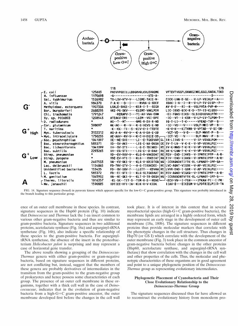
ence of an outer cell membrane in these species. In contrast,signature sequences in the Hsp60 protein (Fig. 10) indicatethat Deinococcus and Thermus lack the 1-aa insert common tovarious other gram-negative bacteria and thus are similar togram-positive bacteria. Signature sequences in two additionalproteins, acetolactate synthase (Fig. 16a) and asparginyl-tRNAsynthetase (Fig. 16b), also indicate a specific relationship ofthese species to the gram-positive bacteria. For asparginyl-tRNA synthetase, the absence of the insert in the proteobac-terium Helicobacter pylori is surprising and may represent acase of horizontal gene transfer.
The above results showing a grouping of the Deinococcus-Thermus genera with either gram-positive or gram-negativebacteria, based on signature sequences in different proteins,are not conflicting but, instead, suggest that the members ofthese genera are probably derivatives of intermediates in thetransition from the gram-positive to the gram-negative groupof prokaryotes and hence possess some characteristics of eachgroup. The presence of an outer cell membrane in these or-ganisms, together with a thick cell wall in the case of Deino-coceaceae, indicates that in the evolution of gram-negativebacteria from a high-G1C gram-positive ancestor, the outermembrane developed first before the changes in the cell wall
took place. It is of interest in this context that in severalmycobacterial species (high-G1C gram-positive bacteria), themembrane lipids are arranged in a highly ordered form, whichmay represent an early stage in the development of outer cellmembrane (18a, 180b). The signature sequences in differentproteins thus provide molecular markers that correlate withthe phenotypic changes in the cell structure. Thus changes inHsp70 (or GS I) which correlate with the development of theouter membrane (Fig. 3) took place in the common ancestor ofgram-negative bacteria before changes in the other proteins(Hsp60, acetolactate synthase, and asparginyl-tRNA syn-thetase) that show correlation with the changes in the cell walland other properties of the cells. Thus, the molecular and phe-notypic characteristics of these organisms are in good agreementand point to a unique phylogenetic position of the Deinococcus-Thermus group as representing evolutionary intermediates.
Phylogenetic Placement of Cyanobacteria and TheirClose Evolutionary Relationship to the
Deinococcus-Thermus Group
The signature sequences discussed thus far have allowed usto reconstruct the evolutionary history from monoderm pro-
FIG. 14. Signature sequence (boxed) in pyruvate kinase which appears specific for the low-G1C gram-positive group. This signature was probably introduced inthe branch leading to this particular group.
1458 GUPTA MICROBIOL. MOL. BIOL. REV.
on May 28, 2019 by guest
http://mm
br.asm.org/
Dow
nloaded from
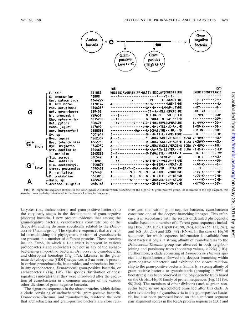
karyotes (i.e., archaebacteria and gram-positive bacteria) tothe very early stages in the development of gram-negative(diderm) bacteria. I now present evidence that among thegram-negative bacteria, cyanobacteria constitute one of thedeepest-branching divisions specifically related to the Deino-coccus-Thermus group. The signature sequences that are help-ful in establishing the phylogenetic position of cyanobacteriaare present in a number of different proteins. These proteinsinclude FtszA, in which a 1-aa insert is present in variousproteobacteria and spirochetes but not in any of the archae-bacteria, gram-positive bacteria, Deinococcus, cyanobacteria,and chloroplast homologs (Fig. 17a). Likewise, in the gluta-mate dehydrogenase (GDH) sequences, a 3-aa insert is presentin various proteobacteria, bacteroides, and spirochetes but notin any cyanobacteria, Deinococcus, gram-positive bacteria, orarchaebacteria (Fig. 17b). The species distribution of thesesignatures indicates that they were introduced after the evolu-tion of cyanobacteria in a common ancestor of the variousother divisions of gram-negative bacteria.
The signature sequences in the above proteins, which definea clade consisting of archaebacteria, gram-positive bacteria,Deinococcus-Thermus, and cyanobacteria, reinforce the viewthat archaebacteria and gram-positive bacteria are close rela-
tives and that within gram-negative bacteria, cyanobacteriaconstitute one of the deepest-branching lineages. This infer-ence is in accordance with the results of detailed phylogeneticstudies based on a number of different gene sequences includ-ing Hsp70 (99, 103), Hsp60 (96, 98, 246), RecA (55, 131, 247),and 16S (33, 250) and 23S (48) rRNAs. In the case of Hsp70sequences, for which sequence information is available frommost bacterial phyla, a strong affinity of cyanobacteria to theDeinococcus-Thermus group was observed in both neighbor-joining and parsimony trees (bootstrap values, .99%) (103).Furthermore, a clade consisting of Deinococcus-Thermus spe-cies and cyanobacteria showed the deepest branching withingram-negative eubacteria and exhibited the closest relation-ship to the gram-positive bacteria. Similarly, a strong affinity ofgram-positive bacteria to cyanobacteria (grouping in 99% ofbootstraps) has been observed in the phylogenetic trees basedon the GroEL-Hsp60 family of protein sequences (Fig. 11) (96,98, 246). The members of other divisions (such as green non-sulfur bacteria and spirochetes) branched after this clade. Aclose relationship of cyanobacteria to the gram-positive bacte-ria has also been proposed based on the significant segmentpair alignment scores in the RecA protein sequences (131) and
FIG. 15. Signature sequence (boxed) in the DNA gyrase A subunit which is specific for the high-G1C gram-positive group. As indicated in the top diagram, thissignature was probably introduced in the branch leading to this group.
VOL. 62, 1998 PHYLOGENY OF PROKARYOTES AND EUKARYOTES 1459
on May 28, 2019 by guest
http://mm
br.asm.org/
Dow
nloaded from

the presence of a G residue at position 1207 in these groups inthe 16S rRNA (250).
In addition to these sequence signatures, which are useful inunderstanding the evolutionary relationships of Deinococcus-Thermus and cyanobacteria to other prokaryotes, a number ofother proteins contain yet another kind of signature sequencethat is unique to only the Deinococcus-Thermus group andcyanobacteria and is not found in any other prokaryotes. In theDnaJ-Hsp40 family of proteins, the homologs from Deinococ-cus-Thermus and cyanobacteria contain a large deletion (68 aa)
which removes the four cysteine-rich repeat domains that arepresent in all other prokaryotic and eukaryotic homologs (Fig.18a) (27). Likewise, in the elongation factor EF-Ts sequences,the homologs from Deinococcus-Thermus and cyanobacteriaharbored a deletion of 55 aa that is not present in otherprokaryotes (Fig. 18b). Two other proteins where signaturesunique to only these groups of prokaryotes are found are theprotein synthesis elongation factor EF-Tu (Fig. 18c) and DNApolymerase I (Fig. 18d) (106). The presence of these uniquelyshared sequence signatures in the Deinococcus-Thermus and
FIG. 16. Signature sequences (boxed) in acetolactate synthase (a) and asparginyl-tRNA synthetase (b) showing a grouping of the Deinococcus-Thermus species witharchaebacteria and gram-positive bacteria. Similar to the Hsp60 protein (Fig. 10), these signatures were introduced in an ancestral gram-negative lineage after thebranching of the Deinococcus-Thermus group.
1460 GUPTA MICROBIOL. MOL. BIOL. REV.
on May 28, 2019 by guest
http://mm
br.asm.org/
Dow
nloaded from
cyanobacterial phyla provides evidence of a close and specificevolutionary relationship between these two groups. Theseresults also suggest that these two groups of organisms had acommon ancestor exclusive of all other prokaryotes. However,this inference is difficult to reconcile with the signature se-quences in other proteins (Hsp60, acetolactate synthase, andasparginyl-tRNA synthetase [Fig. 10 and 16]), which indicatethat cyanobacteria and other gram-negative bacteria had acommon ancestor exclusive of the Deinococcus-Thermus groupand that the Deinococcus-Thermus lineage is more ancestralthan cyanobacteria. To account for these observations, it isnecessary to postulate that cyanobacteria and the Deinococcus-Thermus group are themselves not the direct ancestor of othergram-negative bacteria but branched off from the early ances-tors as shown in the diagram in Fig. 18. Furthermore, to ex-plain the presence of common sequence signatures in thesegroups that are not found in any other prokaryotes, it is nec-essary to postulate that some lateral gene transfers have oc-curred between these groups, as shown by the thin dashedarrow in Fig. 18. The possible significance of such lateral genetransfer events is discussed below (see “Evolutionary relation-ships within prokaryotes: an integrated view based on molec-ular and phenotypic characteristics”).
Signature Sequences Defining Proteobacteria andSome of Their Subdivisions
The proteobacteria, named after the Greek god Proteus andmeaning “capable of assuming many different shapes” (225),comprise one of the largest divisions among gram-negativebacteria. This group of bacteria, also called the “purple bacte-ria and relatives,” exhibits diverse properties and is currentlydefined based mainly on the 16S rRNA phylogeny and signa-ture sequences. Proteobacteria comprise more than 200 generaand have been divided into at least five subclasses: alphathrough epsilon (177). However, the taxonomic relationshipamong proteobacteria, which include a very complex assem-blage of phenotypic and physiological attributes, remains illdefined and has been a cause of concern (177).
In the present work, although the signature sequences thatmay be useful in defining proteobacteria and its subclasseshave not been examined in detail, I have identified some sig-nature sequences that are useful in defining the proteobacte-rial group and some subdivisions within it. The first signaturethat is present in all proteobacteria examined, including mem-bers of all five subdivisions, consists of a 2-aa insert in theHsp70 sequences (Fig. 19a). Interestingly, this signature is alsopresent in the Hsp70 homolog from Thermomicrobium roseum,which is a member of the division “green nonsulfur bacteriaand relatives.” The branching position of the green nonsulfurbacteria, which consists of only a few species (Thermomicro-bium, Herpetosiphon, and Chloroflexus species) in different phy-logenies, including rRNA and Hsp70, has not been satisfacto-rily resolved (103, 184, 250). Hence, the above signaturesequence, which is uniquely shared by various proteobacteriaand a green nonsulfur bacterium but not by any other eubac-terial groups, including cytophagas, flavobacteria, chlamydiae,spirochetes, cyanobacteria, Deinococcus, Thermus, andAquifex, provides the first reliable evidence that some membersof the green nonsulfur group of bacteria show a specific rela-tionship to proteobacteria compared with the other divisions ofeubacteria. The second signature sequence consists of a 4-aainsert in alanyl-tRNA synthetase, which is present in variousspecies examined from the alpha, beta, gamma, and epsilonsubdivisions of proteobacteria (Fig. 19b). Thus far, no se-quences are available for this protein from members of the
delta subdivision or green nonsulfur bacteria. These signaturesequences were probably introduced into a common ancestorof proteobacteria after the branching of chlamydiae, cytopha-gas, and related species (Fig. 19a), and they could be used todefine the proteobacterial group.
Another group of signature sequences that I have identifiedprovides evidence that the members of the beta and gammasubdivisions are distinct from those of the other three subdi-visions. The first of these signature consists of a 4-aa insert ina highly conserved region of Hsp70 (Fig. 20a) that is founduniquely in all members of the beta and gamma subdivisionsthat have been examined. A 1-aa insert in a highly conservedregion, which is specific to only the members of the beta andgamma subdivisions, is also present in DNA gyrase A (Fig.20b). A close affinity of the bacterial species for the beta andgamma subdivisions has also been observed in phylogenetictrees based on a number of genes and proteins, including SSUand LSU rRNA (48, 184, 250), Hsp60 (96, 246), Hsp70 (56,103), RecA (55, 131), and sigma 70 (94). The signature se-quences described above could be used to define the pro-teobacterial group and to distinguish members of the alpha,delta, and epsilon subdivisions (as well as Thermomicrobiumroseum) from those of the beta and gamma subgroups (Fig. 19and 20). These two proteobacterial groups are referred to asproteobacteria-1 and proteobacteria-2, respectively, in the re-mainder of this review.
Nature of the Archaebacterial Group and Its Relationshipto Gram-Positive Bacteria
One of the main premises of current evolutionary thinking isthat archaebacteria comprise a monophyletic group and thatthey are completely distinct from other prokaryotes (54, 183,187, 250, 258). The uniqueness of archaebacteria is indeedsupported by signature sequences in a number of differentgenes and proteins and by the major differences seen betweenarchaebacteria and eubacteria in the information transfer pro-cesses (i.e., replication, transcription, and translation) (9, 47,54, 144, 158, 183, 197). Several other characteristics of archae-bacteria, including the unusual ether-linked nature of theirmembrane lipids (127, 133), are also consistent with this view.While these molecular features and characteristics point to thedifferences between archaebacteria and other prokaryotes, it isessential that we critically examine this relationship to under-stand the significance of these differences and the overall re-lationship of archaebacteria to other prokaryotes.
Within archaebacteria, phylogenetic analyses based onrRNA and EF-1 and EF-2 sequences have identified two maingroups: Crenarchaeota and Euryarchaeota (149, 153, 184, 198,250, 258). These groups are also distinguished from each otherbased on a signature sequence in the EF-1a/Tu protein, iden-tified by Rivera and Lake (198). The Crenarchaeota consistalmost exclusively of sulfur-dependent thermoacidophilic ar-chaebacteria (250, 251, 258), and these genera are referred toas “eocytes” by Lake and coworkers (149, 154, 155, 198). TheEuryarchaeota are phenotypically very diverse and have nospecific physiological attribute. As indicated by Woese (253),this group is a “potpourri of all the archael types” and containsmembers from all phenotypically diverse archaebacteria in-cluding extreme halophiles, methanogens, sulfate-reducing ar-chaebacteria, and sulfur-dependent thermoacidophilic archae-bacteria. It is important to note that within the Euryarchaeota,members of different physiologically diverse archaebacterialphenotypes (halophiles, methanogens, thermoacidophiles, etc.)are not resolved from each other but show polyphyletic branch-ing within three main clusters of methanogens (the Methano-
1461
on May 28, 2019 by guest
http://mm
br.asm.org/
Dow
nloaded from
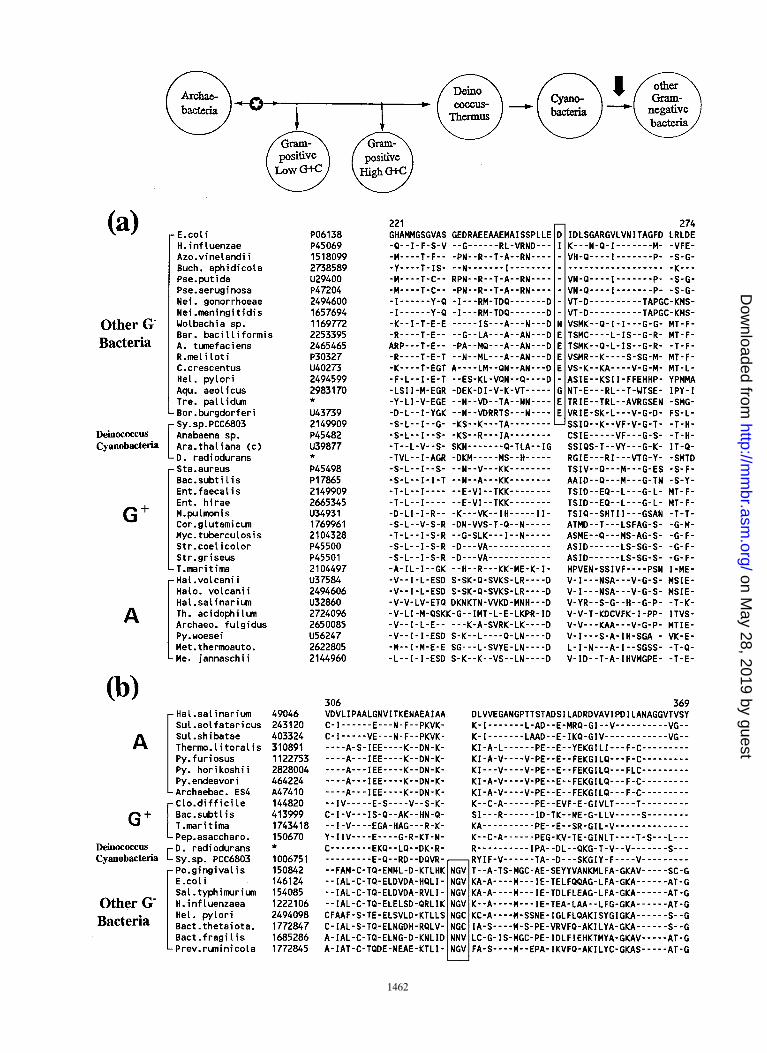
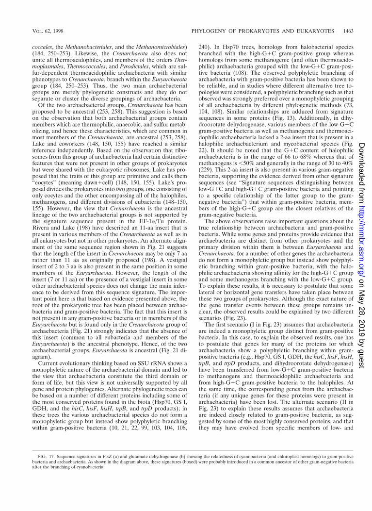
coccales, the Methanobacteriales, and the Methanomicrobiales)(184, 250–253). Likewise, the Crenarchaeota also does notunite all thermoacidophiles, and members of the orders Ther-moplasmales, Thermococcales, and Pyrodictales, which are sul-fur-dependent thermoacidophilic archaebacteria with similarphenotypes to Crenarchaeota, branch within the Euryarchaeotagroup (184, 250–253). Thus, the two main archaebacterialgroups are merely phylogenetic constructs and they do notseparate or cluster the diverse groupings of archaebacteria.
Of the two archaebacterial groups, Crenarchaeota has beenproposed to be ancestral (253, 258). This suggestion is basedon the observation that both archaebacterial groups containmembers which are thermophilic, anaerobic, and sulfur metab-olizing, and hence these characteristics, which are common inmost members of the Crenarchaeota, are ancestral (253, 258).Lake and coworkers (148, 150, 155) have reached a similarinference independently. Based on the observation that ribo-somes from this group of archaebacteria had certain distinctivefeatures that were not present in other groups of prokaryotesbut were shared with the eukaryotic ribosomes, Lake has pro-posed that the traits of this group are primitive and calls them“eocytes” (meaning dawn1cell) (148, 150, 155). Lake’s pro-posal divides the prokaryotes into two groups, one consisting ofonly eocytes and the other encompassing all of the halophiles,methanogens, and different divisions of eubacteria (148–150,155). However, the view that Crenarchaeota is the ancestrallineage of the two archaebacterial groups is not supported bythe signature sequence present in the EF-1a/Tu protein.Rivera and Lake (198) have described an 11-aa insert that ispresent in various members of the Crenarchaeota as well as inall eukaryotes but not in other prokaryotes. An alternate align-ment of the same sequence region shown in Fig. 21 suggeststhat the length of the insert in Crenarchaeota may be only 7 aarather than 11 aa as originally proposed (198). A vestigialinsert of 2 to 3 aa is also present in the same position in somemembers of the Euryarchaeota. However, the length of theinsert (7 or 11 aa) or the presence of a vestigial insert in someother archaebacterial species does not change the main infer-ence to be derived from this sequence signature. The impor-tant point here is that based on evidence presented above, theroot of the prokaryotic tree has been placed between archae-bacteria and gram-positive bacteria. The fact that this insert isnot present in any gram-positive bacteria or in members of theEuryarchaeota but is found only in the Crenarchaeota group ofarchaebacteria (Fig. 21) strongly indicates that the absence ofthis insert (common to all eubacteria and members of theEuryarchaeota) is the ancestral phenotype. Hence, of the twoarchaebacterial groups, Euryarchaeota is ancestral (Fig. 21 di-agram).
Current evolutionary thinking based on SSU rRNA shows amonophyletic nature of the archaebacterial domain and led tothe view that archaebacteria constitute the third domain orform of life, but this view is not universally supported by allgene and protein phylogenies. Alternate phylogenetic trees canbe based on a number of different proteins including some ofthe most conserved proteins found in the biota (Hsp70, GS I,GDH, and the hisC, hisF, hisH, trpB, and trpD products); inthese trees the various archaebacterial species do not form amonophyletic group but instead show polyphyletic branchingwithin gram-positive bacteria (10, 21, 22, 99, 103, 104, 108,
240). In Hsp70 trees, homologs from halobacterial speciesbranched with the high-G1C gram-positive group whereashomologs from some methanogenic (and often thermoacido-philic) archaebacteria grouped with the low-G1C gram-posi-tive bacteria (108). The observed polyphyletic branching ofarchaebacteria with gram-positive bacteria has been shown tobe reliable, and in studies where different alternative tree to-pologies were considered, a polyphyletic branching such as thatobserved was strongly preferred over a monophyletic groupingof all archaebacteria by different phylogenetic methods (73,104, 108). Similar relationships are adduced from signaturesequences in some proteins (Fig. 13). Additionally, in dihy-droorotate dehydrogenase, various members of the low-G1Cgram-positive bacteria as well as methanogenic and thermoaci-dophilic archaebacteria lacked a 2-aa insert that is present in ahalophilic archaebacterium and mycobacterial species (Fig.22). It should be noted that the G1C content of halophilicarchaebacteria is in the range of 66 to 68% whereas that ofmethanogens is ,50% and generally in the range of 30 to 40%(229). This 2-aa insert is also present in various gram-negativebacteria, supporting the evidence derived from other signaturesequences (see “Signature sequences distinguishing betweenlow-G1C and high-G1C gram-positive bacteria and pointingto a specific relationship of the latter group to the gram-negative bacteria”) that within gram-positive bacteria, mem-bers of the high-G1C group are the closest relatives of thegram-negative bacteria.
The above observations raise important questions about thetrue relationship between archaebacteria and gram-positivebacteria. While some genes and proteins provide evidence thatarchaebacteria are distinct from other prokaryotes and theprimary division within them is between Euryarchaeota andCrenarchaeota, for a number of other genes the archaebacteriado not form a monophyletic group but instead show polyphyl-etic branching within gram-positive bacteria, with the halo-philic archaebacteria showing affinity for the high-G1C groupand some methanogens branching with the low-G1C group.To explain these results, it is necessary to postulate that somelateral or horizontal gene transfers have taken place betweenthese two groups of prokaryotes. Although the exact nature ofthe gene transfer events between these groups remains un-clear, the observed results could be explained by two differentscenarios (Fig. 23).
The first scenario (I in Fig. 23) assumes that archaebacteriaare indeed a monophyletic group distinct from gram-positivebacteria. In this case, to explain the observed results, one hasto postulate that genes for many of the proteins for whicharchaebacteria show a polyphyletic branching within gram-positive bacteria (e.g., Hsp70, GS I, GDH, the hisC, hisF, hisH,trpB, and trpD products, and dihydroorotate dehydrogenase)have been transferred from low-G1C gram-positive bacteriato methanogens and thermoacidophilic archaebacteria andfrom high-G1C gram-positive bacteria to the halophiles. Atthe same time, the corresponding genes from the archaebac-teria (if any unique genes for these proteins were present inarchaebacteria) have been lost. The alternate scenario (II inFig. 23) to explain these results assumes that archaebacteriaare indeed closely related to gram-positive bacteria, as sug-gested by some of the most highly conserved proteins, and thatthey may have evolved from specific members of low- and
FIG. 17. Sequence signatures in FtsZ (a) and glutamate dehydrogenase (b) showing the relatedness of cyanobacteria (and chloroplast homologs) to gram-positivebacteria and archaebacteria. As shown in the diagram above, these signatures (boxed) were probably introduced in a common ancestor of other gram-negative bacteriaafter the branching of cyanobacteria.
VOL. 62, 1998 PHYLOGENY OF PROKARYOTES AND EUKARYOTES 1463
on May 28, 2019 by guest
http://mm
br.asm.org/
Dow
nloaded from

FIG. 18. Signature sequences in DnaJ (a), EF-Ts protein (b), EF-Tu protein (c), and DNA polymerase I (d) that are unique to only the Deinococcus-Thermus groupand cyanobacteria. To explain the presence of these signatures (boxed), as well as those in Fig. 10 and 16, it was suggested that these signatures were introduced initiallyinto the branch leading to cyanobacteria (thick arrow) and then laterally transferred to the Deinococcus-Thermus group (thin dashed arrow). The alternate possibility,that these signatures were first introduced into the branch leading to Deinococcus-Thermus and then transferred to cyanobacteria, is also possible. Panels b throughd reproduced from reference 106 with permission of the publisher.
1464 GUPTA MICROBIOL. MOL. BIOL. REV.
on May 28, 2019 by guest
http://mm
br.asm.org/
Dow
nloaded from

high-G1C gram-positive bacteria, as suggested by these phy-logenies and signature sequences, as well as the G1C contentof the halophilic (high-G1C) and methanogenic (low-G1C)archaebacteria (229). In this case, to account for the resultsfrom different gene phylogenies, one has to postulate that thegenes for many functions that indicate a monophyletic natureof archaebacteria were transferred from one or more gram-positive bacteria that originally evolved such changes into oth-ers. This latter scenario, if true, suggests that the earliest pro-karyote was a low-G1C gram-positive bacterium.
Both of these possibilities could explain the observed results,and neither should be dismissed a priori without serious con-sideration. In the past, supporters of the three-domain pro-
posal have favored the first of these possibilities (7, 21, 53, 183,216), and the alternate possibility has not been considered.
Possible Selective Forces Leading to HorizontalGene Transfers
From the signature sequences that I have described thus far,it should be evident that the horizontal gene transfer betweenspecies is not all that common. If this was occurring commonlyand indiscriminately between species, the clear distinction be-tween different phylogenetic groups that we have observedbased on signature sequences in different proteins would nothave been possible. These results are at variance with the
FIG. 18—Continued.
VOL. 62, 1998 PHYLOGENY OF PROKARYOTES AND EUKARYOTES 1465
on May 28, 2019 by guest
http://mm
br.asm.org/
Dow
nloaded from

FIG. 19. Signature sequences (boxed) in Hsp70 (a) and alanyl-tRNA synthetase (b), defining and distinguishing proteobacterial group from all other divisions ofprokaryotes.
1466 GUPTA MICROBIOL. MOL. BIOL. REV.
on May 28, 2019 by guest
http://mm
br.asm.org/
Dow
nloaded from
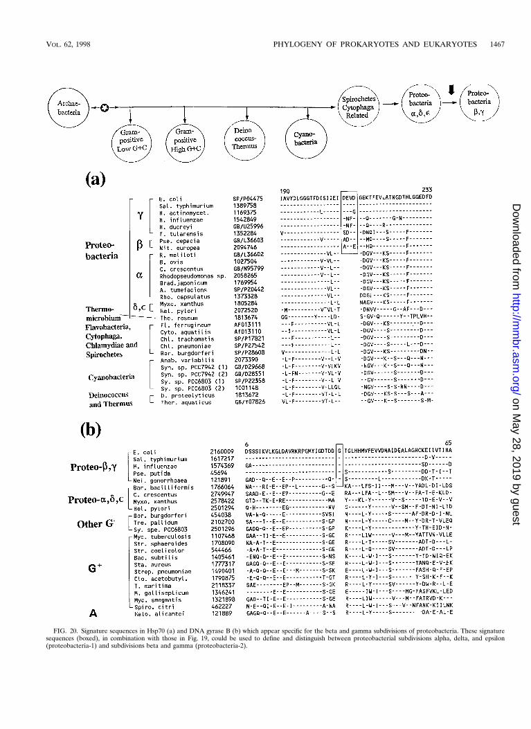
FIG. 20. Signature sequences in Hsp70 (a) and DNA gyrase B (b) which appear specific for the beta and gamma subdivisions of proteobacteria. These signaturesequences (boxed), in combination with those in Fig. 19, could be used to define and distinguish between proteobacterial subdivisions alpha, delta, and epsilon(proteobacteria-1) and subdivisions beta and gamma (proteobacteria-2).
VOL. 62, 1998 PHYLOGENY OF PROKARYOTES AND EUKARYOTES 1467
on May 28, 2019 by guest
http://mm
br.asm.org/
Dow
nloaded from
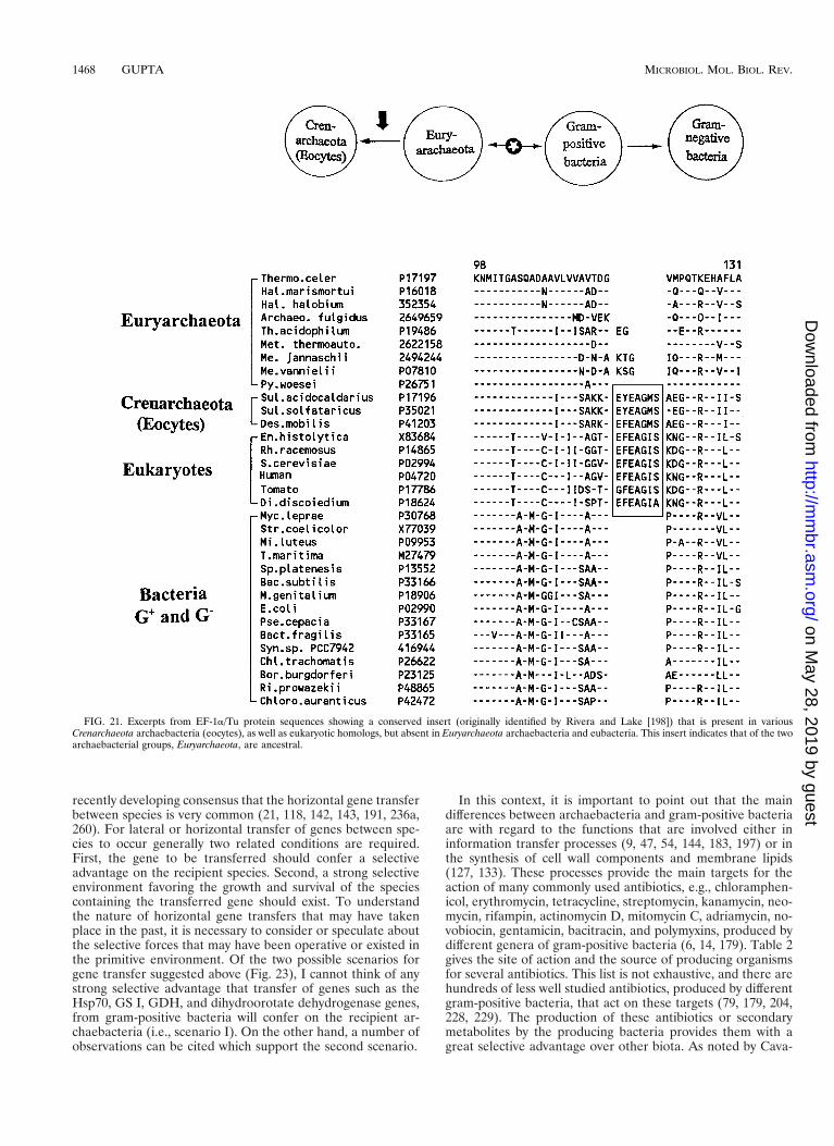
recently developing consensus that the horizontal gene transferbetween species is very common (21, 118, 142, 143, 191, 236a,260). For lateral or horizontal transfer of genes between spe-cies to occur generally two related conditions are required.First, the gene to be transferred should confer a selectiveadvantage on the recipient species. Second, a strong selectiveenvironment favoring the growth and survival of the speciescontaining the transferred gene should exist. To understandthe nature of horizontal gene transfers that may have takenplace in the past, it is necessary to consider or speculate aboutthe selective forces that may have been operative or existed inthe primitive environment. Of the two possible scenarios forgene transfer suggested above (Fig. 23), I cannot think of anystrong selective advantage that transfer of genes such as theHsp70, GS I, GDH, and dihydroorotate dehydrogenase genes,from gram-positive bacteria will confer on the recipient ar-chaebacteria (i.e., scenario I). On the other hand, a number ofobservations can be cited which support the second scenario.
In this context, it is important to point out that the maindifferences between archaebacteria and gram-positive bacteriaare with regard to the functions that are involved either ininformation transfer processes (9, 47, 54, 144, 183, 197) or inthe synthesis of cell wall components and membrane lipids(127, 133). These processes provide the main targets for theaction of many commonly used antibiotics, e.g., chloramphen-icol, erythromycin, tetracycline, streptomycin, kanamycin, neo-mycin, rifampin, actinomycin D, mitomycin C, adriamycin, no-vobiocin, gentamicin, bacitracin, and polymyxins, produced bydifferent genera of gram-positive bacteria (6, 14, 179). Table 2gives the site of action and the source of producing organismsfor several antibiotics. This list is not exhaustive, and there arehundreds of less well studied antibiotics, produced by differentgram-positive bacteria, that act on these targets (79, 179, 204,228, 229). The production of these antibiotics or secondarymetabolites by the producing bacteria provides them with agreat selective advantage over other biota. As noted by Cava-
FIG. 21. Excerpts from EF-1a/Tu protein sequences showing a conserved insert (originally identified by Rivera and Lake [198]) that is present in variousCrenarchaeota archaebacteria (eocytes), as well as eukaryotic homologs, but absent in Euryarchaeota archaebacteria and eubacteria. This insert indicates that of the twoarchaebacterial groups, Euryarchaeota, are ancestral.
1468 GUPTA MICROBIOL. MOL. BIOL. REV.
on May 28, 2019 by guest
http://mm
br.asm.org/
Dow
nloaded from

lier-Smith (31): “Secondary metabolites (antibiotics) are mostoften beneficial to their producers as agents of the chemicalwarfare which is perpetually being waged against competitors,predators and parasites.” Thus, it is quite likely that in theprimitive environment some groups of gram-positive bacteriawere producing antibiotics and others were sensitive to them,so that their survival was at stake. To survive in this environ-ment, the sensitive bacteria had to undergo changes in thetarget sites of the above antibiotics so that their growth was nolonger inhibited (6, 14, 44, 79, 179, 204, 223). There was thusvery strong selection pressure on the genes that were the tar-gets for these antibiotics to undergo changes to survive in theselective environment.
It is possible that under these conditions, after a long periodof (repeated) selection in the primitive environment, assistedby every conceivable sort of stress (83), a resistant strainevolved that had undergone extensive changes in the genesthat are the targets of the above antibiotics. This resistant
strain may have been an ancestor of the present-day archae-bacteria. Once a bacterium has developed a successful strategyto combat the effects of antibiotics, the other sensitive bacteriacan readily acquire the resistance by means of genetic ex-change or horizontal gene transfer from the resistant strain(36, 37, 44, 204, 223). As stated by Cohan (37): “Adaptationsmay be passed from one bacterial species to another, either byhomologous recombination or by plasmid exchange. . . . Thegenes that can be transferred across taxa are necessarily a verysmall set that confer general adaptations which are not limitedto the ecological and genetic context of a particular taxon (e.g.,genes conferring resistance to widely used antibiotics). . . . Asingle genetic exchange in which an adaptation is transferredacross taxa can change forever the course of adaptive evolutionin the recipient species.” This scenario can readily explain whyin phylogenies based on such gene sequences as those encod-ing rRNA, EF-1a/Tu, EF-2/G, etc., phenotypically and physi-ologically diverse groups of monoderm prokaryotes (methano-
FIG. 22. Signature sequence in dihydroorotate dehydrogenase showing the relatedness of halophilic archaebacteria to the high-G1C gram-positive bacteria and ofthe methanogenic and thermoacidophilic archaebacteria to the low-G1C group. The thick arrow indicates that similar to other protein sequences (Fig. 13), thissignature provides evidence that gram-negative bacteria are specifically related to the high-G1C gram-positive group. To explain the results with archaebacterialhomologs, it is necessary to postulate either that there was a lateral gene transfer from high-G1C gram-positive bacteria to the halophilic archaebacteria or that thetwo groups of archaebacteria bear specific relationships to the two divisions of gram-positive bacteria (thin solid arrows). The question mark indicates that these resultsraise questions about the evolutionary relationship between archaebacteria and gram-positive bacteria.
VOL. 62, 1998 PHYLOGENY OF PROKARYOTES AND EUKARYOTES 1469
on May 28, 2019 by guest
http://mm
br.asm.org/
Dow
nloaded from
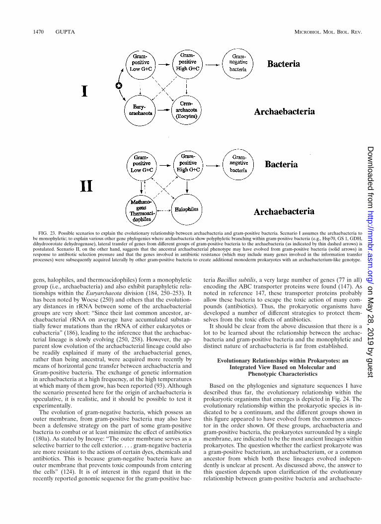
gens, halophiles, and thermoacidophiles) form a monophyleticgroup (i.e., archaebacteria) and also exhibit paraphyletic rela-tionships within the Euryarchaeota division (184, 250–253). Ithas been noted by Woese (250) and others that the evolution-ary distances in rRNA between some of the archaebacterialgroups are very short: “Since their last common ancestor, ar-chaebacterial rRNA on average have accumulated substan-tially fewer mutations than the rRNA of either eukaryotes oreubacteria” (186), leading to the inference that the archaebac-terial lineage is slowly evolving (250, 258). However, the ap-parent slow evolution of the archaebacterial lineage could alsobe readily explained if many of the archaebacterial genes,rather than being ancestral, were acquired more recently bymeans of horizontal gene transfer between archaebacteria andGram-positive bacteria. The exchange of genetic informationin archaebacteria at a high frequency, at the high temperaturesat which many of them grow, has been reported (93). Althoughthe scenario presented here for the origin of archaebacteria isspeculative, it is realistic, and it should be possible to test itexperimentally.
The evolution of gram-negative bacteria, which possess anouter membrane, from gram-positive bacteria may also havebeen a defensive strategy on the part of some gram-positivebacteria to combat or at least minimize the effect of antibiotics(180a). As stated by Inouye: “The outer membrane serves as aselective barrier to the cell exterior. . . . gram-negative bacteriaare more resistant to the actions of certain dyes, chemicals andantibiotics. This is because gram-negative bacteria have anouter membrane that prevents toxic compounds from enteringthe cells” (124). It is of interest in this regard that in therecently reported genomic sequence for the gram-positive bac-
teria Bacillus subtilis, a very large number of genes (77 in all)encoding the ABC transporter proteins were found (147). Asnoted in reference 147, these transporter proteins probablyallow these bacteria to escape the toxic action of many com-pounds (antibiotics). Thus, the prokaryotic organisms havedeveloped a number of different strategies to protect them-selves from the toxic effects of antibiotics.
It should be clear from the above discussion that there is alot to be learned about the relationship between the archae-bacteria and gram-positive bacteria and the monophyletic anddistinct nature of archaebacteria is far from established.
Evolutionary Relationships within Prokaryotes: anIntegrated View Based on Molecular and
Phenotypic Characteristics
Based on the phylogenies and signature sequences I havedescribed thus far, the evolutionary relationship within theprokaryotic organisms that emerges is depicted in Fig. 24. Theevolutionary relationship within the prokaryotic species is in-dicated to be a continuum, and the different groups shown inthis figure appeared to have evolved from the common ances-tor in the order shown. Of these groups, archaebacteria andgram-positive bacteria, the prokaryotes surrounded by a singlemembrane, are indicated to be the most ancient lineages withinprokaryotes. The question whether the earliest prokaryote wasa gram-positive bacterium, an archaebacterium, or a commonancestor from which both these lineages evolved indepen-dently is unclear at present. As discussed above, the answer tothis question depends upon clarification of the evolutionaryrelationship between gram-positive bacteria and archaebacte-
FIG. 23. Possible scenarios to explain the evolutionary relationship between archaebacteria and gram-positive bacteria. Scenario I assumes the archaebacteria tobe monophyletic; to explain various other gene phylogenies where archaebacteria show polyphyletic branching within gram-positive bacteria (e.g., Hsp70, GS 1, GDH,dihydroorotate dehydrogenase), lateral transfer of genes from different groups of gram-positive bacteria to the archaebacteria (as indicated by thin dashed arrows) ispostulated. Scenario II, on the other hand, suggests that the ancestral archaebacterial phenotype may have evolved from gram-positive bacteria (solid arrows) inresponse to antibiotic selection pressure and that the genes involved in antibiotic resistance (which may include many genes involved in the information transferprocesses) were subsequently acquired laterally by other gram-positive bacteria to create additional monoderm prokaryotes with an archaebacterium-like genotype.
1470 GUPTA MICROBIOL. MOL. BIOL. REV.
on May 28, 2019 by guest
http://mm
br.asm.org/
Dow
nloaded from
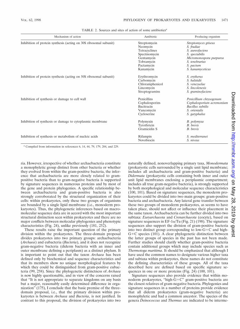
ria. However, irrespective of whether archaebacteria constitutea monophyletic group distinct from other bacteria or whetherthey evolved from within the gram-positive bacteria, the infer-ence that archaebacteria are more closely related to gram-positive bacteria than to gram-negative bacteria is supportedby signature sequences in numerous proteins and by most ofthe gene and protein phylogenies. A specific relationship be-tween archaebacteria and gram-positive bacteria is alsostrongly corroborated by the structural organization of theircells: within prokaryotes, only these two groups of organismsare bounded by a single lipid membrane (i.e., monoderm pro-karyotes). Thus, the phylogenetic inferences based on macro-molecular sequence data are in accord with the most importantstructural distinction seen within prokaryotes and there are nomajor conflicts between molecular phylogenies and phenotypiccharacteristics (Fig. 24), unlike previously (181, 252).
These results raise the important question of the primarydivision within the prokaryotes. The three-domain proposaldivides prokaryotes into two primary groups: archaebacteria(Archaea) and eubacteria (Bacteria), and it does not recognizegram-negative bacteria (diderm bacteria with an inner andouter membrane defining a periplasm) as a distinct phylum. Itis important to point out that the taxon Archaea has beendefined only by biochemical and sequence characteristics andthat its members show no unique morphological features bywhich they could be distinguished from gram-positive eubac-teria (99, 258). Since the phylogenetic distinctness of Archaeais now highly questionable, and in view of the concerns raisedthat “It is not appropriate to separate kingdoms on any basisbut a major, reasonably easily determined difference in orga-nization” (175), I conclude that the basic premise of the three-domain proposal, i.e., that the primary division within pro-karyotes is between Archaea and Bacteria, is not justified. Incontrast to this proposal, the division of prokaryotes into two
naturally defined, nonoverlapping primary taxa, Monodermata(prokaryotic cells surrounded by a single unit lipid membrane;includes all archaebacteria and gram-positive bacteria) andDidermata (prokaryotic cells containing both inner and outerunit lipid membranes enclosing a periplasmic compartment;includes all true gram-negative bacteria), is strongly supportedby both morphological and molecular sequence characteristics(100, 101). Based on signature sequences, the monoderm pro-karyotes could be divided into two main groups: gram-positivebacteria and archaebacteria. Any lateral gene transfer betweenthese two groups of monoderm prokaryotes, as seems to havetaken place, should not affect or influence their placement inthe same taxon. Archaebacteria can be further divided into twosubtaxa: Euryarchaeota and Crenarchaeota (eocyte), based onsignature sequences in EF-1a/Tu (Fig. 21) (198). The signaturesequences also support the division of gram-positive bacteriainto two distinct group corresponding to low-G1C and high-G1C species (101). A clear phylogenetic distinction betweenthe latter groups of species in the past has not been made.Further studies should clarify whether gram-positive bacteriacontain additional groups which may include species such asThermotoga maritima. It should be emphasized that although Ihave used the common names to designate various higher taxaand subtaxa within prokaryotes, these names do not constitutethe defining characteristics of these groups. All of the taxadescribed here are defined based on specific signature se-quences in one or more proteins (Fig. 24) (100, 101).
Signature sequences also provide evidence that within mo-noderm prokaryotes, “high-G1C” gram-positive bacteria arethe closest relatives of gram-negative bacteria. Phylogenies andsignature sequences in a number of proteins provide evidencethat all diderm prokaryotes (gram-negative bacteria) aremonophyletic and had a common ancestor. The species of thegenera Deinococcus and Thermus are indicated to be interme-
TABLE 2. Sources and sites of action of some antibioticsa
Mechanism of action Antibiotic Producing organism
Inhibition of protein synthesis (acting on 30S ribosomal subunit) Streptomycin Streptomyces griseusNeomycin S. fradiaeTetracyclines S. aureofaciensSpectinomycin S. spectabilisGentamycin Micromonospora purpureaTobramycin S. tenebrariusPactamycin S. pactumKanamycin S. kanamyceticus
Inhibition of protein synthesis (acting on 50S ribosomal subunit) Erythromycin S. erytherusCarbomycin S. halstidiiChloramphenicol S. venezuelaeLincomycin S. lincolenesisStreptogramins S. graminofaciens
Inhibition of synthesis or damage to cell wall Penicillins Penicillium chrysogenumCephalosporins Cephalosporium acremoniumBacitracin Bacillus subtilisVancomycin S. orientalisCycloserine S. garyphalus
Inhibition of synthesis or damage to cytoplasmic membrane Polymyxin B. polymyxaTyrothricin B. brevisGramicidin S B. brevis
Inhibition of synthesis or metabolism of nucleic acids Rifampin S. mediterraneiNovobiocin S. niveus
a Compiled from information in references 6, 14, 44, 79, 179, 204, and 229.
VOL. 62, 1998 PHYLOGENY OF PROKARYOTES AND EUKARYOTES 1471
on May 28, 2019 by guest
http://mm
br.asm.org/
Dow
nloaded from
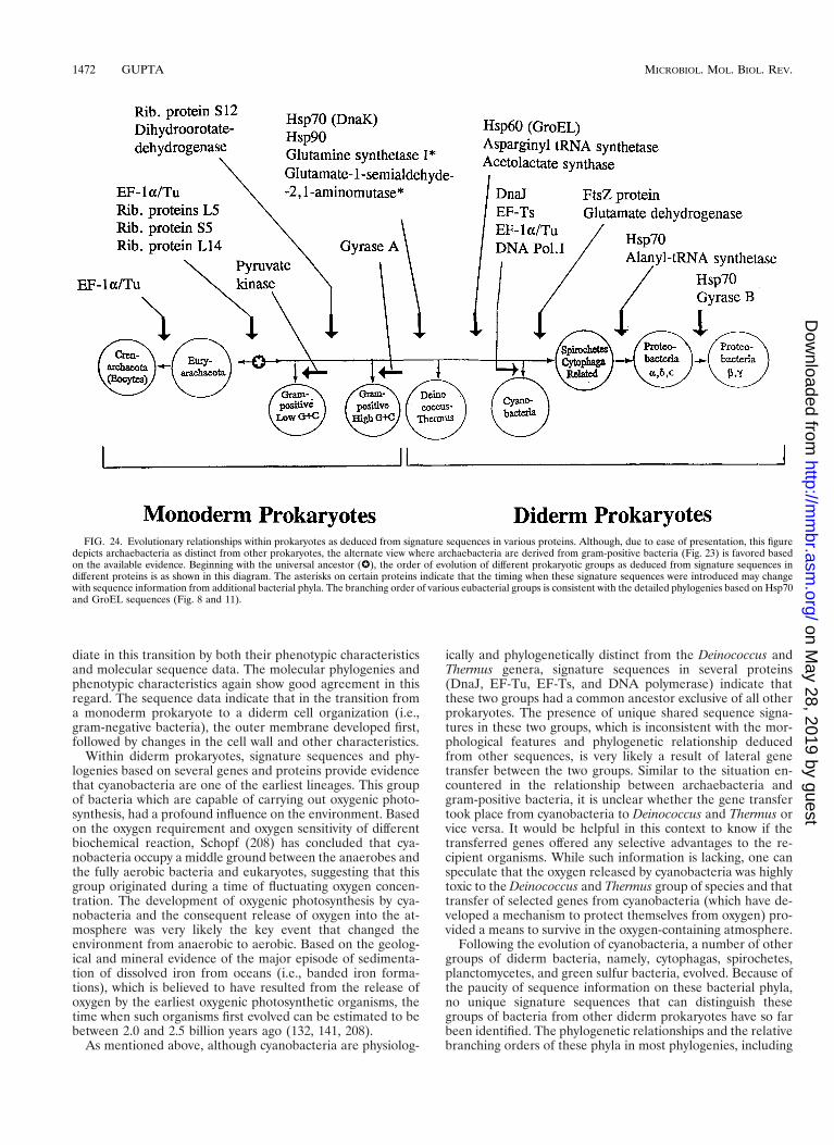
diate in this transition by both their phenotypic characteristicsand molecular sequence data. The molecular phylogenies andphenotypic characteristics again show good agreement in thisregard. The sequence data indicate that in the transition froma monoderm prokaryote to a diderm cell organization (i.e.,gram-negative bacteria), the outer membrane developed first,followed by changes in the cell wall and other characteristics.
Within diderm prokaryotes, signature sequences and phy-logenies based on several genes and proteins provide evidencethat cyanobacteria are one of the earliest lineages. This groupof bacteria which are capable of carrying out oxygenic photo-synthesis, had a profound influence on the environment. Basedon the oxygen requirement and oxygen sensitivity of differentbiochemical reaction, Schopf (208) has concluded that cya-nobacteria occupy a middle ground between the anaerobes andthe fully aerobic bacteria and eukaryotes, suggesting that thisgroup originated during a time of fluctuating oxygen concen-tration. The development of oxygenic photosynthesis by cya-nobacteria and the consequent release of oxygen into the at-mosphere was very likely the key event that changed theenvironment from anaerobic to aerobic. Based on the geolog-ical and mineral evidence of the major episode of sedimenta-tion of dissolved iron from oceans (i.e., banded iron forma-tions), which is believed to have resulted from the release ofoxygen by the earliest oxygenic photosynthetic organisms, thetime when such organisms first evolved can be estimated to bebetween 2.0 and 2.5 billion years ago (132, 141, 208).
As mentioned above, although cyanobacteria are physiolog-
ically and phylogenetically distinct from the Deinococcus andThermus genera, signature sequences in several proteins(DnaJ, EF-Tu, EF-Ts, and DNA polymerase) indicate thatthese two groups had a common ancestor exclusive of all otherprokaryotes. The presence of unique shared sequence signa-tures in these two groups, which is inconsistent with the mor-phological features and phylogenetic relationship deducedfrom other sequences, is very likely a result of lateral genetransfer between the two groups. Similar to the situation en-countered in the relationship between archaebacteria andgram-positive bacteria, it is unclear whether the gene transfertook place from cyanobacteria to Deinococcus and Thermus orvice versa. It would be helpful in this context to know if thetransferred genes offered any selective advantages to the re-cipient organisms. While such information is lacking, one canspeculate that the oxygen released by cyanobacteria was highlytoxic to the Deinococcus and Thermus group of species and thattransfer of selected genes from cyanobacteria (which have de-veloped a mechanism to protect themselves from oxygen) pro-vided a means to survive in the oxygen-containing atmosphere.
Following the evolution of cyanobacteria, a number of othergroups of diderm bacteria, namely, cytophagas, spirochetes,planctomycetes, and green sulfur bacteria, evolved. Because ofthe paucity of sequence information on these bacterial phyla,no unique signature sequences that can distinguish thesegroups of bacteria from other diderm prokaryotes have so farbeen identified. The phylogenetic relationships and the relativebranching orders of these phyla in most phylogenies, including
FIG. 24. Evolutionary relationships within prokaryotes as deduced from signature sequences in various proteins. Although, due to ease of presentation, this figuredepicts archaebacteria as distinct from other prokaryotes, the alternate view where archaebacteria are derived from gram-positive bacteria (Fig. 23) is favored basedon the available evidence. Beginning with the universal ancestor (W), the order of evolution of different prokaryotic groups as deduced from signature sequences indifferent proteins is as shown in this diagram. The asterisks on certain proteins indicate that the timing when these signature sequences were introduced may changewith sequence information from additional bacterial phyla. The branching order of various eubacterial groups is consistent with the detailed phylogenies based on Hsp70and GroEL sequences (Fig. 8 and 11).
1472 GUPTA MICROBIOL. MOL. BIOL. REV.
on May 28, 2019 by guest
http://mm
br.asm.org/
Dow
nloaded from
rRNA (184, 250), Hsp70 (Fig. 8) (103, 108) and GroEL (Fig.11) (96, 246), are not resolved. However, these groups of pro-karyotes consistently branch in between cyanobacteria andproteobacteria (Fig. 8 and 11). The placement of these bacte-rial phyla in a group between cyanobacteria and proteobacte-ria-1 (the alpha, delta, and epsilon subdivisions) can be confi-dently made based on the signature sequences in the FtsZ andGDH proteins (Fig. 15) on the one hand and Hsp70 andalanyl-tRNA synthetase on the other (Fig. 19). Although atpresent I have placed all these bacteria in a single group, it islikely that as further sequence information becomes available,additional signature sequences that make clear distinction be-tween these groups and clarify the evolutionary relationshipsbetween these phyla will be identified.
Signature sequences in proteins also define and provide dis-tinction between two different groups corresponding to pro-teobacteria. One group consists of the alpha, delta, and epsilonsubdivisions, whereas the other consists of the beta and gammasubdivisions. The association of Thermomicrobium with thefirst group is surprising, and it remains to be determinedwhether other members of the green nonsulfur group (Herpe-tosiphon and Chloroflexus) also show such a relationship. Thegroup consisting of the beta and gamma proteobacteria, basedupon signature sequences in different proteins, appears to haveevolved most recently among all the various prokaryotic groupsor divisions. In earlier work, specific amino acid substitutionsin Hsp60 protein that distinguish the alpha proteobacteriafrom other subdivisions have also been described (96). It isexpected that additional signature sequences providing furtherdistinctions between proteobacterial groups will be uncoveredin future studies.
The evolutionary picture reconstructed here based upon sig-nature sequences in different proteins (Fig. 24) is in accor-dance with the phylogenies derived from a number of highlyconserved proteins (Fig. 8 and 11).
EVOLUTIONARY RELATIONSHIP BETWEENEUKARYOTES AND PROKARYOTES
Based on the purported evolutionary relationships amongthe prokaryotic organisms just considered, we can now ask howthe eukaryotic organisms are related to the prokaryotes. Theevolutionary relationship between prokaryotes and eukaryoteshas been studied based on a large number of sequences andother characteristics (21, 249a, 263), and I do not plan toprovide an exhaustive list of these studies or characteristics.Instead, my objective here is to determine what kind of pro-posal or model for the origin of eukaryotic cells can bestaccount for most of the available molecular sequence data aswell as other relevant information.
Some Critical Assumptions in StudyingProkaryote-Eukaryote Relationships
As discussed above (see “Current evolutionary perspec-tive”), the three-domain proposal postulates that the eu-karyotes and archaebacteria had a common ancestor exclusiveof any eubacteria (258), in other words, that the nuclear ge-nome of the ancestral eukaryotic cell (exclusive of organellargenes) directly descended from an archaebacterial cell. A spe-cific relationship of the eukaryotes to archaebacteria, as sug-gested in this proposal, is based on the rooting derived fromthe duplicated gene sequences for EF-1a/Tu and EF-2/G (andthe a and b subunits of F- and V-ATPases) (81, 126). Althoughthe subsequent discovery of V-ATPases in bacteria and anF-ATPase in an archaebacterium have called into question the
rooting based on ATPase sequences (69, 118), the rootingbased on EF-1a/Tu and EF-2/G proteins is widely acceptedand continues to have a major influence on the field (7, 126).A similar rooting of the universal tree between archaebacteriaand eubacteria has been derived based on homologous amino-acyl (isoleucyl, valyl-, and leucyl) tRNA synthetase sequences(20).
However, the question of the root of the universal tree is ahotly debated and very contentious issue (49, 53, 69, 129, 205).Depending upon the protein sequences that are considered,different types of relationships among the three primary groups(archaebacteria, eubacteria, and eukaryotes) could be ob-served. These include (i) all three groups equidistant fromeach other, (ii) archaebacteria and bacteria closely related toeach other compared to the eukaryotic homologs, (iii) archae-bacteria as specific relatives of eukaryotes and eubacteriadistantly related to them, and (iv) a specific relationship ofeukaryotic homologs to eubacteria as compared to the archae-bacteria (49, 53, 69, 85, 197a, 205). Since all of the indicatedrelationships are strongly supported for different proteins, ra-tionally it is difficult to choose among them unless it is postu-lated that extensive lateral gene transfer occurred betweenspecies to support one relationship in preference to the other.
This controversy, in my view, has stemmed in large part froma very basic and profound assumption that the eukaryotic cellshave evolved from prokaryotes by normal evolutionary mech-anisms (mutations, recombination, etc.). However, as empha-sized by many prominent biologists (28, 34, 46, 159, 168, 173,237), the transition from prokaryotes to eukaryotes representsa major discontinuity in the evolutionary history. If the pro-karyote-to-eukaryote transition came about by normal evolu-tionary mechanisms, then given the enormity of the structuraland molecular differences between these two cell types, thistransformation must have occurred over a very long periodinvolving numerous intermediate species, each developing lim-ited selective advantages and evolving certain eukaryotic char-acteristics. However, there is no evidence (living or fossil) forthe existence of any such “intermediate” organisms, despitethe great diversity of the prokaryotic and eukaryotic organismsthat preceded or followed this major change. However, if thetransition from prokaryote to eukaryote did not come about bya normal evolutionary mechanism but instead resulted fromsome unusual event such as fusion and integration of genomesfrom different prokaryotes, any attempt to root the eukaryotictree based on any one particular gene or even a set of genes(e.g., the EF-1a/Tu, EF-2/G, and aminoacyl-tRNA synthetasesgenes) will provide information about the origin of that gene(or sets of genes) and not of the eukaryotic cell. In view ofthese considerations, the proper approach to understandingthe origin of the eukaryotic cell from prokaryotic ancestors, inmy view, would be to examine the relationship of differenteukaryotic genes to prokaryotic homologs without any priorassumptions and then to suggest a model which is consistentwith most of the data. This is the approach I have followed inthis review.
Most Genes for the Information Transfer ProcessesAre Derived from Archaebacteria
For a number of the gene and protein sequences originallystudied, namely, EF-1a/Tu, EF-2/G, RNA polymerase II andIII subunits and F- and V-type ATPases, the eukaryotic ho-mologs exhibited greater similarity to archaebacteria than toeubacteria (81, 126, 196). A close and specific relationship ofarchaebacteria to the eukaryotic homologs is also supported bya number of other genes e.g., ribosomal proteins, DNA poly-
VOL. 62, 1998 PHYLOGENY OF PROKARYOTES AND EUKARYOTES 1473
on May 28, 2019 by guest
http://mm
br.asm.org/
Dow
nloaded from

merase B, TATA binding proteins, transcription factors IIBand IIIB, TCP-1 chaperone (54, 96, 98, 134, 158, 203, 213),most of which are involved in aspects of transcription andtranslation. In the past 2 or 3 years, due to the completesequencing of the genomes of a number of bacterial and ar-chaebacterial species and a eukaryotic species, Saccharomycescerevisiae (15, 26, 45, 66, 72, 73, 80, 119, 128, 138, 147, 215,242), a much larger database has become available to examinethe relationships among species in the three domains. Detailedanalyses of the archaebacterium Methanococcus jannaschii se-quences indicate that 44% of its gene products showed a closerrelationship to eubacteria whereas about 13% of the proteinsshowed a closer relationship to the eukaryotic homologs (144).The rest of the proteins showed approximately the same levelof similarity to the eubacterial and eukaryotic homologs. Animportant understanding that resulted from such analyses isthat the vast majority of genes for which the archaebacterialhomologs exhibited greater similarity to eukaryotes were re-lated to the information transfer processes such as replication,transcription, and translation (9, 47, 54, 144, 158, 183, 197). Infact, for many genes involved in DNA replication and tran-scription, no eubacterial homologs have been found (54, 158,183, 197). Thus, in terms of their informational transfer ma-chineries “archaea look very eukaryotic” (54).
A close and specific relationship between archaebacteriaand eukaryotes for the information transfer processes is alsoreadily apparent from signature sequences in many proteins.EF-1a/Tu (Fig. 7a) and ribosomal proteins L5 (Fig. 7b) and S5(Fig. 7c) provide examples where all archaebacterial and eu-karyotic homologs contain prominent shared signature se-quences not found in any eubacterial homologs. The proteinEF-1a/Tu contains another important signature sequenceidentified by Rivera and Lake (198). This signature sequenceconsists of a 7-aa insert that is uniquely present in the eocytesor Crenarchaeota group of archaebacteria and all eukaryotichomologs but is not found in the Euryarchaeota division ofarchaebacteria (Fig. 21). This signature provides evidence thatwithin archaebacteria, the eocyte archaebacteria are the clos-est relatives of eukaryotes (Fig. 25) (198). A specific relation-ship of the eocyte archaebacteria to eukaryotic homologs isalso strongly supported by the detailed phylogenetic studiesbased on EF-1a/Tu sequences (7, 21, 112). Based on the aboveobservations, it is now indisputable that many of the eukaryoticnuclear genes, particularly those related to the informationtransfer machinery, are of archaebacterial origin. In relation tothe origin of eukaryotic cells, the key question now becomeswhether all of the eukaryotic nuclear-cytosolic genome (i.e.,exclusive of organelles) are derived from archaebacteria orwhether other groups of prokaryotes (i.e., eubacteria) alsomade significant contributions.
Hsp70 Provides the Clearest Example of the Contribution ofEubacteria to the Nuclear-Cytosolic Genome
The question of establishment of any eubacterial contribu-tion to the eukaryotic nuclear-cytosolic genome is far moredifficult than connecting archaebacteria and eukaryotes. (Notethat the term “nuclear-cytosolic” as used in this review refersto those genes and proteins which originated with the forma-tion of the ancestral eukaryotic cell.) The main difficulty lies inthe fact that in contrast to archaebacteria, which have contrib-uted only to the nuclear genome, two classes of eukaryotic cellorganelle genomes, mitochondria and plastids, were derivedfrom eubacteria in later endosymbiotic acquisitions (90, 92,159). Most organellar genes were later transferred to the nu-
cleus. Thus, eukaryotes often have multiple homologs of pro-teins with sequence similarity to eubacteria. For most se-quences in the databases, information that distinguishesnuclear-cytosolic genes from organellar homologs is lacking.Thus, in many cases it is difficult to know whether a giveneukaryotic homolog corresponds to an organellar gene or to anuclear-cytosolic gene product. The presence of multiple genesinside eukaryotes also raises the possibility that genes for somepresumed nuclear-cytosolic proteins are in fact derived fromorganellar genes by means of horizontal transfer followed bydivergence (135, 165). Furthermore, many eukaryotes harborbacterial endosymbionts, and some of the eubacterial genescould be derived from them by horizontal transfers. Thus, ithas proven difficult to establish whether a given eubacterialgene present in eukaryotic cells is of nuclear-cytosolic origin oris derived from other sources. However, in recent years, theenlarged sequence database, in conjunction with extensivecharacterization of many eukaryotic protein families at themolecular, biochemical, subcellular localization, and phyloge-netic levels, has provided specific examples where these prob-lems can be clearly resolved.
The Hsp70 protein, discussed above, provides the best-stud-ied examples of such proteins (17, 99, 102, 108). The Hsp70homologs have been sequenced and characterized from abroad range of prokaryotic and eukaryotic organisms coveringthe entire evolutionary spectrum. In eukaryotic cells, specificHsp70 homologs are found in various cellular compartments,including the cytosol, endoplasmic reticulum (ER), mitochon-dria, and chloroplasts (17, 41, 102, 108). The homologs presentin different compartments are well characterized both bio-chemically and by cellular localization studies (2, 214, 221).Most importantly, global alignment of Hsp70 sequences fromprokaryotic, eukaryotic, and organellar sources shows that thedifferent types of Hsp70 homologs are readily and unambigu-ously distinguished from each other based on specific signaturesequences (99, 102, 105, 108). The eukaryotic nuclear-cytosolicHsp70s contain a large number of unique amino acid substi-tutions and sequence signatures not found in any of the pro-karyotic or organellar homologs. Figure 26 gives an excerptfrom Hsp70 alignment showing some of the important se-quence signatures. As seen in this figure, all the eukaryoticcytosolic and ER homologs, including those from the earliest-diverging eukaryotic lineage such as Giardia, contain two sig-nature sequences (a 4-aa deletion marked ② and a 1-aa insertmarked ③) that are not present in any prokaryotic or organel-lar Hsp70s. Since these signatures are not present in any mi-tochondrial, hydrogenosomal (from Trichomona vaginalis), orprokaryotic homologs, they could not have been derived fromthe latter groups by means of horizontal gene transfer. Thesesignatures are thus uniquely eukaryotic, and they were proba-bly introduced into the common ancestor of eukaryotic cells atthe time of its origin. The homologs containing these sequencesignatures thus are nuclear-cytosolic in origin.
The inference from the above signature sequences that theeukaryotic nuclear-cytosolic homologs are altogether distinctfrom organellar homologs is strongly reinforced by the phylo-genetic analysis based on Hsp70 sequences (Fig. 27). In phy-logenetic trees based on Hsp70 sequences, the nuclear-cytoso-lic homologs consistently form a distinct monophyletic clade(100% of the time by different phylogenetic methods) branch-ing within gram-negative bacteria but showing no relationshipto the organellar homologs (i.e., mitochondria, hydrogeno-some, or chloroplasts) (56, 102–104, 108). If the mitochondrialand nuclear-cytosolic homologs were paralogous sequencesthat originated by a gene duplication event and subsequentdivergence, one would expect them to form distinct but related
1474 GUPTA MICROBIOL. MOL. BIOL. REV.
on May 28, 2019 by guest
http://mm
br.asm.org/
Dow
nloaded from
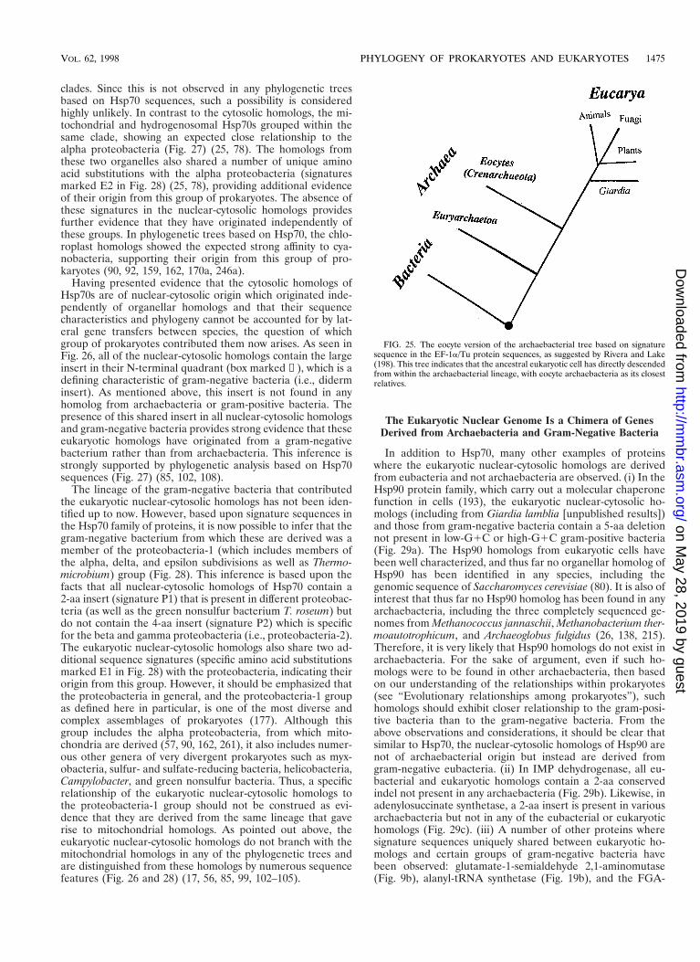
clades. Since this is not observed in any phylogenetic treesbased on Hsp70 sequences, such a possibility is consideredhighly unlikely. In contrast to the cytosolic homologs, the mi-tochondrial and hydrogenosomal Hsp70s grouped within thesame clade, showing an expected close relationship to thealpha proteobacteria (Fig. 27) (25, 78). The homologs fromthese two organelles also shared a number of unique aminoacid substitutions with the alpha proteobacteria (signaturesmarked E2 in Fig. 28) (25, 78), providing additional evidenceof their origin from this group of prokaryotes. The absence ofthese signatures in the nuclear-cytosolic homologs providesfurther evidence that they have originated independently ofthese groups. In phylogenetic trees based on Hsp70, the chlo-roplast homologs showed the expected strong affinity to cya-nobacteria, supporting their origin from this group of pro-karyotes (90, 92, 159, 162, 170a, 246a).
Having presented evidence that the cytosolic homologs ofHsp70s are of nuclear-cytosolic origin which originated inde-pendently of organellar homologs and that their sequencecharacteristics and phylogeny cannot be accounted for by lat-eral gene transfers between species, the question of whichgroup of prokaryotes contributed them now arises. As seen inFig. 26, all of the nuclear-cytosolic homologs contain the largeinsert in their N-terminal quadrant (box marked ①), which is adefining characteristic of gram-negative bacteria (i.e., diderminsert). As mentioned above, this insert is not found in anyhomolog from archaebacteria or gram-positive bacteria. Thepresence of this shared insert in all nuclear-cytosolic homologsand gram-negative bacteria provides strong evidence that theseeukaryotic homologs have originated from a gram-negativebacterium rather than from archaebacteria. This inference isstrongly supported by phylogenetic analysis based on Hsp70sequences (Fig. 27) (85, 102, 108).
The lineage of the gram-negative bacteria that contributedthe eukaryotic nuclear-cytosolic homologs has not been iden-tified up to now. However, based upon signature sequences inthe Hsp70 family of proteins, it is now possible to infer that thegram-negative bacterium from which these are derived was amember of the proteobacteria-1 (which includes members ofthe alpha, delta, and epsilon subdivisions as well as Thermo-microbium) group (Fig. 28). This inference is based upon thefacts that all nuclear-cytosolic homologs of Hsp70 contain a2-aa insert (signature P1) that is present in different proteobac-teria (as well as the green nonsulfur bacterium T. roseum) butdo not contain the 4-aa insert (signature P2) which is specificfor the beta and gamma proteobacteria (i.e., proteobacteria-2).The eukaryotic nuclear-cytosolic homologs also share two ad-ditional sequence signatures (specific amino acid substitutionsmarked E1 in Fig. 28) with the proteobacteria, indicating theirorigin from this group. However, it should be emphasized thatthe proteobacteria in general, and the proteobacteria-1 groupas defined here in particular, is one of the most diverse andcomplex assemblages of prokaryotes (177). Although thisgroup includes the alpha proteobacteria, from which mito-chondria are derived (57, 90, 162, 261), it also includes numer-ous other genera of very divergent prokaryotes such as myx-obacteria, sulfur- and sulfate-reducing bacteria, helicobacteria,Campylobacter, and green nonsulfur bacteria. Thus, a specificrelationship of the eukaryotic nuclear-cytosolic homologs tothe proteobacteria-1 group should not be construed as evi-dence that they are derived from the same lineage that gaverise to mitochondrial homologs. As pointed out above, theeukaryotic nuclear-cytosolic homologs do not branch with themitochondrial homologs in any of the phylogenetic trees andare distinguished from these homologs by numerous sequencefeatures (Fig. 26 and 28) (17, 56, 85, 99, 102–105).
The Eukaryotic Nuclear Genome Is a Chimera of GenesDerived from Archaebacteria and Gram-Negative Bacteria
In addition to Hsp70, many other examples of proteinswhere the eukaryotic nuclear-cytosolic homologs are derivedfrom eubacteria and not archaebacteria are observed. (i) In theHsp90 protein family, which carry out a molecular chaperonefunction in cells (193), the eukaryotic nuclear-cytosolic ho-mologs (including from Giardia lamblia [unpublished results])and those from gram-negative bacteria contain a 5-aa deletionnot present in low-G1C or high-G1C gram-positive bacteria(Fig. 29a). The Hsp90 homologs from eukaryotic cells havebeen well characterized, and thus far no organellar homolog ofHsp90 has been identified in any species, including thegenomic sequence of Saccharomyces cerevisiae (80). It is also ofinterest that thus far no Hsp90 homolog has been found in anyarchaebacteria, including the three completely sequenced ge-nomes from Methanococcus jannaschii, Methanobacterium ther-moautotrophicum, and Archaeoglobus fulgidus (26, 138, 215).Therefore, it is very likely that Hsp90 homologs do not exist inarchaebacteria. For the sake of argument, even if such ho-mologs were to be found in other archaebacteria, then basedon our understanding of the relationships within prokaryotes(see “Evolutionary relationships among prokaryotes”), suchhomologs should exhibit closer relationship to the gram-posi-tive bacteria than to the gram-negative bacteria. From theabove observations and considerations, it should be clear thatsimilar to Hsp70, the nuclear-cytosolic homologs of Hsp90 arenot of archaebacterial origin but instead are derived fromgram-negative eubacteria. (ii) In IMP dehydrogenase, all eu-bacterial and eukaryotic homologs contain a 2-aa conservedindel not present in any archaebacteria (Fig. 29b). Likewise, inadenylosuccinate synthetase, a 2-aa insert is present in variousarchaebacteria but not in any of the eubacterial or eukaryotichomologs (Fig. 29c). (iii) A number of other proteins wheresignature sequences uniquely shared between eukaryotic ho-mologs and certain groups of gram-negative bacteria havebeen observed: glutamate-1-semialdehyde 2,1-aminomutase(Fig. 9b), alanyl-tRNA synthetase (Fig. 19b), and the FGA-
FIG. 25. The eocyte version of the archaebacterial tree based on signaturesequence in the EF-1a/Tu protein sequences, as suggested by Rivera and Lake(198). This tree indicates that the ancestral eukaryotic cell has directly descendedfrom within the archaebacterial lineage, with eocyte archaebacteria as its closestrelatives.
VOL. 62, 1998 PHYLOGENY OF PROKARYOTES AND EUKARYOTES 1475
on May 28, 2019 by guest
http://mm
br.asm.org/
Dow
nloaded from
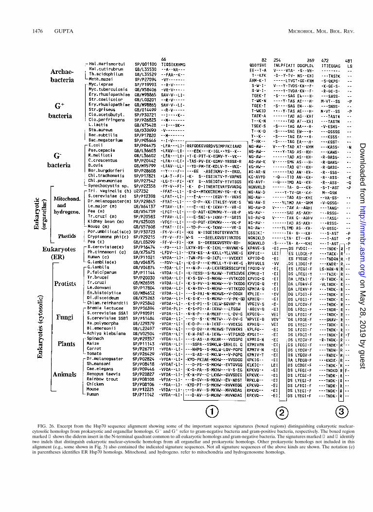
FIG. 26. Excerpt from the Hsp70 sequence alignment showing some of the important sequence signatures (boxed regions) distinguishing eukaryotic nuclear-cytosolic homologs from prokaryotic and organellar homologs. G2 and G1 refer to gram-negative bacteria and gram-positive bacteria, respectively. The boxed regionmarked ① shows the diderm insert in the N-terminal quadrant common to all eukaryotic homologs and gram-negative bacteria. The signatures marked ② and ③ identifytwo indels that distinguish eukaryotic nuclear-cytosolic homologs from all organellar and prokaryotic homologs. Other prokaryotic homologs not included in thisalignment (e.g., some shown in Fig. 3) also contained the indicated signature sequences. Not all signature sequences of the above kinds are shown. The notation (e)in parentheses identifies ER Hsp70 homologs. Mitochond. and hydrogeno. refer to mitochondria and hydrogenosome homologs.
1476 GUPTA MICROBIOL. MOL. BIOL. REV.
on May 28, 2019 by guest
http://mm
br.asm.org/
Dow
nloaded from
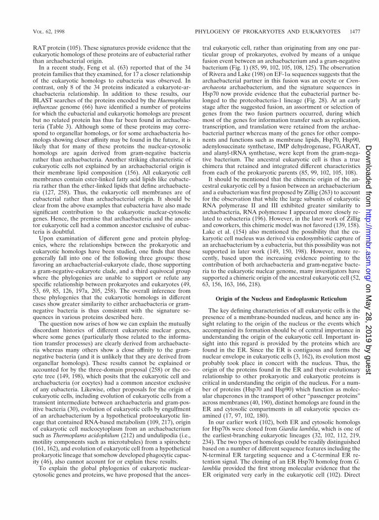
RAT protein (105). These signatures provide evidence that theeukaryotic homologs of these proteins are of eubacterial ratherthan archaebacterial origin.
In a recent study, Feng et al. (63) reported that of the 34protein families that they examined, for 17 a closer relationshipof the eukaryotic homologs to eubacteria was observed. Incontrast, only 8 of the 34 proteins indicated a eukaryote-ar-chaebacteria relationship. In addition to these results, ourBLAST searches of the proteins encoded by the Haemophilusinfluenzae genome (66) have identified a number of proteinsfor which the eubacterial and eukaryotic homologs are presentbut no related protein has thus far been found in archaebac-teria (Table 3). Although some of these proteins may corre-spond to organellar homologs, or for some archaebacteria ho-mologs showing closer affinity may be found in the future, it islikely that for many of these proteins the nuclear-cytosolichomologs are again derived from gram-negative bacteriarather than archaebacteria. Another striking characteristic ofeukaryotic cells not explained by an archaebacterial origin istheir membrane lipid composition (156). All eukaryotic cellmembranes contain ester-linked fatty acid lipids like eubacte-ria rather than the ether-linked lipids that define archaebacte-ria (127, 258). Thus, the eukaryotic cell membranes are ofeubacterial rather than archaebacterial origin. It should beclear from the above examples that eubacteria have also madesignificant contribution to the eukaryotic nuclear-cytosolicgenes. Hence, the premise that archaebacteria and the ances-tor eukaryotic cell had a common ancestor exclusive of eubac-teria is doubtful.
Upon examination of different gene and protein phylog-enies, where the relationships between the prokaryotic andeukaryotic homologs have been studied, one finds that thesegenerally fall into one of the following three groups: thosefavoring an archaebacterial-eukaryote clade, those supportinga gram-negative-eukaryote clade, and a third equivocal groupwhere the phylogenies are unable to support or refute anyspecific relationship between prokaryotes and eukaryotes (49,53, 69, 85, 126, 197a, 205, 258). The overall inference fromthese phylogenies that the eukaryotic homologs in differentcases show greater similarity to either archaebacteria or gram-negative bacteria is thus consistent with the signature se-quences in various proteins described here.
The question now arises of how we can explain the mutuallydiscordant histories of different eukaryotic nuclear genes,where some genes (particularly those related to the informa-tion transfer processes) are clearly derived from archaebacte-ria whereas many others show a close affinity to the gram-negative bacteria (and it is unlikely that they are derived fromorganellar homologs). These results cannot be explained oraccounted for by the three-domain proposal (258) or the eo-cyte tree (149, 198), which posits that the eukaryotic cell andarchaebacteria (or eocytes) had a common ancestor exclusiveof any eubacteria. Likewise, other proposals for the origin ofeukaryotic cells, including evolution of eukaryotic cells from atransient intermediate between archaebacteria and gram-pos-itive bacteria (30), evolution of eukaryotic cells by engulfmentof an archaebacterium by a hypothetical protoeukaryotic lin-eage that contained RNA-based metabolism (109, 217), originof eukaryotic cell nucleocytoplasm from an archaebacteriumsuch as Thermoplams acidophilum (212) and undulipodia (i.e.,motility components such as microtubules) from a spirochete(161, 162), and evolution of eukaryotic cell from a hypotheticalprokaryotic lineage that somehow developed phagocytic capac-ity (46), also cannot account for or explain these results.
To explain the global phylogenies of eukaryotic nuclear-cytosolic genes and proteins, we have proposed that the ances-
tral eukaryotic cell, rather than originating from any one par-ticular group of prokaryotes, evolved by means of a uniquefusion event between an archaebacterium and a gram-negativebacterium (Fig. 1) (85, 99, 102, 105, 108, 125). The observationof Rivera and Lake (198) on EF-1a sequences suggests that thearchaebacterial partner in this fusion was an eocyte or Cren-archaeota archaebacterium, and the signature sequences inHsp70 now provide evidence that the eubacterial partner be-longed to the proteobacteria-1 lineage (Fig. 28). At an earlystage after the suggested fusion, an assortment or selection ofgenes from the two fusion partners occurred, during whichmost of the genes for information transfer such as replication,transcription, and translation were retained from the archae-bacterial partner whereas many of the genes for other compo-nents and functions such as membrane lipids, Hsp70, Hsp90,adenylosuccinate synthetase, IMP dehydrogenase, FGARAT,and alanyl-tRNA synthetase, were kept from the gram-nega-tive bacterium. The ancestral eukaryotic cell is thus a truechimera that retained and integrated different characteristicsfrom each of the prokaryotic parents (85, 99, 102, 105, 108).
It should be mentioned that the chimeric origin of the an-cestral eukaryotic cell by a fusion between an archaebacteriumand a eubacterium was first proposed by Zillig (263) to accountfor the observation that while the large subunits of eukaryoticRNA polymerase II and III exhibited greater similarity toarchaebacteria, RNA polymerase I appeared more closely re-lated to eubacteria (196). However, in the later work of Zilligand coworkers, this chimeric model was not favored (139, 158).Lake et al. (154) also mentioned the possibility that the eu-karyotic cell nucleus was derived via endosymbiotic capture ofan archaebacterium by a eubacteria, but this possibility was notsupported in later work (149, 150, 198). However, more re-cently, based upon the increasing evidence pointing to thecontribution of both archaebacteria and gram-negative bacte-ria to the eukaryotic nuclear genome, many investigators havesupported a chimeric origin of the ancestral eukaryotic cell (52,63, 156, 163, 166, 218).
Origin of the Nucleus and Endoplasmic Reticulum
The key defining characteristics of all eukaryotic cells is thepresence of a membrane-bounded nucleus, and hence any in-sight relating to the origin of the nucleus or the events whichaccompanied its formation should be of central importance inunderstanding the origin of the eukaryotic cell. Important in-sight into this regard is provided by the proteins which arefound in the ER. Since the ER is contiguous and forms thenuclear envelope in eukaryotic cells (3, 162), its evolution mostprobably took place in concert with the nucleus. Thus, theorigin of the proteins found in the ER and their evolutionaryrelationship to other prokaryotic and eukaryotic proteins iscritical in understanding the origin of the nucleus. For a num-ber of proteins (Hsp70 and Hsp90) which function as molec-ular chaperones in the transport of other “passenger proteins”across membranes (40, 190), distinct homologs are found in theER and cytosolic compartments in all eukaryotic species ex-amined (17, 97, 102, 180).
In our earlier work (102), both ER and cytosolic homologsfor Hsp70s were cloned from Giardia lamblia, which is one ofthe earliest-branching eukaryotic lineages (32, 102, 112, 219,234). The two types of homologs could be readily distinguishedbased on a number of different sequence features including theN-terminal ER targeting sequence and a C-terminal ER re-tention signal. The cloning of an ER Hsp70 homolog from G.lamblia provided the first strong molecular evidence that theER originated very early in the eukaryotic cell (102). Direct
VOL. 62, 1998 PHYLOGENY OF PROKARYOTES AND EUKARYOTES 1477
on May 28, 2019 by guest
http://mm
br.asm.org/
Dow
nloaded from
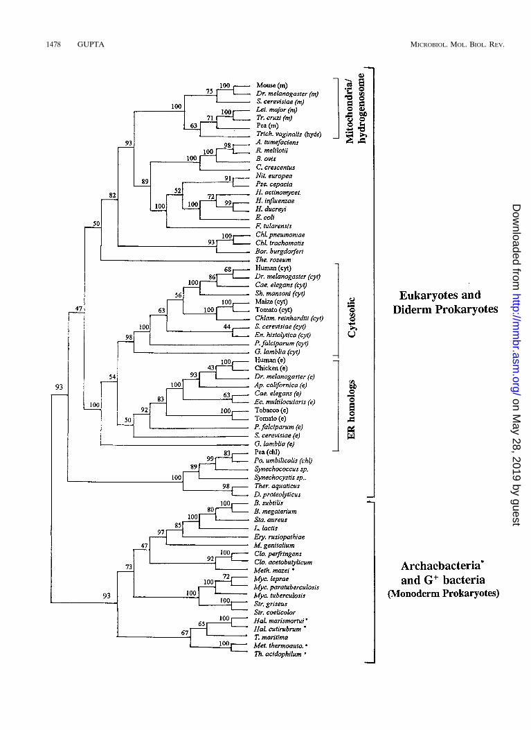
1478 GUPTA MICROBIOL. MOL. BIOL. REV.
on May 28, 2019 by guest
http://mm
br.asm.org/
Dow
nloaded from
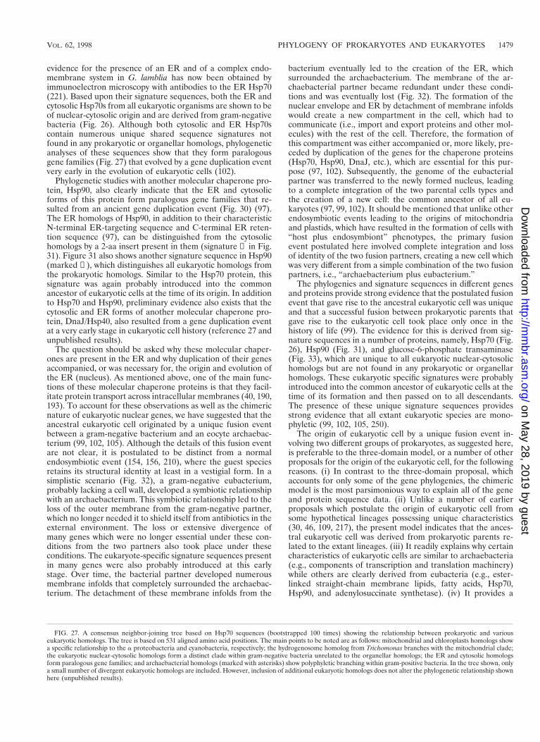
evidence for the presence of an ER and of a complex endo-membrane system in G. lamblia has now been obtained byimmunoelectron microscopy with antibodies to the ER Hsp70(221). Based upon their signature sequences, both the ER andcytosolic Hsp70s from all eukaryotic organisms are shown to beof nuclear-cytosolic origin and are derived from gram-negativebacteria (Fig. 26). Although both cytosolic and ER Hsp70scontain numerous unique shared sequence signatures notfound in any prokaryotic or organellar homologs, phylogeneticanalyses of these sequences show that they form paralogousgene families (Fig. 27) that evolved by a gene duplication eventvery early in the evolution of eukaryotic cells (102).
Phylogenetic studies with another molecular chaperone pro-tein, Hsp90, also clearly indicate that the ER and cytosolicforms of this protein form paralogous gene families that re-sulted from an ancient gene duplication event (Fig. 30) (97).The ER homologs of Hsp90, in addition to their characteristicN-terminal ER-targeting sequence and C-terminal ER reten-tion sequence (97), can be distinguished from the cytosolichomologs by a 2-aa insert present in them (signature ② in Fig.31). Figure 31 also shows another signature sequence in Hsp90(marked ①), which distinguishes all eukaryotic homologs fromthe prokaryotic homologs. Similar to the Hsp70 protein, thissignature was again probably introduced into the commonancestor of eukaryotic cells at the time of its origin. In additionto Hsp70 and Hsp90, preliminary evidence also exists that thecytosolic and ER forms of another molecular chaperone pro-tein, DnaJ/Hsp40, also resulted from a gene duplication eventat a very early stage in eukaryotic cell history (reference 27 andunpublished results).
The question should be asked why these molecular chaper-ones are present in the ER and why duplication of their genesaccompanied, or was necessary for, the origin and evolution ofthe ER (nucleus). As mentioned above, one of the main func-tions of these molecular chaperone proteins is that they facil-itate protein transport across intracellular membranes (40, 190,193). To account for these observations as well as the chimericnature of eukaryotic nuclear genes, we have suggested that theancestral eukaryotic cell originated by a unique fusion eventbetween a gram-negative bacterium and an eocyte archaebac-terium (99, 102, 105). Although the details of this fusion eventare not clear, it is postulated to be distinct from a normalendosymbiotic event (154, 156, 210), where the guest speciesretains its structural identity at least in a vestigial form. In asimplistic scenario (Fig. 32), a gram-negative eubacterium,probably lacking a cell wall, developed a symbiotic relationshipwith an archaebacterium. This symbiotic relationship led to theloss of the outer membrane from the gram-negative partner,which no longer needed it to shield itself from antibiotics in theexternal environment. The loss or extensive divergence ofmany genes which were no longer essential under these con-ditions from the two partners also took place under theseconditions. The eukaryote-specific signature sequences presentin many genes were also probably introduced at this earlystage. Over time, the bacterial partner developed numerousmembrane infolds that completely surrounded the archaebac-terium. The detachment of these membrane infolds from the
bacterium eventually led to the creation of the ER, whichsurrounded the archaebacterium. The membrane of the ar-chaebacterial partner became redundant under these condi-tions and was eventually lost (Fig. 32). The formation of thenuclear envelope and ER by detachment of membrane infoldswould create a new compartment in the cell, which had tocommunicate (i.e., import and export proteins and other mol-ecules) with the rest of the cell. Therefore, the formation ofthis compartment was either accompanied or, more likely, pre-ceded by duplication of the genes for the chaperone proteins(Hsp70, Hsp90, DnaJ, etc.), which are essential for this pur-pose (97, 102). Subsequently, the genome of the eubacterialpartner was transferred to the newly formed nucleus, leadingto a complete integration of the two parental cells types andthe creation of a new cell: the common ancestor of all eu-karyotes (97, 99, 102). It should be mentioned that unlike otherendosymbiotic events leading to the origins of mitochondriaand plastids, which have resulted in the formation of cells with“host plus endosymbiont” phenotypes, the primary fusionevent postulated here involved complete integration and lossof identity of the two fusion partners, creating a new cell whichwas very different from a simple combination of the two fusionpartners, i.e., “archaebacterium plus eubacterium.”
The phylogenies and signature sequences in different genesand proteins provide strong evidence that the postulated fusionevent that gave rise to the ancestral eukaryotic cell was uniqueand that a successful fusion between prokaryotic parents thatgave rise to the eukaryotic cell took place only once in thehistory of life (99). The evidence for this is derived from sig-nature sequences in a number of proteins, namely, Hsp70 (Fig.26), Hsp90 (Fig. 31), and glucose-6-phosphate transaminase(Fig. 33), which are unique to all eukaryotic nuclear-cytosolichomologs but are not found in any prokaryotic or organellarhomologs. These eukaryotic specific signatures were probablyintroduced into the common ancestor of eukaryotic cells at thetime of its formation and then passed on to all descendants.The presence of these unique signature sequences providesstrong evidence that all extant eukaryotic species are mono-phyletic (99, 102, 105, 250).
The origin of eukaryotic cell by a unique fusion event in-volving two different groups of prokaryotes, as suggested here,is preferable to the three-domain model, or a number of otherproposals for the origin of the eukaryotic cell, for the followingreasons. (i) In contrast to the three-domain proposal, whichaccounts for only some of the gene phylogenies, the chimericmodel is the most parsimonious way to explain all of the geneand protein sequence data. (ii) Unlike a number of earlierproposals which postulate the origin of eukaryotic cell fromsome hypothetical lineages possessing unique characteristics(30, 46, 109, 217), the present model indicates that the ances-tral eukaryotic cell was derived from prokaryotic parents re-lated to the extant lineages. (iii) It readily explains why certaincharacteristics of eukaryotic cells are similar to archaebacteria(e.g., components of transcription and translation machinery)while others are clearly derived from eubacteria (e.g., ester-linked straight-chain membrane lipids, fatty acids, Hsp70,Hsp90, and adenylosuccinate synthetase). (iv) It provides a
FIG. 27. A consensus neighbor-joining tree based on Hsp70 sequences (bootstrapped 100 times) showing the relationship between prokaryotic and variouseukaryotic homologs. The tree is based on 531 aligned amino acid positions. The main points to be noted are as follows: mitochondrial and chloroplasts homologs showa specific relationship to the a proteobacteria and cyanobacteria, respectively; the hydrogenosome homolog from Trichomonas branches with the mitochondrial clade;the eukaryotic nuclear-cytosolic homologs form a distinct clade within gram-negative bacteria unrelated to the organellar homologs; the ER and cytosolic homologsform paralogous gene families; and archaebacterial homologs (marked with asterisks) show polyphyletic branching within gram-positive bacteria. In the tree shown, onlya small number of divergent eukaryotic homologs are included. However, inclusion of additional eukaryotic homologs does not alter the phylogenetic relationship shownhere (unpublished results).
VOL. 62, 1998 PHYLOGENY OF PROKARYOTES AND EUKARYOTES 1479
on May 28, 2019 by guest
http://mm
br.asm.org/
Dow
nloaded from
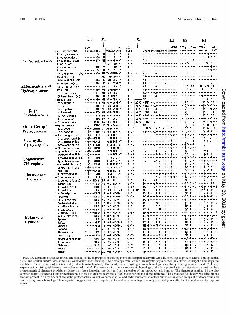
FIG. 28. Signature sequences (boxed and shaded) in the Hsp70 protein showing the relationship of eukaryotic cytosolic homologs to proteobacteria-1 group (alpha,delta, and epsilon subdivisions as well as Thermomicrobium roseum). The homologs from various prokaryotic phyla as well as different eukaryotic homologs areidentified. The notations (m), (c), (e), and (h) denote mitochondrial, chloroplast, ER, and hydrogenosome homologs, respectively. The signatures P1 and P2 identifysequences that distinguish between proteobacteria-1 and -2. The presence in all nuclear-cytosolic homologs of the 2-aa proteobacteria-1 signature but not the 4-aaproteobacteria-2 signature provides evidence that these homologs are derived from a member of the proteobacteria-1 group. The signatures marked E1 are alsocommon to proteobacteria-1 and proteobacteria-2 as well as eukaryotic cytosolic Hsp70s, supporting the above inference. The signatures E2 identify two substitutionsthat are present in all members of the alpha proteobacteria as well as mitochondrial and hydrogenosome homologs but absent in other groups of proteobacteria andeukaryotic cytosolic homologs. These signature suggest that the eukaryotic nuclear-cytosolic homologs have originated independently of mitochondria and hydrogeno-somes.
1480 GUPTA MICROBIOL. MOL. BIOL. REV.
on May 28, 2019 by guest
http://mm
br.asm.org/
Dow
nloaded from
plausible explanation for the origin of the eukaryotic cell nu-cleus and endomembrane systems. (v) Although the enormousstructural differences between the eukaryotic and prokaryoticcell types (169) and the absence of any intermediates in thistransition, cannot be readily explained by normal evolutionarymechanisms, this major evolutionary discontinuity can be ex-plained by an origin of the eukaryotic cell by fusion of twodifferent groups of prokaryotes. (vi) Doolittle and coworkers(51, 63) have inferred that the eukaryotic species divergedfrom either archaebacteria or eubacteria about 2 Ga ago basedon genetic distances between protein sequences. Althoughthese estimates involve many assumptions (84) (see “Molecu-lar phylogenies: assumptions, limitations, and pitfalls”), theinferences derived are consistent with the present model.
Did Mitochondria and the First Eukaryotic Cell Originatefrom the Same Fusion Event?
In the past, it has been generally accepted that the ancestraleukaryotic cell lacked mitochondria, which were acquired in alater endosymbiotic event (90, 92, 159, 162, 258). However, therecent finding of certain glycolytic and fermentation enzymes,i.e., glyceraldehyde-3-phosphate dehydrogenase, triosephos-phate isomerase, pyruvate:ferredoxin oxidoreductase, ferre-doxin, and alcohol dehydrogenase E, and other mitochondri-on-specific proteins (e.g., Hsp60 and mitHsp70) in a number ofprotist phyla, namely, Parabasala (e.g., Trichomonas vaginalis),Archamoebae (e.g., Entamoeba, Pelomyxa), Microsporidia(Vairimorpha nectarix), and Diplomonands (Giardia), whichwere previously thought to lack mitochondria, has suggestedthat the mitochondrial endosymbiosis occurred much earlierthan was previously suspected (25, 35, 52, 78, 113a, 120, 123,135, 165, 199–201, 218). Based on these studies, while the exacttime when mitochondria originated will no doubt be movedconsiderably earlier, the interpretation of these results con-cerning the origin of eukaryotic cells requires caution. Thephylogenies based on glycolytic and fermentation enzymes areambiguous. For glyceraldehyde-3-phosphate dehydrogenase,multiple homologs are present in both prokaryotic and eukary-otic species and their relationships to each other is not clear(116, 165, 201). The phylogenies of pyruvate:ferredoxin oxi-doreductase, ferredoxin, and alcohol dehydrogenase E haveled Rosenthal et al. (201) to conclude that the eukaryotic genesfor these proteins in early-branching protists were derivedfrom bacteria by means of horizontal gene transfers.
More credible evidence for the presence of mitochondrialgenes in early-branching protists comes from the study of theheat shock molecular chaperone proteins Hsp60 (or Cpn60)and Hsp70 (25, 35, 78, 120, 123, 199, 200, 218), where thephylogenies and the relationship between different homologsare well understood (17, 96, 98, 105, 108, 246). In eukaryotes,Hsp60 genes are derived primarily from organellar genomes(i.e., mitochondria and chloroplasts) and no close homologs ofnuclear-cytosolic origin are known (96, 98). Thus, the presenceof any Hsp60 gene in a species is generally taken as evidencethat it once contained mitochondria. However, it is importantto point out that mitochondrial Hsp60 possesses no uniquesequence characteristic, except for the presence of a N-termi-nal mitochondrial targeting presequence (MTP), by which itcould be distinguished from bacterial homologs. In contrast toHsp60, all eukaryotes contain a number of distinct Hsp70 ho-mologs. The mitochondrial Hsp70 homologs, in species whichcontain mitochondria, are clearly distinguished from nuclear-cytosolic homologs by signature sequences and phylogeneticbranching patterns (Fig. 26 and 27), but they are indistinguish-able from the alpha proteobacterial homologs, except for the
presence of a N-terminal MTP (214). Thus, the main basis forconcluding that a given Hsp60 or Hsp70 homolog, from aspecies lacking mitochondria, is of mitochondrial origin isbased on three lines of evidence: (i) localization of the ho-molog to a subcellular compartment that may be related tomitochondria (i.e., hydrogenosomes), (ii) the presence of char-acteristic MTP sequences found in mitochondrial homologs,and (iii) branching of the homologs with the mitochondrialclade in phylogenetic trees. Of these three lines of evidences,in my view the first two are more reliable indicators of mito-chondrial origin. Since many protist species, including Giardialamblia, harbor intracellular bacterial symbionts and/or sur-face-attached bacteria (1, 160), some of which could be derivedfrom the same group of prokaryotes as mitochondria, based onthe branching pattern of the homolog with the mitochondrialclade alone, the possibility that the observed gene is either abacterial contaminant or derived from bacteria via horizontalgene transfer cannot be excluded.
Examined in this light, there is good evidence that Hsp60 orHsp70 genes identified in Trichomonas vaginalis are of mito-chondrial origin. These genes are localized in hydrogenosomeswhich, based on biochemical criteria, are related to mitochon-dria (172); some of these genes contain targeting sequencesimilar to those found in mitochondria; and they branch con-sistently with mitochondria in independent studies (25, 78,199). The homologs for these proteins in Entamoeba histolyticaand Vairimorpha nectarix are also probably of mitochondrialorigin, since in addition to their branching with the mitochon-drial clade, they contain MTP-like sequences (35, 120). How-ever, evidence for the ancestral presence of mitochondria indiplomonads from studies on G. lamblia (200, 218), whichconstitutes one of the earliest-branching lineages in many genephylogenies (32, 102, 113, 219, 234), must be viewed withcaution. Evidence for the presence of mitochondria in thisprotist is based mainly on the branching of Hsp60 homologwith the mitochondrial clade (200). The cloned gene containsno N-terminal MTP sequence characteristic of mitochondriaor other related organelles. In this context, it should be pointedout that the presence of an Hsp60-related protein in Giardiawas first suggested in our work based on cross-reactivity ofHsp60 antibodies to a giardial protein (222). However, ourcloning studies with Giardia (unpublished results) resulted inthe isolation of a novel Hsp60 gene that has all the character-istics of a bacterial rather than a mitochondrial gene: (i) it lacksany upstream targeting sequence characteristic of organellargenes, and (ii) it hybridized to Giardia cultures grown understandard conditions but showed no hybridization when thecells were grown in the presence of antibiotics such as strep-tomycin. It should be emphasized that in these studies nobacterial contaminant could be detected by light or electronmicroscopic investigation or by staining with Hoechst 33258,indicating the cryptic nature of these bacteria (reference 222and unpublished results). These observations emphasize theneed for caution in interpreting the results of finding mito-chondrion-like genes in the earliest-branching eukaryotic lin-eages for the origin of eukaryotic cells.
The question may now be considered whether the primaryfusion event that led to the origin of the eukaryotic cell wasidentical to or distinct from the one that gave rise to mitochon-dria (52, 129, 166). In view of the recent findings, it is clear thatthe endosymbiotic event leading to the acquisition of mito-chondria took place much earlier than was previously believed(29, 32, 218). However, the available data in my view stillstrongly indicate that mitochondrial endosymbiosis was distinctfrom the primary fusion event that gave rise to the ancestraleukaryotic cell. Some key observations which support this con-
VOL. 62, 1998 PHYLOGENY OF PROKARYOTES AND EUKARYOTES 1481
on May 28, 2019 by guest
http://mm
br.asm.org/
Dow
nloaded from

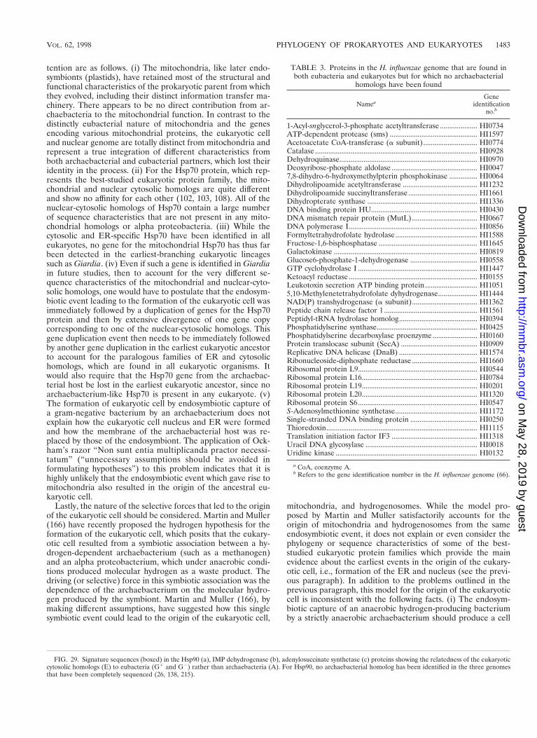
tention are as follows. (i) The mitochondria, like later endo-symbionts (plastids), have retained most of the structural andfunctional characteristics of the prokaryotic parent from whichthey evolved, including their distinct information transfer ma-chinery. There appears to be no direct contribution from ar-chaebacteria to the mitochondrial function. In contrast to thedistinctly eubacterial nature of mitochondria and the genesencoding various mitochondrial proteins, the eukaryotic celland nuclear genome are totally distinct from mitochondria andrepresent a true integration of different characteristics fromboth archaebacterial and eubacterial partners, which lost theiridentity in the process. (ii) For the Hsp70 protein, which rep-resents the best-studied eukaryotic protein family, the mito-chondrial and nuclear cytosolic homologs are quite differentand show no affinity for each other (102, 103, 108). All of thenuclear-cytosolic homologs of Hsp70 contain a large numberof sequence characteristics that are not present in any mito-chondrial homologs or alpha proteobacteria. (iii) While thecytosolic and ER-specific Hsp70 have been identified in alleukaryotes, no gene for the mitochondrial Hsp70 has thus farbeen detected in the earliest-branching eukaryotic lineagessuch as Giardia. (iv) Even if such a gene is identified in Giardiain future studies, then to account for the very different se-quence characteristics of the mitochondrial and nuclear-cyto-solic homologs, one would have to postulate that the endosym-biotic event leading to the formation of the eukaryotic cell wasimmediately followed by a duplication of genes for the Hsp70protein and then by extensive divergence of one gene copycorresponding to one of the nuclear-cytosolic homologs. Thisgene duplication event then needs to be immediately followedby another gene duplication in the earliest eukaryotic ancestorto account for the paralogous families of ER and cytosolichomologs, which are found in all eukaryotic organisms. Itwould also require that the Hsp70 gene from the archaebac-terial host be lost in the earliest eukaryotic ancestor, since noarchaebacterium-like Hsp70 is present in any eukaryote. (v)The formation of eukaryotic cell by endosymbiotic capture ofa gram-negative bacterium by an archaebacterium does notexplain how the eukaryotic cell nucleus and ER were formedand how the membrane of the archaebacterial host was re-placed by those of the endosymbiont. The application of Ock-ham’s razor “Non sunt entia multiplicanda practor necessi-tatum” (“unnecessary assumptions should be avoided informulating hypotheses”) to this problem indicates that it ishighly unlikely that the endosymbiotic event which gave rise tomitochondria also resulted in the origin of the ancestral eu-karyotic cell.
Lastly, the nature of the selective forces that led to the originof the eukaryotic cell should be considered. Martin and Muller(166) have recently proposed the hydrogen hypothesis for theformation of the eukaryotic cell, which posits that the eukary-otic cell resulted from a symbiotic association between a hy-drogen-dependent archaebacterium (such as a methanogen)and an alpha proteobacterium, which under anaerobic condi-tions produced molecular hydrogen as a waste product. Thedriving (or selective) force in this symbiotic association was thedependence of the archaebacterium on the molecular hydro-gen produced by the symbiont. Martin and Muller (166), bymaking different assumptions, have suggested how this singlesymbiotic event could lead to the origin of the eukaryotic cell,
mitochondria, and hydrogenosomes. While the model pro-posed by Martin and Muller satisfactorily accounts for theorigin of mitochondria and hydrogenosomes from the sameendosymbiotic event, it does not explain or even consider thephylogeny or sequence characteristics of some of the best-studied eukaryotic protein families which provide the mainevidence about the earliest events in the origin of the eukary-otic cell, i.e., formation of the ER and nucleus (see the previ-ous paragraph). In addition to the problems outlined in theprevious paragraph, this model for the origin of the eukaryoticcell is inconsistent with the following facts. (i) The endosym-biotic capture of an anaerobic hydrogen-producing bacteriumby a strictly anaerobic archaebacterium should produce a cell
TABLE 3. Proteins in the H. influenzae genome that are found inboth eubacteria and eukaryotes but for which no archaebacterial
homologs have been found
NameaGene
identificationno.b
1-Acyl-snglycerol-3-phosphate acetyltransferase .................... HI0734ATP-dependent protease (sms) ............................................... HI1597Acetoacetate CoA-transferase (a subunit)............................. HI0774Catalase ....................................................................................... HI0928Dehydroquinase.......................................................................... HI0970Deoxyribose-phosphate aldolase .............................................. HI00477,8-dihydro-6-hydroxymethylpterin phosphokinase ............... HI0064Dihydrolipoamide acetyltransferase ........................................ HI1232Dihydrolipoamide succinyltransferase ..................................... HI1661Dihydropterate synthase ........................................................... HI1336DNA binding protein HU......................................................... HI0430DNA mismatch repair protein (MutL) ................................... HI0667DNA polymerase I..................................................................... HI0856Formyltetrahydrofolate hydrolase............................................ HI1588Fructose-1,6-bisphosphatase ..................................................... HI1645Galactokinase ............................................................................. HI0819Glucose6-phosphate-1-dehydrogenase .................................... HI0558GTP cyclohydrolase I ................................................................ HI1447Ketoacyl reductase ..................................................................... HI0155Leukotoxin secretion ATP binding protein ............................ HI10515,10-Methylenetetrahydrofolate dyhydrogenase..................... HI1444NAD(P) transhydrogenase (a subunit)................................... HI1362Peptide chain release factor 1 .................................................. HI1561Peptidyl-tRNA hydrolase homolog.......................................... HI0394Phosphatidylserine synthase...................................................... HI0425Phosphatidylserine decarboxylase proenzyme........................ HI0160Protein translocase subunit (SecA) ......................................... HI0909Replicative DNA helicase (DnaB) .......................................... HI1574Ribonucleoside-diphosphate reductase ................................... HI1660Ribosomal protein L9................................................................ HI0544Ribosomal protein L16.............................................................. HI0784Ribosomal protein L19.............................................................. HI0201Ribosomal protein L20.............................................................. HI1320Ribosomal protein S6................................................................ HI0547S-Adenosylmethionine synthetase............................................ HI1172Single-stranded DNA binding protein .................................... HI0250Thioredoxin................................................................................. HI1115Translation initiation factor IF3 .............................................. HI1318Uracil DNA glycosylase ............................................................ HI0018Uridine kinase ............................................................................ HI0132
a CoA, coenzyme A.b Refers to the gene identification number in the H. influenzae genome (66).
FIG. 29. Signature sequences (boxed) in the Hsp90 (a), IMP dehydrogenase (b), adenylosuccinate synthetase (c) proteins showing the relatedness of the eukaryoticcytosolic homologs (E) to eubacteria (G1 and G2) rather than archaebacteria (A). For Hsp90, no archaebacterial homolog has been identified in the three genomesthat have been completely sequenced (26, 138, 215).
VOL. 62, 1998 PHYLOGENY OF PROKARYOTES AND EUKARYOTES 1483
on May 28, 2019 by guest
http://mm
br.asm.org/
Dow
nloaded from

which should also be a strict anaerobe; however, to my knowl-edge, no free-living eukaryotic organism is strictly anaerobic.(ii) Since this fusion took place in an oxygenic atmosphere(based upon the evolutionary position of proteobacteria in theprokaryotic lineage [see “Evolutionary relationships amongeukaryotes”]), an anaerobic cell will be at a great selectivedisadvantage under such conditions. (iii) Since endosymbioticassociation between hydrogen-producing bacteria and hydro-gen-dependent archaebacteria is indicated to be very common,it does not explain the uniqueness of the fusion event. (iv)Molecular sequence data indicate that among archaebacteria,the eocyte group of archaebacteria (i.e., thermoacidophilic)and not methanogens are the closest relatives of eukaryotes(198).
In contrast to the hydrogen hypothesis, which posits hydro-gen dependence as the major selective force, I propose that thetwo major selective forces that had a profound influence in
shaping the evolutionary history of life were (i) the antibioticselection pressure, which probably led to the evolution of botharchaebacteria and the diderm prokaryotes (see “Possible se-lective forces leading to horizontal gene transfers”) and (ii)oxygen sensitivity of the organisms when the atmospherechanged from anaerobic to aerobic (208). In my view, a com-bination of these two selective forces led to the association andultimate fusion of an antibiotic-resistant archaebacterium withan oxygen-tolerant eubacterium to produce a novel eukaryoticcell which was antibiotic resistant and oxygen tolerant. Thisscenario explains why, during the gene assortment process inthe ancestral eukaryotic cell, most of the genes for the infor-mation transfer processes (which provide the main targets fordifferent antibiotics) were retained from the archaebacterialpartner whereas a large number of genes for the metabolicprocesses were acquired from the eubacterial parent. To ac-count for the uniqueness of the fusion event, it is likely that the
FIG. 30. Neighbor-joining distance tree based on Hsp90 sequences indicating that the cytosolic and ER resident forms of these protein form paralogous genefamilies, which resulted from a gene duplication event very early in the history of eukaryotic cells. The bootstrap scores out of 1,000 replicates are shown. Reproducedfrom reference 97 with permission of the publisher.
1484 GUPTA MICROBIOL. MOL. BIOL. REV.
on May 28, 2019 by guest
http://mm
br.asm.org/
Dow
nloaded from
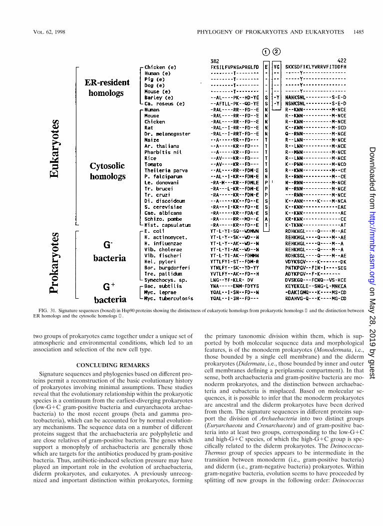
two groups of prokaryotes came together under a unique set ofatmospheric and environmental conditions, which led to anassociation and selection of the new cell type.
CONCLUDING REMARKSSignature sequences and phylogenies based on different pro-
teins permit a reconstruction of the basic evolutionary historyof prokaryotes involving minimal assumptions. These studiesreveal that the evolutionary relationship within the prokaryoticspecies is a continuum from the earliest-diverging prokaryotes(low-G1C gram-positive bacteria and euryarchaeota archae-bacteria) to the most recent groups (beta and gamma pro-teobacteria), which can be accounted for by normal evolution-ary mechanisms. The sequence data on a number of differentproteins suggest that the archaebacteria are polyphyletic andare close relatives of gram-positive bacteria. The genes whichsupport a monophyly of archaebacteria are generally thosewhich are targets for the antibiotics produced by gram-positivebacteria. Thus, antibiotic-induced selection pressure may haveplayed an important role in the evolution of archaebacteria,diderm prokaryotes, and eukaryotes. A previously unrecog-nized and important distinction within prokaryotes, forming
the primary taxonomic division within them, which is sup-ported by both molecular sequence data and morphologicalfeatures, is of the monoderm prokaryotes (Monodermata, i.e.,those bounded by a single cell membrane) and the didermprokaryotes (Didermata, i.e., those bounded by inner and outercell membranes defining a periplasmic compartment). In thatsense, both archaebacteria and gram-positive bacteria are mo-noderm prokaryotes, and the distinction between archaebac-teria and eubacteria is misplaced. Based on molecular se-quences, it is possible to infer that the monoderm prokaryotesare ancestral and the diderm prokaryotes have been derivedfrom them. The signature sequences in different proteins sup-port the division of Archaebacteria into two distinct groups(Euryarchaeota and Crenarchaeota) and of gram-positive bac-teria into at least two groups, corresponding to the low-G1Cand high-G1C species, of which the high-G1C group is spe-cifically related to the diderm prokaryotes. The Deinococcus-Thermus group of species appears to be intermediate in thetransition between monoderm (i.e., gram-positive bacteria)and diderm (i.e., gram-negative bacteria) prokaryotes. Withingram-negative bacteria, evolution seems to have proceeded bysplitting off new groups in the following order: Deinococcus
FIG. 31. Signature sequences (boxed) in Hsp90 proteins showing the distinctness of eukaryotic homologs from prokaryotic homologs ① and the distinction betweenER homologs and the cytosolic homologs ②.
VOL. 62, 1998 PHYLOGENY OF PROKARYOTES AND EUKARYOTES 1485
on May 28, 2019 by guest
http://mm
br.asm.org/
Dow
nloaded from
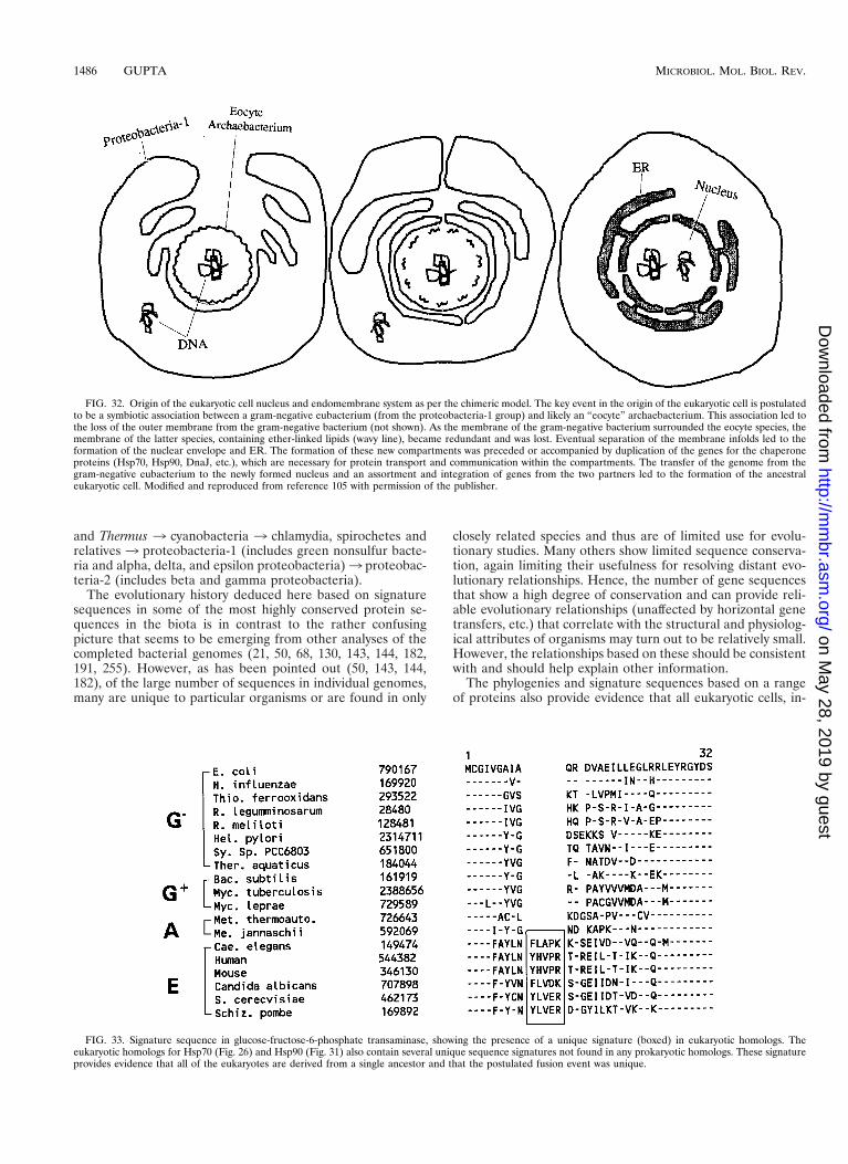
and Thermus 3 cyanobacteria 3 chlamydia, spirochetes andrelatives 3 proteobacteria-1 (includes green nonsulfur bacte-ria and alpha, delta, and epsilon proteobacteria)3 proteobac-teria-2 (includes beta and gamma proteobacteria).
The evolutionary history deduced here based on signaturesequences in some of the most highly conserved protein se-quences in the biota is in contrast to the rather confusingpicture that seems to be emerging from other analyses of thecompleted bacterial genomes (21, 50, 68, 130, 143, 144, 182,191, 255). However, as has been pointed out (50, 143, 144,182), of the large number of sequences in individual genomes,many are unique to particular organisms or are found in only
closely related species and thus are of limited use for evolu-tionary studies. Many others show limited sequence conserva-tion, again limiting their usefulness for resolving distant evo-lutionary relationships. Hence, the number of gene sequencesthat show a high degree of conservation and can provide reli-able evolutionary relationships (unaffected by horizontal genetransfers, etc.) that correlate with the structural and physiolog-ical attributes of organisms may turn out to be relatively small.However, the relationships based on these should be consistentwith and should help explain other information.
The phylogenies and signature sequences based on a rangeof proteins also provide evidence that all eukaryotic cells, in-
FIG. 32. Origin of the eukaryotic cell nucleus and endomembrane system as per the chimeric model. The key event in the origin of the eukaryotic cell is postulatedto be a symbiotic association between a gram-negative eubacterium (from the proteobacteria-1 group) and likely an “eocyte” archaebacterium. This association led tothe loss of the outer membrane from the gram-negative bacterium (not shown). As the membrane of the gram-negative bacterium surrounded the eocyte species, themembrane of the latter species, containing ether-linked lipids (wavy line), became redundant and was lost. Eventual separation of the membrane infolds led to theformation of the nuclear envelope and ER. The formation of these new compartments was preceded or accompanied by duplication of the genes for the chaperoneproteins (Hsp70, Hsp90, DnaJ, etc.), which are necessary for protein transport and communication within the compartments. The transfer of the genome from thegram-negative eubacterium to the newly formed nucleus and an assortment and integration of genes from the two partners led to the formation of the ancestraleukaryotic cell. Modified and reproduced from reference 105 with permission of the publisher.
FIG. 33. Signature sequence in glucose-fructose-6-phosphate transaminase, showing the presence of a unique signature (boxed) in eukaryotic homologs. Theeukaryotic homologs for Hsp70 (Fig. 26) and Hsp90 (Fig. 31) also contain several unique sequence signatures not found in any prokaryotic homologs. These signatureprovides evidence that all of the eukaryotes are derived from a single ancestor and that the postulated fusion event was unique.
1486 GUPTA MICROBIOL. MOL. BIOL. REV.
on May 28, 2019 by guest
http://mm
br.asm.org/
Dow
nloaded from
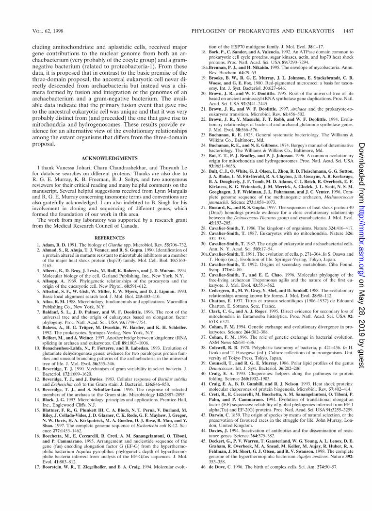
cluding amitochondriate and aplastidic cells, received majorgene contributions to the nuclear genome from both an ar-chaebacterium (very probably of the eocyte group) and a gram-negative bacterium (related to proteobacteria-1). From thesedata, it is proposed that in contrast to the basic premise of thethree-domain proposal, the ancestral eukaryotic cell never di-rectly descended from archaebacteria but instead was a chi-mera formed by fusion and integration of the genomes of anarchaebacterium and a gram-negative bacterium. The avail-able data indicate that the primary fusion event that gave riseto the ancestral eukaryotic cell was unique and that it was veryprobably distinct from (and preceded) the one that gave rise tomitochondria and hydrogenosomes. These results provide ev-idence for an alternative view of the evolutionary relationshipsamong the extant organisms that differs from the three-domainproposal.
ACKNOWLEDGMENTS
I thank Vanessa Johari, Charu Chandrashekhar, and Thuyanh Lefor database searches on different proteins. Thanks are also due toR. G. E. Murray, K. B. Freeman, B. J. Soltys, and two anonymousreviewers for their critical reading and many helpful comments on themanuscript. Several helpful suggestions received from Lynn Margulisand R. G. E. Murray concerning taxonomic terms and conventions arealso gratefully acknowledged. I am also indebted to B. Singh for hisinvolvement in cloning and sequencing of different genes, whichformed the foundation of our work in this area.
The work from my laboratory was supported by a research grantfrom the Medical Research Council of Canada.
REFERENCES
1. Adam, R. D. 1991. The biology of Giardia spp. Microbiol. Rev. 55:706–732.2. Ahmad, S., R. Ahuja, T. J. Venner, and R. S. Gupta. 1990. Identification of
a protein altered in mutants resistant to microtubule inhibitors as a memberof the major heat shock protein (hsp70) family. Mol. Cell. Biol. 10:5160–5165.
3. Alberts, B., D. Bray, J. Lewis, M. Raff, K. Roberts, and J. D. Watson. 1994.Molecular biology of the cell. Garland Publishing, Inc., New York, N.Y.
4. Allsopp, A. 1969. Phylogenetic relationships of the procaryota and theorigin of the eucaryotic cell. New Phytol. 68:591–612.
5. Altschul, S. F., W. Gish, W. Miller, E. W. Myers, and D. J. Lipman. 1990.Basic local alignment search tool. J. Mol. Biol. 215:403–410.
6. Atlas, R. M. 1988. Microbiology: fundamentals and applications. MacmillanPublishing Co., New York, N.Y.
7. Baldauf, S. L., J. D. Palmer, and W. F. Doolittle. 1996. The root of theuniversal tree and the origin of eukaryotes based on elongation factorphylogeny. Proc. Natl. Acad. Sci. USA 93:7749–7754.
8. Balows, A., H. G. Truper, M. Dworkin, W. Harder, and K. H. Schleifer.1992. The prokaryotes. Springer-Verlag, New York, N.Y.
9. Belfort, M., and A. Weiner. 1997. Another bridge between kingdoms: tRNAsplicing in archaea and eukaryotes. Cell 89:1003–1006.
10. Benachenhou-Lahfa, N., P. Forterre, and B. Labedan. 1993. Evolution ofglutamate dehydrogenase genes: evidence for two paralogous protein fam-ilies and unusual branching patterns of the archaebacteria in the universaltree of life. J. Mol. Evol. 36:335–346.
11. Beveridge, T. J. 1990. Mechanism of gram variability in select bacteria. J.Bacteriol. 172:1609–1620.
12. Beveridge, T. J., and J. Davies. 1983. Cellular response of Bacillus subtilisand Escherichia coli to the Gram stain. J. Bacteriol. 156:846–858.
13. Beveridge, T. J., and S. Schultze-Lam. 1996. The response of selectedmembers of the archaea to the Gram stain. Microbiology 142:2887–2895.
14. Black, J. G. 1993. Microbiology: principles and applications. Prentice-Hall,Inc., Englewood Cliffs, N.J.
15. Blattner, F. R., G. Plunkett III, C. A. Bloch, N. T. Perna, V. Burland, M.Riley, J. Collado-Vides, J. D. Glasner, C. K. Rode, G. F. Mayhew, J. Gregor,N. W. Davis, H. A. Kirkpatrick, M. A. Goeden, D. J. Rose, B. Mau, and Y.Shao. 1997. The complete genome sequence of Escherichia coli K-12. Sci-ence 277:1453–1462.
16. Bocchetta, M., E. Ceccarelli, R. Creti, A. M. Sanangelantoni, O. Tiboni,and P. Cammarano. 1995. Arrangement and nucleotide sequence of thegene (fus) encoding elongation factor G (EF-G) from the hyperthermo-philic bacterium Aquifex pyrophilus: phylogenetic depth of hyperthermo-philic bacteria inferred from analysis of the EF-G/fus sequences. J. Mol.Evol. 41:803–812.
17. Boorstein, W. R., T. Ziegelhoffer, and E. A. Craig. 1994. Molecular evolu-
tion of the HSP70 multigene family. J. Mol. Evol. 38:1–17.18. Bork, P., C. Sander, and A. Valencia. 1992. An ATPase domain common to
prokaryotic cell cycle proteins, sugar kinases, actin, and hsp70 heat shockproteins. Proc. Natl. Acad. Sci. USA 89:7290–7294.
18a.Brennan, P. J., and H. Nikaido. 1995. The envelope of mycobacteria. Annu.Rev. Biochem. 64:29–63.
19. Brooks, B. W., R. G. E. Murray, J. L. Johnson, E. Stackebrandt, C. R.Woese, and G. E. Fox. 1980. Red-pigmented micrococci: a basis for taxon-omy. Int. J. Syst. Bacteriol. 30:627–646.
20. Brown, J. R., and W. F. Doolittle. 1995. Root of the universal tree of lifebased on ancient aminoacyl-tRNA synthetase gene duplications. Proc. Natl.Acad. Sci. USA 92:2441–2445.
21. Brown, J. R., and W. F. Doolittle. 1997. Archaea and the prokaryote-to-eukaryote transition. Microbiol. Rev. 61:456–502.
22. Brown, J. R., Y. Masuchi, F. T. Robb, and W. F. Doolittle. 1994. Evolu-tionary relationships of bacterial and archaeal glutamine synthetase genes.J. Mol. Evol. 38:566–576.
23. Buchanan, R. E. 1925. General systematic bacteriology. The Williams &Wilkins Co., Baltimore, Md.
24. Buchanan, R. E., and N. E. Gibbons. 1974. Bergey’s manual of deteminativebacteriology, The Williams & Wilkins Co., Baltimore, Md.
25. Bui, E. T., P. J. Bradley, and P. J. Johnson. 1996. A common evolutionaryorigin for mitochondria and hydrogenosomes. Proc. Natl. Acad. Sci. USA93:9651–9656.
26. Bult, C. J., O. White, G. J. Olsen, L. Zhou, R. D. Fleischmann, G. G. Sutton,J. A. Blake, L. M. FitzGerald, R. A. Clayton, J. D. Gocayne, A. R. Kerlavage,B. A. Dougherty, J. F. Tomb, M. D. Adams, C. I. Reich, R. Overbeek, E. F.Kirkness, K. G. Weinstock, J. M. Merrick, A. Glodek, J. L. Scott, N. S. M.Geoghagen, J. F. Weidman, J. L. Fuhrmann, and J. C. Venter. 1996. Com-plete genome sequence of the methanogenic archaeon, Methanococcusjannaschii. Science 273:1058–1073.
27. Bustard, K., and R. S. Gupta. 1997. The sequences of heat shock protein 40(DnaJ) homologs provide evidence for a close evolutionary relationshipbetween the Deinococcus-Thermus group and cyanobacteria. J. Mol. Evol.45:193–205.
28. Cavalier-Smith, T. 1986. The kingdoms of organisms. Nature 324:416–417.29. Cavalier-Smith, T. 1987. Eukaryotes with no mitochondria. Nature 326:
332–333.30. Cavalier-Smith, T. 1987. The origin of eukaryotic and archaebacterial cells.
Ann. N. Y. Acad. Sci. 503:17–54.30a.Cavalier-Smith, T. 1991. The evolution of cells, p. 271–304. In S. Osawa and
T. Honjo (ed.), Evolution of life. Springer-Verlag, Tokyo, Japan.31. Cavalier-Smith, T. 1992. Origins of secondary metabolism. Ciba Found.
Symp. 171:64–80.32. Cavalier-Smith, T., and E. E. Chao. 1996. Molecular phylogeny of the
free-living archezoan Trepomonas agilis and the nature of the first eu-karyote. J. Mol. Evol. 43:551–562.
33. Cedergren, R., M. W. Gray, Y. Abel, and D. Sankoff. 1988. The evolutionaryrelationships among known life forms. J. Mol. Evol. 28:98–112.
34. Chatton, E. 1937. Titres et travaux scientifiques (1906–1937) de EdouardChatton. E. Sottano, Sete, France.
35. Clark, C. G., and A. J. Roger. 1995. Direct evidence for secondary loss ofmitochondria in Entamoeba histolytica. Proc. Natl. Acad. Sci. USA 92:6518–6521.
36. Cohan, F. M. 1994. Genetic exchange and evolutionary divergence in pro-karyotes. Science 264:382–388.
37. Cohan, F. M. 1996. The role of genetic exchange in bacterial evolution.ASM News 62:631–636.
38. Colewell, R. R. 1970. Polyphasic taxonomy of bacteria, p. 421–436. In H.Iizuka and T. Hasegawa (ed.), Culture collections of microorganisms. Uni-versity of Tokyo Press, Tokyo, Japan.
39. Counsell, T., and R. G. E. Murray. 1986. Polar lipid profiles of the genusDeinococcus. Int. J. Syst. Bacteriol. 36:202–206.
40. Craig, E. A. 1993. Chaperones: helpers along the pathways to proteinfolding. Science 260:1902–1903.
41. Craig, E. A., B. D. Gambill, and R. J. Nelson. 1993. Heat shock proteins:molecular chaperones of protein biogenesis. Microbiol. Rev. 57:402–414.
42. Creti, R., E. Ceccarelli, M. Bocchetta, A. M. Sanangelantoni, O. Tiboni, P.Palm, and P. Cammarano. 1994. Evolution of translational elongationfactor (EF) sequences: reliability of global phylogenies inferred from EF-1alpha(Tu) and EF-2(G) proteins. Proc. Natl. Acad. Sci. USA 91:3255–3259.
43. Darwin, C. 1859. The origin of species by means of natural selection, or thepreservation of favoured races in the struggle for life. John Murray, Lon-don, United Kingdom.
44. Davies, J. 1994. Inactivation of antibiotics and the dissemination of resis-tance genes. Science 264:375–382.
45. Deckert, G., P. V. Warren, T. Gaasterland, W. G. Young, A. L. Lenox, D. E.Graham, R. Overbeek, M. A. Snead, M. Keller, M. Aujay, R. Huber, R. A.Feldman, J. M. Short, G. J. Olsen, and R. V. Swanson. 1998. The completegenome of the hyperthermophilic bacterium Aquifex aeolicus. Nature 392:353–358.
46. de Duve, C. 1996. The birth of complex cells. Sci. Am. 274:50–57.
VOL. 62, 1998 PHYLOGENY OF PROKARYOTES AND EUKARYOTES 1487
on May 28, 2019 by guest
http://mm
br.asm.org/
Dow
nloaded from
47. Dennis, P. P. 1997. Ancient ciphers: translation in Archaea. Cell 89:1007–1010.
48. De Rijk, P., Y. Van de Peer, I. Van den Broeck, and R. De Wachter. 1995.Evolution according to large ribosomal subunit RNA. J. Mol. Evol. 41:366–375.
49. Doolittle, R. F. 1995. Of archae and eo: what’s in a name? Proc. Natl. Acad.Sci. USA 92:2421–2423.
50. Doolittle, R. F. 1998. Microbial genomes opened up. Nature 392:339–342.51. Doolittle, R. F., D. F. Feng, S. Tsang, G. Cho, and E. Little. 1996. Deter-
mining divergence times of the major kingdoms of living organisms with aprotein clock. Science 271:470–477.
52. Doolittle, W. F. 1998. A paradigm gels shifty. Nature 392:15–16.53. Doolittle, W. F., and J. R. Brown. 1994. Tempo, mode, the progenote, and
the universal root. Proc. Natl. Acad. Sci. USA 91:6721–6728.54. Edgell, D. R., and W. F. Doolittle. 1997. Archaea and the origin(s) of DNA
replication proteins. Cell 89:995–998.55. Eisen, J. A. 1995. The RecA protein as a model molecule for molecular
systematic studies of bacteria: comparison of trees of RecAs and 16SrRNAs from the same species. J. Mol. Evol. 41:1105–1123.
56. Falah, M., and R. S. Gupta. 1994. Cloning of the hsp70 (dnaK) genes fromRhizobium meliloti and Pseudomonas cepacia: phylogenetic analyses of mi-tochondrial origin based on a highly conserved protein sequence. J. Bacte-riol. 176:7748–7753.
57. Falah, M., and R. S. Gupta. 1997. Phylogenetic analysis of mycoplasmasbased on Hsp70 sequences: cloning of the dnaK (hsp70) gene region ofMycoplasma capricolum. Int. J. Syst. Bacteriol. 47:38–45.
58. Felsenstein, J. 1982. Numerical methods for inferring evolutionary trees. Q.Rev. Biol. 57:379–404.
59. Felsenstein, J. 1985. Confidence limits in phylogenies: an approach usingthe bootstrap. Evolution 39:783–791.
60. Felsenstein, J. 1988. Phylogenies from molecular sequences: inference andreliability. Ann. Rev. Genet. 22:521–565.
61. Felsenstein, J. 1996. Inferring phylogenies from protein sequences by par-simony, distance, and likelihood methods. Methods Enzymol. 266:418–427.
62. Felsenstein, J. 1997. Cases in which parsimony and compatibility methodswill be positively misleading. Syst. Zool. 27:401–410.
63. Feng, D. F., G. Cho, and R. F. Doolittle. 1997. Determining divergencetimes with a protein clock: update and reevaluation. Proc. Natl. Acad. Sci.USA 94:13028–13033.
64. Fitch, W. M. 1997. Toward defining the course of evolution: minimumchange for a specified tree topology. Syst. Zool. 20:406–416.
65. Fitch, W. M., and E. Margoliash. 1967. Construction of phylogentic trees:a method based on mutational distances as estimated from cytochrome csequences is of general applicability. Science 155:279–284.
65a.Flaherty, K. M., D. B. McKay, W. Kabsch, and K. C. Holmes. 1991. Sim-ilarity of the three-dimensional structures of actin and the ATPase frag-ment of a 70-kDa heat shock cognate protein. Proc. Natl. Acad. Sci. USA88:5041–5045.
66. Fleischmann, R. D., M. D. Adams, O. White, R. A. Clayton, E. F. Kirkness,A. R. Kerlavage, C. J. Bult, J. F. Tomb, B. A. Dougherty, and J. M. Merrick.1995. Whole-genome random sequencing and assembly of Haemophilusinfluenzae Rd. Science 269:496–512.
67. Forterre, P. 1996. A hot topic: the origin of hyperthermophiles. Cell 85:789–792.
68. Forterre, P. 1997. Protein versus rRNA: problems in rooting the universaltree of life. ASM News 63:89–95.
69. Forterre, P. 1997. Archaea: what can we learn from their sequences. Curr.Opin. Genet. Dev. 7:764–770.
70. Forterre, P., N. Benachenhou-Lahfa, F. Confalonieri, M. Duguet, C. Elie,and B. Labedan. 1992. The nature of the last universal ancestor and theroot of the tree of life, still open questions. Biosystems 28:15–32.
71. Fox, G. E., E. Stackebrandt, R. B. Hespell, J. Gibson, J. Maniloff, T. A.Dyer, R. S. Wolfe, W. E. Balch, R. S. Tanner, L. J. Magrum, L. B. Zablen,R. Blakemore, R. Gupta, L. Bonen, B. J. Lewis, D. A. Stahl, K. R. Luehrsen,K. N. Chen, and C. R. Woese. 1980. The phylogeny of prokaryotes. Science209:457–463.
72. Fraser, C. M., S. Casjens, W. M. Huang, G. G. Sutton, R. Clayton, R.Lathigra, O. White, K. A. Ketchum, R. Dodson, E. K. Hickey, M. Gwinn, B.Dougherty, J. F. Tomb, R. D. Fleischmann, D. Richardson, J. Peterson,A. R. Kerlavage, J. Quackenbush, S. Salzberg, M. Hanson, R. Van Vugt, N.Palmer, M. D. Adams, and J. Gocayne. 1997. Genomic sequence of a Lymedisease spirochaete, Borrelia burgdorferi. Nature 390:580–586.
73. Fraser, C. M., J. D. Gocayne, O. White, M. D. Adams, R. A. Clayton, R. D.Fleischmann, C. J. Bult, A. R. Kerlavage, G. Sutton, and J. M. Kelley. 1995.The minimal gene complement of Mycoplasma genitalium. Science 270:397–403.
74. Frye, N. 1990. Words with power, p. 129. Harcourt Brace Jovanovich, NewYork, N.Y.
75. Fuerst, J. A. 1995. The Planctomycetes: emerging models for microbialecology, evolution and cell biology. Microbiology 141:1493–1506.
76. Galley, K. A., B. Singh, and R. S. Gupta. 1992. Cloning of HSP70 (dnaK)gene from Clostridium perfringens using a general polymerase chain reac-
tion based approach. Biochim. Biophys. Acta 1130:203–208.77. Galtier, N., and M. Gouy. 1995. Inferring phylogenies from DNA sequences
of unequal base compositions. Proc. Natl. Acad. Sci. USA 92:11317–11321.78. Germot, A., H. Philippe, and H. Le Guyader. 1996. Presence of a mito-
chondrial-type 70-kDa heat shock protein in Trichomonas vaginalis sug-gests a very early mitochondrial endosymbiosis in eukaryotes. Proc. Natl.Acad. Sci. USA 93:14614–14617.
79. Glasby, J. S. 1979. Encyclopedia of antibiotics. John Wiley & Sons, Inc.,New York, N.Y.
80. Goffeau, A., B. G. Barrell, H. Bussey, R. W. Davis, B. Dujon, H. Feldmann,F. Galibert, J. D. Hoheisel, C. Jacq, M. Johnston, E. J. Louis, H. W. Mewes,Y. Murakami, P. Philippsen, H. Tettelin, and S. G. Oliver. 1996. Life with6000 genes. Science 274:546, 563–567.
81. Gogarten, J. P., H. Kibak, P. Dittrich, L. Taiz, E. J. Bowman, B. J. Bowman,M. F. Manolson, R. J. Poole, T. Date, and T. Oshima. 1989. Evolution of thevacuolar H1-ATPase: implications for the origin of eukaryotes. Proc. Natl.Acad. Sci. USA 86:6661–6665.
82. Gogarten, J. P., T. Starke, H. Kibak, J. Fishman, and L. Taiz. 1992.Evolution and isoforms of V-ATPase subunits. J. Exp. Biol. 172:137–147.
83. Gogarten-Boekels, M., E. Hilario, and J. P. Gogarten. 1995. The effects ofheavy meteorite bombardment on the early evolution—the emergence ofthe three domains of life. Origins Life Evol. Biosphere 25:251–264.
84. Golding, B. 1996. Evolution: when was life’s first branch point? Curr. Biol.6:679–682.
85. Golding, G. B., and R. S. Gupta. 1995. Protein-based phylogenies supporta chimeric origin for the eukaryotic genome. Mol. Biol. Evol. 12:1–6.
86. Gouy, M., and W. H. Li. 1989. Phylogenetic analysis based on rRNAsequences supports the archaebacterial rather than the eocyte tree. Nature339:145–147.
87. Gouy, M., and W. H. Li. 1989. Molecular phylogeny of the kingdomsAnimalia, Plantae, and Fungi. Mol. Biol. Evol. 6:109–122.
88. Gram, C. 1884. Ueber die isolierte farbung der Schizomyceten in Schnittund Trockenpraparaten. Fortschr. Med. 2:185–189.
89. Gray, M. W. 1988. Organelle origins and ribosomal RNA. Biochem. CellBiol. 66:325–348.
90. Gray, M. W. 1992. The endosymbiont hypothesis revisited. Int. Rev. Cytol.141:233–357.
91. Gray, M. W. 1996. The third form of life. Nature 383:299–300.92. Gray, M. W., and W. F. Doolittle. 1982. Has the endosymbiont hypothesis
been proven? Microbiol. Rev. 46:1–42.93. Grogan, D. W. 1996. Exchange of genetic information at extremely high
temperatures in the archaeon Sulfolobus acidocaldarius. J. Bacteriol. 178:3207–3211.
94. Gruber, T. M., and D. A. Bryant. 1997. Molecular systematic studies ofeubacteria, using s70-type sigma factors of group 1 and group 2. J. Bacte-riol. 179:1734–1747.
95. Gupta, R. S. 1990. Sequence and structural homology between a mouseT-complex protein TCP-1 and the ‘chaperonin’ family of bacterial (GroEL,60-65 kDa heat shock antigen) and eukaryotic proteins. Biochem. Int.20:833–841.
96. Gupta, R. S. 1995. Evolution of the chaperonin families (Hsp60, Hsp10 andTcp-1) of proteins and the origin of eukaryotic cells. Mol. Microbiol. 15:1–11.
97. Gupta, R. S. 1995. Phylogenetic analysis of the 90 kD heat shock family ofprotein sequences and an examination of the relationship among animals,plants, and fungi species. Mol. Biol. Evol. 12:1063–1073.
98. Gupta, R. S. 1996. Evolutionary relationships of chaperonins, p. 27–64. InR. J. Ellis (ed.), The chaperonins. Academic Press, Inc., New York, N.Y.
99. Gupta, R. S. 1997. Protein phylogenies and signature sequences: evolution-ary relationships within prokaryotes and between prokaryotes and eu-karyotes. Antonie Leeuwenhoek 72:49–61.
100. Gupta, R. S. 1998. Life’s third domain (Archaea): an established fact or anendangered paradigm? A new proposal for classification of organismsbased on protein sequences and cell structure. Theor. Popul. Biol. 54:91–104.
101. Gupta, R. S. 1998. What are archaebacteria: life’s third domain or mono-derm prokaryotes related to Gram-positive bacteria? A new proposal forthe classification of prokaryotic organisms. Mol. Microbiol. 29:695–708.
102. Gupta, R. S., K. Aitken, M. Falah, and B. Singh. 1994. Cloning of Giardialamblia heat shock protein HSP70 homologs: implications regarding originof eukaryotic cells and of endoplasmic reticulum. Proc. Natl. Acad. Sci.USA 91:2895–2899.
103. Gupta, R. S., K. Bustard, M. Falah, and D. Singh. 1997. Sequencing of heatshock protein 70 (DnaK) homologs from Deinococcus proteolyticus andThermomicrobium roseum and their integration in a protein-based phylog-eny of prokaryotes. J. Bacteriol. 179:345–357.
104. Gupta, R. S., and G. B. Golding. 1993. Evolution of HSP70 gene and itsimplications regarding relationships between archaebacteria, eubacteria,and eukaryotes. J. Mol. Evol. 37:573–582.
105. Gupta, R. S., and G. B. Golding. 1996. The origin of the eukaryotic cell.Trends Biochem. Sci. 21:166–171.
106. Gupta, R. S., and V. Johari. 1998. Signature sequences in diverse proteins
1488 GUPTA MICROBIOL. MOL. BIOL. REV.
on May 28, 2019 by guest
http://mm
br.asm.org/
Dow
nloaded from
provide evidence of a close evolutionary relationship between the Deino-coccus-Thermus group and cyanobacteria. J. Mol. Evol. 46:716–720.
107. Gupta, R. S., and B. Singh. 1992. Cloning of the HSP70 gene from Halobac-terium marismortui: relatedness of archaebacterial HSP70 to its eubacterialhomologs and a model for the evolution of the HSP70 gene. J. Bacteriol.174:4594–4605.
108. Gupta, R. S., and B. Singh. 1994. Phylogenetic analysis of 70 kD heat shockprotein sequences suggests a chimeric origin for the eukaryotic cell nucleus.Curr. Biol. 4:1104–1114.
109. Hartman, H. 1984. The origin of the eukaryotic cell. Speculations Sci.Technol. 7:77–81.
110. Hasegawa, M., and M. Fujiwara. 1993. Relative efficiencies of the maxi-mum likelihood, maximum parsimony, and neighbor-joining methods forestimating protein phylogeny. Mol. Phylogenet. Evol. 2:1–5.
111. Hasegawa, M., and T. Hashimoto. 1993. Ribosomal RNA trees misleading?Nature 361:23.
112. Hashimoto, T., and M. Hasegawa. 1996. Origin and early evolution ofeukaryotes inferred from the amino acid sequences of translation elonga-tion factors 1alpha/Tu and 2/G. Adv. Biophys. 32:73–120.
113. Hashimoto, T., Y. Nakamura, F. Nakamura, T. Shirakura, J. Adachi, N.Goto, K. Okamoto, and M. Hasegawa. 1994. Protein phylogeny gives arobust estimation for early divergences of eukaryotes: phylogenetic place ofa mitochondria-lacking protozoan, Giardia lamblia. Mol. Biol. Evol. 11:65–71.
113a.Hashimoto, T., L. B. Sanchez, T. Shirakura, M. Muller, and M. Hasegawa.1998. Secondary loss of mitochondria in Giardia lamblia and Trichomonasvaginalis revealed by valyl-tRNA synthetase phylogeny. Proc. Natl. Acad.Sci. USA 95:6860–6865.
114. Hensel, R., P. Zwicki, S. Fabry, J. Lang, and P. Palm. 1997. Sequencecomparison of glyceraldehyde-3-phosphate dehydrogenases from the threeurkingdoms: evolutionary implications. Can. J. Microbiol. 35:81–85.
115. Hensell, R., W. Demharter, O. Kandler, R. M. Kroppenstedt, and E. Stackebrandt. 1986. Chemotaxonomic and molecular-genetic studies of the genusThermus: evidence for a phylogenetic relationship of Thermus aquaticus andThermus ruber to the genus Deinococcus. Int. J. Syst. Bacteriol. 36:444–453.
116. Henze, K., A. Badr, M. Wettern, R. Cerff, and W. Martin. 1995. A nucleargene of eubacterial origin in Euglena gracilis reflects cryptic endosymbiosesduring protist evolution. Proc. Natl. Acad. Sci. USA 92:9122–9126.
117. Higgins, D. G., and P. M. Sharp. 1988. CLUSTAL: a package for perform-ing multiple sequence alignments on a microcomputer. Gene 73:237–244.
118. Hilario, E., and J. P. Gogarten. 1993. Horizontal transfer of ATPasegenes—the tree of life becomes a net of life. Biosystems 31:111–119.
119. Himmelreich, R., H. Hilbert, H. Plagens, E. Pirkl, B. C. Li, and R. Herr-mann. 1996. Complete sequence analysis of the genome of the bacteriumMycoplasma pneumoniae. Nucleic Acids Res. 24:4420–4449.
120. Hirt, R. P., B. Healy, C. R. Vossbrinck, E. U. Canning, and T. M. Embley.1997. A mitochondrial Hsp70 orthologue in Vairimorpha necatrix: molecularevidence that microsporidia once contained mitochondria. Curr. Biol.7:995–998.
121. Holt, J. G., N. R. Krieg, P. H. A. Sneath, J. T. Staley, and S. T. Williams.1994. Bergey’s manual of determinative bacteriology, 9th ed. The Williams& Wilkins Co., Baltimore, Md.
122. Hori, H., and S. Osawa. 1987. Origin and evolution of Organisms as de-duced from 5S ribosomal RNA sequences. Mol. Biol. Evol. 4:445–472.
123. Horner, D. S., R. P. Hirt, S. Kilvington, D. Lloyd, and T. M. Embley. 1996.Molecular data suggest an early acquisition of the mitochondrion endosym-biont. Proc. R. Soc. London Ser. B 263:1053–1059.
124. Inouye, M. 1979. What is the outer membrane? p. 1–12. In M. Inouye (ed.),Bacterial outer membranes: biogenesis and functions. John Wiley & Sons,Inc., New York, N.Y.
125. Irwin, D. M. 1994. Molecular evolution. Who are the parents of eu-karyotes? Curr. Biol. 4:1115–1117.
126. Iwabe, N., K. Kuma, M. Hasegawa, S. Osawa, and T. Miyata. 1989. Evo-lutionary relationship of archaebacteria, eubacteria, and eukaryotes in-ferred from phylogenetic trees of duplicated genes. Proc. Natl. Acad. Sci.USA 86:9355–9359.
127. Kandler, O., and H. Konig. 1993. Cell envelopes of archaea: structure andchemistry, p. 223–259. In M. Kates, D. J. Kushner, and A. T. Matheson(ed.), The biochemistry of Archaea (Archaebacteria). Elsevier Science Pub-lishers B.V. New York, N.Y.
128. Kaneko, T., S. Sato, H. Kotani, A. Tanaka, E. Asamizu, Y. Nakamura, N.Miyajima, M. Hirosawa, M. Sugiura, S. Sasamoto, T. Kimura, T. Hosouchi,A. Matsuno, A. Muraki, N. Nakazaki, K. Naruo, S. Okumura, S. Shimpo,C. Takeuchi, T. Wada, A. Watanabe, M. Yamada, M. Yasuda, and S.Tabata. 1996. Sequence analysis of the genome of the unicellular cyanobac-terium Synechocystis sp. strain PCC6803. II. Sequence determination of theentire genome and assignment of potential protein-coding regions. DNARes. 3:109–136.
129. Karlin, S., J. Mrazek, and A. M. Campbell. 1997. Compositional biases ofbacterial genomes and evolutionary implications. J. Bacteriol. 179:3899–3913.
130. Karlin, S., and J. Mrazek. 1997. Compositional differences within and
between eukaryotic genomes. Proc. Natl. Acad. Sci. USA 94:10227–10232.131. Karlin, S., G. M. Weinstock, and V. Brendel. 1995. Bacterial classifications
derived from RecA protein sequence comparisons. J. Bacteriol. 177:6881–6893.
132. Kasting, J. F. 1993. Earth’s early atmosphere. Science 259:920–926.133. Kates, M. 1992. Archaebacterial lipids: structure, biosynthesis and function.
Biochem. Soc. Symp. 58:51–72.134. Keeling, P. J., and W. F. Doolittle. 1995. Archaea: narrowing the gap
between prokaryotes and eukaryotes. Proc. Natl. Acad. Sci. USA 92:5761–5764.
135. Keeling, P. J., and W. F. Doolittle. 1997. Evidence that eukaryotic triose-phosphate isomerase is of alpha-proteobacterial origin. Proc. Natl. Acad.Sci. USA 94:1270–1275.
136. Kimura, M. 1983. The neutral theory of molecular evolution. CambridgeUniversity Press, Cambridge, England.
137. Kishino, H., and M. Hasegawa. 1989. Evaluation of the maximum likeli-hood estimate of the evolutionary tree topologies from DNA sequencedata, and the branching order in hominoidea. J. Mol. Evol. 29:170–179.
138. Klenk, H. P., R. A. Clayton, J. F. Tomb, O. White, K. E. Nelson, K. A.Ketchum, R. J. Dodson, M. Gwinn, E. K. Hickey, J. D. Peterson, D. L.Richardson, A. R. Kerlavage, D. E. Graham, N. C. Kyrpides, R. D. Fleisch-mann, J. Quackenbush, N. H. Lee, G. G. Sutton, S. Gill, E. F. Kirkness,B. A. Dougherty, K. McKenney, M. D. Adams, and B. Loftus. 1997. Thecomplete genome sequence of the hyperthermophilic, sulphate-reducingarchaeon Archaeoglobus fulgidus. Nature 390:364–370.
139. Klenk, H. P., and W. Zillig. 1994. DNA-dependent RNA polymerase sub-unit B as a tool for phylogenetic reconstructions: branching topology of thearchaeal domain. J. Mol. Evol. 38:420–432.
140. Kluyver, A. J., and C. B. van Niel. 1936. Prospects for a natural system ofclassification of bacteria. Zentbl. Bakteriol. Parasitenkd. Infektionskr. Hyg.Abt. II 94:369–403.
141. Knoll, A. H. 1992. The early evolution of eukaryotes: a geological perspec-tive. Science 256:622–627.
142. Kondratieva, E. N., N. Pfennig, and H. G. Truper. 1992. The PhototrophicProkaryotes, p. 312–330. In A. Balows, H. G. Truper, M. Dworkin, W.Harder, and K. H. Schleifer (ed.), The prokaryotes, 2nd ed. Springer-Verlag, New York, N.Y.
143. Koonin, E. V., and M. Y. Galperin. 1997. Prokaryotic genomes: the emerg-ing paradigm of genome-based microbiology. Curr. Opin. Genet. Dev.7:757–763.
144. Koonin, E. V., A. R. Mushegian, M. Y. Galperin, and D. R. Walker. 1997.Comparison of archaeal and bacterial genomes: computer analysis of pro-tein sequences predicts novel functions and suggests a chimeric origin forthe archaea. Mol. Microbiol. 25:619–637.
145. Koonin, E. V., A. R. Mushegian, and K. E. Rudd. 1996. Sequencing andanalysis of bacterial genomes. Curr. Biol. 6:404–416.
146. Kristensen, T., R. Lopez, and H. Prydz. 1992. An estimate of the sequenc-ing error frequency in the DNA sequence databases. DNA Seq. 2:343–346.(Erratum, 3:337, 1993.)
146a.Kumada, Y., E. Takano, K. Nagaoka, and C. J. Thompson. 1990. Strepto-myces hygroscopicus has two glutamine synthetase genes. J. Bacteriol. 172:5343–5351.
147. Kunst, F., N. Ogasawara, I. Moszer, A. M. Albertini, G. Alloni, V. Azevedo,M. G. Bertero, P. Bessieres, A. Bolotin, S. Borchert, R. Borriss, L. Boursier,A. Brans, M. Braun, S. C. Brignell, S. Bron, S. Brouillet, C. V. Bruschi, B.Caldwell, V. Capuano, N. M. Carter, S. K. Choi, J. J. Codani, and I. F.Connerton. 1997. The complete genome sequence of the Gram-positivebacterium Bacillus subtilis. Nature 390:249–256.
148. Lake, J. A. 1985. Evolving ribosome structure: domains in archaebacteria,eubacteria, eocytes and eukaryotes. Annu. Rev. Biochem. 54:507–530.
149. Lake, J. A. 1988. Origin of the eukaryotic nucleus determined by rate-invariant analysis of rRNA sequences. Nature 331:184–186.
150. Lake, J. A. 1991. Tracing origins with molecular sequences: metazoan andeukaryotic beginnings. Trends Biochem. Sci. 16:46–50.
151. Lake, J. A. 1991. The order of sequence alignment can bias the selection oftree topology. Mol. Biol. Evol. 8:378–385.
152. Lake, J. A. 1994. Reconstructing evolutionary trees from DNA and proteinsequences: paralinear distances. Proc. Natl. Acad. Sci. USA 91:1455–1459.
153. Lake, J. A., M. W. Clark, E. Henderson, S. P. Fay, M. Oakes, A. Scheinman,J. P. Thornber, and R. A. Mah. 1985. Eubacteria, halobacteria, and theorigin of photosynthesis: the photocytes. Proc. Natl. Acad. Sci. USA 82:3716–3720.
154. Lake, J. A., E. Henderson, M. W. Clark, and A. T. Matheson. 1982. Map-ping evolution with ribosome structure: intralineage constancy and inter-lineage variation. Proc. Natl. Acad. Sci. USA 79:5948–5952.
155. Lake, J. A., E. Henderson, M. Oakes, and M. W. Clark. 1984. Eocytes: anew ribosome structure indicates a kingdom with a close relationship toeukaryotes. Proc. Natl. Acad. Sci. USA 81:3786–3790.
156. Lake, J. A., and M. C. Rivera. 1994. Was the nucleus the first endosymbi-ont? Proc. Natl. Acad. Sci. USA 91:2880–2881.
157. Lancy, P., Jr., and R. G. E. Murray. 1978. The envelope of Micrococcusradiodurans: isolation, purification and preliminary analysis of the wall
VOL. 62, 1998 PHYLOGENY OF PROKARYOTES AND EUKARYOTES 1489
on May 28, 2019 by guest
http://mm
br.asm.org/
Dow
nloaded from

layers. Can. J. Microbiol. 24:162–176.158. Langer, D., J. Hain, P. Thuriaux, and W. Zillig. 1995. Transcription in
archaea: similarity to that in eucarya. Proc. Natl. Acad. Sci. USA 92:5768–5772.
159. Margulis, L. 1970. Origin of eukaryotic cells. Yale University Press, NewHaven, Conn.
160. Margulis, L. 1992. Symbiosis theroy: cells as microbial communities, p.149–172. In L. Margulis and L. Olendzenski (ed.), Environmental evolu-tion: effects of the origin and evolution of life on planet earth. The MITPress, Cambridge, Mass.
161. Margulis, L. 1992. Biodiversity: molecular biological domains, symbiosisand kingdom origins. Biosystems 27:39–51.
162. Margulis, L. 1993. Symbiosis in cell evolution. W. H. Freeman & Co., NewYork, N.Y.
163. Margulis, L. 1996. Archaeal-eubacterial mergers in the origin of Eukarya:phylogenetic classification of life. Proc. Natl. Acad. Sci. USA 93:1071–1076.
164. Margulis, L., and K. V. Schwartz. 1988. Five kingdoms—an illustratedguide to the phyla of life on Earth. W. H. Freeman & Co., New York, N.Y.
165. Martin, W., H. Brinkmann, C. Savonna, and R. Cerff. 1993. Evidence for achimeric nature of nuclear genomes: eubacterial origin of eukaryotic glyc-eraldehyde-3-phosphate dehydrogenase genes. Proc. Natl. Acad. Sci. USA90:8692–8696.
166. Martin, W., and M. Muller. 1998. The hydrogenosome hypothesis for thefirst eukaryote. Nature 392:37–41.
167. Mayr, E. 1942. Systematics and the origin of species. Columbia UniversityPress, New York, N.Y.
168. Mayr, E. 1968. The role of systematics in biology. Science 159:595–599.169. Mayr, E. 1990. A natural system of organisms. Nature 348:491.170. Meyer, T. E., M. A. Cusanovich, and M. D. Kamen. 1986. Evidence against
use of bacterial amino acid sequence data for construction of all-inclusivephylogenetic trees. Proc. Natl. Acad. Sci. USA 83:217–220.
170a.Morden, C. W., C. F. Delwiche, M. Kuhsel, and J. D. Palmer. 1992. Genephylogenies and the endosymbiotic origin of plastids. Biosystems 28:75–90.
171. Morell, V. 1996. Life’s last domain. Science 273:1043–1045.172. Muller, M. 1993. The hydrogenosome. J. Gen. Microbiol. 139:2879–2889.173. Murray, R. G. E. 1968. Microbial structure as an aid to microbial classifi-
cation and taxonomy. Spisy Prirodoved. Fak. Univ. J. E. Purkyne Brne43:249–252.
174. Murray, R. G. E. 1984. Kingdom Procaryotae, p. 34–36. In N. R. Krieg andJ. G. Holt (ed.), Bergey’s manual of systematic bacteriology, vol. 1. TheWilliams & Wilkins Co., Baltimore, Md.
175. Murray, R. G. E. 1984. The higher taxa, or, a place for everything . . .? p.31–34. In N. R. Krieg and J. G. Holt (ed.), Bergey’s manual of systematicbacteriology, vol. 1. The Williams & Wilkins Co., Baltimore, Md.
176. Murray, R. G. E. 1986. Family II. Deinococcaceae Brooks and Murray 1981,356VP, p. 1035–1043. In P. H. A. Sneath, N. S. Mair, M. E. Sharpe, and J. G.Holt (ed.), Bergey’s manual of systematic bacteriology, vol. 2. The Williams& Wilkins Co., Baltimore, Md.
177. Murray, R. G. E., D. J. Brenner, R. R. Colwell, P. De Vos, M. Goodfellow,P. A. D. Grimont, N. Pfennig, E. Stackebrandt, and G. A. Zavarzin. 1990.Report of the Ad Hoc Committee on Approaches to Taxonomy within theProteobacteria. Int. J. Syst. Bacteriol. 40:213–215.
178. Nei, M. 1991. Relative efficiencies of different tree-making methods formolecular data, p. 90–128. In M. M. Miyamoto and J. Cracraft (ed.),Phylogenetic analysis of DNA sequences. Oxford University Press, NewYork, N.Y.
179. Neu, H. C. 1992. The crisis in antibiotic resistance. Science 257:1064–1072.180. Nicholson, R. C., D. B. Williams, and L. A. Moran. 1990. An essential
member of the HSP70 gene family of Saccharomyces cerevisiae is homolo-gous to immunoglobulin heavy chain binding protein. Proc. Natl. Acad. Sci.USA 87:1159–1163.
180a.Nikaido, H. 1994. Prevention of drug access to bacterial targets: permeabil-ity barriers and active efflux. Science 264:382–387.
180b.Nikaido, H., S.-H. Kim, and E. Y. Rosenberg. 1993. Physical organization oflipids in the cell wall of Mycobacterium chelonae. Mol. Microbiol. 8:1025–1030.
181. Olsen, G. J., and C. R. Woese. 1993. Ribosomal RNA: a key to phylogeny.FASEB J. 7:113–123.
182. Olsen, G. J., and C. R. Woese. 1996. Lessons from an Archaeal genome:what are we learning from Methanococcus jannaschii? Trends Genet. 12:377–379.
183. Olsen, G. J., and C. R. Woese. 1997. Archaeal genomics: an overview. Cell89:991–994.
184. Olsen, G. J., C. R. Woese, and R. Overbeek. 1994. The winds of (evolution-ary) change: breathing new life into microbiology. J. Bacteriol. 176:1–6.
185. Opperman, T., and J. P. Richardson. 1994. Phylogenetic analysis of se-quences from diverse bacteria with homology to the Escherichia coli rhogene. J. Bacteriol. 176:5033–5043.
186. Pace, N. R. 1991. Origin of life—facing up to the physical setting. Cell65:531–533.
187. Pace, N. R. 1997. A molecular view of microbial diversity and the biosphere.Science 276:734–740.
188. Pace, N. R., G. J. Olsen, and C. R. Woese. 1986. Ribosomal RNA phylogenyand the primary lines of evolutionary descent. Cell 45:325–326.
189. Pace, N. R., D. A. Stahl, D. J. Lane, and G. J. Olsen. 1986. The analysis ofnatural microbial populations by ribosomal RNA sequences, p. 1–55. InK. C. Marshall (ed.), Advances in microbial ecology. Plenum Press, NewYork, N.Y.
190. Pelham, H. R. B. 1989. Heat shock and the sorting of luminal ER proteins.EMBO J. 8:3171–3176.
191. Pennisi, E. 1998. Genome data shake tree of life. Science 280:672–674.192. Perry, J. J., and J. T. Staley. 1996. Microbiology: dynamics and diversity.
Saunders College Publishing, Philadelphia, Pa.193. Pratt, W. B. 1993. The role of heat shock proteins in regulating the function,
folding and trafficking of the glucocorticoid receptor. J. Biol. Chem. 268:21455–21458.
194. Prevot, A. R. 1940. Manuel de classification et de determination des bac-teres anaerobies. Masson et Cie, Paris, France.
195. Pringsheim, E. G. 1949. The relationship between bacteria and Myxophy-cae. Bacteriol. Rev. 13:47–98.
196. Puhler, G., H. Leffers, F. Gropp, P. Palm, H. P. Klenk, F. Lottspeich, R. A.Garrett, and W. Zillig. 1989. Archaebacterial DNA-dependent RNA poly-merases testify to the evolution of the eukaryotic nuclear genome. Proc.Natl. Acad. Sci. USA 86:4569–4573.
197. Reeve, J. N., K. Sandman, and C. J. Daniels. 1997. Archaeal histones,nucleosomes, and transcription initiation. Cell 89:999–1002.
197a.Ribeiro, S., and G. B. Golding. 1998. The mosaic nature of the eukaryoticnucleus. Mol. Biol. Evol. 15:779–788.
198. Rivera, M. C., and J. A. Lake. 1992. Evidence that eukaryotes and eocyteprokaryotes are immediate relatives. Science 257:74–76.
199. Roger, A. J., C. G. Clark, and W. F. Doolittle. 1996. A possible mitochon-drial gene in the early-branching amitochondriate protist Trichomonas vagi-nalis. Proc. Natl. Acad. Sci. USA 93:14618–14622.
200. Roger, A. J., S. G. Svard, J. Tovar, C. G. Clark, M. W. Smith, F. D. Gillin,and M. L. Sogin. 1998. A mitochondrial-like chaperonin 60 gene in Giardialamblia: evidence that diplomonads once harbored an endosymbiont re-lated to the progenitor of mitochondria. Proc. Natl. Acad. Sci. USA 95:229–234.
201. Rosenthal, B., Z. Mai, D. Caplivski, S. Ghosh, H. De La Vega, T. Graf, andJ. Samuelson. 1997. Evidence for the bacterial origin of genes encodingfermentation enzymes of the amitochondriate protozoan parasite Entamoe-ba histolytica. J. Bacteriol. 179:3736–3745.
202. Rothschild, L. J., M. A. Ragan, A. W. Coleman, P. Heywood, and S. A.Gerbi. 1986. Are rRNA sequence comparisons the Rosetta stone of phy-logenetics? Cell 47:640.
203. Rowlands, T., P. Baumann, and S. P. Jackson. 1994. The TATA-bindingprotein: a general transcription factor in eukaryotes and archaebacteria.Science 264:1326–1329.
204. Russell, A. D., and I. Chopra. 1990. Understanding antibacterial action andresistance. Ellis Horwood, New York, N.Y.
205. Saccone, C., C. Gissi, C. Lanave, and G. Pesole. 1995. Molecular classifi-cation of living organisms. J. Mol. Evol. 40:273–279.
206. Saier, M. H., Jr. 1979. The role of the cell surface in regulating the internalenvironment, p. 167–227. In J. R. Sokatch and L. N. Ornston (ed.), Thebacteria, vol. VII. Academic Press, Inc., New York, N.Y.
207. Saitou, N., and M. Nei. 1987. The neighbor-joining method: a new methodfor reconstructing phylogenetic trees. Mol. Biol. Evol. 4:406–425.
208. Schopf, J. W. 1978. The evolution of the earliest cells. Sci. Am. 239:110–120.209. Schopf, J. W. 1994. Disparate rates, differing fates: tempo and mode of
evolution changed from the Precambrian to the Phanerozoic. Proc. Natl.Acad. Sci. USA 91:6735–6742.
210. Schubert, I. 1988. Eukaryotic nuclei of endosymbiontic origin? Naturwis-senschaften 75:89–91.
211. Schwartz, R. M., and M. O. Dayhoff. 1978. Origin of prokaryotes, eu-karyotes, mitochondria, and chloroplasts. Science 199:395–403.
212. Searcy, D. G. 1982. Thermoplasma: a primordial cell from a refuse pile.Trends Biochem. Sci. 7:183–185.
213. Shimmin, L. C., C. Ramirez, A. T. Matheson, and P. P. Dennis. 1989.Sequence alignment and evolutionary comparison of the L10 equivalentand L12 equivalent ribosomal proteins from archaebacteria, eubacteria,and eucaryotes. J. Mol. Evol. 29:448–462.
214. Singh, B., B. J. Soltys, Z. C. Wu, H. V. Patel, K. B. Freeman, and R. S.Gupta. 1997. Cloning and some novel characteristics of mitochondrialHsp70 from Chinese hamster cells. Exp. Cell Res. 234:205–216.
215. Smith, D. R., L. A. Doucette-Stamm, C. Deloughery, H. M. Lee, J. Dubois,T. Aldredge, R. Bashirzadeh, D. Blakely, R. Cook, K. Gilbert, D. Harrison,L. Hoang, P. Keagle, W. Lumm, B. Pothier, D. Y. Qiu, R. Spadafora, R.Vicaire, Y. Wang, J. Wierzbowski, R. Gibson, N. Jiwani, A. Caruso, and D.Bush. 1997. Complete genome sequence of Methanobacterium thermoau-totrophicum DeltaH: functional analysis and comparative genomics. J. Bac-teriol. 179:7135–7155.
216. Smith, M. W., D. F. Feng, and R. F. Doolittle. 1992. Evolution by acquisi-tion: the case for horizontal gene transfers. Trends Biochem. Sci. 17:489–493.
1490 GUPTA MICROBIOL. MOL. BIOL. REV.
on May 28, 2019 by guest
http://mm
br.asm.org/
Dow
nloaded from
217. Sogin, M. L. 1991. Early evolution and the origin of eukaryotes. Curr. Opin.Genet. Dev. 1:457–463.
218. Sogin, M. L. 1997. History assignment: when was the mitochondrion found.Curr. Opin. Genet. Dev. 7:792–799.
219. Sogin, M. L., J. H. Gunderson, H. J. Elwood, R. A. Alonso, and D. A.Peattie. 1989. Phylogenetic meaning of the kingdom concept: an unusualribosomal RNA from Giardia lamblia. Science 243:75–77.
220. Sokatch, J. R. 1979. Roles of appendages and surface layers in adaptationof bacteria to their environment, p. 229–289. In L. N. Ornston and J. R.Sokatch (ed.), The bacteria, vol. VII. Academic Press, Inc., New York, N.Y.
221. Soltys, B. J., M. Falah, and R. S. Gupta. 1996. Identification of endoplasmicreticulum in the primitive eukaryote Giardia lamblia using cryoelectronmicroscopy and antibody to Bip. J. Cell Sci. 109:1909–1917.
222. Soltys, B. J., and R. S. Gupta. 1994. Presence and cellular distribution of a60-kDa protein related to mitochondrial hsp60 in Giardia lamblia. J. Para-sitol. 80:580–590.
223. Spratt, B. G. 1994. Resistance to antibiotics mediated by target alterations.Science 264:388–393.
224. Stackebrandt, E. 1992. Unifying phylogeny and phenotypic diversity, p.19–47. In A. Balows, H. G. Truper, M. Dworkin, W. Harder, and K. H.Schleifer (ed.), The prokaryotes, 2nd ed. Springer-Verlag, New York, N.Y.
225. Stackebrandt, E., R. G. E. Murray, and H. G. Truper. 1988. Proteobacteriaclassic nov., a name for the phylogenetic taxon that includes the “purplebacteria and their relatives.” Int. J. Syst. Bacteriol. 38:321–325.
226. Stackebrandt, E., and C. R. Woese. 1984. The phylogeny of prokaryotes.Microbiol. Sci. 1:117–122.
227. Stanier, R. Y. 1941. The main outlines of bacterial classification. J. Bacte-riol. 42:437–466.
228. Stanier, R. Y., E. A. Adelberg, and J. L. Ingraham. 1976. The microbialworld. Prentice-Hall, Inc., Englewood Cliffs, N.J.
229. Stanier, R. Y., J. L. Ingraham, M. L. Wheelis, and P. R. Painter. 1987.General microbiology. Macmillan Education Ltd., London, England.
230. Stanier, R. Y., and C. B. van Niel. 1962. The concept of a bacterium. Arch.Mikrobiol. 42:17–35.
231. Steel, M. A., P. J. Lockhart, and D. Penny. 1993. Confidence in evolutionarytrees from biological sequence data. Nature 364:440–442.
232. Stetter, K. O. 1995. Microbial life in hyperthermal environments. ASMNews 61:285–290.
233. Stewart, C.-B. 1993. The powers and pitfalls of parsimony. Nature 361:603–607.
234. Stiller, J. W., and B. D. Hall. 1997. The origin of red algae: implications forplastid evolution. Proc. Natl. Acad. Sci. USA 94:4520–4525.
235. Suzuki, K., M. Goodfellow, and A. G. O’Donnell. 1993. Cell envelopes andclassification, p. 195–250. In M. Goodfellow and A. G. O’Donnell (ed.),Handbook of new bacterial systematics. Academic Press, Inc., New York,N.Y.
236. Swofford, D. L., and G. L. Olsen. 1990. Phylogeny reconstruction, p. 411–501. In D. Hillis and C. Moritz (ed.), Molecular systematics. Sinauer As-sociates, Inc., Sunderland, Mass.
236a.Syvanen, M. 1994. Horizontal gene transfer: evidence and possible conse-quences. Annu. Rev. Genet. 28:237–261.
237. Szathmary, E., and J. M. Smith. 1995. The major evolutionary transitions.Nature 374:227–232.
238. Tateno, Y., N. Takezei, and M. Nei. 1994. Relative efficiencies of the max-imum-likelihood, neighbor-joining, and maximum-parsimony methodswhen substitution rate varies with site. Mol. Biol. Evol. 12:261–277.
239. Tiboni, O., P. Cammarano, and A. M. Sanangelantoni. 1993. Cloning andsequencing of the gene coding glutamine synthetase I from the archaeumPyrococcus woesi: anomalous phylogenies inferred from analysis of archaeland bacterial glutamine synthetase I sequence. J. Bacteriol. 175:2961–2969.
240. Tiboni, O., R. Cantoni, R. Creti, P. Cammarano, and A. M. Sanangelan-toni. 1991. Phylogenetic depth of Thermotoga maritima inferred from anal-ysis of the fus gene: amino acid sequence of elongation factor G andorganization of the Thermotoga str operon. J. Mol. Evol. 33:142–151.
241. Tipper, D. J., and A. Wright. 1979. The structure and biosynthesis ofbacterial cell walls, p. 291–415. In J. R. Sokatch and L. N. Ornston (ed.),The bacteria, vol. VII. Academic Press, Inc., New York, N.Y.
242. Tomb, J. F., O. White, A. R. Kerlavage, R. A. Clayton, G. G. Sutton, R. D.Fleischmann, K. A. Ketchum, H. P. Klenk, S. Gill, B. A. Dougherty, K.Nelson, J. Quackenbush, L. Zhou, E. F. Kirkness, S. Peterson, B. Loftus, D.
Richardson, R. Dodson, H. G. Khalak, A. Glodek, K. McKenney, L. M.Fitzegerald, N. Lee, M. D. Adams, J. C. Venter, et al. 1997. The completegenome sequence of the gastric pathogen Helicobacter pylori. Nature 388:539–547.
243. Trent, J. D., E. Nimmesgern, J. S. Wall, F. U. Hartl, and A. L. Horwich.1991. A molecular chaperone from a thermophilic archaebacterium is re-lated to the eukaryotic protein t-complex polypeptide 1. Nature 354:490–493.
244. Truper, H. G., and K. H. Schleifer. 1992. Prokaryote characterization andidentification, p. 126–148. In A. Balows, H. G. Truper, M. Dworkin, W.Harder, and K. H. Schleifer (ed.), The prokaryotes, 2nd ed. Springer-Verlag, New York, N.Y.
245. van Niel, C. B. 1946. The classification and natural relationships of bacteria.Cold Spring Harbor Symp. Quant. Biol. 11:285–301.
246. Viale, A. M., A. K. Arakaki, F. C. Soncini, and R. G. Ferreyra. 1994.Evolutionary relationships among eubacterial groups as inferred fromGroEL (chaperonin) sequence comparisons. Int. J. Syst. Bacteriol. 44:527–533.
246a.Viale, A. M., and A. K. Arakaki. 1994. The chaperone connection to theorigins of the eukaryotic organelles. FEBS Lett. 341:146–151.
247. Wetmur, J. G., D. M. Wong, B. Ortiz, J. Tong, F. Reichert, and D. H.Gelfand. 1994. Cloning, sequencing, and expression of RecA proteins fromthree distantly related thermophilic eubacteria. J. Biol. Chem. 269:25928–25935.
248. Whittaker, R. H., and L. Margulis. 1978. Protist classification and thekingdoms of organisms. Biosystems 10:3–18.
249. Winefield, C. S., K. J. Farnden, P. H. Reynolds, and C. J. Marshall. 1995.Evolutionary analysis of aspartate aminotransferases. J. Mol. Evol. 40:455–463.
249a.Woese, C. R. 1981. Archaebacteria. Sci. Am. 244:98–122.250. Woese, C. R. 1987. Bacterial evolution. Microbiol. Rev. 51:221–271.251. Woese, C. R. 1991. The use of ribosomal RNA in reconstructing evolution-
ary relationships among bacteria, p. 1–24. In R. K. Selander, A. G. Clark,and T. S. Whittmay (ed.), Evolution at the molecular level. Sinauer Asso-ciates, Inc., Sunderland, Mass.
252. Woese, C. R. 1992. Prokaryote systematics: the evolution of a science, p.3–18. In A. Balows, H. G. Truper, M. Dworkin, W. Harder, and K. H.Schleifer (ed.), The prokaryotes, 2nd ed. Springer-Verlag, New York, N.Y.
253. Woese, C. R. 1993. The Archaea: their history and significance, p. vii–xxix.In M. Kates, D. J. Kushner, and A. T. Matheson (ed.), The biochemistry ofArchaea (Archaebacteria). Elsevier Science Publishers B.V., New York,N.Y.
254. Woese, C. R. 1994. There must be a prokaryote somewhere: microbiology’ssearch for itself. Microbiol. Rev. 58:1–9.
255. Woese, C. R. 1998. The universal ancestor. Proc. Natl. Acad. Sci. USA95:6854–6859.
256. Woese, C. R., and G. E. Fox. 1977. Phylogenetic structure of the prokaryoticdomain: the primary kingdoms. Proc. Natl. Acad. Sci. USA 74:5088–5090.
257. Woese, C. R., R. Gutell, R. Gupta, and H. F. Noller. 1983. Detailed analysisof the higher-order structure of 16S-like ribosomal ribonucleic acids. Mi-crobiol. Rev. 47:621–669.
258. Woese, C. R., O. Kandler, and M. L. Wheelis. 1990. Towards a naturalsystem of organisms: proposal for the domains Archaea, Bacteria, andEucarya. Proc. Natl. Acad. Sci. USA 87:4576–4579.
259. Woese, C. R., L. J. Magrum, R. Gupta, R. B. Siegel, D. A. Stahl, J. Kop, N.Crawford, J. Brosius, R. Gutell, J. J. Hogan, and H. F. Noller. 1980.Secondary structure model for bacterial 16S ribosomal RNA: phylogenetic,enzymatic and chemical evidence. Nucleic Acids Res. 8:2275–2293.
260. Wright, A., and D. J. Tipper. 1979. The outer membrane of gram-negativebacteria, p. 427–485. In J. R. Sokatch and L. N. Ornston (ed.), The bacteria,vol. VII. Academic Press, Inc., New York, N.Y.
261. Yang, D., Y. Oyaizu, H. Oyaizu, G. J. Olsen, and C. R. Woese. 1985.Mitochondrial origins. Proc. Natl. Acad. Sci. USA 82:4443–4447.
262. Zillig, W. 1991. Comparative biochemistry of Archaea and Bacteria. Curr.Opin. Genet. Dev. 1:544–551.
263. Zillig, W., R. Schnabel, and K. O. Stetter. 1985. Archaebacteria and theorigin of the eukaryotic cytoplasm. Curr. Top. Microbiol. Immunol. 114:1–18.
264. Zuckerkandl, E., and L. Pauling. 1965. Molecules as documents of evolu-tionary history. J. Theor. Biol. 8:357–366.
VOL. 62, 1998 PHYLOGENY OF PROKARYOTES AND EUKARYOTES 1491
on May 28, 2019 by guest
http://mm
br.asm.org/
Dow
nloaded from