protein profiling of arabidopsis roots treated with humic ... · silvia quaggiotti2, micaela...
TRANSCRIPT

fpls-09-01812 December 11, 2018 Time: 17:34 # 1
ORIGINAL RESEARCHpublished: 12 December 2018doi: 10.3389/fpls.2018.01812
Edited by:Dominique Job,
Centre National de la RechercheScientifique (CNRS), France
Reviewed by:Andrés Calderín García,
Universidade Federal Rural do Riode Janeiro, Brazil
Athanassios Molassiotis,Aristotle University of Thessaloniki,
Greece
*Correspondence:Giorgio Arrigoni
[email protected] Carletti
†These authors have contributedequally to this work
Specialty section:This article was submitted to
Plant Proteomics,a section of the journal
Frontiers in Plant Science
Received: 14 June 2018Accepted: 21 November 2018Published: 12 December 2018
Citation:Roomi S, Masi A, Conselvan GB,
Trevisan S, Quaggiotti S, Pivato M,Arrigoni G, Yasmin T and Carletti P
(2018) Protein Profiling of ArabidopsisRoots Treated With HumicSubstances: Insights Into
the Metabolic and InteractomeNetworks. Front. Plant Sci. 9:1812.
doi: 10.3389/fpls.2018.01812
Protein Profiling of ArabidopsisRoots Treated With HumicSubstances: Insights Into theMetabolic and Interactome NetworksSohaib Roomi1†, Antonio Masi2†, Giovanni Battista Conselvan2, Sara Trevisan2,Silvia Quaggiotti2, Micaela Pivato2, Giorgio Arrigoni3,4* , Tayyaba Yasmin1 andPaolo Carletti2*
1 Department of Biosciences, COMSATS University Islamabad, Islamabad, Pakistan, 2 Department of Agronomy, Food,Natural Resources, Animals and Environment, University of Padua, Padua, Italy, 3 Proteomics Center, University of Paduaand Azienda Ospedaliera di Padova, Padua, Italy, 4 Department of Biomedical Sciences, University of Padua, Padua, Italy
Background and Aim: Humic substances (HSs) influence the chemical and physicalproperties of the soil, and are also known to affect plant physiology and nutrient uptake.This study aimed to elucidate plant metabolic pathways and physiological processesinfluenced by HS activity.
Methods: Arabidopsis roots were treated with HS for 8 h. Quantitative massspectrometry-based proteomics analysis of root proteins was performed using theiTRAQ (Isobaric Tag for Relative and Absolute Quantification) technique. Out of 902protein families identified and quantified for HS treated vs. untreated roots, 92 proteinshad different relative content. Bioinformatic tools such as STRING, KEGG, IIS andCytoscape were used to interpret the biological function, pathway analysis andvisualization of network amongst the identified proteins.
Results: From this analysis it was possible to evaluate that all of the identified proteinswere functionally classified into several categories, mainly redox homeostasis, responseto inorganic substances, energy metabolism, protein synthesis, cell trafficking, anddivision.
Conclusion: In the present study an overview of the metabolic pathways most modifiedby HS biological activity is provided. Activation of enzymes of the glycolytic pathwayand up regulation of ribosomal protein indicated a stimulation in energy metabolism andprotein synthesis. Regulation of the enzymes involved in redox homeostasis suggest apivotal role of reactive oxygen species in the signaling and modulation of HS-inducedresponses.
Keywords: proteomics, biostimulant, iTRAQ, ubiquitin, cell wall, redox homeostasis
INTRODUCTION
Humic substances (HSs) are complex, heterogeneous, and widely distributed mixtures of organiccompounds with different functional groups and molecular masses. These compounds representthe end products of microbial decomposition and chemical degradation of dead biota and areconsidered the major components of soil organic matter (Nardi et al., 2002) and the most
Frontiers in Plant Science | www.frontiersin.org 1 December 2018 | Volume 9 | Article 1812

fpls-09-01812 December 11, 2018 Time: 17:34 # 2
Roomi et al. Root Proteome of HS-Treated Arabidopsis
abundant naturally occurring organic molecules on Earth (Suttonand Sposito, 2005). Due to the structural complexity they are yetto be separated into pure components (Muscolo et al., 2013) whilethey are traditionally obtained from soils or sediments by meansof dilute base solutions (Schnitzer and Monreal, 2011).
Humic substance influence the chemical and physicalproperties of the soil and its overall health as they participatein many agronomic, environmental, and geochemical processes(Nardi et al., 2009). HS can be used directly on plants at lowconcentrations (Aguiar et al., 2012) to enhance plant growth,yield and nutrient uptake. For this reason, HS constitute acategory of plant biostimulants as defined in du Jardin (2012)and Calvo et al. (2014): studying plant responses to thesecompounds might have a broader impact, elucidating the effectsof biostimulants in general.
The mechanisms of action of HS and other biostimulantsare still debated. HS have been found to affect plant growthin different ways such as interacting with morphological andphysiological processes related to plant growth (Vaughan andMalcom, 1985; Yang et al., 2004; Jindo et al., 2011; Conselvanet al., 2017), lateral root development and root hair formation(Nardi et al., 2009), and root cell elongation (Canellas andOlivares, 2014). HS are also reported to upregulate glycolysis andtricarboxylic acid cycle (TCA) (Conselvan et al., 2018) and nitrateuptake and metabolism (Quaggiotti et al., 2004).
Some authors demonstrated that these positive effects couldbe due to an auxin-like effect (Trevisan et al., 2010; Canellaset al., 2011; Mora et al., 2014), while Nitric oxide signaling(Zandonadi et al., 2010) has been proposed to be involved inHS-induced increase in plasma membrane (PM) H+-ATPaseactivity in the root (Canellas et al., 2002). In parallel with thesestudies other research presented metabolic changes of reactiveoxygen species (ROS) in plants after root application of humicacids (HAs) from vermicompost (García et al., 2012, 2014).Although ROS are mainly recognized as toxic chemical speciesderiving from aerobic metabolism (Mittler et al., 2011), they havebeen designated as signaling molecules involved in transductionmechanisms controlling metabolic processes like plant growthand development (Foreman et al., 2003; Berbara and Garciìa,2014; Mittler, 2017). In this context ROS have been proposed asmediators of the effects of HS in plants (Garcia et al., 2016).
In the last decade, new molecular “omics” approaches havebeen used to characterize the complexity of signal cascadesand biochemical reactions responsible for the beneficial effectsof HS on plant metabolism. Just a few papers (Carlettiet al., 2008; Trevisan et al., 2011; Jannin et al., 2012; Gaoet al., 2015; Conselvan et al., 2018) have been publishedusing these techniques for analyzing plant responses to HS.From the fundamental research perspective, studies on thetranscriptomics and proteomics effects of HS will help to clarifyhow these biostimulants elicit plant growth, nutrient uptake,and stress-tolerance responses. Zandonadi et al. (2013) recentlyevidenced the need of efforts directed toward the developmentof standardized, accessible and cost-effective methods to quantifyand qualify the biostimulant bioactivity. Such studies also wouldoffer the potential to find biological markers to be used duringproduct development (Calvo et al., 2014).
The aim of the present work was to understand thephysiological mechanisms underlying plant responses to HS.Plant growth parameters and protein content were recorded toassess Arabidopsis response to HS treatment. iTRAQ (IsobaricTag for Relative and Absolute Quantification) technique coupledto liquid chromatography-mass spectrometry (LC–MS) analysiswas applied to evaluate the changes in the proteome ofArabidopsis roots following HS treatment. Present results havebeen compared with recent transcriptomic and metabolomicdata obtained in analogous experimental conditions. Finally,quantitative reverse transcription PCR (qRT-PCR) has beenperformed to assess the transcript accumulation of three genesencoding differentially abundant proteins evidenced by iTRAQ.
MATERIALS AND METHODS
Preparation of Humic ExtractThe feces of Nicodrilus [ = Allolobophora (Eisen) = Aporrectodea(Oerley)] caliginosus (Savigny) and Allolobophora rosea (Savigny)(Minelli et al., 1995) were collected from the Ah horizon of anuncultivated couchgrass, Agropyron repens L., growing in soilsclassified as Calcaric Cambisol (CMc-F.A.O. classification) (FAO-UNESCO, 1997). Earthworm culture conditions, HS extraction,and extract purification were conducted as reported in Carlettiet al. (2008). HS extraction was performed with 0.1N KOH.The extract was desalted by using 14 kDa cut-off dialysisVisking (Medicell, London, United Kingdom) tubing againstdistilled water. Subsequently, the extract was desalted on ionexchange Amberlite IR-120 (H+ form), assessed for organiccarbon content, and lyophilized before conducting the followinganalyses. Humic substances chemical characterization can beretrieved in Conselvan et al. (2018).
Plants Growth and Treatment ConditionsArabidopsis (Arabidopsis thaliana) plants were hydroponicallygrown in a growth chamber as described previously (Destroet al., 2011). After vernalization, A. thaliana wild-type seedswere germinated in the dark before being transferred to thehydroponic system. Seedlings were grown in pools containingMurashige and Skoog basal salt mixture (Sigma-Aldrich) solutionfor 28 days, and the medium was changed every 7 days. Growthconditions were: 14 h of light at 20◦C, 10 h of dark at 18◦Cand constant 60% relative humidity. After 28 days of pre-cultivation, plants were partitioned in two hydroponic systembatches: one, containing Murashige and Skoog solution, waskept as control (CTRL), in the other 1 mg C/L of HS fromearthworm feces was added to the solution. After 8 h of treatment,about 200 mg of plant roots (12 plants) were pooled andcollected for each treatment, snap frozen in liquid nitrogen, andimmediately treated for extraction. Three independent biologicalreplicates were performed. Plant fresh root and leaves weightswere recorded during sample collection in triplicate for eachbiological replicate (n = 9). Root images were collected using aflatbed scanner. The primary root length was measured usingthe Image J Image Analysis Software. Three biological replicatesfor each treatment and an ANOVA statistic test were performed
Frontiers in Plant Science | www.frontiersin.org 2 December 2018 | Volume 9 | Article 1812
fpls-09-01812 December 11, 2018 Time: 17:34 # 3
Roomi et al. Root Proteome of HS-Treated Arabidopsis
(n = 30). Analysis of variance (one-way ANOVA) was performedusing the SPSS 23 (IBM, Corp.) software.
Protein Content DeterminationFresh leaf and root samples, previously stored at −80◦C, wereground to a homogenous powder with liquid N2. Proteins wereextracted by homogenizing 0.2 g of root or shoot materialswith 5 mL of 38 mM KH2PO4 and 62 mM K2HPO4, pH 7,at 4◦C. After 2 min, the extract was filtered through threelayers of muslin and centrifuged at 15,000 g for 20 min at4◦C. A 50-µL supernatant sample was incubated with 50 µL ofMilli-Q water and 2.5 mL of 0.00117 M Bradford reagent. Theprotein concentration in the extract was determined according toBradford (1976), after 15 min of incubation, using a Jasco V-530UV/Vis spectro- photometer (Jasco Corporation, Tokyo, Japan)at 595 nm wavelength. Three independent biological replicateswere analyzed three times for a total of nine replicates. Theprotein concentration was expressed as mg of protein per g offresh root or leaf. Analysis of variance (one-way ANOVA) wasperformed using the SPSS 23 (IBM, Corp.) software.
RNA Extraction and cDNA SynthesisRNA was extracted from 12 pooled seedlings, in threeindependent biological repetitions, and extracted using TRIzolreagent (Invitrogen, Thermo Fisher Scientific, Waltham, MAUnited States) as previously described by Trevisan et al. (2011).RNA was quantified with a Nanodrop1000 (Thermo Scientific,Nanodrop Products, Wilmington, DE, United States) and reversetranscribed to cDNA as described by Manoli et al. (2012).
Quantitative Reverse Transcription PCR(qRT-PCR)Quantitative reverse transcription PCR (qRT-PCR) wasperformed as described by Nonis et al. (2007) using the StepOneReal-Time PCR System (Applied Biosystems, Thermo FisherScientific, Waltham, MA, United States). Melting-curve analysisconfirmed the absence of multiple products and primer dimers.The gene-specific primers for AT4G35830, AT1G57720, andAT3G60770 were designed with Primer3 software version0.4.01 (AT4G35830for: GCGTTAGAGAAGCCTGATGG;AT4G35830rev: CTCAACCTGCTTGGGAGAAG; AT1G57720for:CACTCTGTCACCCTTGCTGA, AT1G57720rev:GCATCACCCAACACCTTCTT; AT3G60770for:GCTCAAGACAACCCCTCAAG, AT3G60770rev:TCTCAAGATGCTTGCGGATA).Gene expression values were normalized to the 18S gene andreported as arbitrary units (AUs) of mean normalized expression(Trevisan et al., 2010). Three technical replicates were performedon three independent biological repetitions.
Root Protein ExtractionProtein extraction was performed as described by Lan et al.(2011). Briefly, roots (ca. 150–250 mg) were ground in liquidnitrogen; 50 mL pre-cooled acetone (−20◦C), 10% trichloroaceticacid (TCA) and 0.07% 2-mercaptoethanol were added and mixed
1http://bioinfo.ut.ee/primer3-0.4.0/
vigorously. After 2 h of precipitation at −20◦C, proteins werecollected by centrifuging at 35,000 g (JA-20 rotor; BeckmanCoulter Avanti J-E) at 4◦C for 30 min. The supernatantswere removed, and the protein pellets were washed twicewith cold acetone containing 0.07% 2-mercaptoethanol and1 mM phenylmethanesulfonyl fluoride and a third time withcold acetone without 2-mercaptoethanol. Protein pellets wereextracted using protein extraction buffer composed of 6 M urea,50 mM triethylammonium bicarbonate, pH 8.5, and 2% CHAPS.In addition to the protocol of Lan et al. (2011), 1% PVPP wasadded to the extraction buffer to improve the purification fromimpurities such as polyphenols, and four cycles of sonication(10′′ at 72 Hz, 10′′ pause) were performed to improve proteinsolubilization. Protein extracts were then centrifuged at 19,000 g(JA-20 rotor; Beckman Coulter Avanti J-E) for 20 min at4◦C. Eventually, supernatants were collected, and the proteinconcentrations were determined using a protein Bradford assaykit (Sigma Aldrich). Extracted proteins were then precipitatedovernight with 80% acetone at−20◦C.
In situ Trypsin Digestion and iTRAQLabelingTo further clean the sample from the detergent present in theextraction buffer, protein pellets were loaded into a pre-cast 4–12% SDS gel and the electrophoretic run was stopped as soonas the protein extracts entered the running gel. They were thenexcised from the gels as single, narrow bands, and in situ digestionand peptide extraction were performed according to Resminiet al. (2017). The resulting peptide solution was desalted on aC18 solid-phase extraction cartridge and 1 µg of each samplewas analyzed by LC–MS/MS to check the digestion efficiency(details of the instruments and instrumental methods are givenin the following section). Peptides belonging to the two studiedconditions (control and HS) were labeled with the iTRAQreagents (ABSciex) according to the manufacturer’s instructions,and for the three biological replicates a tag swapping strategywas used following the Latin square experimental design. Priorto mixing the samples in a 1:1 ratio, 1 µg of each sample wasanalyzed separately by LC–MS/MS (details of the instrumentsand instrumental methods are given in the following section).The resulting data were searched against the database, settingthe iTRAQ labeling as a variable modification. All peptides werecorrectly identified as being iTRAQ-modified at the N-terminusand at each lysine residue. The samples were then pooled anddried under vacuum for further analysis.
Strong Cation Exchange FractionationStrong cation exchange chromatography was performed ona strong cation exchange cartridge (AB Sciex) as previouslydescribed (Tolin et al., 2013). The labeled samples were dissolvedin 500 µl of buffer A (10 mM KH2PO4, 25% acetonitrile, pH2.9) and loaded onto the cartridge using a syringe pump with a50 µL/min flow rate. The cartridge was washed three times with500 µL of buffer A. Peptides were eluted in a stepwise mannerwith increasing concentrations of KCl in buffer A. The labeledpeptides were eluted in eight fractions (500 µL per fraction) with
Frontiers in Plant Science | www.frontiersin.org 3 December 2018 | Volume 9 | Article 1812
fpls-09-01812 December 11, 2018 Time: 17:34 # 4
Roomi et al. Root Proteome of HS-Treated Arabidopsis
the following concentrations of KCl in buffer A: 50, 100, 120,140, 160, 180, 200, and 350 mM. The volume of each fractionwas reduced under vacuum to remove acetonitrile. Samples weredesalted using C18 cartridges (Sep-Pack, C18, Waters, Milford,MA, United States) according to the manufacturer’s instructions.Samples were finally dried under vacuum and kept at−20◦C untilMS analysis.
LC–MS/MS AnalysisSamples were re-suspended in H2O/0.1% formic acid and 1 µg ofeach fraction underwent LC–MS/MS analysis. The MS analyseswere conducted with a LTQ-Orbitrap XL mass spectrometer(Thermo Fisher Scientific, Pittsburgh, CA, United States) coupledonline with a nano-HPLC Ultimate 3000 (Dionex – ThermoFisher Scientific). Samples were loaded onto a homemade 10 cmchromatographic column packed into a pico-frit (75 µm id,10 µm tip, New Objectives) with C18 material (ReproSil,300 Å, 3 µm). Peptides were eluted with a linear gradient ofacetonitrile/0.1% formic acid from 3 to 50% in 90 min at aflow rate of 250 nL/min. According to the method described byKocher et al. (2009), the instrument performed a full scan athigh resolution (60,000) on the Orbitrap, followed by MS/MSscans on the three most intense ions with CID fragmentation onthe linear trap. MS/MS scans were performed on the same ionswith higher energy collision dissociation fragmentation (HCD)on the Orbitrap (with a resolution of 7,500) to obtain low massrange data suitable for protein quantification. Peptides reliablyidentified in each sample were inserted in a static exclusionlist that was used to perform (under the same chromatographicand instrumental conditions) a second LC–MS/MS run for eachsample fraction.
Database Search and QuantificationRaw LC–MS/MS files were analyzed using Proteome Discoverer1.4 (Thermo Fisher Scientific). The software was connected toa Mascot Search Engine server, version 2.2.4 (Matrix Science,London, United Kingdom). The spectra were searched againstan A. thaliana database (downloaded from ARATH UniProtdatabase version dated January 2013) with a MudPIT protocol.Enzyme specificity was set to trypsin with two missed cleavages.Peptide and fragment tolerance was set to 10 ppm and 0.6 Da,respectively. Carbamidomethylation of cysteines, 4-plex iTRAQat the N-terminus and Lys were set as fixed modifications, whilemethionine oxidation was selected as a variable modification.Based on the search against the corresponding randomizeddatabase, false discovery rates (FDRs) were calculated by thesoftware.
The data were pre-filtered to exclude MS/MS spectracontaining less than five peaks or with a total ion count below50. All proteins identified with at least two independent uniquepeptides and with FDR ≤ 5% were considered as positive hitsand grouped into protein families according to the principleof maximum parsimony (all relevant information regardingprotein and peptide identification and quantification are reportedin Supplementary Tables S2, S3). The mass spectrometryproteomics data have been deposited to the ProteomeXchangeConsortium via the PRIDE partner repository (Vizcaíno et al.,
2016) with the dataset identifier PXD009989. A two-tailed Z-testwas used to highlight proteins with a significantly (p ≤ 0.05)different abundance in treated vs. control roots. A fold changeof treated to control ≥ 1.3 was set as the threshold for increasedabundance, while a fold change ≤ −1.3 was taken to indicatedecreased protein content.
Bioinformatics AnalysisIdentified proteins were analyzed by means of KEGG Mapper –Search&Color Pathway on-line tool2 (Kanehisa et al., 2016, 2017)against A. thaliana database using UniProt ID as object.
Networks of functionally related proteins were created usingSTRING version 10.5 (Szklarczyk et al., 2017), while the toolGeneCodis3 (Tabas-Madrid et al., 2012) was used to highlightbiological annotations significantly associated to the list ofdifferentially abundant proteins.
Protein interactomes were built with Integrate InteractomeSystem (IIS) platform3 (Carazzolle et al. (2014). Interactomenetwork was built based only on differentially expressed proteins,expanding the network to first neighbors’ nodes (Bernardo et al.,2017) (Figure 3). IIS output networks were visualized andanalyzed using Cytoscape 3.5.1 software (Shannon et al., 2003).
For functional analysis of differentially regulated transcriptsand proteins, targets were classified according to GO terms usingthe PANTHER (Mi et al., 2013). Classification System Graph-based visualization of GO categories and interactive graphs weredeveloped by REVIGO (Supek et al., 2011).
RESULTS
Significant differences in morphological parameters wereevidenced in HS treated Arabidopsis plants compared to control.In particular both root fresh weight and principal root lengthresulted higher following HS treatment. Moreover, total proteincontent (Figures 1D,E) resulted significantly higher in treatedplants roots and aerial parts.
Following iTRAQ labeling, mass spectrometry analysis of HStreated samples of Arabidopsis roots resulted in identificationof 902 different protein groups in the three biologicalreplicates. Supplementary Table S1 lists all proteins withtheir quantification values and all parameters related to MSanalysis. Following statistical analysis, proteins were consideredas significantly altered (p-value ≤ 0.05) with a fold-change ≥ 1.3(up regulated proteins) or with a fold-change ≤ −1.3 (down-regulated proteins). Sixty three identified proteins were found tohave an increased abundance (Table 1); while 29 proteins resultedto have lower abundance compared to the control (Table 2).
STRING analysis (Szklarczyk et al., 2017) reveals that mostaltered proteins are functionally connected (Figure 2) and threemain clusters were identified, namely: protein synthesis, proteinfolding and elongation, energy and metabolism (includingsecondary metabolism).
The differentially expressed proteins were also subjected toGene Ontology via GeneCodis3 platform analysis in order to
2http://www.genome.jp/kegg/tool/map_pathway2.html3http://bioinfo03.ibi.unicamp.br/lnbio/IIS2/index.php
Frontiers in Plant Science | www.frontiersin.org 4 December 2018 | Volume 9 | Article 1812

fpls-09-01812 December 11, 2018 Time: 17:34 # 5
Roomi et al. Root Proteome of HS-Treated Arabidopsis
FIGURE 1 | Percent values (mean ± SE) of leaf (A) and root (B) fresh weights; principal root length (C) and leaf (D) and root (E) protein content of Arabidopsisthaliana (C, control; HS, humic substances treated). Data are mean ± SD, n = 9. Letters indicate significant differences among treatments (p ≤ 0.05) based onone-way ANOVA.
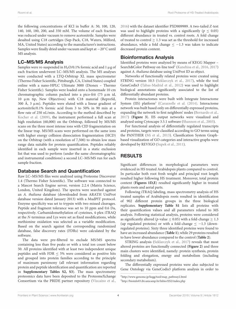
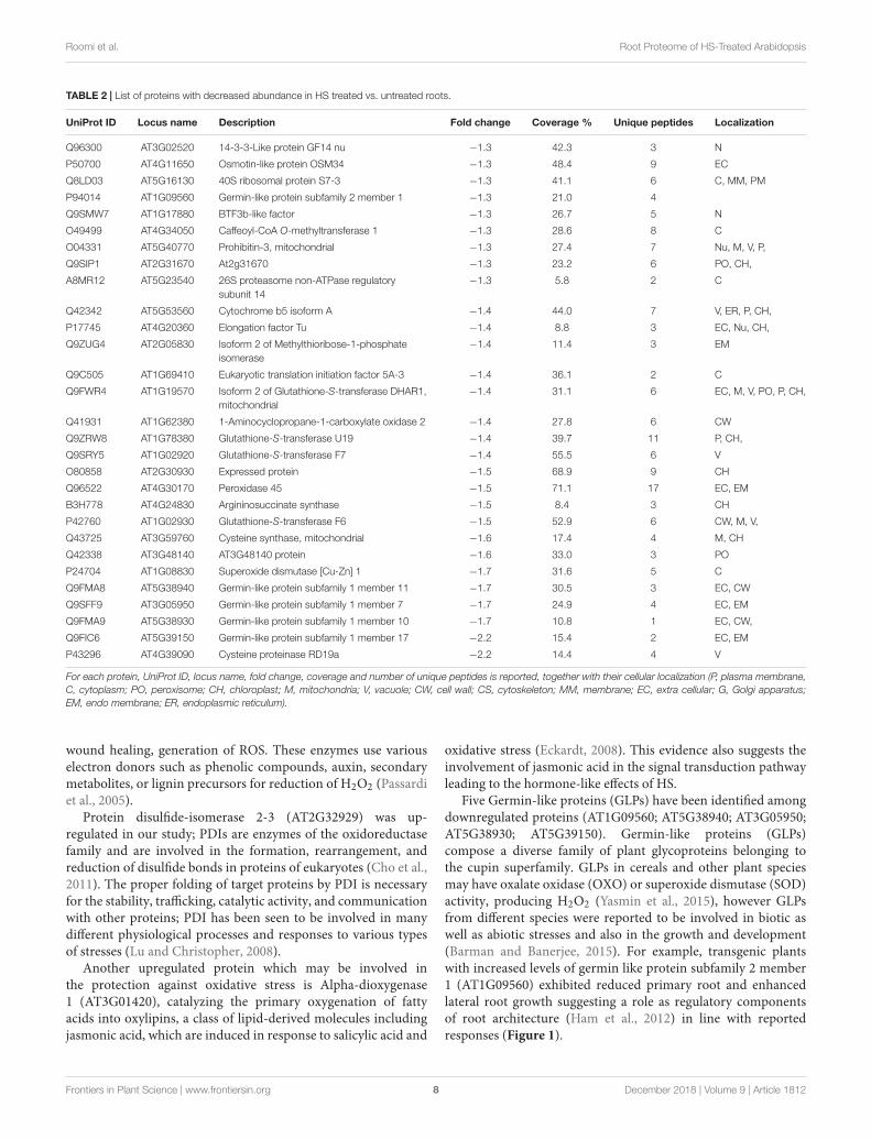
identify major biological processes involved in the response to HS(Tabas-Madrid et al., 2012) (Figure 3).
Functional annotation analysis of the 92 altered proteinsunder high stringency conditions is reported in Figure 3A.Response to salt stress, response to cadmium ion and response tooxidative stress are the most enriched clusters. Other two majorgroups of altered proteins relate to protein folding and responseto heat.
The main molecular functions highlighted by the analysis arecopper and manganese ion binding, nutrient reservoir activity,and protease binding (Figure 3B). Most altered proteins belongto the cytosolic compartment, followed by plasma membrane,plasmodesma, vacuole, and apoplast (Figure 3C).
Pathway analysis and protein interaction network analysiswere carried out to highlight the most regulated pathways andthe interaction amongst the identified proteins. Most informativemaps obtained in KEGG Pathway software are phenylpropanoidbiosynthesis (KEGG:00940), protein processing in endoplasmicreticulum (KEGG:04141), ribosome (KEGG:03010), cysteineand methionine metabolism (KEGG:00270), glyoxylate anddicarboxylate metabolism (KEGG:00630) (SupplementaryFigures S1–S5, respectively).
Three genes encoding for differentially abundant proteinsevidenced in this study, namely AT1G57720, probable elongationfactor 1-gamma; AT4G35830, aconitate hydratase 1 andAT3G60770, 40S ribosomal protein S13-1, were selected tovalidate present results. Transcript levels of these genes weremonitored by means of qPCR (Figure 4). The expressionprofiles of AT1G57720 and AT4G35830 fully confirmedthe protein abundance patterns, evidencing an inductionin transcript accumulation after 2 h of HS provision. Theexpression profile of AT3G60770 was demonstrated tobe not directly affected by the presence of HS (2 h). Inthis case either the duration of the treatment was notsufficient to induce an up-regulation of the transcripts, orthe protein may be controlled at the post-transcriptionallevel, confirming that changes in mRNA expressionprovide only limited insight into changes in proteinexpression.
DISCUSSION
Biostimulants and HSs ability to improve plant growth anddevelopment have been previously reported in different plantspecies (Nardi et al., 2009). In this case study increased proteincontent and higher root fresh weight (Figure 1) confirm thepositive effect of HS treatment in plant physiology. To pinpointthe proteins involved in these responses iTRAQ proteomics hasbeen adopted. However, the complex mechanisms occurring inthe biological systems are not always fully grasped by meansof the simple identification and quantification of proteins froma tissue (Carnielli et al., 2015). Protein–protein interactionsand post-translational modifications (PTMs) are some of thenumerous levels of complexity determining the life span,localization, and activity of a protein. This may play a pivotalrole in regulating the transcriptional changes related to cellularand plant responses to stimuli (Walton et al., 2016). Thus,we explored the applicability of Integrated Interactome System(IIS, Carazzolle et al., 2014) and uploaded the UniProt proteinIDs of differentially abundant Arabidopsis proteins in the IISmodule. IIS networks provide information of proteins thatmight be interacting with the input list. This may contributeto a better understanding of the biological role of the HS-responsive proteins. Interactome network identified in ourexperimental setup evidenced that most of the proteins withdifferent abundance are located in the extracellular; cell wall andplasma membrane (Figure 5) in the GO Cellular Components.This confirms that proteins associated with root cell plasmamembrane can be a target for HS, and changes in their abundancemay be seen as the primary reactions leading to the biologicalresponses reported so far (Carletti et al., 2008).
Twenty-four differentially abundant proteins (Figure 6),evidenced known protein–protein interactions with Ubiquitin(UBQ3). The small ubiquitin molecule attaches to lysine residueson target proteins, leading to the PMT named ubiquitination(Manzano et al., 2008; Igawa et al., 2009; Kim et al., 2013).
Ubiquitinated proteins have several different fates, the mostcommon one being degradation by the 26S proteasome, butchanges in their sub-cellular localization or activity are also
Frontiers in Plant Science | www.frontiersin.org 5 December 2018 | Volume 9 | Article 1812

fpls-09-01812 December 11, 2018 Time: 17:34 # 6
Roomi et al. Root Proteome of HS-Treated Arabidopsis
TABLE 1 | List of proteins with increased abundance in HS treated vs. untreated roots.
UniProt ID Locus name Description Fold change Coverage (%) Unique peptides Localization
Q9XI10 AT1G21680 DPP6 N-terminal domain-like protein 2.3 3.3 3 CW, V, P,
Q56ZI2 AT1G22530 Patellin-2 2.3 26.5 12 P, CH,
Q84WU7 AT3G51330 Aspartyl protease family protein 2.3 5.1 3 P
Q9ZPI1 AT3G11710 Lysine–tRNA ligase 2.1 6.6 4
Q9C8Y9 AT1G66280 Beta-glucosidase 22 2.1 43.7 17 ER, CH,
F4J9K9 AT3G05900 Neurofilament protein-related protein 2.0 34.8 18 C
P51418 AT2G34480 60S ribosomal protein L18a-2 2.0 22.5 6 C, R, P,
F4HV16 AT1G47600 Myrosinase 4 2.0 6.5 3 EM
Q8RX87 AT5G20250 Probable galactinol-sucrosegalactosyltransferase 6
2.0 4.1 3 CH
Q9M8T0 AT3G02880 Probable inactive receptor kinaseAt3g02880
2.0 14.2 8 CW, P,
Q9FJ62 AT5G55480 Probable glycerophosphoryl diesterphosphodiesterase 1
2.0 10.4 7 P
Q9SZ51 AT4G31840 Early nodulin-like protein 15 2.0 22.0 5 P
O82762 AT2G25970 F17H15.1/F17H15.1 2.0 18.0 9 C
Q9STW6 AT4G24280 Heat shock 70 kDa protein 6 1.9 23.4 4 M, CH
Q56WK6 AT1G72150 Patellin-1 1.9 24.3 12 EC, V, P, CH,
Q9S791 AT1G70770 AT1G70770 protein 1.9 7.7 4 ER, P
Q9SR37 AT3G09260 Beta-glucosidase 23 1.8 37.2 13 V
Q9M9K1 AT3G08590 Probable2,3-bisphosphoglycerate-independentphosphoglycerate mutase 2
1.8 12.3 4 EC, C
F4K0F7 AT5G60640 Protein disulfide-isomerase A1 1.8 29.1 13 M, ER, P, CH
O22126 AT2G45470 Fasciclin-like arabinogalactan protein 8 1.8 23.3 9 EC, CW, P, A
Q9C525-2 AT1G66270 Isoform 2 of Beta-glucosidase 21 1.7 37.9 10 V
F4J110 AT3G63460 Protein transport protein SEC31 1.7 4.0 4 C, G, MM
F4JBY2 AT3G60750 Transketolase 1.6 10.8 6 CH
O23006 AT2G17120 LysM domain-containing GPI-anchoredprotein 2
1.6 19.4 8 P
Q9SRH6 AT3G01290 Hypersensitive-induced responseprotein 3
1.6 21.1 6 M, V, P
Q39043 AT5G42020 Mediator of RNA polymerase IItranscription subunit 37f
1.6 49.6 7 CW, Nu, V, ER, P,
F4JWM1 AT5G18380 40S ribosomal protein S16-3 1.6 41.7 6 C, R, CH,
Q42560 AT4G35830 Aconitate hydratase 1 1.5 19.9 9 EC
Q9FVT2 AT1G57720 Probable elongation factor 1-gamma 2 1.5 14.3 7 CW, V
Q9SMT7 AT3G48990 4-Coumarate-CoA ligase-like 10 1.5 21.0 12 EC, CH
F4IB69 AT1G51850 Leucine-rich repeat protein kinasefamily protein
1.5 3.8 4 P
Q9FXA2 AT1G49760 Polyadenylate-binding protein 8 1.50 21.9 11 C, N
O50008 AT5G17920 5-Methyltetrahydropteroyltriglutamate-homocysteinemethyltransferase
1.5 45.6 20 EC, PO, C, P
Q56ZQ3 AT2G14720 Vacuolar-sorting receptor 4 1.5 17.0 10 V, G, P
Q680P8 AT4G33865 40S ribosomal protein S29 1.5 48.2 3 C, R,
C0Z361 AT5G56500 Chaperonin 60 subunit beta 3 1.4 17.1 3 M, CH
P42731 AT4G34110 Polyadenylate-binding protein 2 1.4 26.4 13 C
Q9FJI5 AT5G40760 Glucose-6-phosphate1-dehydrogenase, cytoplasmic isoform2
1.4 20.4 12 C
Q9SRG3 AT1G77510 Protein disulfide isomerase-like 1-2 1.4 32.1 8 ER, P, CH,
P0DH99 AT1G07940 Elongation factor 1-alpha 1 1.4 36.1 17 Nu, M, V, P, CH,
Q1H583 AT1G54000 GDSL esterase/lipase 1.4 50.6 15 V, EC, CW, V, P,
F4KHS2 AT5G59090 Subtilase 4.12 1.4 21.8 14 EC
(Continued)
Frontiers in Plant Science | www.frontiersin.org 6 December 2018 | Volume 9 | Article 1812

fpls-09-01812 December 11, 2018 Time: 17:34 # 7
Roomi et al. Root Proteome of HS-Treated Arabidopsis
TABLE 1 | Continued
UniProt ID Locus name Description Fold change Coverage (%) Unique peptides Localization
P22953 AT5G02500 Heat shock cognate 70 kDa protein 1 1.4 45.8 12 EC, CW, Nu, C, R, P, CH,
Q8L7E3 AT4G20110 Vacuolar-sorting receptor 7 1.3 22.7 14 G, P
P59223 AT3G60770 40S ribosomal protein S13-1 1.3 41.1 7 CW, Nu C, R, CH
P53492 AT5G09810 Actin-7 1.3 40.1 5 CW, Nu, M, CS, P,
Q9LTF2 AT5G52650 40S ribosomal protein S10-3 1.3 37.4 6 CW, C,R
Q9LX13 AT5G10160 (3R)-Hydroxymyristoyl-[acyl carrierprotein] dehydratase-like protein
1.3 13.7 3 CW, CH,
Q9FKK7 AT5G57655 Xylose isomerase 1.3 11.3 6 V, ER, P
O65719 AT3G09440 Heat shock 70 kDa protein 3 1.3 42.2 10 EC, CW, V, C, R, P, CH,
Q9SE60 AT3G59970 Methylenetetrahydrofolate reductase 1 1.3 12.5 4 C
Q9SVG4-2 AT4G20830 Isoform 2 of Reticuline oxidase-likeprotein
1.3 13.7 8 EC, CW, M, V, P
Q94C59 AT1G30690 Patellin-4 1.3 7.2 3 C, P,
O80517 AT2G44790 Uclacyanin-2 1.3 30.2 5 P
Q9SIB9 AT2G05710 Aconitate hydratase 2 1.3 17.7 9 CW, M, P, CH
Q94A28 AT4G26970 Aconitate hydratase 3 1.3 17.9 14 M, CH
O48773 AT2G32920 Protein disulfide-isomerase 2-3 1.3 10.2 4 ER
F4JMJ1 AT4G16660 Heat shock 70 kDa protein 17 1.3 4.3 3 ER
Q9SGH6 AT3G01420 Alpha-dioxygenase 1 1.3 15.2 7 EC
Q9LZ66 AT5G04590 Assimilatory sulfite reductase(ferredoxin)
1.3 17.0 10 CH
Q56YU0 AT3G24503 Aldehyde dehydrogenase family 2member C4
1.3 10.6 4 C
Q9FLQ4 AT5G55070 2-Oxoglutarate dehydrogenasecomplex component E2-1
1.3 8.6 2 M
P52410-2 AT5G46290 Beta-ketoacyl-ACP synthase I 1.3 14.1 5 CH
For each protein, UniProt ID, locus name, fold change, coverage and number of unique peptides is reported, together with their cellular localization (P, plasma membrane,C, cytoplasm; PO, peroxisome; CH, chloroplast; M, mitochondria; V, vacuole; CW, cell wall; CS, cytoskeleton; MM, membrane; EC, extra cellular; G, Golgi apparatus;EM, endo membrane; ER, endoplasmic reticulum).
potential fates. Ubiquitination is known to be involved in plantresponse to stress (Min et al., 2016) and in modulation ofhormone signaling (Vierstra, 2009) as additional non-proteolyticfunction of ubiquitin modification (Stone, 2014). In our study,although not listed among identified proteins, ubiquitin presencein the interactome confirms its role in the responses that weretriggered by HS and suggests that many of the regulatoryprocesses might be controlled at post-translational levels.
In order to decipher the HS treatment-associated metabolicreadjustments in roots, the altered proteins were discussed withreference to functional categories.
Redox HomeostasisWe identified a protein related to redox homeostasis, proteindisulfide isomerase 2 (AT1G77510), whose abundanceincreased in response to HS, while glutathione-S-transferaseU19 (GSTU19) (AT1G78380), glutathione-S-transferaseF7 (AT1G02920), Superoxide dismutase [Cu-Zn] 1, CSD1(AT1G08830) were down-regulated by HS treatment. Reactiveoxygen species such as hydrogen peroxide (H2O2), hydroxylradical (OH−), superoxide (O−2 ) and singlet oxygen (1O2)have been considered as an unavoidable process of normalaerobic metabolism in plants (Dinakar et al., 2010). Differentcellular molecules including proteins, DNA, RNA, and lipidsmay be destroyed by ROS (Shah et al., 2001), but plant cells
have evolved enzymatic and non-enzymatic mechanisms againstthese deleterious effects of ROS (Zhang et al., 2009). Antioxidantenzymes such as catalase CAT (EC 1.11.1.6), CSD (EC 1.15.1.1),and peroxidase POD (EC 1.11.1.7) as well as other antioxidantsmall molecules (glutathione, ascorbic acid, and carotenoids) areused against oxidative stress by plants (Tewari et al., 2008).
Our results confirm the study of García et al. (2012) whichshowed that the antioxidant system of rice roots responds toHAs in a similar way as they do against stress. For example, theactivity of CAT and POX were increased after 8 h of treatmentwith HA. Muscolo et al. (1993) also reported the morphogeneticinfluence of HS on leaf explant of Nicotiana plumbaginifolia dueto stimulation of peroxidases and esterase. The study of Cordeiroet al. (2011) also presents the up-regulation of ROS and CATin maize after the application of HA extracted from Oxisol.However, when applied in combination with environmentalstresses, such as drought (de Vasconcelos et al., 2009) or salinity(Aydin et al., 2012), also opposite effects on these proteins havebeen observed.
Among the identified proteins, peroxidase isoform PER45(AT4G30170) was found. This protein belongs to Class IIIperoxidases, comprising catalytically flexible enzymes with agreat number of isoforms (a total of 73 in Arabidopsis), whichhave been found to regulate a wide range of physiologicalprocesses in plants such as cell wall metabolism, auxin catabolism,
Frontiers in Plant Science | www.frontiersin.org 7 December 2018 | Volume 9 | Article 1812
fpls-09-01812 December 11, 2018 Time: 17:34 # 8
Roomi et al. Root Proteome of HS-Treated Arabidopsis
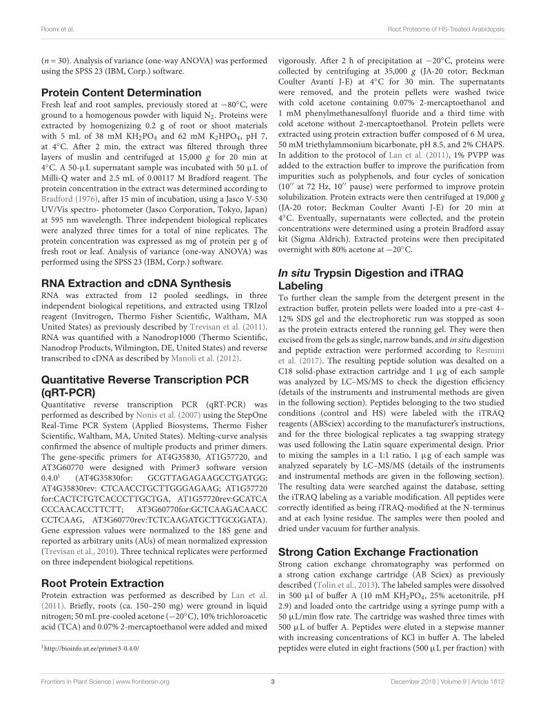
TABLE 2 | List of proteins with decreased abundance in HS treated vs. untreated roots.
UniProt ID Locus name Description Fold change Coverage % Unique peptides Localization
Q96300 AT3G02520 14-3-3-Like protein GF14 nu −1.3 42.3 3 N
P50700 AT4G11650 Osmotin-like protein OSM34 −1.3 48.4 9 EC
Q8LD03 AT5G16130 40S ribosomal protein S7-3 −1.3 41.1 6 C, MM, PM
P94014 AT1G09560 Germin-like protein subfamily 2 member 1 −1.3 21.0 4
Q9SMW7 AT1G17880 BTF3b-like factor −1.3 26.7 5 N
O49499 AT4G34050 Caffeoyl-CoA O-methyltransferase 1 −1.3 28.6 8 C
O04331 AT5G40770 Prohibitin-3, mitochondrial −1.3 27.4 7 Nu, M, V, P,
Q9SIP1 AT2G31670 At2g31670 −1.3 23.2 6 PO, CH,
A8MR12 AT5G23540 26S proteasome non-ATPase regulatorysubunit 14
−1.3 5.8 2 C
Q42342 AT5G53560 Cytochrome b5 isoform A −1.4 44.0 7 V, ER, P, CH,
P17745 AT4G20360 Elongation factor Tu −1.4 8.8 3 EC, Nu, CH,
Q9ZUG4 AT2G05830 Isoform 2 of Methylthioribose-1-phosphateisomerase
−1.4 11.4 3 EM
Q9C505 AT1G69410 Eukaryotic translation initiation factor 5A-3 −1.4 36.1 2 C
Q9FWR4 AT1G19570 Isoform 2 of Glutathione-S-transferase DHAR1,mitochondrial
−1.4 31.1 6 EC, M, V, PO, P, CH,
Q41931 AT1G62380 1-Aminocyclopropane-1-carboxylate oxidase 2 −1.4 27.8 6 CW
Q9ZRW8 AT1G78380 Glutathione-S-transferase U19 −1.4 39.7 11 P, CH,
Q9SRY5 AT1G02920 Glutathione-S-transferase F7 −1.4 55.5 6 V
O80858 AT2G30930 Expressed protein −1.5 68.9 9 CH
Q96522 AT4G30170 Peroxidase 45 −1.5 71.1 17 EC, EM
B3H778 AT4G24830 Argininosuccinate synthase −1.5 8.4 3 CH
P42760 AT1G02930 Glutathione-S-transferase F6 −1.5 52.9 6 CW, M, V,
Q43725 AT3G59760 Cysteine synthase, mitochondrial −1.6 17.4 4 M, CH
Q42338 AT3G48140 AT3G48140 protein −1.6 33.0 3 PO
P24704 AT1G08830 Superoxide dismutase [Cu-Zn] 1 −1.7 31.6 5 C
Q9FMA8 AT5G38940 Germin-like protein subfamily 1 member 11 −1.7 30.5 3 EC, CW
Q9SFF9 AT3G05950 Germin-like protein subfamily 1 member 7 −1.7 24.9 4 EC, EM
Q9FMA9 AT5G38930 Germin-like protein subfamily 1 member 10 −1.7 10.8 1 EC, CW,
Q9FIC6 AT5G39150 Germin-like protein subfamily 1 member 17 −2.2 15.4 2 EC, EM
P43296 AT4G39090 Cysteine proteinase RD19a −2.2 14.4 4 V
For each protein, UniProt ID, locus name, fold change, coverage and number of unique peptides is reported, together with their cellular localization (P, plasma membrane,C, cytoplasm; PO, peroxisome; CH, chloroplast; M, mitochondria; V, vacuole; CW, cell wall; CS, cytoskeleton; MM, membrane; EC, extra cellular; G, Golgi apparatus;EM, endo membrane; ER, endoplasmic reticulum).
wound healing, generation of ROS. These enzymes use variouselectron donors such as phenolic compounds, auxin, secondarymetabolites, or lignin precursors for reduction of H2O2 (Passardiet al., 2005).
Protein disulfide-isomerase 2-3 (AT2G32929) was up-regulated in our study; PDIs are enzymes of the oxidoreductasefamily and are involved in the formation, rearrangement, andreduction of disulfide bonds in proteins of eukaryotes (Cho et al.,2011). The proper folding of target proteins by PDI is necessaryfor the stability, trafficking, catalytic activity, and communicationwith other proteins; PDI has been seen to be involved in manydifferent physiological processes and responses to various typesof stresses (Lu and Christopher, 2008).
Another upregulated protein which may be involved inthe protection against oxidative stress is Alpha-dioxygenase1 (AT3G01420), catalyzing the primary oxygenation of fattyacids into oxylipins, a class of lipid-derived molecules includingjasmonic acid, which are induced in response to salicylic acid and
oxidative stress (Eckardt, 2008). This evidence also suggests theinvolvement of jasmonic acid in the signal transduction pathwayleading to the hormone-like effects of HS.
Five Germin-like proteins (GLPs) have been identified amongdownregulated proteins (AT1G09560; AT5G38940; AT3G05950;AT5G38930; AT5G39150). Germin-like proteins (GLPs)compose a diverse family of plant glycoproteins belonging tothe cupin superfamily. GLPs in cereals and other plant speciesmay have oxalate oxidase (OXO) or superoxide dismutase (SOD)activity, producing H2O2 (Yasmin et al., 2015), however GLPsfrom different species were reported to be involved in biotic aswell as abiotic stresses and also in the growth and development(Barman and Banerjee, 2015). For example, transgenic plantswith increased levels of germin like protein subfamily 2 member1 (AT1G09560) exhibited reduced primary root and enhancedlateral root growth suggesting a role as regulatory componentsof root architecture (Ham et al., 2012) in line with reportedresponses (Figure 1).
Frontiers in Plant Science | www.frontiersin.org 8 December 2018 | Volume 9 | Article 1812

fpls-09-01812 December 11, 2018 Time: 17:34 # 9
Roomi et al. Root Proteome of HS-Treated Arabidopsis
FIGURE 2 | STRING analysis of 92 differentially expressed proteins following HS treatment, visualizing the presence of three major clusters. Colors indicate differentbiological function: red, protein synthesis; blue, protein folding and elongation; green, energy and metabolism (including secondary metabolism). Labels reportprotein Gene Names.
Many studies have demonstrated the regulatory role of ROSin many signaling pathways in plants both in redox homeostasisand under biotic stress (Mittler et al., 2011; Mittler, 2017). Thediscussed results on proteins involved in redox homeostasis,in particular SOD and glutathione-S-transferase, indicate aninvolvement in the response mechanisms induced by HS. Theseresponses do not rule out the participation of the known hormonalsignaling cascades in HS-induced responses, but suggests thatROS may play a pivotal role in regulating metabolic pathwaysrelated to plant growth, in synergy with auxin or NO stimuli.HS regulation of ROS metabolism in roots has been previouslydescribed (García et al., 2012, Garcia et al., 2016): present results
are in agreement with the description of a situation of mild stressin HS-treated roots triggering the physiological responses leadingto higher biomass and protein production (Olaetxea et al., 2018).
Energy Metabolism/RespirationA number of differentially abundant enzymes/proteins identifiedfrom Arabidopsis roots were found to be involved in plant energymetabolism.
Some of these enzymes are related to carbohydrate metabolismincluding glycolysis, TCA and pentose phosphate pathway andwere more abundant such as aconitase 1 (AT4G35830), aconitase2 (AT2G05710), aconitase 3 (AT4G26970), glucose-6-phosphate
Frontiers in Plant Science | www.frontiersin.org 9 December 2018 | Volume 9 | Article 1812
fpls-09-01812 December 11, 2018 Time: 17:34 # 10
Roomi et al. Root Proteome of HS-Treated Arabidopsis
FIGURE 3 | Functional annotation of the 92 differentially expressed proteins following HS treatment performed with GeneCodis 3. (A) Biological process; (B)molecular function; (C) cellular components; (D) KEGG pathways analysis.
1-dehydrogenase (AT5G40760). These results are resumed inKEGG Pathway (Supplementary Figure S3).
The glycolytic pathway is important in plants as it providesfuel for respiration and major carbon skeletons for the synthesis
of various vital compounds such as nucleic acids, aminoacids, fatty acids, isoprenoids, and other secondary metabolites(Plaxton, 1996). The role of glycolysis in opposing various stressesincluding drought, salt, cold, and anoxia has been widely reported
Frontiers in Plant Science | www.frontiersin.org 10 December 2018 | Volume 9 | Article 1812

fpls-09-01812 December 11, 2018 Time: 17:34 # 11
Roomi et al. Root Proteome of HS-Treated Arabidopsis
FIGURE 4 | The relative expression level of AT1G57720, AT4G35830, andAT3G60770 genes in Arabidopsis roots under the whole nutrient (C) or afterHS treatment (HS). The y-axis represents the relative transcript abundanceratios expressed in arbitrary units respect to the T0. Root samples wereharvested 2 h after treatment, and mRNA abundance was determined byRT-qPCR. Error bars represented SD of three independent biologicalreplicates.
in literature (Kosova et al., 2014). Thus, the known HS effect onplant stress relief (Aguiar et al., 2016) can be, at least partially,ascribed to their action on the glycolytic pathway.
One of the up-regulated enzymes in our study is glucose-6-phosphate dehydrogenase (G6PD) (AT5G40760), catalyzing theoxidation of glucose-6-phosphate to 6-phosphogluconate, thekey step in the pentose phosphate pathway (PPP). PPP is theprimary source of NADPH in various biosynthetic processes suchas fatty acid metabolism, integration of nitrogen into amino acid
and resistance against oxidative destruction. An intermediate inPPP, ribose-5-phosphate is used for phenylpropanoid productionthrough shikimate pathway (Scharte et al., 2009). G6PD ishighly regulated: besides transcriptional control, also redoxregulation and cellular NADPH/NADP+ ratio have been shownto regulate the activity of diverse G6PD isoforms (Schurmannand Buchanan, 2008). In a previous study, the low molecularweight humic extracts were found to stimulate the Pi level andenergetic metabolism, resulting specifically in higher glucose-6-phosphate and ATP level (Zancani et al., 2009).
In mitochondria, aconitase (ACO; EC 4.2.1.3) plays animportant role in TCA cycle by catalyzing the isomerizationreaction of citrate to isocitrate via cis-aconitate. The cytosolicisoform is involved in glyoxylate cycle (Moeder et al., 2007). Weidentified three isoforms of aconitase which were all upregulated:aconitase 1 (AT4G35830, cytoplasmic and mitochondrial),aconitase 2 (AT2G05710, mitochondrial) and aconitase 3(AT4G26970, mitochondrial). Adjacent spots of aconitase werealso reported as differentially expressed by Carletti et al. (2008) in2D gels of HS-treated maize roots.
Taken together, the enhanced abundance of the above-mentioned enzymes involved in glycolysis, pentose phosphatepathway, and TCA cycle due to HS may result in an increasein the production of NAD(P)H, ATP and carbon skeletonsneeded for various vital cellular processes such as biosynthesisof macromolecules (proteins, nucleic acids, amino acids, fattyacids, secondary metabolites), which, in turn may explain theknown effect of HS on plant growth. In analogous experimentalconditions, using the same plant species and same HS treatment,metabolomics results evidenced a significative decrease (around−50%) in carbohydrates abundances in roots (Conselvan et al.,2018). Lower contents of fructose, glucose and sucrose represent ametabolic confirmation of the proteomic results in our study, andsubstantiate the hypothesis of enhanced glycolysis in HS-treatedplants.
The stimulation of energy metabolism related enzymesafter HS treatment has been reported previously by manyresearchers: up-regulation of various metabolic processesand signaling pathways associated with plant development(Trevisan et al., 2010; Pizzeghello et al., 2013), stimulationof glycolysis and TCA cycle related enzymes in maize(Nardi et al., 2007). Quaggiotti et al. (2004) also foundthat HS can stimulate carbon and nitrogen metabolism byoverexpression of various enzymes of glycolysis and TCAcycle.
Cell Wall MetabolismCell wall is contained in the outermost extracellular matrixin plant cells and its regulation is important for proper sizeand shape, mechanical resistance, interaction with environment,defense against pathogens, development and growth (Reiter,2002). Cell wall is the first compartment getting in contactwith the exogenous agents, thus unsurprisingly its proteome isaltered by the HS-treatment. In our study, many of identifiedenzymes related to cell wall metabolism were up-regulated(Figure 2), as for example the enzyme glycerophosphodiesterphosphodiesterase (GDPD) (AT5G55480) (EC 3.1.4.46). This
Frontiers in Plant Science | www.frontiersin.org 11 December 2018 | Volume 9 | Article 1812

fpls-09-01812 December 11, 2018 Time: 17:34 # 12
Roomi et al. Root Proteome of HS-Treated Arabidopsis
FIGURE 5 | Interactome of 92 up- or down-regulated proteins detected in Arabidopsis roots treated with HS. The interactome created with IIS website includedproteins with interactions between the input protein dataset and first-neighbor proteins. Proteins were visualized with Cytoscape 3.5.1 and they were distributedaccording to Cellular Components (GO) classification. Nodes marked in green were associated with significantly up-regulated proteins, red nodes denoteddown-regulated proteins and grey nodes were first-neighbor proteins. Labels report protein UniProt ID.
FIGURE 6 | Interactome of 24 differentially abundant proteins detected inArabidopsis roots treated with HS with known direct interaction with Ubiquitinin IIS database. Proteins were visualized with Cytoscape 3.4.0 and they weredistributed with Ubiquitin (Ubq3) as central node. Nodes marked in greenwere associated with significantly up-regulated proteins, red nodes denoteddown-regulated proteins. Labels report protein UniProt ID.
enzyme plays a vital role in many physiological processesin living organisms, by converting glycerophosphodiester toglycerol-3-phosphate and alcohols during glycerol metabolism(Cheng et al., 2011). GDPD and its homologs have beenfound to be involved in cell wall organization and inroot hair morphogenesis in Arabidopsis (Hayashi et al.,2008).
These evidences point to readjustments in cell wallcomposition which are likely required in remodeling andredefining the root organ size, architecture and root hairmorphogenesis when stimulated by HS, as found in Trevisanet al. (2010).
Protein Synthesis, Folding, andDegradationThe involvement of HS in stimulation of protein synthesisin plants has been previously observed in many studies: inArabidopsis roots (Trevisan et al., 2011), maize roots (Carlettiet al., 2008), and guava leaves (Dantas et al., 2007). Canellas et al.(2002) also observed that the HA and Indole Acetic Acid (IAA)groups may be able to access receptors and resulted in activationof protein synthesis in maize roots.
Our identified proteins comprise 40 and 60S ribosomalproteins (RPs) mostly up regulated (AT2G34480, AT5G18380,AT4G33865, AT3G60770, AT5G52650 up-regulated; AT5G16130down-regulated). Ribosomes are the basic and essentialcomponents of every cell and catalyze numerous transpeptidalesterase reactions during protein synthesis. Ribosomal proteins(RPs) are not only vital for protein synthesis but also play acentral role in cell division, growth, and metabolism. The role ofRPs as regulatory components in addition to their housekeepingfunction in developmental processes has been indicated byvarious mutational studies (Byrne, 2009).
Ribosomal proteins are regulated by various growthregulators: for example, the application of BAP (cytokinin)and IAA (auxin) increased the transcription of RPS15aF whileabscisic acid (ABA) treatment decreased it. Abiotic stresses liketemperature and mechanical stress increased the transcript ofRPS15aA, RPS15aD, and RPS15aF (Hulm et al., 2005).
Frontiers in Plant Science | www.frontiersin.org 12 December 2018 | Volume 9 | Article 1812
fpls-09-01812 December 11, 2018 Time: 17:34 # 13
Roomi et al. Root Proteome of HS-Treated Arabidopsis
FIGURE 7 | Continued
Frontiers in Plant Science | www.frontiersin.org 13 December 2018 | Volume 9 | Article 1812

fpls-09-01812 December 11, 2018 Time: 17:34 # 14
Roomi et al. Root Proteome of HS-Treated Arabidopsis
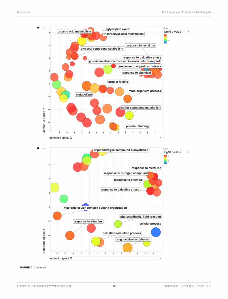
FIGURE 7 | REViGO Scatterplot of the Enriched GO Terms representatives for the differentially regulated proteins (A) and DEGs (B) isolated by Trevisan et al. (2010).Bubble color indicates the p-value (legend in upper right-hand corner), the two ends of the colors are red and blue, depicting lower- and higher p-values respectively.Size indicates the relative frequency of the GO term in the underlying reference database. Bubbles of more general terms are larger.
This could suggest an overall increase in ribosome productionin HS treated root, which is consistent with enhancedprotein synthesis. Protein contents, both in root and leaves(Figures 1D,E) corroborate these proteomic results. Moreover,enhanced protein synthesis is also confirmed by the lowerconcentration of free amino acids in HS-treated plant rootsgrown in analogous conditions. All identified free amino acidswere less abundant (ranging from −30 to −60%) compared tocontrol plants (Conselvan et al., 2018). Decrease in free aminoacid content, paired with higher abundance of ribosomal proteinsand higher total protein content account for plant root responsesleading to higher biomass production (Figure 1B).
The enzyme lysyl-tRNA synthetase (AT3G11710), which isup-regulated in our experiment, has a key role in conversion ofgenetic information from mRNA to protein by catalyzing theformation of lysyl-tRNA (Freist and Gauss, 1995). The enzymeis also found to be linked with many other secondary functionssuch as activation of gene expression (Lee et al., 2004), and byserving as a cytokine (Park et al., 2005).
Our data report an increase of two isoforms of poly(A)-binding protein (PABP2, AT1G49760; AT4G34110). Afterattaching with 3′ end of mRNA, PABP interacts with eukaryotictranslation initiation factor 4F complex enhancing the translationprocess inside the cell (Sachs and Varani, 2000). The role of PABPis also found to be correlated with nuclear export of mRNAs(Brune et al., 2005) and stability (Behm-Ansmant et al., 2007). InA. thaliana, a total of eight different isoforms of PABP have beencharacterized (Belostotsky, 2003).
Various heat shock protein cognates (AT4G24280,AT3G09440) were also over produced in response to HS.The 70-kD heat shock proteins (Hsp70s) are found in allcellular compartments of almost all organisms and have beenfound to be crucial for protein folding, protein translocation,and stress responses (Latijnhouwers et al., 2010). One of theup-regulated Hsp70s have been described in Arabidopsis asstromal isoforms cpHsc70-1 (At4g24280). In one knockout studya mutation in cpHsc70-1 resulted in abnormal leaves, impairedroot development and retardation in growth (Su and Li, 2008).
Plant proteasomes (26S proteasomes) contain twosubparticles: the core particle (CP) or 20S proteasome whereproteins are degraded, and the regulatory particle (RP) or 19Sproteasome. 26S proteasomes are in charge of the ATP-dependentdegradation of ubiquitin tagged proteins (Sadanandom et al.,2012) including normal, mutated, misfolded, and damagedproteins (Kurepa and Smalle, 2008; Tolin et al., 2013). Theubiquitin-proteasome system plays a role in nearly all aspectsof cell homeostasis including plant development, responseto plant hormones (Sullivan et al., 2003) and signaling inresponse to abiotic and biotic stimuli (Smalle and Vierstra,2004). Moreover, the 20S particle can also degrade proteins ina ubiquitin-independent manner, mainly for oxidized proteins(Foyer and Allen, 2003). In this study HS treatment resulted
in differential abundance of a proteasome subunit such as 26Sproteasome non-ATPase regulatory subunit 14 (AT5G23540).The involvement of the proteasome complex may be seen as acomponent of cell metabolic remodeling in the response to HStreatment, coherently with the view that the proteolytic capacityof a cell is the result of a careful balancing act that reflectsenvironmental conditions and developmental stage (Kurepa andSmalle, 2008).
26S proteasome is ubiquitin and ATP dependent, andis involved in protein degradation in the nucleus and thecytosol (Voges et al., 1999; Pickart and Cohen, 2004).Various catalytic activities such as trypsin-like, chymotrypsin-like peptidylglutamyl-peptide hydrolase are performed by 20Score particle of 26S proteasome (Groll et al., 1997; Voges et al.,1999).
Cell Trafficking and DivisionOur proteomic data identified proteins related to cell vesicletrafficking and growth such as Actin-7 (AT5G09810), Patellin-1(AT1G72150), Patellin-2 (AT1G22530), Patellin-4 (AT1G30690),all of which were up-regulated. One other protein involvedin actin metabolism was also found to be upregulated,namely GDPDL4 (AT5G55480), reported to be involved inactin nucleation (Ma et al., 2012). The involvement of actinin plant responses to HS has been already highlighted inprevious works (Carletti et al., 2008) as well as transportprocesses and vesicles trafficking related genes (Trevisan et al.,2011).
The role of actin in cellular processes is diverse and rangesfrom cell division and morphogenesis to cell motility (Pollardand Cooper, 2009). Actin is also found to be important fortip growth (polarized cell extension) in plants (Menand et al.,2007), whose implications are of particular relevance in thisstudy.
Patellin is a phosphoinositide-binding protein that playsa role in membrane trafficking during the expansion andmaturation stages of cytokinesis, in particular cell-plateformation (McMichael and Bednarek, 2013).
Response to Inorganic SubstancesIn this study, we identified a number of proteins as responses toinorganic substances that were affected by HS treatment. Many ofthese are also involved in redox homeostasis, such as glutathione-S-transferase 1 (AT1G02930), Peroxidase 45 (AT4G30170), and[Cu-Zn] Superoxide dismutase (AT1G08830) (details in redoxhomeostasis paragraph).
The group also contains some of the enzymes of glycolysisand TCA cycle as aconitate hydratase 2 (AT2G05710), Aconitatehydratase 3 (AT4G26970). Similarly, some other identifiedproteins of diverse function were over expressed as heat shockcognate 70 kDa protein 1 (AT5G02500), heat shock cognate
Frontiers in Plant Science | www.frontiersin.org 14 December 2018 | Volume 9 | Article 1812

fpls-09-01812 December 11, 2018 Time: 17:34 # 15
Roomi et al. Root Proteome of HS-Treated Arabidopsis
70 kDa protein 3 (AT3G09440), protein disulfide-isomerase 2(AT1G77510).
Heat ResponseHumic substance caused a significant increase in abundanceof two heat responsive proteins in this study: heat shock70 kDa protein 6 (AT4G24280); heat shock 70 kDa protein 3(AT3G09440).
The 70-kD heat shock proteins (Hsp70s) are molecularchaperones involved in a variety of cellular processes includingprotein folding, protein transport across membranes, modulationof protein activity, regulation of protein degradation, andprevention of irreversible protein aggregation. Plant Hsp70sare encoded by a multiple-gene family (Su and Li, 2008).It is well-known that Hsps are ubiquitous proteins foundin plant and animal cells, which were initially describedto be involved in heat shock, but they are known tobe induced by a wide variety of stresses, including cold,drought, salt, UV-light, wound, and biotic stresses (Wen et al.,2017).
Integration With Previous ResultsIn this work we have provided a thorough proteomic analysisin Arabidopsis roots treated with HS obtained at our laboratory,following a standardized procedure for extraction, purification,and characterization providing a product with homogeneous andstable properties over time.
Identical experimental conditions in terms of HS quality,concentration, exposure time, growth chamber settings(temperature, humidity, daylength) were previously used totreat Arabidopsis plants also in another study, aimed at studyingthe effects of HS on gene expression by a transcriptomic approachbased on the detection of cDNA-AFLP markers (Trevisan et al.,2011).
Gene ontology enrichment analysis was used to comparethe effect exerted by HS on proteome and transcriptomeregulation. The terms along with their p-values were furthersummarized independently by the REViGO4 reductionanalysis tool that condenses the GO description by removingredundant terms. Results of these further reductions arevisualized in Figure 6. An encouragingly simple relationshipbetween changes of transcripts and proteins and changesin downstream biological functions could be inferred bythe graphs. Analysis on transcripts retrieved 32 GO terms,whilst the enrichment analysis on dysregulated proteinsidentified 116 terms. Despite the differences in the amountof enriched terms, the comprehensive proteome (Figure 7A)and transcriptome (Figure 7B) datasets demonstrated thatseveral of the enriched GO terms (carbohydrate metabolicprocess, organic substance biosynthetic process, response tochemical, response to oxidative stress, response to abioticstimulus, translation) are included in both the enriched groups,suggesting some correlation between transcript and proteinlevels.
4http://revigo.irb.hr/
CONCLUSION
With the present work, an overview of metabolic pathwaysinfluenced by HS activity is presented, only in part previouslyobserved. Our results, also in accordance with previouslypublished metabolomic data, point to the activation of enzymesinvolved in glycolysis, pentose phosphate pathway and TCAcycle to support the production of NAD(P)H, ATP, and carbonskeletons needed for various vital cellular processes. Stimulationof energy metabolism may explain the known beneficial effectsof HS on plant growth. Up-regulation of ribosomal proteinsand actin are representative of a co-ordinately enhanced proteinsynthesis, folding, trafficking and transport across membraneswhich is required to sustain growth. Our findings also point toreadjustments in cell wall composition which are required in rootremodeling and root hair morphogenesis. The regulation of ROS-related enzymes indicates that these compounds play a pivotalrole in response to HS stimulus, possibly acting as a regulatorymechanism to coordinate the other responses leading to growthenhancement.
The results discussed in this study should represent a newframework in the development of a new model mechanism,considering ROS as a chemical species of great importance in theaction of HS on plants.
AUTHOR CONTRIBUTIONS
SR and PC wrote the manuscript with contribution from allauthors. GC and SR analyzed the data. MP performed theprotein extraction and purification. GA performed all proteomicsexperiments and statistical analysis. AM and TY contributed inthe interpretation of the results and revised the manuscript. STand SQ performed the transcriptome analyses. PC conceived theoriginal idea and supervised the project. All authors discussed theresults and contributed to the final manuscript.
FUNDING
This project was funded by University of Padua grantCPDA112850. Grant (372239-1-2012-1-PT-ERA MUNDUS-EMMA21) awarded to SR for his Ph.D. research at the Universityof Padova, Italy. Ph.D. grant for GC funded by MIUR “L170.” PC was financially supported by the University of PadovaDOR1883089/18.
ACKNOWLEDGMENTS
The authors gratefully acknowledge Dr. Diego Pizzeghello fortechnical help in HS extraction and purification. They gratefullyacknowledge Dr. Annarita Tretin for the kind support in proteinextraction and purification. The authors wish to thank the“Cassa di Risparmio di Padova e Rovigo” Holding (Cariparo)for funding the acquisition of the LTQ-Orbitrap XL massspectrometer.
Frontiers in Plant Science | www.frontiersin.org 15 December 2018 | Volume 9 | Article 1812

fpls-09-01812 December 11, 2018 Time: 17:34 # 16
Roomi et al. Root Proteome of HS-Treated Arabidopsis
SUPPLEMENTARY MATERIAL
The Supplementary Material for this article can be found onlineat: https://www.frontiersin.org/articles/10.3389/fpls.2018.01812/full#supplementary-material
FIGURE S1 | Regulatory changes on the pathways of cysteine and methioninemetabolism, with highlighted up- regulated proteins (green) and down-regulatedproteins (red). Labels report EC numbers. The image was obtained by KEGGplatform (https://www.kegg.jp; see reference in the text).
FIGURE S2 | Regulatory changes on the pathways of glyoxylate anddicarboxylate metabolism, with highlighted up- regulated proteins (green). Labelsreport EC numbers. The image was obtained by KEGG platform(https://www.kegg.jp; see reference in the text).
FIGURE S3 | Regulatory changes on the pathways on phenylpropanoidbiosynthesis, with highlighted up- regulated proteins (green) and down-regulatedproteins (red). Labels report EC numbers. The image was obtained by KEGGplatform (https://www.kegg.jp; see reference in the text).
FIGURE S4 | Regulatory changes on ribosome proteins, with highlighted up-regulated proteins (green) and down-regulated proteins (red). Labels report ECnumbers. The image was obtained by KEGG platform (https://www.kegg.jp; seereference in the text).
FIGURE S5 | Regulatory changes on protein processing in endoplasmicreticulum, with highlighted up- regulated proteins (green) and down-regulatedproteins (red). Labels report EC numbers. The image was obtained by KEGGplatform (https://www.kegg.jp; see reference in the text).
TABLE S1 | List of differentially abundant proteins. Table lists all proteins witha significantly different abundance in HS vs CTRL samples. Uniprot accessionnumber, gene name, protein description, quantification values relative to the 3replicates, fold change, and p value are reported.
TABLE S2 | List of all identified peptides. Table lists all peptides identified bythe LC-MS/MS analysis together with all relevant parameters required to assesspeptide identification and quantification confidence.
TABLE S3 | List of all identified proteins. Table lists all proteins identified withhigh confidence by the LC-MS/MS analysis, together with all relevant parametersrequired to assess protein identification and quantification confidence.
REFERENCESAguiar, N. O., Medici, L. O., Olivares, F. L., Dobbss, L. B., Torres-Netto, A.,
Silva, S. F., et al. (2016). Metabolic profile and antioxidant responses duringdrought stress recovery in sugarcane treated with humic acids and endophyticdiazotrophic bacteria. Ann. Appl. Biol. 168, 203–213. doi: 10.1111/aab.12256
Aguiar, N. O., Olivares, F. L., Novotny, E. H., Dobbss, L. B., Balmori, D. M.,Santos-Juìnior, L. G., et al. (2012). Bioactivity of humic acids isolated fromvermicomposts at different maturation stages. Plant Soil 362, 161–174. doi:10.1007/s11104-012-1277-5
Aydin, A., Kant, C., and Turan, M. (2012). Humic acid application alleviate salinitystress of bean (Phaseolus vulgaris L.) plants decreasing membrane leakage. Afr.J. Agric. Res. 7, 1073–1086. doi: 10.5897/AJAR10.274
Barman, A. R., and Banerjee, J. (2015). Versatility of germin-like proteins in theirsequences, expressions, and functions. Funct. Integr. Genomics 15, 533–548.doi: 10.1007/s10142-015-0454-z
Behm-Ansmant, I., Branlant, C., and Motorin, Y. (2007). The Saccharomycescerevisiae Pus2 protein encoded by YGL063w ORF is a mitochondrial tRNA:927/28-synthase. RNA 13, 1641–1647. doi: 10.1261/rna.605607
Belostotsky, D. A. (2003). Unexpected complexity of poly(A)-binding protein genefamilies in flowering plants: three conserved lineages that are at least 200 millionyears old and possible auto- and cross-regulation. Genetics 163, 311–319.
Berbara, R. L. L., and Garciìa, A. C. (2014). “Humic substances and plantdefense metabolism,” in Physiological Mechanisms and Adaptation Strategies inPlants Under Changing Environment, Vol. 1, eds P. Ahmad and M. R. Wani(New York, NY: Springer Science+Business Media), 297–319.
Bernardo, L., Morcia, C., Carletti, P., Ghizzoni, R., Badeck, F. W., Rizza, F., et al.(2017). Proteomic insight into the mitigation of wheat root drought stress byarbuscular mycorrhizae. J. Proteomics 169, 21–32. doi: 10.1016/j.jprot.2017.03.024
Bradford, M. M. (1976). A rapid and sensitive method for the quantitationof microgram quantities of protein utilizing the principle of protein-dye binding. Anal. Biochem. 72, 248–254. doi: 10.1016/0003-2697(76)90527-3
Brune, C., Munchel, S. E., Fischer, N., Podtelejnikov, A. V., and Weis, K. (2005).Yeast poly(A)-binding protein Pab1 shuttles between the nucleus and thecytoplasm and functions in mRNA export. RNA 11, 517–531. doi: 10.1261/rna.7291205
Byrne, M. E. (2009). A role for the ribosome in development. Trends Plant Sci. 14,512–519. doi: 10.1016/j.tplants.2009.06.009
Calvo, P., Nelson, L., and Kloepper, J. W. (2014). Agricultural uses of plantbiostimulants. Plant Soil 383, 3–41. doi: 10.1007/S11104-014-2131-8
Canellas, L. P., Dantas, D. J., Aguiar, N. O., Peres, L. E. P., Zsogon, A., Olivares,F. L., et al. (2011). Probing the hormonal activity of fractionated molecular
humic components in tomato auxin mutants. Ann. Appl. Biol. 159, 202–211.doi: 10.1111/j.1744-7348.2011.00487.x
Canellas, L. P., and Olivares, F. L. (2014). Physiological responses to humicsubstances as plant growth promoter. Chem. Biol. Technol. Agric. 1:3. doi:10.1186/2196-5641-1-3
Canellas, L. P., Olivares, F. L., Okorokova-Facanha, A. L., and Facanha,A. R. (2002). Humic acids isolated from earthworm compost enhance rootelongation, lateral root emergence, and plasma membrane H+-ATPase activityin maize roots. Plant Physiol. 130, 1951–1957. doi: 10.1104/pp.007088
Carazzolle, M. F., de Carvalho, L. M., Slepicka, H. H., Vidal, R. O., Pereira, G. A. G.,Kobarg, J., et al. (2014). IIS - Integrated Interactome System: a web-basedplatform for the annotation, analysis and visualization of protein-metabolite-gene-drug interactions by integrating a variety of data sources and tools. PLoSOne 9:e100385. doi: 10.1371/journal.pone.0100385
Carletti, P., Masi, A., Spolaore, B., De Laureto, P. P., De Zorzi, M., Turetta, L.,et al. (2008). Protein expression changes in maize roots in response to humicsubstances. J. Chem. Ecol. 34, 804–818. doi: 10.1007/s10886-008-9477-4
Carnielli, C. M., Winck, F. V., and Leme, A. F. P. (2015). Functional annotationand biological interpretation of proteomics data. Biochim. Biophys. Acta 1854,46–54. doi: 10.1016/j.bbapap.2014.10.019
Cheng, Y., Zhou, W., El Sheery, N. I., Peters, C., Li, M., Wang, X., et al. (2011).Characterization of the Arabidopsis glycerophosphodiester phosphodiesterase(GDPD) family reveals a role of the plastid-localized AtGDPD1 in maintainingcellular phosphate homeostasis under phosphate starvation. Plant J. 66, 781–795. doi: 10.1111/j.1365-313X.2011.04538.x
Cho, E. J., Yuen, C. Y., Kang, B. H., Ondzighi, C. A., Staehelin, L. A., andChristopher, D. A. (2011). Protein disulfide isomerase-2 ofArabidopsismediatesprotein folding and localizes to both the secretory pathway and nucleus, whereit interacts with maternal effect embryo arrest factor. Mol. Cells 32, 459–475.doi: 10.1007/s10059-011-0150-3
Conselvan, G. B., Fuentes, D., Merchant, A., Peggion, C., Francioso, O., andCarletti, P. (2018). Effects of humic substances and indole-3-acetic acid onArabidopsis sugar and amino acid metabolic profile. Plant Soil 426, 17–32.doi: 10.1007/s11104-018-3608-7
Conselvan, G. B., Pizzeghello, D., Francioso, O., Di Foggia, M., Nardi, S., andCarletti, P. (2017). Biostimulant activity of humic substances extracted fromleonardites. Plant Soil 420, 119–134. doi: 10.1007/s11104-017-3373-z
Cordeiro, F. C., Santa-Catarina, C., Silveira, V., and de Souza, S. R. (2011). Humicacid effect on catalase activity and the generation of reactive oxygen speciesin corn (Zea mays). Biosci. Biotechnol. Biochem. 75, 70–74. doi: 10.1271/bbb.100553
Dantas, B. F., Pereira, M. S., Ribeiro, L. D., Maia, J. L. T., and Bassoi, L. H. (2007).Effect of humic substances and weather conditions on leaf biochemical changesof fertigated guava tree, during orchard establishment. Rev. Bras. Frutic. 29,632–638. doi: 10.1590/s0100-29452007000300040
Frontiers in Plant Science | www.frontiersin.org 16 December 2018 | Volume 9 | Article 1812
fpls-09-01812 December 11, 2018 Time: 17:34 # 17
Roomi et al. Root Proteome of HS-Treated Arabidopsis
de Vasconcelos, A. C. F., Zhang, X. Z., Ervin, E. H., and Kiehl, J. D.(2009). Enzymatic antioxidant responses to biostimulants in maize andsoybean subjected to drought. Sci. Agric. 66, 395–402. doi: 10.1590/S0103-90162009000300015
Destro, T., Prasad, D., Martignago, D., Bernet, I. L., Trentin, A. R., Renu, I. K.,et al. (2011). Compensatory expression and substrate inducibility of gamma-glutamyl transferase GGT2 isoform in Arabidopsis thaliana. J. Exp. Bot. 62,805–814. doi: 10.1093/jxb/erq316
Dinakar, C., Abhaypratap, V., Yearla, S. R., Raghavendra, A. S., and Padmasree, K.(2010). Importance of ROS and antioxidant system during the beneficialinteractions of mitochondrial metabolism with photosynthetic carbonassimilation. Planta 231, 461–474. doi: 10.1007/s00425-009-1067-3
du Jardin, P. (2012). The Science of Plant Biostimulants – a Bibliographic Analysis.Available at: http://ec.europa.eu/enterprise/sectors/chemicals/files/fertilizers/final_report_bio_2012_en.pd
Eckardt, N. A. (2008). Oxylipin signaling in plant stress responses. Plant Cell 20,495–497. doi: 10.1105/tpc.108.05948
FAO-UNESCO (1997). World Soil Map, Revised Legend. Wageningen: FAO.Foreman, J., Demidchik, V., Bothwell, J. H. F., Mylona, P., Miedema, H., Torres,
M. A., et al. (2003). Reactive oxygen species produced by NADPH oxidaseregulate plant cell growth. Nature 422, 442–446. doi: 10.1038/nature01485
Foyer, C. H., and Allen, J. F. (2003). Lessons from redox signaling in plants.Antioxid. Redox Signal. 5, 3–5. doi: 10.1089/152308603321223487
Freist, W., and Gauss, D. H. (1995). Lysyl-transfer-RNA synthetase. Biol. Chem.Hoppe Seyler 376, 451–472.
Gao, T. G., Xu, Y. Y., Jiang, F., Li, B. Z., Yang, J. S., Wang, E. T., et al.(2015). Nodulation characterization and proteomic profiling of Bradyrhizobiumliaoningense CCBAU05525 in response to water-soluble humic materials. Sci.Rep. 5:10836. doi: 10.1038/srep10836
Garcia, A. C., Olaetxea, M., Santos, L. A., Mora, V., Baigorri, R., Fuentes, M., et al.(2016). Involvement of hormone- and ROS-signaling pathways in the beneficialaction of humic substances on plants growing under normal and stressingconditions. Biomed Res. Int. 2016:3747501. doi: 10.1155/2016/3747501
García, A. C., Santos, L. A., Izquierdo, F. G., Rumjanek, V. M., Castro, R. N.,dos Santos, F. S., et al. (2014). Potentialities of vermicompost humic acids toalleviate water stress in rice plants (Oryza sativa L.). J. Geochem. Explor. 136,48–54. doi: 10.1016/j.gexplo.2013.10.005
García, A. C., Santos, L. A., Izquierdo, F. G., Sperandio, M. V. L., Castro, R. N.,and Berbara, R. L. L. (2012). Vermicompost humic acids as an ecologicalpathway to protect rice plant against oxidative stress. Ecol. Eng. 47, 203–208.doi: 10.1016/j.ecoleng.2012.06.011
Groll, M., Ditzel, L., Löwe, J., Stock, D., Bochtler, M., Bartunik, H. D., et al. (1997).Structure of 20S proteasome from yeast at 2.4Å resolution. Nature 386:463.doi: 10.1038/386463a0
Ham, B.-K., Li, G., Kang, B.-H., Zeng, F., and Lucas, W. J. (2012).Overexpression of Arabidopsis plasmodesmata germin-like proteins disruptsroot growth and development. Plant Cell 24, 3630–3648. doi: 10.1105/tpc.112.101063
Hayashi, S., Ishii, T., Matsunaga, T., Tominaga, R., Kuromori, T., Wada, T., et al.(2008). The glycerophosphoryl diester phosphodiesterase-like proteins SHV3and its homologs play important roles in cell wall organization. Plant CellPhysiol. 49, 1522–1535. doi: 10.1093/pcp/pcn120
Hulm, J. L., McIntosh, K. B., and Bonham-Smith, P. C. (2005). Variation intranscript abundance among the four members of the Arabidopsis thalianaRIBOSOMAL PROTEIN S15a gene family. Plant Sci. 169, 267–278. doi: 10.1016/j.plantsci.2005.04.001
Igawa, T., Fujiwara, M., Takahashi, H., Sawasaki, T., Endo, Y., Seki, M.,et al. (2009). Isolation and identification of ubiquitin-related proteinsfrom Arabidopsis seedlings. J. Exp. Bot. 60, 3067–3073. doi: 10.1093/jxb/erp134
Jannin, L., Arkoun, M., Ourry, A., Laîneì, P., Goux, D., Garnica, M., et al.(2012). Microarray analysis of humic acid effects on Brassica napus growth:involvement of N, C and S metabolisms. Plant Soil 359, 297–319. doi: 10.1007/s11104-012-1191-x
Jindo, K., Martim, S. A., Navarro, E. C., Peìrez-Alfocea, F., Hernandez, T.,Garcia, C., et al. (2011). Root growth promotion by humic acids fromcomposted and non-composted urban organic wastes. Plant Soil 353, 209–220.doi: 10.1007/s11104-011-1024-3
Kanehisa, M., Furumichi, M., Tanabe, M., Sato, Y., and Morishima, K. (2017).KEGG: new perspectives on genomes, pathways, diseases and drugs. NucleicAcids Res. 45, D353–D361. doi: 10.1093/nar/gkw1092
Kanehisa, M., Sato, Y., Kawashima, M., Furumichi, M., and Tanabe, M. (2016).KEGG as a reference resource for gene and protein annotation. Nucleic AcidsRes. 44, D457–D462. doi: 10.1093/nar/gkv1070
Kim, D.-Y., Scalf, M., Smith, L. M., and Vierstra, R. D. (2013). Advanced proteomicanalyses yield a deep catalog of ubiquitylation targets in Arabidopsis. Plant Cell25, 1523–1540. doi: 10.1105/tpc.112.108613
Kocher, T., Pichler, P., Schutzbier, M., Stingl, C., Kaul, A., Teucher, N., et al. (2009).High precision quantitative proteomics using iTRAQ on an LTQ orbitrap: anew Mass spectrometric method combining the benefits of all. J. Proteome Res.8, 4743–4752. doi: 10.1021/pr900451u
Kosova, K., Vitamvas, P., and Prasil, I. T. (2014). Proteomics of stress responses inwheat and barley-search for potential protein markers of stress tolerance. Front.Plant Sci. 5:711. doi: 10.3389/fpls.2014.00711
Kurepa, J., and Smalle, J. A. (2008). Structure, function and regulation of plantproteasomes. Biochimie 90, 324–335. doi: 10.1016/j.biochi.2007.07.019
Lan, P., Li, W., Wen, T. N., Shiau, J. Y., Wu, Y. C., Lin, W., et al. (2011). iTRAQprotein profile analysis of Arabidopsis roots reveals new aspects critical for ironhomeostasis. Plant Physiol. 155, 821–834. doi: 10.1104/pp.110.169508
Latijnhouwers, M., Xu, X. M., and Moller, S. G. (2010). Arabidopsis stromal 70-kDa heat shock proteins are essential for chloroplast development. Planta 232,567–578. doi: 10.1007/s00425-010-1192-z
Lee, S. W., Cho, B. H., Park, S. G., and Kim, S. (2004). Aminoacyl-tRNA synthetasecomplexes: beyond translation. J. Cell Sci. 117, 3725–3734. doi: 10.1242/jcs.01342
Lu, D. P., and Christopher, D. A. (2008). Endoplasmic reticulum stress activates theexpression of a sub-group of protein disulfide isomerase genes and AtbZIP60modulates the response in Arabidopsis thaliana. Mol. Genet. Genomics 280,199–210. doi: 10.1007/s00438-008-0356-z
Ma, B., Qian, D., Nan, Q., Tan, C., An, L., and Xiang, Y. (2012). Arabidopsisvacuolar H+- ATPase (V-ATPase) B subunits are involved in actin cytoskeletonremodeling via binding to, bundling, and stabilizing F-actin. J. Biol. Chem. 287,19008–19017. doi: 10.1074/jbc.m111.281873
Manoli, A., Sturaro, A., Trevisan, S., Quaggiotti, S., and Nonis, A. (2012).Evaluation of candidate reference genes for qPCR in maize. J. Plant Physiol.169, 807–815. doi: 10.1016/j.jplph.2012.01.019
Manzano, C., Abraham, Z., Loìpez-Torrejoìn, G., and Del Pozo, J. C. (2008).Identification of ubiquitinated proteins in Arabidopsis. Plant Mol. Biol. 68,145–158. doi: 10.1007/s11103-008-9358-9
McMichael, C. M., and Bednarek, S. Y. (2013). Cytoskeletal and membranedynamics during higher plant cytokinesis. New Phytol. 197, 1039–1057. doi:10.1111/nph.12122
Menand, B., Yi, K. K., Jouannic, S., Hoffmann, L., Ryan, E., Linstead, P., et al.(2007). An ancient mechanism controls the development of cells with a rootingfunction in land plants. Science 316, 1477–1480. doi: 10.1126/science.1142618
Mi, H., Muruganujan, A., Casagrande, J. T., and Thomas, P. D. (2013). Large-scalegene function analysis with the PANTHER classification system. Nat. Protoc. 8,1551–1566. doi: 10.1038/nprot.2013.092
Min, H. W., Chen, C. X., Wei, S. W., Shang, X. L., Sun, M. Y., Xia, R., et al. (2016).Identification of drought tolerant mechanisms in maize seedlings based ontranscriptome analysis of recombination inbred lines. Front. Plant Sci. 7:1080.doi: 10.3389/fpls.2016.01080
Minelli, A., Omodeo, P., Rota, E., and Sambugar, B. (1995). “Annelida Clitellata, Aphenomenon,” in Checklist Delle Specie Della Fauna Italiana, eds A. Minelli, S.Rufo, and S. La Posta (Bologna: Calderoni), 12–13.
Mittler, R. (2017). ROS are good. Trends Plant Sci. 22, 11–19. doi: 10.1016/j.tplants.2016.08.002
Mittler, R., Vanderauwera, S., Suzuki, N., Miller, G., Tognetti, V. B., Vandepoele, K.,et al. (2011). ROS signaling: the new wave? Trends Plant Sci. 16, 300–309.doi: 10.1016/j.tplants.2011.03.007
Moeder, W., Del Pozo, O., Navarre, D. A., Martin, G. B., and Klessig, D. F. (2007).Aconitase plays a role in regulating resistance to oxidative stress and cell deathin Arabidopsis and Nicotiana benthamiana. Plant Mol. Biol. 63, 273–287. doi:10.1007/s11103-006-9087-x
Mora, V., Bacaicoa, E., Baigorri, R., Zamarreno, A. M., and Garcia-Mina, J. M.(2014). NO and IAA key regulators in the shoot growth promoting action
Frontiers in Plant Science | www.frontiersin.org 17 December 2018 | Volume 9 | Article 1812

fpls-09-01812 December 11, 2018 Time: 17:34 # 18
Roomi et al. Root Proteome of HS-Treated Arabidopsis
of humic acid in Cucumis sativus L. J. Plant Growth Regul. 33, 430–439. doi:10.1007/s00344-013-9394-9
Muscolo, A., Felici, M., Concheri, G., and Nardi, S. (1993). Effects of earthwormhumic substances on esterase and peroxidase-activity during growth of leafexplants of Nicotiana plumbaginifolia. Biol. Fertil. Soil 15, 127–131. doi: 10.1007/bf00336430
Muscolo, A., Sidari, M., and Nardi, S. (2013). Humic substance: relationshipbetween structure and activity. Deeper information suggests univocal findings.J. Geochem. Explor. 129, 57–63. doi: 10.1016/j.gexplo.2012.10.012
Nardi, S., Carletti, P., Pizzeghello, D., and Muscolo, A. (2009). “Biological activitiesof humic substances,” in Biophysico-Chemical Processes in EnvironmentalSystems, eds P. M. Huang and N. Senesi (Hoboken, NJ: John Wiley & Sons).
Nardi, S., Muscolo, A., Vaccaro, S., Baiano, S., Spaccini, R., and Piccolo, A. (2007).Relationship between molecular characteristics of soil humic fractions andglycolytic pathway and krebs cycle in maize seedlings. Soil Biol. Biochem. 39,3138–3146. doi: 10.1016/j.soilbio.2007.07.006
Nardi, S., Pizzeghello, D., Muscolo, A., and Vianello, A. (2002). Physiologicaleffects of humic substances on higher plants. Soil Biol. Biochem. 34, 1527–1536.doi: 10.1016/S0038-0717(02)00174-8
Nonis, A., Ruperti, B., Falchi, R., Casatta, E., Enferadi, S. T., and Vizzotto, G. (2007).Differential expression and regulation of a neutral invertase encoding gene frompeach (Prunus persica): evidence for a role in fruit development. Physiol. Plant.129, 436–446. doi: 10.1111/j.1399-3054.2006.00832.x
Olaetxea, M., De Hita, D., Garcia, C. A., Fuentes, M., Baigorri, R., Mora, V., et al.(2018). Hypothetical framework integrating the main mechanisms involvedin the promoting action of rhizospheric humic substances on plant root- andshoot- growth. Appl. Soil Ecol. 123, 521–537. doi: 10.1016/j.apsoil.2017.06.007
Park, S. G., Ewalt, K. L., and Kim, S. (2005). Functional expansion ofaminoacyl-tRNA synthetases and their interacting factors: new perspectives onhousekeepers. Trends Biochem. Sci. 30, 569–574. doi: 10.1016/j.tibs.2005.08.004
Passardi, F., Cosio, C., Penel, C., and Dunand, C. (2005). Peroxidases have morefunctions than a Swiss army knife. Plant Cell Rep. 24, 255–265. doi: 10.1007/s00299-005-0972-6
Pickart, C. M., and Cohen, R. E. (2004). Proteasomes and their kin: proteases in themachine age. Nat. Rev. Mol. Cell Biol. 5, 177–187. doi: 10.1038/nrm1336
Pizzeghello, D., Francioso, O., Ertani, A., Muscolo, A., and Nardi, S. (2013).Isopentenyladenosine and cytokinin-like activity of different humic substances.J. Geochem. Explor. 129, 70–75. doi: 10.1016/j.gexplo.2012.10.007
Plaxton, W. C. (1996). The organization and regulation of plant glycolysis. Annu.Rev. Plant Physiol. Plant Mol. Biol. 47, 185–214. doi: 10.1146/annurev.arplant.47.1.185
Pollard, T. D., and Cooper, J. A. (2009). Actin, a central player in cell shape andmovement. Science 326, 1208–1212. doi: 10.1126/science.1175862
Quaggiotti, S., Ruperti, B., Pizzeghello, D., Francioso, O., Tugnoli, V., and Nardi, S.(2004). Effect of low molecular size humic substances on nitrate uptake andexpression of genes involved in nitrate transport in maize (Zea mays L.). J. Exp.Bot. 55, 803–813. doi: 10.1093/jxb/erh085
Reiter, W.-D. (2002). Biosynthesis and properties of the plant cell wall. Curr. Opin.Plant Biol. 5, 536–542. doi: 10.1016/s1369-5266(02)00306-0
Resmini, G., Rizzo, S., Franchin, C., Zanin, R., Penzo, C., Pegoraro, S.,et al. (2017). HMGA1 regulates the Plasminogen activation system in thesecretome of breast cancer cells. Sci. Rep. 7:11768. doi: 10.1038/s41598-017-11409-4
Sachs, A. B., and Varani, G. (2000). Eukaryotic translation initiation: there are (atleast) two sides to every story. Nat. Struct. Biol. 7, 356–361. doi: 10.1038/75120
Sadanandom, A., Bailey, M., Ewan, R., Lee, J., and Nelis, S. (2012). The ubiquitin-proteasome system: central modifier of plant signalling. New Phytol. 196, 13–28.doi: 10.1111/j.1469-8137.2012.04266.x
Scharte, J., Schon, H., Tjaden, Z., Weis, E., and von Schaewen, A. (2009). Isoenzymereplacement of glucose-6-phosphate dehydrogenase in the cytosol improvesstress tolerance in plants. Proc. Natl. Acad. Sci. U.S.A. 106, 8061–8066. doi:10.1073/pnas.0812902106
Schnitzer, M., and Monreal, C. M. (2011). Quo vadis soil organic matter research?A biological link to the chemistry of humification. Adv. Agron. 113, 139–213.doi: 10.1016/B978-0-12-386473-4.00003-8
Schurmann, P., and Buchanan, B. B. (2008). The ferredoxin/thioredoxin system ofoxygenic photosynthesis. Antioxid. Redox Signal. 10, 1235–1273. doi: 10.1089/ars.2007.1931
Shah, K., Kumar, R. G., Verma, S., and Dubey, R. S. (2001). Effect of cadmium onlipid peroxidation, superoxide anion generation and activities of antioxidantenzymes in growing rice seedlings. Plant Sci. 161, 1135–1144. doi: 10.1016/s0168-9452(01)00517-9
Shannon, P., Markiel, A., Ozier, O., Baliga, N. S., Wang, J. T., Ramage, D.,et al. (2003). Cytoscape: a software environment for integrated models ofbiomolecular interaction networks. Genome Res. 13, 2498–2504. doi: 10.1101/gr.1239303
Smalle, J., and Vierstra, R. D. (2004). The ubiquitin 26S proteasome proteolyticpathway. Annu. Rev. Plant Biol. 55, 555–590. doi: 10.1146/annurev.arplant.55.031903.141801
Stone, S. L. (2014). The role of ubiquitin and the 26S proteasome in plant abioticstress signaling. Front. Plant Sci. 5:135. doi: 10.3389/fpls.2014.00135
Su, P.-H., and Li, H.-M. (2008). Arabidopsis stromal 70-kD heat shock proteinsare essential for plant development and important for thermotolerance ofgerminating seeds. Plant Physiol. 146, 1231–1241. doi: 10.1104/pp.107.114496
Sullivan, J. A., Shirasu, K., and Deng, X. W. (2003). The diverse roles of ubiquitinand the 26S proteasome in the life of plants. Nat. Rev. Gen. 4, 948–958. doi:10.1038/nrg1228
Supek, F., Bošnjak, M., Škunca, N., and Šmuc, T. (2011). REVIGO summarizesand visualizes long lists of Gene Ontology terms. PLoS One 6:e21800. doi:10.1371/journal.pone.0021800
Sutton, R., and Sposito, G. (2005). Molecular structure in soil humic substances:the new view. Environ. Sci. Technol. 39, 9009–9015. doi: 10.1021/es050778q
Szklarczyk, D., Morris, J. H., Cook, H., Kuhn, M., Wyder, S., Simonovic, M.,et al. (2017). The STRING database in 2017: quality-controlled protein-proteinassociation networks, made broadly accessible. Nucleic Acids Res. 45, D362–D368. doi: 10.1093/nar/gkw937
Tabas-Madrid, D., Nogales-Cadenas, R., and Pascual-Montano, A. (2012).GeneCodis3: a non- redundant and modular enrichment analysis tool forfunctional genomics. Nucleic Acids Res. 40, W478–W483. doi: 10.1093/nar/gks402
Tewari, A., Singh, R., Singh, N. K., and Rai, U. N. (2008). Amelioration ofmunicipal sludge by Pistia stratiotes L.: role of antioxidant enzymes indetoxification of metals. Bioresour. Technol. 99, 8715–8721. doi: 10.1016/j.biortech.2008.04.018
Tolin, S., Arrigoni, G., Trentin, A. R., Veljovic-Jovanovic, S., Pivato, M.,Zechman, B., et al. (2013). Biochemical and quantitative proteomicsinvestigations in Arabidopsis ggt1 mutant leaves reveal a role for the gamma-glutamyl cycle in plant’s adaptation to environment. Proteomics 13, 2031–2045.doi: 10.1002/pmic.201200479
Trevisan, S., Botton, A., Vaccaro, S., Vezzaro, A., Quaggiotti, S., and Nardi, S.(2011). Humic substances affect Arabidopsis physiology by altering theexpression of genes involved in primary metabolism, growth and development.Environ. Exp. Bot. 74, 45–55. doi: 10.1016/j.envexpbot.2011.04.017
Trevisan, S., Pizzeghello, D., Ruperti, B., Francioso, O., Sassi, A., Palme, K., et al.(2010). Humic substances induce lateral root formation and expression of theearly auxin- responsive IAA19 gene and DR5 synthetic element in Arabidopsis.Plant Biol. 12, 604–614. doi: 10.1111/j.1438-8677.2009.00248.x
Vaughan, D., and Malcom, R. E. (1985). “Influence of humic substances on growthand physiological processes,” in Soil Organic Matter and Biological Activity,eds D. Vaughan and R. E. Malcom (Dordrecht: Martinus Nijhoff), 37–76. doi:10.1007/978-94-009-5105-1_2
Vierstra, R. D. (2009). The ubiquitin-26S proteasome system at the nexus of plantbiology. Nat. Rev. Mol. Cell Biol. 10, 385–397. doi: 10.1038/nrm2688
Vizcaíno, J. A., Csordas, A., del-Toro, N., Dianes, J. A., Griss, J., Lavidas, I., et al.(2016). 2016 update of the PRIDE database and its related tools. Nucleic AcidsRes. 44, D447–D456. doi: 10.1093/nar/gkv1145
Voges, D., Zwickl, P., and Baumeister, W. (1999). The 26S Proteasome: AMolecular Machine Designed for Controlled Proteolysis. Annu. Rev. Biochem.68, 1015–1068. doi: 10.1146/annurev.biochem.68.1.1015
Walton, A., Stes, E., Cybulski, N., Van Bel, M., Inþigo, S., Durand, A. N., et al.(2016). It’s time for some “Site”-seeing: novel tools to monitor the ubiquitinlandscape inArabidopsis thaliana. Plant Cell 28, 6–16. doi: 10.1105/tpc.15.00878
Wen, F., Wu, X., Li, T., Jia, M., Liu, X., Li, P., et al. (2017). Genome-wide surveyof heat shock factors and heat shock protein 70s and their regulatory networkunder abiotic stresses in Brachypodium distachyon. PLoS One 12:e0180352.doi: 10.1371/journal.pone.0180352
Frontiers in Plant Science | www.frontiersin.org 18 December 2018 | Volume 9 | Article 1812
fpls-09-01812 December 11, 2018 Time: 17:34 # 19
Roomi et al. Root Proteome of HS-Treated Arabidopsis
Yang, P., Fu, H., Walker, J., Papa, C. M., Smalle, J., Ju, Y. M., et al. (2004).Purification of the Arabidopsis 26 S proteasome: biochemical and molecularanalyses revealed the presence of multiple isoforms. J. Biol. Chem. 279, 6401–6413. doi: 10.1074/jbc.M311977200
Yasmin, T., Mumtaz, A., Mahmood, T., Hyder, M. Z., and Naqvi, S. M. S. (2015).A germin- like protein gene of rice increased superoxide dismutase activityin transformed tobacco. Biol. Plant. 59, 456–462. doi: 10.1007/s10535-015-0524-z
Zancani, M., Petrussa, E., Krajnaìkovaì, J., Casolo, V., Spaccini, R., Piccolo, A.,et al. (2009). Effect of humic acids on phosphate level and energetic metabolismof tobacco BY-2 suspension cell cultures. Environ. Exp. Bot. 65, 287–295. doi:10.1016/j.envexpbot.2008.09.012
Zandonadi, D. B., Santos, M. P., Busato, J. G., Peres, L. E. P., and Facanha, A. R.(2013). Plant physiology as affected by humified organic matter. Theor. Exp.Plant Physiol. 25, 12–25. doi: 10.1590/S2197-00252013000100003
Zandonadi, D. B., Santos, M. P., Dobbss, L. B., Olivares, F. L., Canellas, L. P.,Binzel, M. L., et al. (2010). Nitric oxide mediates humic acids-induced rootdevelopment and plasma membrane H+-ATPase activation. Planta 231, 1025–1036. doi: 10.1007/s00425-010-1106-0
Zhang, L., Li, Y., Xing, D., and Gao, C. (2009). Characterization of mitochondrialdynamics and subcellular localization of ROS reveal that HsfA2 alleviatesoxidative damage caused by heat stress in Arabidopsis. J. Exp. Bot. 60, 2073–2091. doi: 10.1093/jxb/erp078
Conflict of Interest Statement: The authors declare that the research wasconducted in the absence of any commercial or financial relationships that couldbe construed as a potential conflict of interest.
The handling Editor is currently co-organizing a Research Topic with one of theauthors, AM, and confirms the absence of any other collaboration.
Copyright © 2018 Roomi, Masi, Conselvan, Trevisan, Quaggiotti, Pivato, Arrigoni,Yasmin and Carletti. This is an open-access article distributed under the termsof the Creative Commons Attribution License (CC BY). The use, distribution orreproduction in other forums is permitted, provided the original author(s) and thecopyright owner(s) are credited and that the original publication in this journalis cited, in accordance with accepted academic practice. No use, distribution orreproduction is permitted which does not comply with these terms.
Frontiers in Plant Science | www.frontiersin.org 19 December 2018 | Volume 9 | Article 1812