protein-reactive natural products · of protein-reactive natural products that target prokaryotic...
TRANSCRIPT

Natural Products ChemistryDOI: 10.1002/anie.200500900
Protein-Reactive Natural ProductsCarmen Drahl, Benjamin F. Cravatt,* and Erik J. Sorensen*
AngewandteChemie
Keywords:enzymes · inhibitors · molecular probes ·natural products · structure–activity relationships
B. F. Cravatt, E. J. Sorensen, and C. DrahlReviews
5788 www.angewandte.org � 2005 Wiley-VCH Verlag GmbH & Co. KGaA, Weinheim Angew. Chem. Int. Ed. 2005, 44, 5788 – 5809

1. Introduction
The sequencing of the human genome has transformedthe way in which scientists think about biology. However,there is a vast gulf between the wealth of gene sequenceinformation and our knowledge of gene function. Researchersare now confronting the task of understanding the cellularand molecular functions of thousands of predicted geneproducts. The genome gives rise to the proteome, and it is thecombinatorial interactions among proteins that make livingorganisms so complex at the molecular level. Elucidating thethree-dimensional structures and cellular functions of allproteins encoded by prokaryotic and eukaryotic genomes anddeciphering the architectures of protein–protein interactionnetworks of cells and tissues are among the great challengesand opportunities facing new generations of life scientists. Toreach this understanding, the development of new technolo-gies and concepts to expedite global analyses of proteinfunction is required.[1] A growing number of researchlaboratories are exploring biology with chemistry-basedstrategies that are capable of yielding insight into the role ofindividual proteins in complex biological systems. The field ofchemical synthesis has often played a major role in thisprocess, perhaps most visibly through facilitating the identi-fication of protein targets of bioactive natural products.[2]
For centuries, natural products have been used formedicinal purposes.[3] Life forms that lack immune systems,in particular, biosynthesize natural products of unparalleledstructural diversity, some of which modulate biologicalfunction with exquisite specificity. It is tantalizing to considerthe wealth of secondary metabolites that remains undiscov-ered. Microorganisms, for example, are a fertile and diversesource for new chemical entities, and researchers are activelydeveloping ways to exploit their metabolic diversity.[4] Aprovocative subset of biologically active natural products isendowed with electrophilic functional groups that covalentlymodify nucleophilic residues in specific protein targets.
Lipstatin,[5] fumagillin,[6] and microcystin[7] embody thechemistry of the carbonyl group, the epoxide, and theelectron-deficient alkene, respectively, and are prominentexamples of protein-reactive natural products. These andrelated secondary metabolites are important because theyhave yielded insight into the cellular functions of keyenzymes. Most natural products that covalently modifyproteins possess structural features that render them chemi-cally reactive, whereas others bear latent reactivity; by posingas innocuous substrates, they are activated only by catalyticturnover in their enzyme targets.[8]
Protein-reactive natural products are highly attractive asmolecular probes for protein activity profiling experiments,because they provide information about enzyme active sitesin complex proteomes. Moreover, the diversity of mecha-nisms employed by reactive natural products to target enzymeactive sites can serve as a valuable guide for the de novodesign of affinity agents for the proteomic profiling of specificclasses of enzymes. Such activity-based protein profiling(ABPP) endeavors[9] provide a more direct readout ofenzyme activity in proteomes, as opposed to inferring thiscritical parameter from mRNA or protein levels, neither ofwhich reflect the myriad post-translational mechanisms thatregulate enzyme function in vivo.[10]
[*] Prof. B. F. CravattThe Skaggs Institute for Chemical Biology andThe Departments of Chemistry and Cell BiologyThe Scripps Research Institute10550 North Torrey Pines Road, La Jolla, CA 92037 (USA)Fax: (+1)858-784-8023E-mail: [email protected]
C. Drahl, Prof. E. J. SorensenDepartment of ChemistryPrinceton UniversityPrinceton, NJ 08544 (USA)Fax: (+1)609-258-1980E-mail: [email protected]
Researchers in the post-genome era are confronted with the dauntingtask of assigning structure and function to tens of thousands ofencoded proteins. To realize this goal, new technologies are emergingfor the analysis of protein function on a global scale, such as activity-based protein profiling (ABPP), which aims to develop active site-directed chemical probes for enzyme analysis in whole proteomes. Forthe pursuit of such chemical proteomic technologies, it is helpful toderive inspiration from protein-reactive natural products. Naturalproducts use a remarkably diverse set of mechanisms to covalentlymodify enzymes from distinct mechanistic classes, thus providing awellspring of chemical concepts that can be exploited for the design ofactive-site-directed proteomic probes. Herein, we highlight severalexamples of protein-reactive natural products and illustrate how theirmechanisms of action have influenced and continue to shape theprogression of chemical proteomic technologies like ABPP.
From the Contents
1. Introduction 5789
2. Natural Products that TargetCatalytic Nucleophiles inEnzyme Active Sites 5790
3. Natural Products that TargetNon-Nucleophilic Residues inEnzyme Active Sites 5794
4. Targeting NonenzymaticProteins 5802
5. Summary and Outlook 5803
Enzyme InhibitorsAngewandte
Chemie
5789Angew. Chem. Int. Ed. 2005, 44, 5788 – 5809 � 2005 Wiley-VCH Verlag GmbH & Co. KGaA, Weinheim

Nature engages protein targets with reactive small mol-ecules in many ways, and the number of natural products thatcovalently modify proteins is likely very large. In coming togrips with this subject, we have chosen to address selectedreactive natural products for which the protein targets arewell-characterized, and we have placed particular emphasison natural products that exert their activities in eukaryoticsystems. For a more detailed discussion of classical examplesof protein-reactive natural products that target prokaryoticenzymes, such as the b-lactam family of antibiotics, whichinhibit peptidases involved in cell wall biosynthesis, thereader is referred to some authoritative review articles.[11]
Furthermore, natural products such as DNA-alkylatingagents that covalently modify non-protein biomoleculeshave been extensively reviewed elsewhere, and are notdiscussed herein.[12] Finally, throughout this review, weattempt to emphasize themes that may be useful in conceivingnovel chemical proteomics probes; as will become apparent,the target of natural product action need not be a catalyticnucleophile, or for that matter even an enzyme. Strategiesother than the general electrophile–nucleophile interactionoffer valuable lessons that may be harnessed by chemicalbiologists to further expand the proteome space amenable toanalysis by ABPP.[13] From the examples below, we hope it isevident that bioactive natural products provide key tools andconcepts that can be exploited for the characterization ofprotein function on a global scale.
2. Natural Products that Target Catalytic Nucleo-philes in Enzyme Active Sites
Many natural products have cleverly exploited thecatalytic mechanisms of enzymes to elicit selective, covalentinhibition. In this section, we highlight representative naturalproducts that modify key catalytic residues in enzyme activesites.
2.1. Lipstatin
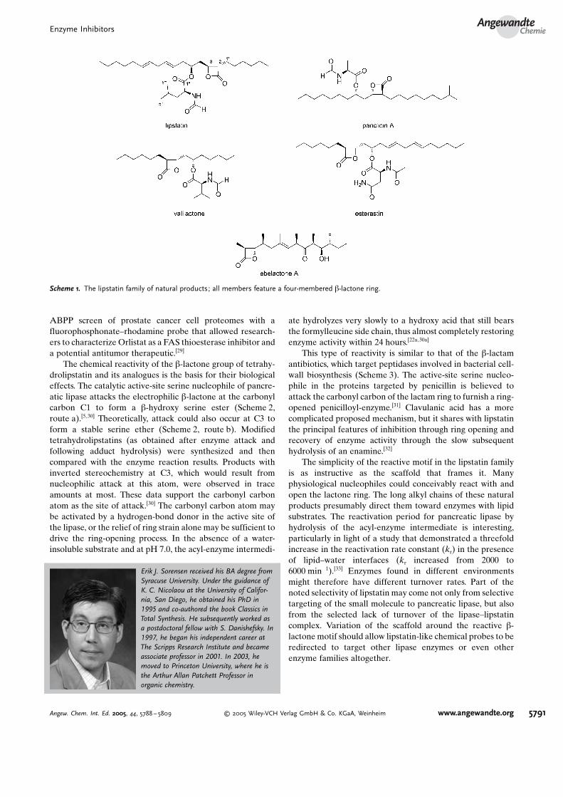
Lipstatin was isolated from Streptomyces toxytricini on thebasis of its potent, selective, and irreversible inhibition ofpancreatic lipase (Scheme 1).[14] The structure of lipstatin wasdetermined by a combination of spectroscopic and chemicalmethods,[15] and was confirmed by chemical synthesis.[16]
Lipstatin features a b-lactone ring with two linear carbonchains of six and thirteen atoms, respectively. The longer ofthe two is doubly unsaturated and contains an N-formylleu-cine ester side chain. Structural analysis revealed a strikingsimilarity between lipstatin and esterastin, a reported inhib-itor of esterase which features a different amino acid sidechain,N-acetylasparagine.[17] Other members of this b-lactoneenzyme inhibitor family include the panclicins,[18] the ebelac-tones,[19] and valilactone (Scheme 1).[20] The research group ofBacher later found that lipstatin is derived biosyntheticallyfrom 3-hydroxytetradeca-5,8-dienoic acid,[21] which formsfrom a condensation of two mycolic acid[22] components of14 and 8 carbon atoms, both of which originate from fatty acidcatabolism. Protons at the C2 and C3 positions, and oneproton at C4 are derived from water.[21c] This is in contrastwith the initial assumption that lipstatin had a polyketideorigin; early [13C]acetate feeding experiments were incon-clusive.[21d] A total synthesis of lipstatin from (S)-N-formyl-leucine and dimethyl-(S)-(�)-malate was recently de-scribed,[23] and there is also a comprehensive body of syntheticwork on tetrahydrolipstatin,[16,24] which is obtained by thecatalytic hydrogenation of lipstatin.[15]
Pancreatic lipase is the target of lipstatin and its deriva-tives. This lipase possesses an active-site charge-relay systemsimilar to serine proteases; it features the catalytic triad ofHis263, Asp176, and Ser152.[25] Pancreatic lipase is respon-sible for the hydrolysis of dietary triacylglycerols to fatty acidsand monoacylglycerols.[26] This catabolic process is critical forproper fat absorption; inhibition of pancreatic lipase activityresults in the passage of fats through the stool. Tetrahydro-lipstatin limits the absorption of dietary fat by blocking theactivity of pancreatic lipase, as well as carboxylester lipase,human milk lipase, and gastric lipase.[22a] Now available underthe trade names orlistat or xenical, tetrahydrolipstatin wasapproved by the Food and Drug Administration in March1999 and has been successful in helping obese patients loseweight.[26b,27] Obesity affects approximately one third of theU.S. population,[26b] and many problems are thought to stemfrom this chronic condition, including diabetes mellitustype II. This disease may be prevented or brought undercontrol with an Orlistat regimen.[28]
Tetrahydrolipstatin is also an inhibitor of the thioesterasedomain of fatty acid synthase (FAS), an enzyme associatedwith tumor cell proliferation.[29] Administration of orlistat toprostate tumor cells causes apoptosis; the drug also haltsgrowth of xenograft tumors in mice. Interestingly, it was an
Carmen Drahl obtained her BA in chemistryat Drew University in 2002. She received herMA at Princeton University in the laboratoryof G. McLendon. Currently, she is conduct-ing graduate studies at Princeton under thedirection of E. J. Sorensen. Her researchinterests include the synthesis and biologicalevaluation of reactive natural products. Sheis the recipient of a Barry M. Goldwater Fel-lowship, as well as predoctoral fellowshipsfrom Eli Lilly and the National ScienceFoundation.
Benjamin Cravatt studied biological sciences(BS) and history (BA) at Stanford Univer-sity. He then conducted research with D.Boger and R. Lerner and received his PhDfrom The Scripps Research Institute (TSRI)in 1996. He joined the faculty at TSRI in1997 as a member of the Skaggs Institutefor Chemical Biology and the departmentsof Cell Biology and Chemistry. His researchgroup is developing and applying new tech-nologies to elucidate the roles of enzymes inthe physiological and pathological processesof the nervous system and in cancer.
B. F. Cravatt, E. J. Sorensen, and C. DrahlReviews
5790 www.angewandte.org � 2005 Wiley-VCH Verlag GmbH & Co. KGaA, Weinheim Angew. Chem. Int. Ed. 2005, 44, 5788 – 5809
ABPP screen of prostate cancer cell proteomes with afluorophosphonate–rhodamine probe that allowed research-ers to characterize Orlistat as a FAS thioesterase inhibitor anda potential antitumor therapeutic.[29]
The chemical reactivity of the b-lactone group of tetrahy-drolipstatin and its analogues is the basis for their biologicaleffects. The catalytic active-site serine nucleophile of pancre-atic lipase attacks the electrophilic b-lactone at the carbonylcarbon C1 to form a b-hydroxy serine ester (Scheme 2,route a).[5, 30] Theoretically, attack could also occur at C3 toform a stable serine ether (Scheme 2, route b). Modifiedtetrahydrolipstatins (as obtained after enzyme attack andfollowing adduct hydrolysis) were synthesized and thencompared with the enzyme reaction results. Products withinverted stereochemistry at C3, which would result fromnucleophilic attack at this atom, were observed in traceamounts at most. These data support the carbonyl carbonatom as the site of attack.[30] The carbonyl carbon atom maybe activated by a hydrogen-bond donor in the active site ofthe lipase, or the relief of ring strain alone may be sufficient todrive the ring-opening process. In the absence of a water-insoluble substrate and at pH 7.0, the acyl-enzyme intermedi-
ate hydrolyzes very slowly to a hydroxy acid that still bearsthe formylleucine side chain, thus almost completely restoringenzyme activity within 24 hours.[22a, 30a]
This type of reactivity is similar to that of the b-lactamantibiotics, which target peptidases involved in bacterial cell-wall biosynthesis (Scheme 3). The active-site serine nucleo-phile in the proteins targeted by penicillin is believed toattack the carbonyl carbon of the lactam ring to furnish a ring-opened penicilloyl-enzyme.[31] Clavulanic acid has a morecomplicated proposed mechanism, but it shares with lipstatinthe principal features of inhibition through ring opening andrecovery of enzyme activity through the slow subsequenthydrolysis of an enamine.[32]
The simplicity of the reactive motif in the lipstatin familyis as instructive as the scaffold that frames it. Manyphysiological nucleophiles could conceivably react with andopen the lactone ring. The long alkyl chains of these naturalproducts presumably direct them toward enzymes with lipidsubstrates. The reactivation period for pancreatic lipase byhydrolysis of the acyl-enzyme intermediate is interesting,particularly in light of a study that demonstrated a threefoldincrease in the reactivation rate constant (kr) in the presenceof lipid–water interfaces (kr increased from 2000 to6000 min�1).[33] Enzymes found in different environmentsmight therefore have different turnover rates. Part of thenoted selectivity of lipstatin may come not only from selectivetargeting of the small molecule to pancreatic lipase, but alsofrom the selected lack of turnover of the lipase–lipstatincomplex. Variation of the scaffold around the reactive b-lactonemotif should allow lipstatin-like chemical probes to beredirected to target other lipase enzymes or even otherenzyme families altogether.
Erik J. Sorensen received his BA degree fromSyracuse University. Under the guidance ofK. C. Nicolaou at the University of Califor-nia, San Diego, he obtained his PhD in1995 and co-authored the book Classics inTotal Synthesis. He subsequently worked asa postdoctoral fellow with S. Danishefsky. In1997, he began his independent career atThe Scripps Research Institute and becameassociate professor in 2001. In 2003, hemoved to Princeton University, where he isthe Arthur Allan Patchett Professor inorganic chemistry.
Scheme 1. The lipstatin family of natural products; all members feature a four-membered b-lactone ring.
Enzyme InhibitorsAngewandte
Chemie
5791Angew. Chem. Int. Ed. 2005, 44, 5788 – 5809 � 2005 Wiley-VCH Verlag GmbH & Co. KGaA, Weinheim www.angewandte.org

2.2. Lactacystin
Lactacystin (Scheme 4) was isolated by Omura and co-workers from a Streptomyces strain in a screen for naturalproducts that induce neurite outgrowth.[34] Its interestingstructure was elucidated by X-ray crystallography and NMRspectroscopy,[35] and confirmed by the total syntheses carriedout by several research groups.[36] Lactacystin is biosynthe-sized from three units: leucine, isobutyrate (and/or valine),and cysteine.[37]
Further investigations showed that lactacystin does notmimic nerve growth factor;[38] instead, it reacts specificallywith the proteasome, binding covalently to the hydroxy group
of the highly conserved N-terminal threonine residue toinhibit the chymotrypsin-like and trypsin-like proteasomeactivities in irreversible fashion.[38b] The proteasome is anabundant ATP-dependent cellular protease that degradesdamaged proteins.[39] With few exceptions, proteasomes act onproteins that are tagged for destruction through the covalentattachment of the small protein ubiquitin.[39] The proteasomeconsists of a barrel-like core, in which controlled proteolysistakes place. The core is capped at each end by a regulatoryprotein.[39] The N-terminal threonine residue modified bylactacystin serves as the active-site nucleophile of the protea-some.[38b,40] The proteasome lacks a catalytic triad, butrequires a basic group to accept a proton from threonine in
Scheme 2. Two possible mechanisms for the covalent modification of pancreatic lipase by lipstatin. The reaction occurs exclusively at theb-lactone carbonyl group.
Scheme 3. Principal features of b-lactam antibiotic reactivity.
B. F. Cravatt, E. J. Sorensen, and C. DrahlReviews
5792 www.angewandte.org � 2005 Wiley-VCH Verlag GmbH & Co. KGaA, Weinheim Angew. Chem. Int. Ed. 2005, 44, 5788 – 5809

the transition state. This is most likely the job of thea-amino group of Thr1, which is ideally situated toaccept a proton in the crystal structure of theproteasome in complex with lactacystin.[41] Alterna-tively, the conserved Lys33 residue may fulfill thisrole, since mutation of this residue abolishes pro-teasome activity.[42] Lactacystin inhibits neither theserine proteases trypsin and chymotrypsin, nor thecysteine proteases papain, the calpains, or cathe-psin B;[38b] it does, however, covalently inhibit theserine protease cathepsin A.[43] As a fairly selectiveproteasome inhibitor, lactacystin has proven to be avaluable probe to study the cellular role of the ubiquitin–proteasome pathway.[44]
Different families of proteasome inhibitors have beendeveloped over the years to better understand the ubiquitin–proteasome system. The earliest synthetic inhibitors, the C-terminal peptide aldehydes, showed too little selectivity forthe proteasome over serine and cysteine proteases.[40] Certaintripeptides modified at the C terminus by a vinyl sulfone orglyoxal act as mechanism-based inhibitors of the proteaso-me.[39b,45] Boronic acid peptides (in which the aldehydepharmacophore has been replaced by a boronate group) aremore potent and selective for the proteasome. The flagshipboronate compound, PS-341,[40, 46] was recently clinicallyapproved for the treatment of multiple myeloma (velcade(bortezomib), Scheme 4). This validates the proteasome as atherapeutic target for at least this type of cancer andpotentially other forms as well.[47] Unlike lactacystin, how-ever, Velcade reversibly inhibits the chymotrypsin-like activ-ity of the proteasome.[40] The most clinically advancedlactacystin analogue is PS-519 (Scheme 4), a cell-permeablevariant that features an n-propyl substitution at C7. Thiscompound is more potent than the natural product and hasentered phase II clinical trials for acute stroke.[40,48]
The reactivity of lactacystin is brought about by its latentb-lactone group, termed clasto-lactacystin b-lactone or omur-alide (Scheme 4).[49] Derivatives that cannot form the b-lactone are inactive. Omuralide is formed in the extracellularfluid from cyclization (lactonization) of lactacystin with loss
of the N-acetylcysteine leaving group.The resulting omuralide is more cell-permeable than the parent com-pound,[40, 49] and it inhibits proteasomeactivities 15 to 20-fold faster thanlactacystin.[38] Carbon atom C4 of thisintermediate lactone is electrophilicand is presumed to undergo attack bythe hydroxy group of the catalyticthreonine residue[44b] to yield an ester-linked omuralide–proteasome adduct(Scheme 5). The “masking” of thereactive b-lactone in its open thioesterform is important, as it may helplactacystin avoid spurious targets. Sev-eral research groups have shown thatlactacystin labels all active proteasomeb subunits.[39b,50] Interestingly, it was
demonstrated that replacement of the catalytic N-terminalthreonine group with serine produced a fivefold increase inthe rate of proteasome inactivation by omuralide. Thisreplacement also increased the rate of hydrolysis of theacyl-enzyme intermediate.[51] This result may explain whyomuralide and lactacystin inhibit very few serine proteases, asserine–omuralide adducts may undergo rapid hydrolysis torelieve inhibition.[44a] It is instructive to note how the activityand selectivity of lactacystin are exquisitely controlled byintrinsic chemical reactivity.
The structure of the proteasome from Saccharomycescerevisiae co-crystallized with omuralide[41] was determinedby X-ray analysis, and confirmed the covalent linkagebetween the threonine side chain hydroxy group and omur-alide. The N-terminal amino group is not modified by thenatural product. Omuralide makes four hydrogen-bond con-tacts with the main chain of the proteasome catalytic subunit.The C9 isopropyl moiety of omuralide projects into ahydrophobic pocket of the proteasome to lend additionalbinding affinity. Corey and co-workers have shown thatreplacement of the omuralide C9 isopropyl group with aphenyl group abolishes all activity.[52] Interestingly, salino-sporamide A (Scheme 4), a natural product 35-fold morepotent than omuralide, possesses a cyclohexene moiety inplace of the isopropyl group, yet it shares the bicyclic ringstructure of omuralide.[53] Feling and co-workers suggestedthat this may be indicative of the different ways by whichomuralide and salinosporamide A interact with the protea-
Scheme 4. Lactacystin and related proteasome inhibitors. Velcade is a synthetic inhibitorclinically approved for the treatment of multiple myeloma.
Scheme 5. Lactacystin is lactonized to its cell-permeable, protein-reactive derivative,omuralide, which then covalently inactivates the proteasome.
Enzyme InhibitorsAngewandte
Chemie
5793Angew. Chem. Int. Ed. 2005, 44, 5788 – 5809 � 2005 Wiley-VCH Verlag GmbH & Co. KGaA, Weinheim www.angewandte.org

some.[53] Corey and co-workers recently reported the firstenantiospecific total synthesis of salinosporamide A[54] with aprocedure that yields sufficient material to carry out biolog-ical analyses of the efficacy of salinosporamide A as apotential anticancer agent. Moreover, congeners of salino-sporamide A were recently prepared for the possible treat-ment of proteasome-mediated disease.[55] It will be interestingto learn whether salinosporamide A has improved upon thedelicately balanced chemistry of lactacystin.
2.3. E-64
In 1978, the Hanada group pursued a specific inhibitor ofcysteine proteases to expedite investigation of proteasebiological functions. They succeeded in isolating a highlypotent and irreversible inhibitor, E-64, from the moldAspergillus japonicus.[56] The structure of E-64 was deter-mined principally through classical chemical reactivity test-ing, IR spectroscopy, and 1H NMR spectroscopy, and wassubsequently confirmed by a chemical synthesis of itsenantiomer.[57] The structure contains agmatine, the argininedecarboxylation product 1-amino-4-guanidinobutane(Scheme 6). To identify an E-64 pharmacophore, a compre-
hensive library of derivatives and fragments was synthesized.Both the epoxide and the carboxyl group derived from l-trans-(S,S)-epoxysuccinic acid were found to be indispensablefor inhibition of papain.[58] Notably, other peptidyl epoxidenatural products that act as covalent inhibitors of theproteasome have also been identified, including epoxomi-cin[59] and eponemicin[60] (Scheme 6).
In addition to papain, E-64 is a covalent inhibitor ofseveral other cysteine proteases, including cathepsins B, H,and L, stem bromelain, and ficin.[61] The revelation that thehigh affinity, active-site-directed reversible inhibitor leupep-tin decreased the rate of E-64 binding cemented the idea thatE-64 is an active-site-directed inhibitor of cysteine pro-teases.[61] These proteases exert their catalytic activity withthe assistance of an active-site ion pair between cysteine andhistidine residues.[62] In papain, these residues are Cys25 andHis159.[62]
Aberrant cysteine protease activity leads to many diseasestates. Inflammatory and traumatic processes, muscular dys-
trophy, AlzheimerFs disease, cancer, and cataract formationall have a putative link to cysteine protease dysfunction.[63] Inaddition, because cysteine proteases are a critical componentof the Plasmodium life cycle, they are regarded as viabletargets for potential antimalarial drugs.[64] In an effort toimprove the cell permeability of leads, the agmatine groupwas replaced with neutral moieties, such as isoamylamine inthe case of E-64c (Scheme 6).[65] In 1986, the ethyl ester of E-64c was removed from phase III clinical trials in Japan formuscular dystrophy indications because its efficacy did notmeet expectations, and the drug covalently modified proteinsof other mechanistic classes.[63d,66] However, by virtue of itsinhibition of calpain, this same E-64 derivative has found usein eye drops for the prevention and treatment of cataracts.[67]
E-64 is still frequently used to facilitate discovery andclassification of newly isolated papain-class enzymes.[68]
Molecular probes for ABPP inspired by the E-64 motifhave proven to be of great value for tracking the activities ofboth animal and plant cysteine proteases in whole proteomes,and in the design and screening of inhibitors for theseenzymes.[68a,b] For example, a screen of plant extracts revealedcysteine protease activity for three proteins that had not beenpreviously characterized.[68a] In a comparative screen ofmature and senescent leaf extracts, ABPP demonstrated
that a change in mRNA transcript levels does notalways correlate with protease activity.[68a] In addition,a cell-permeable E-64 probe revealed that increasedcathepsin activity is associated with the angiogenicvasculature and invasive fronts of carcinomas.[69]
Based on these results, administration of a broad-spectrum cathepsin inhibitor was performed, whichimpaired tumor growth and invasiveness.
The reaction of the E-64 epoxide with papain waselucidated by 13C NMR spectroscopy.[70] Attack by thecatalytic cysteine nucleophile occurs at the epoxide C2atom.[70,71] Backside attack in SN2 fashion inverts theconfiguration at C2 of the product thioether(Scheme 7). The key contacts between cysteine pro-teases and E-64 that are responsible for situating theinhibitorFs epoxide group for regioselective nucleo-
philic attack have been extensively characterized in a series ofcrystal structures.[57, 71,72] In the structure of the E-64–papaincomplex, the N-terminal carboxyl group of E-64 is positionedin the oxyanion hole, while the carbonyl group adjacent to theepoxide group interacts with the histidine catalytic base.[71]
The mechanistic characterization of the interaction betweenE-64 and cysteine proteases of the papain class is an excellentexample of the power of convergent research efforts inchemical synthesis, classical biochemistry, and structuralbiology.
3. Natural Products that Target “Non-Nucleophilic”Residues in Enzyme Active Sites
Several natural products inhibit enzymes through thecovalent modification of active-site residues that are notessential for catalysis. Although some of these residuesappear to perform supportive catalytic roles, they do not
Scheme 6. E-64 and other l-trans-(S,S)-epoxysuccinic acid derivatives.
B. F. Cravatt, E. J. Sorensen, and C. DrahlReviews
5794 www.angewandte.org � 2005 Wiley-VCH Verlag GmbH & Co. KGaA, Weinheim Angew. Chem. Int. Ed. 2005, 44, 5788 – 5809

participate as nucleophiles directly; other residues seemaltogether superfluous for catalysis. In either case, however,covalent modification of these residues results in enzymeinactivation. Herein, we highlight selected examples ofnatural products that target “non-nucleophilic” residues inenzyme active sites.
3.1. Fumagillin
A 1949 report demonstrated that concentrates of Asper-gillus fumigatus cultures possessed antibiotic activity againstStaphylococcus aureus.[73] Tarbell and co-workers character-ized the chemical behavior of the active component, fuma-gillin, including the acid sensitivity of its characteristicspiroepoxide unit.[74] The constitution and stereochemistryof fumagillin were confirmed by X-ray crystallography(Scheme 8).[75] In 1972, Corey and Snider described theirelegant studies that culminated in the first chemical synthesisof this natural product.[76] Subsequent work has produced a26-step synthesis of (�)-fumagillol,[77] the saponificationproduct of fumagillin, and a 13-step racemic synthesis offumagillol.[78] Fumagillin and its close relatives ovalicin[79] andFR65814 (Scheme 8)[80] possess a rare sesquiterpene carbonskeleton that has been shown by labeling experiments tooriginate biosynthetically from b-trans-bergamotene, whicharises from farnesyl pyrophosphate.[81] Other natural productsnot necessarily in the fumagillin family, but which contain aspiroepoxide moiety include curvularol[82] and FR901464.[83]
Fumagillin and other members of its family targetmethionine aminopeptidase 2 (MetAP-2), but not the closelyrelated enzyme methionine aminopeptidase 1 (MetAP-
1).[6b,84] Methionine aminopeptidases are metalloproteasesresponsible for co-translational removal of the N-terminalmethionine residue in specific protein targets.[6b,85] Theseenzymes were originally thought to use cobalt as the active-site metal center, but more recent studies indicate thatmanganese is likely the physiologically relevant cofactor.[86] Inthe enzyme active site, the metal ions are coordinated byAsp251, Asp262, His331, Glu364, Glu459, and a watermolecule.[87] The nucleophilicity of the water molecule isenhanced by coordination to the manganese ion; it attacks thescissile amide bond of the substrate.[88] MetAP-mediatedremoval of methionine is necessary for post-translationalmodifications such as myristoylation.[89] A single amino acidresidue in MetAP-2, Ala362, confers sensitivity to ovalicin,and a Thr362Ala mutation of the human MetAP-1 geneinserted into yeast results in ovalicin-sensitive yeast colo-nies.[90]
The connection between inhibition of MetAP-2 and themedicinal utility of fumagillin appears complex and remainsunclear.[91] It has been speculated that a connection exists atthe level of protein myristoylation.[92] Without removal of N-terminal methionine residues, proteins cannot be myristoy-lated in vivo, which in turn, could disrupt their localizationand function. Therefore, the hypothesis is that fumagillinindirectly causes cell-cycle arrest by interfering with myris-toylation, which leads to aberrant subcellular localization ofproteins that are not yet identified.[92] Despite a mechanism ofaction that remains enigmatic, fumagillin is firmly establishedas a potent antiparasitic agent.[93] During the 1950s, fumagillinwas used for treatment of amoebiasis in humans, caused byEndamoeba histolytica, and is still used today for Nosemadisease in honeybees, caused by the protozoan Nosema
Scheme 7. The active-site cysteine residue of papain attacks the electrophilic epoxide of E-64 in an SN2 fashion.
Scheme 8. Fumagillin and other protein-reactive spiroepoxides. TNP-470 and CKD-731 are synthetic compounds.
Enzyme InhibitorsAngewandte
Chemie
5795Angew. Chem. Int. Ed. 2005, 44, 5788 – 5809 � 2005 Wiley-VCH Verlag GmbH & Co. KGaA, Weinheim www.angewandte.org
apis.[93a,b] More recently, fumagillin has shown efficacy againstmicrosporidial keratoconjunctivitis of the eye, caused by asingle-celled fungus.[94] Fumagillin and its semisyntheticderivative TNP-470 (Scheme 8) are the only treatments formicrosporidiosis in HIV-positive patients[95] and block thein vitro growth of Plasmodium falciparum and Leishmaniadonavani, the organisms that cause malaria and leishmaniasis,respectively.[96] The broad anti-infective activity of fumagillindoes not come without a price; it is toxic to mammals whichindicates its low selectivity for parasitic over mammalianMetAP-2.[97]
The research group of Folkman stimulated a renaissanceof interest in fumagillin with their discovery that this naturalproduct inhibits tumor-induced angiogenesis.[98] TNP-470 isalso a promising small-molecule inhibitor of angiogenesis andhas progressed into clinical trials as a potential anticancerdrug.[99] By using a homology modeling approach from thecrystal structure of MetAP-2, Han and co-workers designedCKD-731 (Scheme 8), an analogue that is 1000-fold morepotent against endothelial cell proliferation than TNP-470.[100]
New data suggest that MetAP-2 may not be the biolog-ically relevant target of fumagillin, and that MetAP-2function is not necessary for the growth of endothelialcells.[101] However, this study could not completely rule outa functional role for MetAP-2, because knockdown of thepeptidase by siRNA was incomplete. The authors raised theintriguing possibility that another MetAP family membermay exist in humans that could also act as a target forfumagillin.[101]
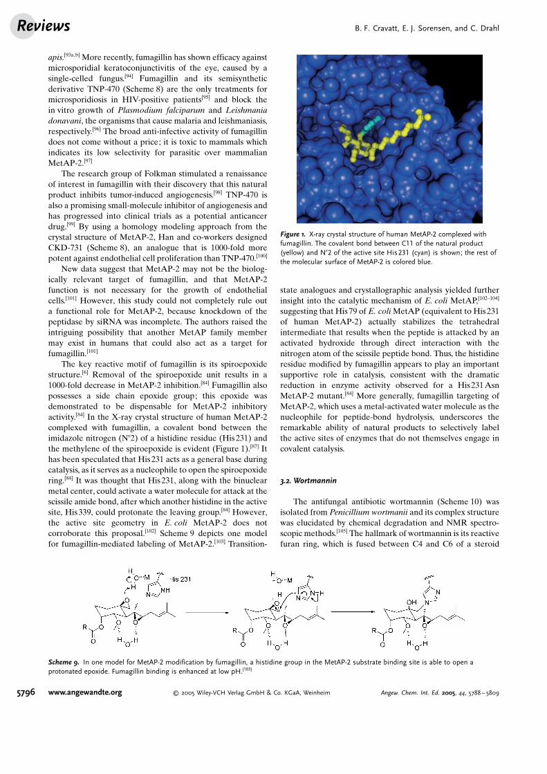
The key reactive motif of fumagillin is its spiroepoxidestructure.[6] Removal of the spiroepoxide unit results in a1000-fold decrease in MetAP-2 inhibition.[84] Fumagillin alsopossesses a side chain epoxide group; this epoxide wasdemonstrated to be dispensable for MetAP-2 inhibitoryactivity.[84] In the X-ray crystal structure of human MetAP-2complexed with fumagillin, a covalent bond between theimidazole nitrogen (Ne2) of a histidine residue (His231) andthe methylene of the spiroepoxide is evident (Figure 1).[87] Ithas been speculated that His231 acts as a general base duringcatalysis, as it serves as a nucleophile to open the spiroepoxidering.[84] It was thought that His231, along with the binuclearmetal center, could activate a water molecule for attack at thescissile amide bond, after which another histidine in the activesite, His339, could protonate the leaving group.[84] However,the active site geometry in E. coli MetAP-2 does notcorroborate this proposal.[102] Scheme 9 depicts one modelfor fumagillin-mediated labeling of MetAP-2.[103] Transition-
state analogues and crystallographic analysis yielded furtherinsight into the catalytic mechanism of E. coli MetAP,[102–104]
suggesting that His79 of E. coliMetAP (equivalent to His231of human MetAP-2) actually stabilizes the tetrahedralintermediate that results when the peptide is attacked by anactivated hydroxide through direct interaction with thenitrogen atom of the scissile peptide bond. Thus, the histidineresidue modified by fumagillin appears to play an importantsupportive role in catalysis, consistent with the dramaticreduction in enzyme activity observed for a His231AsnMetAP-2 mutant.[84] More generally, fumagillin targeting ofMetAP-2, which uses a metal-activated water molecule as thenucleophile for peptide-bond hydrolysis, underscores theremarkable ability of natural products to selectively labelthe active sites of enzymes that do not themselves engage incovalent catalysis.
3.2. Wortmannin
The antifungal antibiotic wortmannin (Scheme 10) wasisolated from Penicillium wortmanii and its complex structurewas elucidated by chemical degradation and NMR spectro-scopic methods.[105] The hallmark of wortmannin is its reactivefuran ring, which is fused between C4 and C6 of a steroid
Scheme 9. In one model for MetAP-2 modification by fumagillin, a histidine group in the MetAP-2 substrate binding site is able to open aprotonated epoxide. Fumagillin binding is enhanced at low pH.[103]
Figure 1. X-ray crystal structure of human MetAP-2 complexed withfumagillin. The covalent bond between C11 of the natural product(yellow) and Ne2 of the active site His231 (cyan) is shown; the rest ofthe molecular surface of MetAP-2 is colored blue.
B. F. Cravatt, E. J. Sorensen, and C. DrahlReviews
5796 www.angewandte.org � 2005 Wiley-VCH Verlag GmbH & Co. KGaA, Weinheim Angew. Chem. Int. Ed. 2005, 44, 5788 – 5809

framework.[106] This feature is also found in viridin, deme-thoxyviridin, the marine-derived pp60v-src protein tyrosinekinase inhibitors halenaquinone and halenaquinol, and hibis-cone C (gmelofuran), which is found in the heartwood ofGmelina arborea and the Jamaican Blue Mahoe(Scheme 10).[107]
Wortmannin[108] and its structural relative viridin[109] arebiosynthesized from the triterpenoid alcohol lanosterol[110]
and are potent cell-permeable inhibitors of the lipid kinasephosphatidylinositol 3-kinase (PI 3-kinase). This enzyme is acentral component of two major receptor-mediated signaltransduction pathways:[111] the G-protein-coupled receptorpathway and the receptor tyrosine kinase pathway. PI 3-kinase phosphorylates at position 3 of the inositol ring ofphosphatidylinositol (PtdIns), leading to the production ofthe second messengers PtdIns-3-phosphate, PtdIns-3,4-bisphosphate, and PtdIns-3,4,5-triphosphate. The precisenature of the PI 3-kinase catalytic mechanism is unknown.[112]
In porcine PI 3-kinase, contact with the a- and b-phosphategroups of ATP is mediated by the conserved Lys833 andSer806 side chains, respectively. An early mechanistic modelinvolved deprotonation of the lipid substrate hydroxy groupby Asp946 to generate a nucleophile that attacks the g-phosphate of ATP. Although mutations of this amino acidabolish activity, the crystal structure of PI 3-kinase indicatesthat this particular aspartate is not positioned properly for arole as catalytic base. Another residue, His948, has beenoffered as an alternative base, but there is no experimentalevidence to support this claim.[112b] Alternatively, PI 3-kinasemay not possess a base catalyst. Instead, it may operatethrough a dissociative transition state.[113]
In addition to its well-studied lipid kinase target, wort-mannin has been demonstrated to inhibit other serine/threonine kinases, including the mammalian target of rapa-
mycin (mTOR), DNA-dependent protein kinase (DNA-PK),[114] myosin light chain kinase (MLCK), the gentamicinresistance enzyme AAC(6’)-APH(2’’), and a membrane-bound PI 4-kinase.[115]
PI 3-kinases are involved in a large number of fundamen-tal cellular processes, including apoptosis, proliferation, cell
motility, and adhesion, and have beenimplicated in the malignant transforma-tion of cells. Increased levels of PI 3-kinase products have been observed incolorectal tumors and in breast can-cers.[116] It was also reported thatdephosphorylation of PI 3-kinase prod-ucts suppresses tumor formation.[117] Asinhibitors of PI 3-kinase in vertebrates,wortmannin and its structural relativesare considered to have potential astherapeutic agents for the treatment ofhuman neoplasms and other diseasestates such as diabetes, inflammation,platelet aggregation, atherosclerosis,and osteoporosis. A semisynthetic wort-mannin library yielded ten compoundsthat were advanced to pharmacokineticand toxicity studies.[118] PX-866(Scheme 10), a diallylamino wortman-nin derivative lacking the furan ring,demonstrated good pharmacokineticsand toxicity and exhibited prolongedinhibition of PI 3-kinase in vivo. Thiscompound also augmented the antitu-
mor effects of the established chemotherapeutic agentcisplatin.[119] It was speculated that loss of the furan ring wasredeemed by improved chemical stability or a better fit in theATP-binding pocket.[118]
Amine nucleophiles react efficiently with the doublyactivated and highly electrophilic strained furan ring ofwortmannin by attacking C20 to give vinylogous carbamatesthrough an addition–elimination mechanism.[120] Wipf and co-workers recently synthesized a library of 94 C20-substitutedsemisynthetic wortmannin derivatives by reaction of thenatural product with a wide variety of amine and thiolnucleophiles.[118] Wymann and co-workers discovered that thischemistry also occurs in the active site of PI 3-kinase.[121] Thee-amino group of Lys802 of PI 3-kinase (Lys833 in porcinePI 3-kinase)[122] attacks C20 of wortmannin which results infuran ring-opening to form a vinylogous carbamate(Scheme 11). This covalent bond-forming event irreversiblyinhibits the enzyme. The acid lability of the vinylogouscarbamate group complicated efforts to isolate wortmannin-labeled peptides. Fortunately, this group could be reducedwith sodium cyanoborohydride to give a considerably morestable amine–wortmannin adduct. ATP, adenine, and FSBA(5’-para-fluorosulfonylbenzoyladenosine) each at 1 mm inter-fered with the alkylation of PI 3-kinase by wortmannin whenadded before the inhibitor, although substances containingnucleophilic amino acid side chain groups had no effect at thesame concentration. FSBA is a derivative of adenine thatreacts covalently with nucleophilic amino acids and has been
Scheme 10. The wortmannin family of steroidal natural products. PX-866 was synthesized fromwortmannin.
Enzyme InhibitorsAngewandte
Chemie
5797Angew. Chem. Int. Ed. 2005, 44, 5788 – 5809 � 2005 Wiley-VCH Verlag GmbH & Co. KGaA, Weinheim www.angewandte.org

used to map nucleotide binding sites.[121] The PI 3-kinasesubstrate PtdIns-4,5-bisphosphate also effectively competeswith wortmannin binding. These findings ledWymann and co-workers to conclude that the wortmannin target site in PI 3-kinase is in a nucleotide binding site in proximity to thesubstrate binding site. These postulates were confirmed by theX-ray crystal structure of a complex between wortmannin andporcine PI 3-kinase.[122]
The role of Lys802, as mentioned above, may be thestabilization of the a-phosphate of ATP in the binding pocket,as has been speculated for Lys72 in protein kinase A(PKA).[121] Lysine 72 anchors the nonlabile phosphategroups of ATP and neutralizes their charges by a saltbridge. Replacement of Lys72 with Ala in yeast PKA leadsto an 800-fold decrease in the Vmax value.
[123] Whereas Lys72of PKA and perhaps Lys802 of PI3-kinase appear importantfor enzyme activity, they probably participate in a primarilystructural role rather than through direct involvement incatalysis. The protein microenvironment may be responsiblefor an enhanced nucleophilicity of the targeted lysine;alternatively, the positioning of wortmannin in kinase activesites may preclude productive interactions with catalyticresidues. Regardless, the potency of wortmannin shouldencourage consideration of cofactor binding sites as targetsfor small-molecule probes. This may expand the menu ofproteins amenable to functional proteomic methods likeABPP. Indeed, quite recently, a rhod-amine-tagged wortmannin analoguewas synthesized and used in proteo-mic studies to identify mammalianpolo-like kinase as an additionaltarget of this natural product.[124]
This kinase is an important proteinfor mitosis that is known to be over-expressed in various human cancers.The wortmannin-based probe suc-cessfully detected kinase activitychanges due to treatment with drugsand in different stages of the cellcycle. Importantly, these results sug-gest that wortmannin is a good leadfor the development of polo-likekinase inhibitors for cancer therapy.
3.3. Aspirin
In a letter to the Right Honorable George,Earl of Macclesfield and President of the RoyalSociety, the Reverend Edward Stone wrote onApril 25, 1763: “There is a bark of an Englishtree, which I have found by experience to be apowerful astringent, and very efficacious incuring aguish and intermitting disorders.”[125]
Stone was not the first to observe the feverand pain-reducing properties of the extracts ofthe willow tree. In the year 200 B.C., the Greekphysician Hippocrates prescribed willow barkand leaves for pain relief. Willow leaves weredescribed by the Roman Pliny the Elder in hisNatural History. Today, a modified version of
salicylic acid, the active constituent in willow bark, namedacetylsalicylic acid or aspirin (Scheme 12),[126] is one of themost widely used remedies in the world.
Salicylic acid and structurally related secondary metabo-lites, the salicylates, are major defensive compounds that arepresent in nearly all higher plants,[127] and deter the feeding ofa variety of herbivores.[127] The biosynthesis of salicylic acid(Scheme 13) may be traced to the phenylpropanoid path-way,[127] which begins with the formation of trans-cinnamicacid through the phenylalanine ammonia lyase-catalyzedelimination of ammonia from phenylalanine.[127a] From thispoint, two pathways are possible, and different plant speciesmay use either or both.[127b] The first involves the removal of atwo-carbon unit from cinnamic acid, leading to benzoic acid,followed by ortho-hydroxylation to salicylic acid. The second
Scheme 11. Lysine 802 in PI 3-kinase opens the doubly activated furan ring of wortmannin by anaddition–elimination mechanism to yield a vinylogous carbamate.
Scheme 12. Aspirin and salicylic acid, the natural product from whichthe drug was first synthetically derived.
Scheme 13. Two biosynthetic pathways for salicylic acid in plants.
B. F. Cravatt, E. J. Sorensen, and C. DrahlReviews
5798 www.angewandte.org � 2005 Wiley-VCH Verlag GmbH & Co. KGaA, Weinheim Angew. Chem. Int. Ed. 2005, 44, 5788 – 5809

reverses the order of the two steps, resulting first in ortho-coumaric acid. The decarboxylation step is thought toproceed in a fashion similar to the b-oxidation events offatty acid breakdown.[127] This was supported by studies inwhich the decarboxylation step in cell-free plant extracts wasfound to be ATP and CoA-dependent.[128]
In 1860, Hermann Kolbe of Marburg University inGermany carried out a laboratory synthesis of salicylic acidand its sodium salt from phenol, carbon dioxide, and sodium.KolbeFs student, Friedrich von Heyden, established a factoryfor the bulk production of salicylates in Radebeul, nearDresden. The unpleasant side effect of salicylate, gastricirritation, revealed itself quite rapidly as the drug becamewidely available. Felix Hoffmann, a chemist at Bayer,ameliorated the problem in 1897 by synthesizing a moreeasily tolerated derivative, acetylsalicylic acid.[129] HeinrichDreser, the director of research at Bayer, coined the term“aspirin” from a combination of SpirsJure (salicylic acid) withan initial “a” for acetyl, and a patent was granted in 1899.
For nearly a century, the molecular mechanism of aspirinremained a mystery. However, in 1971, research by Vaneindicated that aspirin inhibits prostaglandin biosynthesis.[130]
This seminal finding explained both the beneficial propertiesand side effects of aspirin, and culminated in the awarding of ashare of the 1982 Nobel Prize in Physiology or Medicine toVane.[131] The prostaglandins are paracrine hormones that acton cells proximal to their place of synthesis. A trauma to cellsresults in an increase in local biosynthesis of prostaglandins,which cause elevated body temperature, inflammation, andpain.[132] Aspirin was found to acetylate and irreversiblyinactivate prostaglandin endoperoxide (PGG/H) synthase,also termed cyclooxygenase (COX).[133] In addition, aspirinacetylates human hemoglobin and human serum albumin.[134]
The COX enzyme has two known isoforms, the constit-utive COX-1 and the inducible COX-2.[135] COX enzymescatalyze the first two steps in the metabolism of arachidonicacid to prostaglandins. The cyclooxygenase activity incorpo-rates two oxygen atoms into arachidonic acid to produceprostaglandin G2 (PGG2). PGG2 is then subjected to theperoxidase section of these enzymes, which catalyzes areduction of the hydroperoxy group of PGG2 to the hydroxyendoperoxide, PGH2.
[133b,136]
It is recognized that aspirin inhibits the cyclooxygenaseactivity of the COX enzymes, but not their peroxidaseactivity.[137] Aspirin appears to exert its inhibitory effects bydisrupting substrate (arachidonic acid) binding to the cyclo-oxygenase active site, as the serine residue acetylated byaspirin (Ser530 in COX-1, Ser516 in COX-2) is not critical forcatalysis.[133b]
Studies of active-site COX residues have provided insightinto the mechanism of COX enzymes, as well as that of aspirinacetylation. Themechanism of acetyl transfer involves severalamino acids in the COX active site, including conservedtyrosine and arginine residues (Scheme 14).[138] In 1990,spectral and biochemical studies revealed that Tyr385 isessential for COX-1 cyclooxygenase activity.[139] Structurally,this tyrosine is properly positioned to perform the rate-limiting step of cyclooxygenation: abstraction of the 13-pro-Shydrogen atom from arachidonate.[139,140] A Tyr385Phemutant of COX-2 displays virtually no serine acetylation byaspirin, and a Tyr348Phe mutant displays reduced activityand aspirin acetylation.[141] The latter result confirms theimportance of Tyr348, which forms a hydrogen bond toTyr385 and helps to position the substrate. Arginine 120 ofCOX-1 is an important residue for both substrate recognitionand imparting sensitivity to inhibitors that contain a freecarboxylic acid.[142] Analysis of a crystal structure of COX-1suggests that the guanidino moiety interacts with the arach-idonate carboxylate group.[143] Furthermore, reversible inhib-itors with a free carboxylic acid, such as flurbiprofen, wereineffective against COX-1 enzymes with a mutation atArg120.[142,143] The corresponding COX-2 Arg-to-Glnmutant was later found to have a 73% reduction ofacetylation of Ser516 in COX-2.[141]
Based on the above experimental data, researchers havedescribed a best possible mechanism for serine acetylation byaspirin (Scheme 14). The amino acid numbering schemepresented in the following paragraph corresponds to COX-1.Initial interaction of the carboxylate group of aspirin withArg120 stabilizes its location in the substrate-binding pocket.The hydroxy groups of Tyr385 and Tyr348 then act as anextended hydrogen-bonding network. This directs the acetylgroup of aspirin toward Ser530, and increases the reactivity ofaspirin by stabilizing the developing negative charge of the
Scheme 14. An intricate network of hydrogen bonds and other interactions position aspirin to acetylate Ser530 of cyclooxygenase-1.
Enzyme InhibitorsAngewandte
Chemie
5799Angew. Chem. Int. Ed. 2005, 44, 5788 – 5809 � 2005 Wiley-VCH Verlag GmbH & Co. KGaA, Weinheim www.angewandte.org

tetrahedral addition intermediate. The possibility of transienttyrosine acetylation was excluded by testing the acetylation ofa Ser530Ala mutant, for which no O-acetyltyrosine wasdetected.[141] These data are all corroborated by the X-raycrystal structure of COX-1 inactivated by bromoaspirin(bromoacetylsalicylic acid). Bromoaspirin was used so thatits location could be identified definitively in a relatively low-resolution (3.4 L) crystal structure from the presence of theheavy bromine atom. The phenolic oxygen atom of Tyr385 iswithin hydrogen-bonding distance to the acetyl group.[144]
In summary, an elaborate scheme of hydrogen bondingand ionic interactions serves to position aspirin in the COXactive site, increasing its effective concentration and enhan-cing its reactivity, which leads to the selective acetylation ofSer530. The X-ray structure also lends insight into theinhibitory effect of aspirin acetylation. Ser530 lies in ahighly conserved region[133b] along a hydrophobic channel thatleads from the surface of the enzyme to the catalytic tyrosine.The aspirin acetyl group extends outward into the channel,perhaps in a manner sufficient to prevent arachidonic acidfrom reaching the active site.[133b,144]
Finally, it is important to note that salicylic acid alsopossesses anti-inflammatory effects, but does not itselfcovalently modify COX enzymes. Discussion concerning theprecise mechanism of action of salicylate is ongoing.[145] Thus,the conversion of this natural product to its semisyntheticderivative aspirin had the fortuitous effects of conferringmechanistic irreversibility and anointing principal proteintargets for therapeutic intervention.
3.4. Microcystin
Cyanobacteria in the blue-green algae blooms of con-taminated drinking water have been the cause of many animaldeaths. Beginning in 1878,[146] it became evident that bloomsof the generaOscillatoria, Anabaena, Nostoc, andMicrocystisin freshwater and marine environments were to blame forrelated incidents of acute toxicity.[147] The active components,a family of potent hepatotoxins, were isolated and a vigorousresearch effort initiated toward determining their struc-
ture.[148] Although the peptidic nature of the toxins wasconfirmed in 1959,[149] their amino acid compositions were notdetermined until many years later.[150] The first completestructure was determined by NMR spectroscopy and MS in1984,[151] and was soon followed by characterization of otherfamily members.[152] The toxins are cyclic heptapeptides withvariable l-amino acid residues at two positions labeled X andY. They contain an Adda residue, an unusual aromatic b-amino acid featuring an unsaturated alkyl chain (Scheme 15).An unambiguous determination of the configuration of Addawas published in 1988, confirming the structure as(2S,3S,8S,9S)-3-amino-9-methoxy-2,6,8-trimethyl-10-phenyl-(4E,6E)-decadienoic acid.[153] Various names were present inthe literature for Adda-containing cyclic peptide naturalproducts, but ultimately the name microcystin was chosen(Scheme 15).[154]
The microcystin family comprises more than sixty mem-bers;[155] the vast majority differ in the nature of two variableamino acids, or in the methylation states of the dehydroala-nine and/or aspartic acid groups. In naming new microcystins,two letters are appended to abbreviate amino acids at thevariable positions. Biosynthetic studies on microcystins wereparticularly focused on the identification of the origin of thecarbon atoms in the Adda unit and the methylaspartic acidresidue. Methylaspartic acid originates from acetyl-CoA andpyruvic acid via a methylsuccinic acid intermediate, and theAdda unit derives from acetate; certain methyl groups aremethionine-derived.[156] Typically, microcystins are biosynthe-sized non-ribosomally.[157] Analyses of microcystin synthetasegenes have yielded genes for polyketide synthases.[158]
Researchers also sought to understand the molecularnature of the toxicity of microcystins, and not long after thestructures of these natural products were revealed, it wasdetermined that they are potent inhibitors of serine/threoninephosphatases 1 and 2A,[7,147b,159] and other less-thoroughlycharacterized phosphatase family members.[160] Reversibleprotein phosphorylation is a critical mechanism for theregulation of cellular processes.[161] More than 98% of proteinphosphorylation in eukaryotic cells occurs on serine andthreonine residues.[160] Serine/threonine phosphatases 1 and2A (PP1 and PP2A) have been implicated in the regulation of
Scheme 15. Two prototypical microcystin family members; positions X and Y may represent any of several different amino acids. Ndha=N-methyldehydroalanine.
B. F. Cravatt, E. J. Sorensen, and C. DrahlReviews
5800 www.angewandte.org � 2005 Wiley-VCH Verlag GmbH & Co. KGaA, Weinheim Angew. Chem. Int. Ed. 2005, 44, 5788 – 5809

such diverse events as glycogen metabolism, synaptic plasti-city, cell-cycle progression, embryogenesis, apoptosis andsmooth-muscle contraction.[162] The structures, activities, andinhibition properties of PP1 and PP2A have been reviewedextensively.[163] Significantly, these enzymes do not possess acatalytic nucleophile; instead, they activate a water moleculefor catalysis with protein-bound metal ion cofactors.[163]
Indiscriminate inhibition of the many essential functionsperformed by PP1 and PP2A may contribute to the severetoxicity of the microcystins; between one and two microgramsconstitutes a lethal intraperitoneal dose in mice.[147b] Althoughthe broad toxicity of microcystins undermines their potentialas therapeutic agents, research groups have synthesizedvariants and truncated versions of the toxin family to betterunderstand the properties that confer toxicity and specific-ity.[164] So far, only one total synthesis of a microcystin familymember has been completed.[165] These natural products havefacilitated the functional characterization of phosphatasesand phosphatase–protein complexes in proteomes.[159b,166] Amicrocystin-LR–rhodamine conjugate reacted with anddetected numerous serine/threonine phosphatases in a crudecell lysate sample, and could record changes in phosphataseactivity levels.[166] These probes expanded the list of enzymefamilies amenable to functional proteomic analysis.
Several bioactive natural products, such as leptomy-cin B,[167] isoavenaciolide,[168] and pironetin[169] have beenshown to act as carbon-based electrophiles, similar to thea,b-unsaturated carbonyl element of the methyl-dehydroala-nine residue of microcystin. In 1995, two research groupssimultaneously published work confirming the conjugateaddition chemistry of the dehydroalanine moiety of micro-cystin with PP1 at Cys273 near the enzyme C terminus.[7,159a]
In PP2A, the analogous Cys266 is the targeted residue.[159a]
The amino acid sequence Ser-Ala-Pro-Asn-Tyr-Cys, whichincludes the cysteine residue modified by microcystin, ishighly conserved in Ser/Thr phosphatases.[7] The covalentlinkage between the various microcystins and PP1 is stable toheat and acid. However, covalent binding of microcystin-YR
to phosphatases was not observed when the proteins weredenatured with sodium dodecyl sulfate prior to the addition ofthe natural product, indicating that the interaction dependson an intact active site.[7]
Microcystins bind to PP1 with subnanomolar affinity;[170]
in the crystal structure of microcystin-LR bound to PP1, theAdda side chain makes contacts with a conserved hydro-phobic groove, and the carboxyl group of glutamate and theadjacent carbonyl oxygen atom coordinate the metal-bindingsite indirectly through water molecules.[171] The leucine sidechain of the toxin is situated in a loop of the C-terminaldomain; in addition, the carboxyl group of methylaspartatehydrogen bonds to Arg96, a residue crucial for interactionswith phosphate groups on substrates. Hence, access to theactive site is completely blocked.
The extensive interactions between microcystins andphosphatase active sites suggest that covalent reactivitycontributes only partly to the mode of action of these naturalproducts. Microcystins immobilized to a Sepharose beadthrough an aminoethanethiol adduct retained inhibitoryactivity over PP1 and PP2A.[159b] Microcystins-LR, YR, andRR were converted synthetically to their glutathione andcysteine conjugates; both of these thiols reacted with thedehydroalanine group, yet the 1,4-addition products retaineda measure of in vivo toxicity (LD50= 38 mgkg�1 for micro-cystin-LR; LD50= 630 mgkg�1 for its glutathione conju-gate).[171] Notably, such adducts may not be resistant towardretro-Michael reaction in vivo, and reversible adduct forma-tion may explain the observed biological activity.[172] Theseresults imply that the dehydroalanine residue probably doesnot play a role in early enzyme–inhibitor interactions.
Mechanistic studies by Craig and co-workers confirmed atwo-step mechanism for irreversible inhibition by micro-cystins (Scheme 16).[173] In the first step, the rapid noncova-lent binding and inhibition occurs with all the contactsdetailed above. The slower covalent modification of Cys273occurs over the course of hours.[173] The tendency for theconjugate addition reaction is likely enhanced by the high
Scheme 16. Microcystin inactivates PP1 prior to the slow covalent attachment to Cys273. However, the network of noncovalent interactions isessential for a successful conjugate addition.
Enzyme InhibitorsAngewandte
Chemie
5801Angew. Chem. Int. Ed. 2005, 44, 5788 – 5809 � 2005 Wiley-VCH Verlag GmbH & Co. KGaA, Weinheim www.angewandte.org

effective concentration of microcystin in the well-foldedbinding pocket, as evidenced by the control reaction withdenatured protein discussed above. It is also not out of thequestion that the nucleophilicity of the target cysteine may beenhanced in its local protein environment. Regardless, it isimportant to note that mutagenesis studies have found thatCys273 is not required for catalysis; in fact, the activity of aCys273Ser mutant is indistinguishable from that of the wild-type enzyme.[174] Thus, microcystins provide a salient exampleof how an intricate architecture of noncovalent interactionswith enzymes can facilitate covalent modification on non-catalytic active-site residues. Notably, similar strategies havebeen employed to convert tight-binding reversible inhibitorsinto covalent inactivators of the EGF receptor[175a–c] and p90ribosomal protein S6 kinase[175d] by the introduction ofelectrophilic groups in proximity to cysteine residues in theactive sites of these proteins.
4. Targeting Nonenzymatic Proteins
Although most protein-reactive natural products targetthe active sites of enzymes, there are exceptions. Here, wehighlight one such natural product, leptomycin B, whichcovalently modifies a receptor protein involved in nucleartransport.
4.1. Leptomycin B
A Streptomyces strain isolated from soil samples yieldedtwo novel antifungal antibiotics. The active components werepurified, isolated, and termed leptomycins A and B.[176] These
compounds constitute part of a family of structurally similarnatural products, including callystatin A, ratjadone, kazusa-mycin, anguinomycin, leptofuranin and leptolstatin(Scheme 17).[177] Structures of the leptomycins were deter-mined by NMR spectroscopy, MS, and IR spectroscopy.[178]
They feature an unsaturated fatty acid with a terminal d-lactone ring. Leptomycin B has a polyketide origin; it may betraced back to four acetate units, seven propionate units, andone butyrate unit.[179] Leptomycin B interferes with DNAsynthesis stages of the cell cycle,[180] presumably a result of itsinhibitory effect on nuclear transport.[181] A nanomolar bind-ing partner for leptomycin B was discovered and namedchromosome maintenance region 1 (CRM1).[182] CRM1, alsoknown as exportin 1, is an evolutionarily conserved receptorfor the leucine-rich nuclear export signal (NES)[183] ofproteins and is an essential mediator of NES-dependentnuclear export of proteins and ribonucleoprotein complexesin eukaryotic cells.[182,184]
In conjunction with CRM1, the protein Rev allowspartially processed HIV mRNAs to be transported outsidethe nucleus for translation, enabling the HIV virus to hijackthe cellular protein synthesis machinery.[182, 185] Agents likeleptomycin B that block the interaction of Rev with nucleartransport proteins have the potential to inhibit HIV-1replication.[186] Leptomycin B has also been used to accumu-late the tumor suppressor p53 in the nucleus.[187] Thissubcellular localization leads to p53 activation, cell-cyclearrest, and apoptosis. Hence, leptomycin could represent anovel therapeutic approach for HIV and cancer treat-ment.[187,188]
Leptomycin B binds to CRM1 and covalently modifiesCys529 in a central conserved region of the protein, whichinhibits recognition of the NES; ratjadone was recently found
Scheme 17. The leptomycin family of natural products.
B. F. Cravatt, E. J. Sorensen, and C. DrahlReviews
5802 www.angewandte.org � 2005 Wiley-VCH Verlag GmbH & Co. KGaA, Weinheim Angew. Chem. Int. Ed. 2005, 44, 5788 – 5809

to share this mechanism (Scheme 18).[167,181,186a,189] Research-ers found that a single mutation, Cys529Ser, in Schizosac-charomyces pombe was sufficient to greatly reduce sensitivityto leptomycin B.[167,190] To determine which portion of the
natural product serves as the pharmacophore, Kobayashi andcolleagues performed extensive studies on a series ofanalogues of callystatin. The a,b-unsaturated lactone wasfound to be the crucial reactive element.[191] Other researchgroups reduced the lactone moiety to a saturated analogue[192]
or formed a Michael adduct with nitromethane;[185a] both ofthese events greatly decreased inhibition.[192] Furthermore,N-acetylcysteine methyl ester and leptomycin B reacted to givea stable 1,4-addition product, as determined by NMRspectroscopy.[167] All these results lend credence to the ideathat leptomycin binds covalently through an attack on itselectrophilic a,b-unsaturated lactone function by the thiolgroup of CRM1 Cys529 (Scheme 18).
The role of Cys529 in CRM1 is not yet clear. This residuedoes not seem to be necessary for CRM1 to function. TheCys529Ser mutant mediates nuclear export just as effectivelyas wild-type CRM1, and it is not temperature-sensitive.[167]
Moreover, wild-type Saccharomyces cerevisiae CRM1 has athreonine in place of cysteine at the analogous position.[167]
It is difficult to speculate further about the nature of theCRM1–leptomycin B interaction, as a structure of the com-plex has yet to be determined.[188, 193] This information iscrucial to the development of new drug candidates for theinhibition of nuclear translocation. Leptomycin B itself has ahigh toxicity profile, as demonstrated in a phase I clinical trial,and was not recommended for further development.[194]
Several syntheses of leptomycin B and its relatives havebeen carried out in an effort to understand the function of thisfamily of compounds.[191a,195] The premise that some of thesereagents might selectively shut down transport of specificmRNA molecules is enticing and warrants further research.
In summary, CRM1 is a particularly notable target for aprotein-reactive natural product, because this protein is not
an enzyme. Despite this, leptomycin B labels Cys529 withexquisite specificity, suggesting that this residue resides in astructured small-molecule binding site. One intriguing possi-bility is that the central conserved region of CRM1 contains ahydrophobic pocket that is capable of interacting with bothleucine-rich NESs and the distinct architecture of leptomy-cin B in a manner similar to enzyme active sites that bindsubstrates and inhibitors that often share little structuralhomology.[167]
5. Summary and Outlook
In this review, we have attempted to highlight the diversityof mechanisms by which natural products engage andcovalently modify proteins and protein active sites. Thespecific compounds discussed are meant to serve as repre-sentatives of the different classes of protein-reactive naturalproducts discovered so far, rather than as a comprehensivelist. Notably, the reactivity of these natural products is notrestricted to a specific type of amino acid, but rather can occuron a range of active-site residues, including serine (lipstatin,aspirin), threonine (lactacystin), lysine (wortmannin), cys-teine (E-64, microcystin), and histidine (fumagillin). More-over, these residues do not need to serve a catalytic functionin the target enzyme class (for example, Cys273 in PP1) or, forthat matter, to be part of an enzyme active site (as is the casefor Cys529 in CRM1). This underscores the creative ability ofnature to exploit “spurious” nucleophiles in small-moleculebinding sites for the selective modification of proteins.Although all the natural products discussed herein inactivatetheir cognate protein targets by modifying amino acidresidues, other mechanisms of enzyme inhibition are possible.For example, gabaculine (Scheme 19), a rare amino acid
isolated from the mold Streptomyces toyocaensis,[196] inacti-vates several related aminotransferases by covalently reactingwith the enzymeFs pyridoxal phosphate cofactor(Table 1).[8c,197]
What lessons might be learned from protein-reactivenatural products that could assist in the design of chemicalprobes for the burgeoning field of activity-based proteinprofiling (ABPP)?[9] ABPP is a functional proteomic methodthat aims to develop active-site-directed probes that labelmany enzymes in parallel, thereby facilitating their collectivefunctional characterization in samples of high biologicalcomplexity. The application of ABPP methods has so farenabled the discovery of enzyme activities that are up-regulated in diseases like cancer,[69,198] obesity,[199] andmalaria;[200] it has also enabled the design of selectiveinhibitors for these enzymes.[201]
Scheme 18. The a,b-unsaturated lactone of leptomycin reacts byconjugate addition with Cys529 of exportin.
Scheme 19. The natural product gabaculine is a conformationallyconstrained GABA analogue. GABA=g-aminobutyric acid.
Enzyme InhibitorsAngewandte
Chemie
5803Angew. Chem. Int. Ed. 2005, 44, 5788 – 5809 � 2005 Wiley-VCH Verlag GmbH & Co. KGaA, Weinheim www.angewandte.org

The profound influence of protein-reactive natural prod-ucts on the maturation of ABPP can be discerned at severallevels. First, and perhaps most clear, many ABPP probes arethemselves derivatives of natural products, including biotin/fluorophore-tagged reagents that target cysteine proteases(based on E-64),[68a,b,201b] phosphatases (based on microcys-tin),[166] and kinases (based on wortmannin).[124] A secondclass of ABPP probes also borrows a strategy of naturalproducts by modifying conserved catalytic nucleophiles inenzyme active sites. These probes, which include fluoro-phosphonate and electrophilic ketone reagents for serine[202]
and cysteine[203] hydrolases, respectively, typically exhibit abroad target spectrum within a given family of enzymes. EvenABPP probes that incorporate photoreactive (rather thanelectrophilic) groups could be considered as followers of theprecedents set by natural products, as they too exploit class-selective functional groups to bind conserved elements ofenzyme active sites.[204] Finally, the principles derived fromprotein-reactive natural products have also inspired theemergence of a third approach for ABPP that explores theproteome reactivity of structurally diverse libraries of electro-philic agents.[199,205] In this combinatorial, or “nondirected”approach for ABPP, probe libraries have been designed tocontain carbon electrophiles, based in large part on the richdiversity of natural products that have exploited this versatilereactive group, to label enzyme active sites (for example,fumagillin, wortmannin, and microcystin). To date, nondir-ected ABPP efforts have identified active-site proteomicsprobes for more than 10 mechanistically distinct enzymeclasses.[9,199] Remarkably, most of these enzymes lack cognateaffinity agents, underscoring the value of nondirected ABPPfor the discovery of novel molecular probes of enzymefunction.
Looking forward, we anticipate that our understanding ofthe full spectrum of mechanisms employed by naturalproducts to target protein active sites is still far fromcomplete. Given the central role that natural products haveplayed in the elucidation of the biochemical and cell-biological activities of proteins, we strongly advocate thesustained pursuit of these special pharmacological agents.
Their discovery and characterization should continue to offerfascinating insight into the seemingly endless array of small-molecule–protein interactions that occur in nature. Thelessons learned from these studies should in turn guidechemical biologists in their efforts to achieve a paramountobjective of post-genomic science—the design of selectivesmall-molecule probes for the functional analysis of everyprotein.
We thank K. Barglow and Dr. E. C. Taylor for critical readingof this manuscript, and Dr. M. H. Bracey for assistance withthe generation of figures. This research was supported by theNIH (CA087660), NIGMS (GM065483), a Bristol Myers-Squibb Unrestricted Grant in Synthetic Organic Chemistry, theSkaggs Institute for Chemical Biology, and Princeton Univer-sity. C.D. gratefully acknowledges predoctoral fellowshipsfrom Eli Lilly and the National Science Foundation.
Received: March 10, 2005
[1] a) A. Pandey, M. Mann, Nature 2000, 405, 837 – 846; b) N. L.Anderson, N. G. Anderson, Electrophoresis 1998, 19, 1853 –1861.
[2] a) D. T. Hung, T. F. Jamison, S. L. Schreiber, Chem. Biol. 1996,3, 623 – 639; b) R. F. Standaert, A. Galat, G. L. Verdine, S. L.Schreiber, Nature 1990, 346, 671 – 674.
[3] K. Nakanishi in Comprehensive Natural Products Chemistry,Vol. 1 (Eds.: D. Barton, K. Nakanishi, O. Meth-Cohn, U.Sankawa), Elsevier, New York, 1999, pp. xxiii-xl.
[4] N. Kasanah, M. T. Hamann, Expert Opin. Invest. Drugs 2004, 5,827 – 837.
[5] P. Hadvary, W. Sidler, W. Meister, W. Vetter, H. Wolfer, J. Biol.Chem. 1991, 266, 2021 – 2027.
[6] a) W. T. Lowther, D. A. McMillen, A. M. Orville, B. W. Mat-thews, Proc. Natl. Acad. Sci. USA 1998, 95, 12153 – 12157; b) N.Sin, L. H. Meng, M. Q. W. Wang, J. J. Wen, W. G. Bornmann,C. M. Crews, Proc. Natl. Acad. Sci. USA 1997, 94, 6099 – 6103.
[7] R. W. Mackintosh, K. N. Dalby, D. G. Campbell, P. T. W.Cohen, P. Cohen, C. Mackintosh, FEBS Lett. 1995, 371, 236 –240.
[8] a) R. B. Silverman, Methods Enzymol. 1995, 249, 240 – 283;b) R. R. Rando, Pharmacol. Rev. 1984, 36, 111 – 142; c) C.Walsh, Tetrahedron 1982, 38, 871 – 909.
[9] a) A. E. Speers, B. F. Cravatt, ChemBioChem 2004, 5, 41 – 47;b) G. C. Adam, E. J. Sorensen, B. F. Cravatt, Mol. Cell.Proteomics 2002, 1, 781 – 790; c) B. F. Cravatt, E. J. Sorensen,Curr. Opin. Chem. Biol. 2000, 4, 663 – 668.
[10] P. R. Graves, T. A. J. Haystead,Microbiol. Mol. Biol. Rev. 2002,66, 39 – 63.
[11] a) J. F. Fisher, S. Q. Meroueh, S. Mobashery, Chem. Rev. 2005,105, 395 – 424; b) S. J. Coulthurst, A. M. L. Barnard, G. P. C.Salmond, Nat. Rev. Microbiol. 2005, 3, 295 – 306; c) B. F.Gherman, S. D. Goldberg, V. W. Cornish, R. A. Friesner, J.Am. Chem. Soc. 2004, 126, 7652 – 9664; d) J. R. Knowles, Acc.Chem. Res. 1985, 18, 97 – 104.
[12] a) D. L. Boger, R. M. Garbaccio, Acc. Chem. Res. 1999, 32,1043 – 1052; b) K. S. Gates in Comprehensive Natural ProductsChemistry, Vol. 7 (Eds.: D. Barton, K. Nakanishi, O. Meth-Cohn, E. T. Kool), Elsevier, New York, 1999, pp. 491 – 552;c) D. L. Boger, D. S. Johnson, Angew. Chem. 1996, 108, 1542 –1580; Angew. Chem. Int. Ed. Engl. 1996, 35, 1438 – 1474.
[13] J. A. Gerlt, Nat. Biotechnol. 2002, 20, 786 – 787.
Table 1: Representative protein-reactive natural products and theirtargets.
Naturalproduct
Target Labeledresidue
Catalyticnucleophile?
lipstatin pancreatic lipase Ser152 yeslactacystin proteasome
b subunitsThr1 yes
E-64 papain(Cys proteases)
Cys25 yes
fumagillin MetAP-2 His -231 nowortmannin PI 3-kinase Lys802 noaspirin COX-1 Ser530 no
COX-2 Ser516 nomicrocystin PP1 Cys273 no
PP2A Cys266 noleptomycin B CRM1 Cys529 nogabaculine aminotransferases PLP cofactor[a] no
[a] PLP=pyridoxal phosphate.
B. F. Cravatt, E. J. Sorensen, and C. DrahlReviews
5804 www.angewandte.org � 2005 Wiley-VCH Verlag GmbH & Co. KGaA, Weinheim Angew. Chem. Int. Ed. 2005, 44, 5788 – 5809

[14] a) P. Hadvary, H. Lengsfeld, H. Wolfer, Biochem. J. 1988, 256,357 – 361; b) E. K. Weibel, P. Hadvary, E. Hochuli, E. Kupfer,H. Lengsfeld, J. Antibiot. 1987, 40, 1081 – 1086.
[15] E. Hochuli, E. Kupfer, R. Maurer, W. Meister, Y. Mercadal, K.Schmidt, J. Antibiot. 1987, 40, 1086 – 1091.
[16] a) P. Barbier, F. Schneider,Helv. Chim. Acta 1987, 70, 196 – 202;b) P. Barbier, F. Schneider, U. Widmer, Helv. Chim. Acta 1987,70, 1412 – 1418.
[17] H. Umezawa, T. Aoyagi, T. Hazato, K. Uotani, F. Kojima, M.Hamada, T. Takeuchi, J. Antibiot. 1978, 31, 639 – 641.
[18] K. Yoshinari, M. Aoki, T. Ohtsuka, N. Nakayama, Y. Itezono,M. Mutoh, J. Watanabe, K. Yokose, J. Antibiot. 1994, 47, 1376 –1384.
[19] a) K. Uotani, H. Naganawa, S. Kondo, T. Aoyagi, H. Umezawa,J. Antibiot. 1982, 35, 1495 – 1499; b) K. Uotani, H. Naganawa, T.Aoyagi, H. Umezawa, J. Antibiot. 1982, 35, 1670 – 1674; c) H.Umezawa, T. Aoyagi, K. Uotani, M. Hamada, T. Takeuchi, S.Takahashi, J. Antibiot. 1980, 33, 1594 – 1596.
[20] M. Kitahara, M. Asano, H. Naganawa, K. Maeda, M. Hamada,T. Aoyagi, H. Umezawa, Y. Iitaka, H. Nakamura, J. Antibiot.1987, 40, 1647 – 1650.
[21] a) C. A. Schuhr, W. Eisenreich, M. Goese, P. Stohler, W.Weber,E. Kupfer, A. Bacher, J. Org. Chem. 2002, 67, 2257 – 2262; b) M.Goese, W. Eisenreich, E. Kupfer, P. Stohler, W. Weber, H. G.Leuenberger, A. Bacher, J. Org. Chem. 2001, 66, 4673 – 4678;c) M. Goese, W. Eisenreich, E. Kupfer, W. Weber, A. Bacher, J.Biol. Chem. 2000, 275, 21192 – 21196; d) W. Eisenreich, E.Kupfer, W. Weber, A. Bacher, J. Biol. Chem. 1997, 272, 867 –874.
[22] a) B. Borgstrom, Biochim. Biophys. Acta 1988, 962, 308 – 316;b) F. H. Stodola, A. Lesuk, R. J. Anderson, J. Biol. Chem. 1938,126, 505 – 513.
[23] A. Pommier, J. M. Pons, P. J. Kocienski, J. Org. Chem. 1995, 60,7334 – 7339.
[24] a) J. A. Bodkin, E. J. Humphries, M. D. McLeod, TetrahedronLett. 2003, 44, 2869 – 2872; b) A. K. Ghosh, S. Fidanze, Org.Lett. 2000, 2, 2405 – 2407; c) P. J. Parsons, J. K. Cowell, Synlett2000, 107 – 109; d) A. K. Ghosh, C. F. Liu, Chem. Commun.1999, 1743 – 1744; e) C. Lowe, J. C. Vederas, Org. Prep. Proced.Int. 1995, 27, 305 – 346; f) A. Pommier, J. M. Pons, P. I.Kocienski, L. Wong, Synthesis 1994, 1294 – 1300.
[25] F. K. Winkler, A. DFArcy, W. Hunziker, Nature 1990, 343, 771 –774.
[26] a) M. E. Lowe, J. Lipid Res. 2002, 43, 2007 – 2016; b) A. M.Heck, J. A. Yanovski, K. A. Calis, Pharmacotherapy 2000, 20,270 – 279; c) M. E. Lowe, Annu. Rev. Nutr. 1997, 17, 141 – 158;d) S. Ransac, Y. Gargouri, F. Marguet, G. Buono, C. Beglinger,P. Hildebrand, H. Lengsfeld, P. Hadvary, R. Verger, MethodsEnzymol. 1997, 286, 190 – 231.
[27] a) P. Hollander, Prim Care. 2003, 30, 427 – 440; b) K. H. Lucas,B. Kaplan-Machlis, Ann. Pharmacother. 2001, 35, 314 – 328;c) A. Ballinger, Expert Opin. Pharmacother. 2000, 1, 841 – 847.
[28] a) L. M. Prisant, J. Clin. Pharmacol. 2004, 44, 406 – 413; b) D. E.Kelley, M. Jneidi, Expert Opin. Pharmacother. 2002, 3, 599 –605; c) L. J. Aronne, Prog. Cardiovasc. Nurs. 2001, 16, 98 – 106,115; d) G. M. Keating, B. Jarvis, Drugs 2001, 61, 2107 – 2119.
[29] S. J. Kridel, F. Axelrod, N. Rozenkrantz, J. W. Smith, CancerRes. 2004, 64, 2070 – 2075.
[30] a) H. Stalder, G. Oesterhelt, Helv. Chim. Acta 1992, 75, 1593 –1603; b) Q. Q. Luthipeng, H. P. Marki, P. Hadvary, FEBS Lett.1992, 299, 111 – 115; c) H. Stalder, P. R. Schneider, G. Oester-helt, Helv. Chim. Acta 1990, 73, 1022 – 1036.
[31] D. J. Waxman, J. L. Strominger, Annu. Rev. Biochem. 1983, 52,825 – 869.
[32] C. Therrien, R. C. Levesque, FEMS Microbiol. Rev. 2000, 24,251 – 262.
[33] A. Lookene, N. Skotlova, G. Olivecrona, Eur. J. Biochem. 1994,222, 395 – 403.
[34] S. Omura, T. Fujimoto, K. Otoguro, K. Matsuzaki, R. Mor-iguchi, H. Tanaka, Y. Sasaki, J. Antibiot. 1991, 44, 113 – 116.
[35] S. Omura, K. Matsuzaki, T. Fujimoto, K. Kosuge, T. Furuya, S.Fujita, A. Nakagawa, J. Antibiot. 1991, 44, 117 – 118.
[36] a) C. E. Masse, A. J. Morgan, J. Adams, J. S. Panek, Eur. J. Org.Chem. 2000, 2513 – 2528; b) E. J. Corey, W.-D. Z. Li, G. A.Reichard, J. Am. Chem. Soc. 1998, 120, 2330 – 2336; c) E. J.Corey, W. D. Li, T. Nagamitsu, Angew. Chem. 1998, 110, 1784 –1787; Angew. Chem. Int. Ed. 1998, 37, 1676 – 1679; d) N. Chida,J. Takeoka, K. Ando, N. Tsutsumi, S. Ogawa, Tetrahedron 1997,53, 16287 – 16298; e) H. Uno, J. E. Baldwin, I. Churcher, A. T.Russell, Synlett 1997, 390 – 392; f) T. Nagamitsu, T. Sunazuka,H. Tanaka, S. Omura, P. A. Sprengeler, A. B. Smith, J. Am.Chem. Soc. 1996, 118, 3584 – 3590; g) N. Chida, J. Takeoka, N.Tsutsumi, S. Ogawa, J. Chem. Soc. Chem. Commun. 1995, 793 –794; h) H. Uno, J. E. Baldwin, A. T. Russell, J. Am. Chem. Soc.1994, 116, 2139 – 2140; i) T. Sunazuka, T. Nagamitsu, K.Matsuzaki, H. Tanaka, S. Omura, A. B. Smith, J. Am. Chem.Soc. 1993, 115, 5302 – 5302; j) E. J. Corey, G. A. Reichard, J.Am. Chem. Soc. 1992, 114, 10677 – 10678.
[37] a) A. Nakagawa, M. Kainosho, S. Omura, Pure Appl. Chem.1994, 66, 2411 – 2413; b) A. Nakagawa, S. Takahashi, K. Uchida,K. Matsuzaki, S. Omura, A. Nakamura, N. Kurihara, T.Nakamatsu, Y. Miyake, K. Take, M. Kainosho, TetrahedronLett. 1994, 35, 5009 – 5012.
[38] a) L. R. Dick, A. A. Cruikshank, L. Grenier, F. D. Melandri,S. L. Nunes, R. L. Stine, J. Biol. Chem. 1996, 271, 7273 – 7276;b) G. Fenteany, R. F. Standaert, W. S. Lane, S. Choi, E. J. Corey,S. L. Schreiber, Science 1995, 268, 726 – 731; c) G. Fenteany,R. F. Standaert, G. A. Reichard, E. J. Corey, S. L. Schreiber,Proc. Natl. Acad. Sci. USA 1994, 91, 3358 – 3362.
[39] a) B. Alberts, A. Johnson, J. Lewis, M. Raff, K. Roberts, P.Walter, Molecular Biology of the Cell, 4th ed. , GarlandScience, New York, 2002, pp. 358 – 359; b) M. Bogyo, M.Gaczynska, H. L. Ploegh, Biopolymers 1997, 43, 269 – 280.
[40] P. J. Elliott, T. M. Zollner, W. H. Boehncke, J. Mol. Med. 2003,81, 235 – 245.
[41] M. Groll, L. Ditzel, J. Loewe, D. Stock, M. Bochter, H. D.Bartunik, R. Huber, Nature 1997, 386, 463 – 471.
[42] a) E. SeemRller, A. Lupas, D. Stock, J. LSwe, R. Huber, H.Baumeister, Science 1995, 268, 579 – 582; b) O. Coux, K.Tanaka, A. L. Goldberg, Annu. Rev. Biochem. 1996, 65, 801 –847.
[43] a) L. Kozlowski, T. Stoklosa, S. Omura, C. Wojcik, M. Z.Wojtukiewicz, K. Worowski, H. Ostrowska, Tumor Biol. 2001,22, 211 – 215; b) H. Ostrowska, C. Wojcik, S. Wilk, S. Omura, L.Kozlowski, T. Stoklosa, K. Worowski, P. Radziwon, Int. J.Biochem. Cell Biol. 2000, 32, 747 – 757; c) H. Ostrowska, C.Wojcik, S. Omura, K. Worowski, Biochem. Biophys. Res.Commun. 1997, 234, 729 – 732.
[44] a) A. F. Kisselev, A. L. Goldberg, Chem. Biol. 2001, 8, 739 –758; b) G. Fenteany, S. L. Schreiber, J. Biol. Chem. 1998, 273,8545 – 8548.
[45] a) D. H. Lee, A. L. Goldberg, Trends Cell Biol. 1998, 8, 397 –403; b) M. Bogyo, J. S. McMaster, M. Gaczynska, D. Tortorella,A. L. Goldberg, H. L. Ploegh, Proc. Natl. Acad. Sci. USA 1997,94, 6629 – 6634; c) M. C. Sousa, B. M. Kessler, H. J. Overkleeft,R. B. McKay, J. Mol. Biol. 2002, 318, 779 – 785.
[46] G. Martinelli, P. Tosi, E. Ottaviani, S. Soverini, S. Tura,Haematologica 2001, 86, 908 – 917.
[47] R. Twombly, J. Natl. Cancer Inst. 2003, 95, 845.[48] F. Soucy, L. Grenier, M. L. Behnke, A. T. Destree, T. A.
McCormack, J. Adams, L. Plamondon, J. Am. Chem. Soc.1999, 121, 9967 – 9976.
Enzyme InhibitorsAngewandte
Chemie
5805Angew. Chem. Int. Ed. 2005, 44, 5788 – 5809 � 2005 Wiley-VCH Verlag GmbH & Co. KGaA, Weinheim www.angewandte.org

[49] a) E. J. Corey, W.-D. Z. Li, Chem. Pharm. Bull. 1999, 47, 1 – 10;b) L. R. Dick, A. A. Cruikshank, A. T. Destree, L. Grenier,T. A. McCormack, F. D. Melandri, S. L. Nunes, V. J. Palom-bella, L. A. Parent, L. Plamondon, R. L. Stein, J. Biol. Chem.1997, 272, 182 – 188.
[50] A. Craiu, M. Gaczynska, T. Akopian, C. F. Gramm, G.Fenteany, A. L. Goldberg, K. L. Rock, J. Biol. Chem. 1997,272, 13437 – 13445.
[51] A. F. Kisselev, Z. Songyang, A. L. Goldberg, J. Biol. Chem.2000, 275, 14831 – 14837.
[52] E. J. Corey, W. D. Z. Li, T. Nagamitsu, G. Fenteany, Tetrahe-dron 1999, 55, 3305 – 3316.
[53] R. H. Feling, G. O. Buchanan, T. J. Mincer, C. A. Kaufmann,P. R. Jensen, W. Fenical, Angew. Chem. 2003, 115, 369 – 371;Angew. Chem. Int. Ed. 2003, 42, 355 – 357.
[54] L. R. Reddy, P. Saravanan, E. J. Corey, J. Am. Chem. Soc. 2004,126, 6230 – 6231.
[55] M. Stadler, S. Seip, H. Mueller, A. Mayer-Bartschmid, M.-A.Bruenig, J. Benet-Buchholz, H. Togame, R. Dodo, P. Reinemer,K. Bacon, K. Fuchikami, S. Matsukawa, K. Urbahns, PCT Int.Appl. 2004, 79 pp. CODEN: PIXXD2 WO 2004071382 A220040826 CAN 141:224070 AN 2004:701941.
[56] K. Hanada, M. Tamai, M. Yamagishi, S. Ohmura, J. Sawada, I.Tanaka, Agric. Biol. Chem. 1978, 42, 523 – 528.
[57] K. Hanada, M. Tamai, S. Ohmura, J. Sawada, T. Seki, I. Tanaka,Agric. Biol. Chem. 1978, 42, 529 – 536.
[58] K. Hanada, M. Tamai, S. Morimoto, T. Adachi, S. Ohmura, J.Sawada, I. Tanaka, Agric. Biol. Chem. 1978, 42, 537 – 541.
[59] a) L. H. Meng, R. Mohan, B. H. B. Kwok, M. Elofsson, N. Sin,C. M. Crews, Proc. Natl. Acad. Sci. USA 1999, 96, 10403 –10408; b) N. Sin, K. B. Kim, M. Elofsson, L. H. Meng, H.Auth, B. H. B. Kwok, C. M. Crews, Bioorg. Med. Chem. Lett.1999, 9, 2283 – 2288.
[60] L. H. Meng, B. H. B. Kwok, N. Sin, C. M. Crews, Cancer Res.1999, 59, 2798 – 2801.
[61] A. J. Barrett, A. A. Kembhavi, M. A. Brown, H. Kirschke,C. G. Knight, M. Tamai, K. Hanada, Biochem. J. 1982, 201,189 – 198.
[62] M. J. Harrison, N. A. Burton, I. H. Hillier, J. Am. Chem. Soc.1997, 119, 12285 – 12291.
[63] a) M. Lee, R. Fridman, S. Mobashery, Chem. Soc. Rev. 2004, 33,401 – 409; b) A. Baruch, D. Greenbaum, E. T. Levy, P. A.Nielsen, N. B. Gilula, N. M. Kumar, M. Bogyo, J. Biol. Chem.2001, 276, 28999 – 29006; c) J. G. Tidball, M. J. Spencer, Int. J.Biochem. Cell Biol. 2000, 32, 1 – 5; d) H. H. Otto, T. Schirmeis-ter, Chem. Rev. 1997, 97, 133 – 171.
[64] B. K. Na, T. S. Kim, P. J. Rosenthal, J. K. Lee, Y. Kong,Parasitol. Res. 2004, 94, 312 – 317.
[65] T. Noda, K. Isogai, N. Katunuma, Y. Tarumoto, M. Ohzeki, J.Biochem. 1981, 90, 893 – 896.
[66] a) Y. Shoji-Kasai, M. Senshu, S. Iwashita, K. Imahori, Proc.Natl. Acad. Sci. USA 1988, 85, 146 – 150; b) M. Tamai, K.Matsumoto, S. Omura, I. Koyama, Y. Ozawa, K. Hanada, J.Pharmacobio-Dyn. 1986, 9, 672 – 677.
[67] a)Drugs Future 1994, 19, 1040; b) P. Mathur, S. K. Gupta, A. R.Wegener, W. Breipohl, M. H. Ahrend, Y. D. Sharma, Y. K.Gupta, R. B. Vajpayee, Curr. Eye Res. 2000, 21, 926 – 933; c) M.Inomata, H. Hayashi, S. Shumiya, S. Kawashima, Y. Ito, J.Biochem. 2000, 128, 771 – 776.
[68] a) R. A. L. van der Hoorn, M. A. Leeuwenburgh, M. Bogyo,M. H. A. J. Joosten, S. C. Peck, Plant Physiol. 2004, 135, 1170 –1178; b) D. Greenbaum, K. F. Medzihradszky, A. Burlingame,M. Bogyo, Chem. Biol. 2000, 7, 569 – 581; c) H. Cheng, C. A.Parish, B. A. Gilbert, R. R. Rando, Biochemistry 1995, 34,16662 – 16671; d) G. Velasco, A. A. Ferrando, X. S. Puente,L. M. Sanchez, C. Lopezotin, J. Biol. Chem. 1994, 269, 27136 –27142.
[69] J. A. Joyce, A. Baruch, K. Chehade, N. Meyer-Morse, E.Giraudo, F. Y. Tsai, D. C. Greenbaum, J. H. Hager, M. Bogyo,D. Hanahan, Cancer Cell 2004, 5, 443 – 453.
[70] Y. Yabe, D. Guillaume, D. H. Rich, J. Am. Chem. Soc. 1988,110, 4043 – 4044.
[71] K. Matsumoto, K.Mizoue, K. Kitamura,W. C. Tse, C. P. Huber,T. Ishida, Biopolymers 1999, 51, 99 – 107.
[72] a) K. Matsumoto, M. Murata, S. Sumiya, K. Mizoue, K.Kitamura, T. Ishida, Biochim. Biophys. Acta 1998, 1383, 93 –100; b) A. Fujishima, Y. Imai, T. Nomura, Y. Fujisawa, Y.Yamamoto, T. Sugawara, FEBS Lett. 1997, 407, 47 – 50; c) B. G.Zhao, C. A. Janson, B. Y. Amegadzie, K. DFAlessio, C. Griffin,C. R. Hanning, C. Jones, J. Kurdyla, M. McQueney, X. Y. Qiu,W. W. Smith, S. S. Abdel-Meguid, Nat. Struct. Biol. 1997, 4,109 – 111; d) N. A. Katerelos, M. A. J. Taylor, M. Scott, P. W.Goodenough, R. W. Pickersgill, FEBS Lett. 1996, 392, 35 – 39;e) K. I. Varughese, Y. Su, D. Cromwell, S. Hasnain, N. H.Xuong, Biochemistry 1992, 31, 5172 – 5176; f) D. Yamamoto, K.Matsumoto, H. Ohishi, T. Ishida, M. Inoue, K. Kitamura, K.Hanada, FEBS Lett. 1990, 263, 134 – 136; g) D. Yamamoto, K.Matsumoto, T. Ishida,M. Inoue, S. Sumiya, K. Kitamura,Chem.Pharm. Bull. 1989, 37, 2577 – 2581.
[73] F. R. Hanson, E. Eble, J. Bacteriol. 1949, 58, 527 – 529.[74] a) J. R. Turner, D. S. Tarbell, Proc. Natl. Acad. Sci. USA 1962,
59, 733 – 735; b) D. S. Tarbell, R. M. Carman, D. D. Chapman,S. E. Cremer, A. D. Cross, K. R. Huffman, M. Kunstmann, N. J.McCorkindale, J. J. G. McNally, A. Rosowsky, F. H. L. Varino,R. L. West, J. Am. Chem. Soc. 1961, 83, 3096 – 3113; c) D. S.Tarbell, R. M. Carman, D. D. Chapman, K. R. Huffman, N. J.McCorkindale, J. Am. Chem. Soc. 1960, 82, 1005 – 1007.
[75] N. McCorkindale, J. G. Sime, Proc. Chem. Soc. London 1961,331.
[76] a) B. B. Snider, PhD thesis, Harvard University 1973 ; b) E. J.Corey, B. B. Snider, J. Am. Chem. Soc. 1972, 94, 2549 – 2550.
[77] a) D. Kim, S. K. Ahn, H. Bae, W. J. Choi, H. S. Kim, Tetrahe-dron Lett. 1997, 38, 4437 – 4440; for other syntheses offumagillin/fumagillol, see: b) O. Bedel, A. Haudrechy, Y.Langlois, Eur. J. Org. Chem. 2004, 3813 – 3819; c) D. F. Taber,T. E. Christos, A. L. Rheingold, I. A. Guzei, J. Am. Chem. Soc.1999, 121, 5589 – 5590.
[78] D. A. Vosburg, S. Weiler, E. J. Sorensen, Angew. Chem. 1999,111, 1024 – 1027; Angew. Chem. Int. Ed. 1999, 38, 971 – 974.
[79] a) P. Bollinger, H. P. Sigg, H. P. Weber, Helv. Chim. Acta 1973,56, 819 – 830; b) H. P. Sigg, H. P. Weber,Helv. Chim. Acta 1968,51, 1395 – 1408.
[80] a) H. Hatanaka, T. Kino, M. Hashimoto, Y. Tsurumi, A.Kuroda, H. Tanaka, T. Goto, M. Okuhara, J. Antibiot. 1988, 41,999 – 1008; b) S. Amano, N. Ogawa, M. Ohtsuka, N. Chida,Tetrahedron 1999, 55, 2205 – 2224.
[81] a) D. E. Cane, D. B. McIlwaine, Tetrahedron Lett. 1987, 28,6545 – 6548; b) D. E. Cane, S. L. Buchwald, J. Am. Chem. Soc.1977, 99, 6132 – 6134; c) D. E. Cane, G. G. S. King, TetrahedronLett. 1976, 17, 4737 – 4740; d) D. E. Cane, R. H. Levin, J. Am.Chem. Soc. 1976, 98, 1183 – 1188; e) D. E. Cane, R. H. Levin, J.Am. Chem. Soc. 1975, 97, 1282 – 1284; f) M. Tanabe, K. T.Suzuki, Tetrahedron Lett. 1974, 15, 4417 – 4420; g) A. J. Birch,S. F. Hussain, J. Chem. Soc. C 1969, 1473 – 1474.
[82] Y. Honda, M. Ueki, G. Okada, R. Onose, R. Usami, K.Horikoshi, H. Osada, J. Antibiot. 2001, 54, 10 – 16.
[83] H. Nakajima, S. Takase, H. Terano, H. Tanaka, J. Antibiot. 1997,50, 96 – 99.
[84] E. C. Griffith, Z. Su, S. Niwayama, C. A. Ramsay, Y. H. Chang,J. O. Liu, Proc. Natl. Acad. Sci. USA 1998, 95, 15183 – 15188.
[85] E. C. Griffith, Z. Su, B. E. Turk, S. P. Chen, Y. H. Chang, Z. C.Wu, K. Biemann, J. O. Liu, Chem. Biol. 1997, 4, 461 – 471.
B. F. Cravatt, E. J. Sorensen, and C. DrahlReviews
5806 www.angewandte.org � 2005 Wiley-VCH Verlag GmbH & Co. KGaA, Weinheim Angew. Chem. Int. Ed. 2005, 44, 5788 – 5809

[86] J. Y. Wang, G. S. Sheppard, P. P. Lou, M. Kawai, C. Park, D. A.Egan, A. Schneider, J. Bouska, R. Lesniewski, J. Henkin,Biochemistry 2003, 42, 5035 – 5042.
[87] S. P. Liu, J. Widom, C. W. Kemp, C. M. Crews, J. Clardy, Science1998, 282, 1324 – 1327.
[88] M. D. Vaughan, P. B. Sampson, J. F. Honek, Curr. Med. Chem.2002, 9, 385 – 409.
[89] a) R. A. Bradshaw, W. W. Brickey, K. W. Walker, TrendsBiochem. Sci. 1998, 23, 263 – 267; b) R. L. Kendall, R.Yamada, R. A. Bradshaw, Methods Enzymol. 1990, 185, 398 –407.
[90] C. M. Brdlik, C. M. Crews, J. Biol. Chem. 2004, 279, 9475 – 9480.[91] a) A. Catalano, M. Romano, I. Robuffo, L. Strizzi, A. Procopio,
Am. J. Pathol. 2001, 159, 721 – 731; b) N. Wernert, A. Stanjek, S.Kiriakidis, A. HRgel, H. C. Jha, R. Mazitschek, A. Giannis,Angew. Chem. 1999, 111, 3432 – 3435; Angew. Chem. Int. Ed.1999, 38, 3228 – 3231; c) B. E. Turk, Z. Su, J. O. Liu, Bioorg.Med. Chem. 1998, 6, 1163 – 1169.
[92] J. Taunton, Chem. Biol. 1997, 4, 493 – 496.[93] a) H. Katznelson, C. A. Jamieson, Science 1952, 115, 70 – 71;
b) J. H. Killough, G. B. Magill, R. C. Smith, Science 1952, 115,71 – 72; c) M. C. McCowen, M. E. Callender, J. F. Lawlis,Science 1951, 113, 202 – 203.
[94] J. H. Wilkins, N. Joshi, T. P. Margolis, V. Cevallos, C. R.Dawson, Eye 1994, 8, 703 – 704.
[95] a) J. M. Molina, M. Tourneur, C. Sarfati, S. Chevret, A.de Gouvello, J. G. Gobert, S. Balkan, F. Derouin, N. Engl. J.Med. 2002, 346, 1963 – 1969; b) J. M. Molina, J. Goguel, C.Sarfati, J. F. Michiels, I. Desportes-Livage, S. Balkan, C.Chastang, L. Cotte, C. Maslo, A. Struxiano, F. Derouin, J. M.Decazes, AIDS 2000, 14, 1341 – 1348.
[96] P. Zhang, D. E. Nicholson, J. M. Bujnicki, X. Z. Su, J. J.Brendle, M. Ferdig, D. E. Kyle, W. K. Milhous, P. K. Chiang,J. Biomed. Sci. 2002, 9, 34 – 40.
[97] a) E. S. Didier, M. E. Stovall, L. C. Green, P. J. Brindley, K.Sestak, P. Didier, Vet. Parasitol. 2004, 126, 145 – 166; b) E. S.Didier, Antimicrob. Agents Chemother. 1997, 41, 1541 – 1546.
[98] D. Ingber, T. Fujita, S. Kishimoto, K. Sudo, T. Kanamaru, H.Brem, J. Folkman, Nature 1990, 348, 555 – 557.
[99] a) G. Bergers, K. Javaherian, K. M. Lo, J. Folkman, D.Hanahan, Science 1999, 284, 808 – 812; b) R. Offodile, T.Walton, M. Lee, A. Stiles, M. Nguyen, Tumori 1999, 85, 51 –53; c) M. Kusaka, K. Sudo, E. Matsutani, Y. Kozai, S. Marui, T.Fujita, D. Ingber, J. Folkman, Br. J. Cancer 1994, 69, 212 – 216;d) T. Yanase, M. Tamura, K. Fujita, S. Kodama, K. Tanaka,Cancer Res. 1993, 53, 2566 – 2570.
[100] C. K. Han, S. K. Ahn, N. S. Choi, R. K. Hong, S. K. Moon, H. S.Chun, S. J. Lee, J. W. Kim, C. I. Hong, D. Kim, J. H. Yoon, K. T.No, Bioorg. Med. Chem. Lett. 2000, 10, 39 – 43.
[101] S. Kim, K. LaMontagne, M. Sabio, S. Sharma, R. W. Versace, N.Yusuff, P. E. Phillips, Cancer Res. 2004, 64, 2984 – 2987.
[102] W. T. Lowther, A. M. Orville, D. T. Madden, S. J. Lim, D. H.Rich, B. W. Matthews, Biochemistry 1999, 38, 7678 – 7688.
[103] C. D. P. Klein, R. Schiffmann, G. Folkers, S. Piana, U. Roth-lisberger, J. Biol. Chem. 2003, 278, 47862 – 47867.
[104] W. T. Lowther, Y. Zhang, P. B. Sampson, J. F. Honek, B. W.Matthews, Biochemistry 1999, 38, 14810 – 14819.
[105] a) J. MacMillan, A. E. Vanstone, S. K. Yeboah, J. Chem. Soc.Perkin Trans. 1 1972, 2892 – 2898; b) J. MacMillan, T. J. Simp-son, A. E. Vanstone, S. K. Yeboah, J. Chem. Soc. PerkinTrans. 1 1972, 2898 – 2903; c) J. MacMillan, T. J. Simpson,S. K. Yeboah, J. Chem. Soc. Chem. Commun. 1972, 1063; d) J.MacMillan, A. E. Vanstone, S. K. Yeboah, J. Chem. Soc. Chem.Commun. 1968, 613 – 614; e) P. W. Brian, P. J. Curtis, H. G.Hemming, G. L. F. Norris, Trans. Br. Mycol. Soc. 1957, 40, 365 –368.
[106] a) P. Wipf, R. J. Halter, Org. Biomol. Chem. 2005, 2053 – 2061;b) J. R. Hanson, Nat. Prod. Rep. 1995, 12, 381 – 384.
[107] a) R. H. Lee, D. L. Slate, R. Moretti, K. A. Alvi, P. Crews,Biochem. Biophys. Res. Commun. 1992, 184, 765 – 772; b) M. A.Ferreira, T. J. King, S. Ali, R. H. Thomson, J. Chem. Soc. PerkinTrans. 1 1980, 249 – 256; c) K. C. Joshi, P. Singh, R. T. Pardasani,A. Pelter, R. S. Ward, R. Reinhardt, Tetrahedron Lett. 1978, 19,4719 – 4722.
[108] a) T. Mizutani, S. Honzawa, S. Tosaki, M. Shibasaki, Angew.Chem. 2002, 114, 4874 – 4876; Angew. Chem. Int. Ed. 2002, 41,4680 – 4682; b) S. Sato, M. Nakada, M. Shibasaki, TetrahedronLett. 1996, 37, 6141 – 6144.
[109] a) P. W. Brian, J. C. McGowan, Nature 1945, 156, 144 – 145;b) For a racemic total synthesis of viridin, see: E. A. Anderson,E. J. Alexanian, E. J. Sorensen,Angew. Chem. 2004, 116, 2032 –2035; Angew. Chem. Int. Ed. 2004, 43, 1998 – 2001.
[110] W. S. Golder, T. R. Watson, J. Chem. Soc. Perkin Trans. 1 1980,422 – 425.
[111] C. P. Downes, A. N. Carter, Cell. Signalling 1991, 3, 501 – 513.[112] a) E. H. Walker, O. Perisic, C. Reid, L. Stephens, R. L.
Williams, Nature 1999, 402, 313 – 320; b) J. Djordjevic, P. C.Driscoll, Trends Biochem. Sci. 2002, 27, 426 – 432.
[113] K. Kim, P. A. Cole, J. Am. Chem. Soc. 1997, 119, 11096 – 11097.[114] a) G. J. Brunn, J. Williams, C. Sabers, G. Wiederrecht, J. C.
Lawrence, R. T. Abraham, EMBO J. 1996, 15, 5256 – 5267; b) S.Boulton, S. Kyle, L. Yalcintepe, B. W. Durkacz, Carcinogenesis1996, 17, 2285 – 2290.
[115] a) D. D. Boehr, W. S. Lane, G. D. Wright, Chem. Biol. 2001, 8,791 – 800; b) S. Corvera, M. P. Czech, Trends Cell Biol. 1998, 8,442 – 446; c) S. Nakanishi, S. Kakita, I. Takahashi, K. Kawahara,E. Tsukuda, T. Sano, K. Yamada, M. Yoshida, H. Kase, Y.Matsuda, Y. Hashimoto, Y. Nonomura, J. Biol. Chem. 1992,267, 2157 – 2163.
[116] a) E. S. Gershtein, V. A. Shatskaya, V. D. Ermilova, N. E.Kushlinsky, M. A. KrasilFnikov, Clin. Chim. Acta 1999, 287,59 – 67; b) W. A. Phillips, F. St. Clair, A. D. Munday, R. J. S.Thomas, C. A. Mitchell, Cancer 1998, 83, 41 – 47.
[117] C. P. Downes, D. Bennett, G. McConnachie, N. R. Leslie, I.Pass, C. MacPhee, L. Patel, A. Gray, Biochem. Soc. Trans. 2001,29, 846 – 851.
[118] P. Wipf, D. J. Minion, R. J. Halter, M. I. Berggren, C. B. Ho,G. G. Chiang, L. Kirkpatrick, R. Abraham, G. Powis, Org.Biomol. Chem. 2004, 2, 1911 – 1920.
[119] N. T. Ihle, R. Williams, S. Chow, W. Chew, M. I. Berggren, G.Paine-Murrieta, D. J. Minion, R. J. Halter, P. Wipf, R. Abra-ham, L. Kirkpatrick, G. Powis,Mol. Cancer Ther. 2004, 3, 763 –772.
[120] a) B. H. Norman, C. Shih, J. E. Toth, J. E. Ray, J. A. Dodge,D. W. Johnson, P. G. Rutherford, R. M. Schultz, J. F. Worzalla,C. J. Vlahos, J. Med. Chem. 1996, 39, 1106 – 1111; b) B. H.Norman, J. Paschal, C. J. Vlahos, Bioorg. Med. Chem. Lett.1995, 5, 1183 – 1186; c) W. Haefliger, Z. Kis, D. Hauser, Helv.Chim. Acta 1975, 58, 1620 – 1633.
[121] M. P. Wymann, G. Bulgarelli-Leva, M. J. Zvelebil, L. Pirola, B.Vanhaesebroeck, M. D. Waterfield, G. Panayotou, Mol. Cell.Biol. 1996, 16, 1722 – 1733.
[122] E. H. Walker, M. E. Pacold, O. Perisic, L. Stephens, P. T.Hawkins, M. P. Wymann, R. L. Williams, Mol. Cell 2000, 6,909 – 919.
[123] Madhusudan, E. A. Trafny, N. H. Xuong, J. A. Adams, L. F.Teneyck, S. S. Taylor, J. M. Sowadski, Protein Sci. 1994, 3, 176 –187.
[124] Y. Liu, K. R. Shreder, W. Gai, S. Corral, D. K. Ferris, J. S.Rosenblum, Chem. Biol. 2005, 12, 99 – 107.
[125] E. Stone, Philos. Trans. R. Soc. London 1763, 53, 195 – 200.[126] a) G. Weissman, Sci. Am. 1991, 264, 84 – 90; b) K. D. Rainsford,
Aspirin and the Salicylates, Thetford, England, 1984.
Enzyme InhibitorsAngewandte
Chemie
5807Angew. Chem. Int. Ed. 2005, 44, 5788 – 5809 � 2005 Wiley-VCH Verlag GmbH & Co. KGaA, Weinheim www.angewandte.org

[127] a) T. M. Ruuhola, M. R. K. Julkunen-Tiitto, Plant Physiol.2000, 122, 895 – 905; b) H. I. Lee, J. Leon, I. Raskin, Proc.Natl. Acad. Sci. USA 1995, 92, 4076 – 4079; c) S. Z. El-Basyouni, D. Chen, R. K. Ibrahim, A. C. Neish, G. H. N.Towers, Phytochemistry 1964, 3, 485 – 492.
[128] G. Alibert, R. Ranjeva, FEBS Lett. 1971, 19, 11 – 14.[129] H. Dreser, Pfluegers Arch. Gesamte Physiol. Menschen Tiere
1899, 76, 306.[130] J. R. Vane, Nature New Biol. 1971, 231, 232 – 235.[131] J. A. Oates, Science 1982, 218, 765 – 768.[132] D. L. Nelson, M. M. Cox, Lehninger Principles of Biochem-
istry, 3rd ed. , Worth, New York, 2000, p. 378.[133] a) W. L. Smith, D. L. Dewitt, T. Shimokawa, Adv. Prostaglan-
din Thromboxane Leukotriene Res. 1991, 21, 77 – 80; b) D. L.Dewitt, E. A. El-Harith, S. A. Kraemer, M. J. Andrews, E. F.Yao, R. L. Armstrong, W. L. Smith, J. Biol. Chem. 1990, 265,5192 – 5198.
[134] a) A. S. L. Xu, Y. O. L. Vida, R. J. Labotka, R. E. London,Biochem. Pharmacol. 2000, 60, 917 – 922; b) J. E.Walker, FEBSLett. 1976, 66, 173 – 175; c) D. Hawkins, R. N. Pinckard, R. S.Farr, Science 1968, 160, 780 – 781.
[135] L. P. Wennogle, H. Liang, J. C. Quintavalla, B. R. Bowen, J.Wasvary, D. B. Miller, A. Allentoff, W. Boyer, M. Kelly, P.Marshall, FEBS Lett. 1995, 371, 315 – 320.
[136] a) S. W. Rowlinson, J. R. Kiefer, J. J. Prusakiewicz, J. L. Pawlitz,K. R. Kozak, A. S. Kalgutkar, W. C. Stallings, R. G. Kurumbail,L. J. Marnett, J. Biol. Chem. 2003, 278, 45763 – 45769; b) W. L.Smith, D. L. DeWitt, R. M. Garavito, Annu. Rev. Biochem.2000, 69, 149 – 182.
[137] F. J. van der Ouderaa, M. Buytenhek, D. H. Nugteren, D. A.Vandorp, Eur. J. Biochem. 1980, 109, 1 – 8.
[138] E. D. Thuresson, K. M. Lakkides, C. J. Rieke, Y. Sun, B. A.Wingerd, R. Micielli, A. M. Mulichak, M. G. Malkowski, R. M.Garavito, W. L. Smith, J. Biol. Chem. 2001, 276, 10347 – 10357.
[139] T. Shimokawa, R. J. Kulmacz, D. L. DeWitt, W. L. Smith, J.Biol. Chem. 1990, 265, 20073 – 20076.
[140] a) R. G. Kurumbail, J. R. Kiefer, L. J. Marnett, Curr. Opin.Struct. Biol. 2001, 11, 752 – 760; b) M. G. Malkowski, S. L.Ginell, W. L. Smith, R. M. Garavito, Science 2000, 289, 1933 –1937.
[141] G. P. Hochgesang, S. W. Rowlinson, L. J. Marnett, J. Am. Chem.Soc. 2000, 122, 6514 – 6515.
[142] J. A. Mancini, D. Riendeau, J. P. Falgueyret, P. J. Vickers, G. P.OFNeill, J. Biol. Chem. 1995, 270, 29372 – 29377.
[143] D. K. Bhattacharyya, M. Lecomte, C. J. Rieke, M. Garavito,W. L. Smith, J. Biol. Chem. 1996, 271, 2179 – 2184.
[144] P. J. Loll, D. Picot, R. M. Garavito, Nat. Struct. Biol. 1995, 2,637 – 643.
[145] R. Amann, B. A. Peskar, Eur. J. Pharmacol. 2002, 447, 1 – 9.[146] G. Francis, Nature 1878, 18, 11 – 12.[147] a) L. Zhou, H. Yu, K. Chen, Biomed. Environ. Sci. 2002, 15,
166 – 171; b) C. Mackintosh, K. A. Beattie, S. Klumpp, P.Cohen, G. A. Codd, FEBS Lett. 1990, 264, 187 – 192.
[148] D. G. Steyn, S. Afr. J. Sci. 1945, 41, 243 – 244.[149] C. T. Bishop, E. F. L. J. Anet, P. R. Gorham, Can. J. Biochem.
Physiol. 1959, 37, 453 – 471.[150] a) S. Santikarn, D. H. Williams, R. J. Smith, S. J. Hammond,
D. P. Botes, A. Tuinman, P. L.Wessels, C. C. Viljoen, H. Kruger,J. Chem. Soc. Chem. Commun. 1983, 652 – 654; b) D. P. Botes,C. C. Viljoen, H. Kruger, P. L. Wessels, D. H. Williams, S. Afr. J.Sci. 1982, 78, 378 – 379; c) T. C. Elleman, I. R. Falconer,A. R. B. Jackson, M. T. Runnegar, Aust. J. Biol. Sci. 1978, 31,209 – 218.
[151] D. P. Botes, A. A. Tuinman, P. L. Wessels, C. C. Viljoen, H.Kruger, D. H. Williams, S. Santikarn, R. J. Smith, S. J. Ham-mond, J. Chem. Soc. Perkin Trans. 1 1984, 2311 – 2318.
[152] a) W. W. Carmichael, J. W. He, J. Eschedor, Z. R. He, Y. M.Juan, Toxicon 1988, 26, 1213 – 1217; b) D. P. Botes, P. L.Wessels, H. Kruger, M. T. C. Runnegar, S. Santikarn, R. J.Smith, J. C. J. Barna, D. H. Williams, J. Chem. Soc. PerkinTrans. 1 1985, 2747 – 2748.
[153] K. L. Rinehart, K. Harada, M. Namikoshi, C. Chen, C. A.Harvis, M. H. G. Munro, J. W. Blunt, P. E. Mulligan, V. R.Beasley, A. M. Dahlem, W. W. Carmichael, J. Am. Chem. Soc.1988, 110, 8557 – 8558.
[154] W. W. Carmichael, V. Beasley, D. L. Bunner, J. N. Eloff, I.Falconer, P. Gorham, K. I. Harada, T. Krishnamurthy, M. J. Yu,R. E. Moore, K. Rinehart, M. Runnegar, O. M. Skulberg, M.Watanabe, Toxicon 1988, 26, 971 – 973.
[155] a) L. A. Lawton, C. Edwards, J. Chromatogr. A 2001, 912, 191 –209; b) M. Namikoshi, B. W. Choi, R. Sakai, F. Sun, K. L.Rinehart, W. W. Carmichael, W. R. Evans, P. Cruz, M. H. G.Munro, J. W. Blunt, J. Org. Chem. 1994, 59, 2349 – 2357; c) M.Craig, T. L. McCready, H. A. Luu, M. A. Smillie, P. Dubord,C. F. B. Holmes, Toxicon 1993, 31, 1541 – 1549.
[156] R. E. Moore, J. L. Chen, B. S. Moore, G. M. L. Patterson, W. W.Carmichael, J. Am. Chem. Soc. 1991, 113, 5083 – 5084.
[157] a) M. Kaebernick, E. Dittmann, T. BSrner, B. A. Neilan, Appl.Environ. Microbiol. 2002, 68, 449 – 455; b) E. Dittmann, B. A.Neilan, T. BSrner, Appl. Microbiol. Biotechnol. 2001, 57, 467 –473; c) L. H. Du, C. Sanchez, B. Shen,Metab. Eng. 2001, 3, 78 –95; d) D. Tillett, E. Dittmann, M. Erhard, H. von Dohren, T.BSrner, B. A. Neilan, Chem. Biol. 2000, 7, 753 – 764.
[158] a) G. Christiansen, J. Fastner, M. Erhard, T. Borner, E.Dittmann, J. Bacteriol. 2003, 185, 564 – 572; b) T. Nishizawa,A. Ueda, M. Asayama, K. Fujii, K. Harada, K. Ochi, M. Shirai,J. Biochem. 2000, 127, 779 – 789.
[159] a) M. Runnegar, N. Berndt, S. M. Kong, E. Y. C. Lee, L. F.Zhang, Biochem. Biophys. Res. Commun. 1995, 216, 162 – 169;b) G. Moorhead, R. W. Mackintosh, N. Morrice, T. Gallagher,C. Mackintosh, FEBS Lett. 1994, 356, 46 – 50; c) R. Matsush-ima, S. Yoshizawa, M. F. Watanabe, K. Harada, M. Furusawa,W. W. Carmichael, H. Fujiki, Biochem. Biophys. Res. Commun.1990, 171, 867 – 874; d) R. E. Honkanen, J. Zwiller, R. E.Moore, S. L. Daily, B. S. Khatra, M. Dukelow, A. L. Boynton,J. Biol. Chem. 1990, 265, 19401 – 19404.
[160] R. E. Honkanen, T. Golden, Curr. Med. Chem. 2002, 9, 2055 –2075.
[161] W. P. Taylor, T. S. Widlanski, Chem. Biol. 1995, 2, 713 – 718.[162] a) P. T. W. Cohen, J. Cell Sci. 2002, 115, 241 – 256; b) D. M.
Virshup, Curr. Opin. Cell Biol. 2000, 12, 180 – 185.[163] a) H. Ceulemans, M. Bollen, Physiol. Rev. 2004, 84, 1 – 39; b) V.
Janssens, J. Goris, Biochem. J. 2001, 353, 417 – 439; c) J.Goldberg, H. B. Huang, Y. G. Kwon, P. Greengard, A. C.Nairn, J. Kuriyan, Nature 1995, 376, 745 – 753; d) S. Shenolikar,Annu. Rev. Cell Biol. 1994, 10, 55 – 86.
[164] B. M. Gulledge, J. B. Aggen, H. B. Huang, A. C. Nairn, A. R.Chamberlin, Curr. Med. Chem. 2002, 9, 1991 – 2003.
[165] J. M. Humphrey, J. B. Aggen, A. R. Chamberlin, J. Am. Chem.Soc. 1996, 118, 11759 – 11770.
[166] K. R. Shreder, Y. Liu, T. Nomanhboy, S. R. Fuller, M. S. Wong,W. Z. Gai, J. Wu, P. S. Leventhal, J. R. Lill, S. Corral,Bioconjugate Chem. 2004, 15, 790 – 798.
[167] N. Kudo, N. Matsumori, H. Taoka, D. Fujiwara, E. P. Schreiner,B. Wolff, M. Yoshida, S. Horinouchi, Proc. Natl. Acad. Sci. USA1999, 96, 9112 – 9117.
[168] K. Ueda, T. Usui, H. Nakayama, M. Ueki, K. Takio, M.Ubukata, H. Osada, FEBS Lett. 2002, 525, 48 – 52.
[169] T. Usui, H. Watanabe, H. Nakayama, Y. Tada, N. Kanoh, M.Kondoh, T. Asao, K. Takio, H. Watanabe, K. Nishikawa, T.Kitahara, H. Osada, Chem. Biol. 2004, 11, 799 – 806.
[170] C. F. B. Holmes, J. T. Maynes, K. R. Perreault, J. F. Dawson,M. N. G. James, Curr. Med. Chem. 2002, 9, 1981 – 1989.
B. F. Cravatt, E. J. Sorensen, and C. DrahlReviews
5808 www.angewandte.org � 2005 Wiley-VCH Verlag GmbH & Co. KGaA, Weinheim Angew. Chem. Int. Ed. 2005, 44, 5788 – 5809

[171] F. Kondo, Y. Ikai, H. Oka, M. Okumura, N. Ishikawa, K.Harada, K. Matsuura, H. Murata, M. Suzuki, Chem. Res.Toxicol. 1992, 5, 591 – 596.
[172] M. van Iersel, PhD Thesis, Wageningen Universiteit 1998.[173] M. Craig, H. A. Luu, T. L. McCready, D. Williams, R. J.
Andersen, C. F. B. Holmes, Biochem. Cell Biol. 1996, 74, 569 –578.
[174] Z. Zhang, S. Zhao, S. Deans-Zirattu, G. Bai, E. Y. Lee, Mol.Cell. Biochem. 1993, 127–128, 113 – 119.
[175] a) J. B. Smaill, H. D. Showalter, H. Zhou, A. J. Bridges, D. J.McNamara, D. W. Fry, J. M. Nelson, V. Sherwood, P. W.Vincent, B. J. Roberts, W. L. Elliott, W. A. Denny, J. Med.Chem. 2001, 44, 429 – 440; b) J. B. Smaill, B. D. Palmer, G. W.Rewcastle, W. A. Denny, D. J. McNamara, E. M. Dobrusin,A. J. Bridges, H. Zhou, H. D. Showalter, R. T. Winters, W. R.Leopold, D. W. Fry, J. M. Nelson, V. Slintak, W. L. Elliot, B. J.Roberts, P. W. Vincent, S. J. Patmore, J. Med. Chem. 1999, 42,1803 – 1815; c) D. W. Fry, A. J. Bridges, W. A. Denny, A.Doherty, K. D. Greis, J. L. Hicks, K. E. Hook, P. R. Keller,W. R. Leopold, J. A. Loo, D. J. McNamara, J. M. Nelson, V.Sherwood, J. B. Smaill, S. Trumpp-Kallmeyer, E. M. Dobrusin,Proc. Natl. Acad. Sci. USA 1998, 95, 12022 – 12027; d) M. S.Cohen, C. Zhang, K. M. Shokat, J. Taunton, Science 2005, 308,1318 – 1321.
[176] T. Hamamoto, S. Gunji, H. Tsuji, T. Beppu, J. Antibiot. 1983, 36,639 – 645.
[177] a) Y. Hayakawa, K. Sohda, H. Seto, J. Antibiot. 1996, 49, 980 –984; b) Y. Hayakawa, K. Sohda, K. Shin-ya, T. Hidaka, H. Seto,J. Antibiot. 1995, 48, 974 – 979; c) K. Gerth, D. Schummer, G.HSfle, H. Irschik, H. Reichenbach, J. Antibiot. 1995, 48, 973 –976; d) D. Schummer, K. Gerth, H. Reichenbach, G. HSfle,Liebigs Ann. 1995, 685 – 688; e) K. Abe, M. Yoshida, S.Horinouchi, T. Beppu, J. Antibiot. 1993, 46, 728 – 734; f) K.Abe, M. Yoshida, H. Naoki, S. Horinouchi, T. Beppu, J.Antibiot. 1993, 46, 735 – 740; g) Y. Hayakawa, K. Adachi, N.Komeshima, J. Antibiot. 1987, 40, 1349 – 1352; h) K. Komiyama,K. Okada, Y. Hirokawa, K. Masuda, S. Tomisaka, I. Umezawa,J. Antibiot. 1985, 38, 224 – 229; i) K. Komiyama, K. Okada, H.Oka, S. Tomisaka, T. Miyano, S. Funayama, I. Umezawa, J.Antibiot. 1985, 38, 220 – 223.
[178] T. Hamamoto, H. Seto, T. Beppu, J. Antibiot. 1983, 36, 646 –650.
[179] T. Hamamoto, T. Uozumi, T. Beppu, J. Antibiot. 1985, 38, 533 –535.
[180] T. Hamamoto, T. Uozumi, T. Beppu, J. Antibiot. 1985, 38, 1573 –1580.
[181] a) T. Meissner, E. Krause, U. Vinkemeier, FEBS Lett. 2004,576, 27 – 30; b) B. Wolff, J. J. Sanglier, Y. Wang, Chem. Biol.1997, 4, 139 – 147.
[182] M. Fukuda, S. Asano, T. Nakamura, M. Adachi, M. Yoshida, M.Yanagida, E. Nishida, Nature 1997, 390, 308 – 311.
[183] E. A. Nigg, Nature 1997, 386, 779 – 787.[184] a) D. Goerlich, U. Kutay, Annu. Rev. Cell Dev. Biol. 1999, 15,
607 – 660; b) B. Ossareh-Nazari, F. Bachelerie, C. Dargemont,Science 1997, 278, 141 – 144; c) K. Stade, C. S. Ford, C. Guthrie,K. Weis, Cell 1997, 90, 1041 – 1050.
[185] a) N. Kudo, B. Wolff, T. Sekimoto, E. P. Schreiner, Y. Yoneda,M. Yanagida, S. Horinouchi, M. Yoshida, Exp. Cell Res. 1998,242, 540 – 547; b) M. Fornerod, M. Ohno, M. Yoshida, I. W.Mattaj, Cell 1997, 90, 1051 – 1060.
[186] a) T. J. Hope, Arch. Biochem. Biophys. 1999, 365, 186 – 191;b) A. Heguy, Front. Biosci. 1997, 2, 283 – 293.
[187] S. Lain, C. Midgley, A. Sparks, E. B. Lane, D. P. Lane, Exp. CellRes. 1999, 248, 457 – 472.
[188] T. R. Kau, P. A. Silver, Drug Discovery Today 2003, 8, 78 – 85.
[189] a) M. KSster, S. Lykke-Andersen, Y. A. Elnakady, K. Gerth, P.Washausen, G. HSfle, F. Sasse, J. Kjems, H. Hauser, Exp. CellRes. 2003, 286, 321 – 331; ratjadone exhibits additional featuresnot observed for leptomycin. It leads to G1 phase arrest asopposed to G1 and G2 arrest shown by leptomycin andcallystatin: b) M. Kalesse, K. P. Chary, M. Quitschalle, A.Burzlaff, C. Kasper, T. Scheper, Chem. Eur. J. 2003, 9, 1129 –1136; the configuration at C10 of ratjadone is crucial for itsactivity: c) M. Kalesse, M. Christmann, U. Bhatt, M. Quit-schalle, E. Claus, A. Saeed, A. Burzlaff, C. Kasper, L. O.Haustedt, E. Hofer, T. Scheper, W. Beil, ChemBioChem 2001,2, 709 – 714; d) A. Burzlaff, M. Kalesse, C. Kasper, T. Scheper,Appl. Microbiol. Biotechnol. 2003, 62, 174 – 179.
[190] M. Neville, M. Rosbash, EMBO J. 1999, 18, 3746 – 3756.[191] a) M. Kalesse, M. Christmann, Synthesis 2002, 981 – 1003; b) N.
Murakami, M. Sugimoto, M. Kobayashi, Bioorg. Med. Chem.2001, 9, 57 – 67; c) N. Murakami, M. Sugimoto, T. Nakajima, M.Kawanishi, Y. Tsutsui, M. Kobayashi, Bioorg. Med. Chem.2000, 8, 2651 – 2661.
[192] M. Kuhnt, F. Bitsch, M. Ponelle, J. J. Sanglier, Y. Wang, B.Wolff, Appl. Environ. Microbiol. 1998, 64, 714 – 720.
[193] K. Weis, Cell 2003, 112, 441 – 451.[194] E. S. Newlands, Br. J. Cancer 1996, 74, 648 – 649.[195] M. Kobayashi, W. Q. Wang, Y. Tsutsui, M. Sugimoto, N.
Murakami, Tetrahedron Lett. 1998, 39, 8291 – 8294.[196] K. Kobayashi, S. Miyazawa, A. Terahara, H. Mishima, H.
Kurihara, Tetrahedron Lett. 1976, 17, 537 – 540.[197] a) S. A. Shah, B. W. Shen, A. T. Brunger, Structure 1997, 5,
1067 – 1075; b) G. Burnett, K. Yonaha, S. Toyama, K. Soda, C.Walsh, J. Biol. Chem. 1980, 255, 428 – 432; c) R. R. Rando, F. W.Bangerter, J. Am. Chem. Soc. 1977, 99, 5141 – 5145; d) R. R.Rando, F. W. Bangerter, Biochem. Biophys. Res. Commun.1977, 76, 1276 – 1281; e) R. R. Rando, Biochemistry 1977, 16,4604 – 4610; f) R. R. Rando, F. W. Bangerter, J. Am. Chem. Soc.1976, 98, 6762 – 6764.
[198] a) N. Jessani, M. Humphrey, W. H. McDonald, S. Niessen, K.Masuda, B. Gangadharan, J. R. Yates, B. M. Mueller, B. F.Cravatt, Proc. Natl. Acad. Sci. USA 2004, 101, 13756 – 13761;b) N. Jessani, Y. S. Liu, M. Humphrey, B. F. Cravatt, Proc. Natl.Acad. Sci. USA 2002, 99, 10335 – 10340.
[199] K. T. Barglow, B. F. Cravatt, Chem. Biol. 2004, 11, 1523 – 1531.[200] D. C. Greenbaum, A. Baruch, M. Grainger, Z. Bozdech, K. F.
Medzihradszky, J. Engel, J. DeRisi, A. A. Holder, M. Bogyo,Science 2002, 298, 2002 – 2006.
[201] a) D. Leung, C. Hardouin, D. L. Boger, B. F. Cravatt, Nat.Biotechnol. 2003, 21, 687 – 691; b) D. Greenbaum,A. Baruch, L.Hayrapetian, Z. Darula, A. Burlingame, K. F. Medzihradszky,M. Bogyo, Mol. Cell. Proteomics 2002, 1, 60 – 68.
[202] a) M. P. Patricelli, D. K. Giang, L. M. Stamp, J. J. Burbaum,Proteomics 2001, 1, 1067 – 1071; b) D. Kidd, Y. S. Liu, B. F.Cravatt, Biochemistry 2001, 40, 4005 – 4015; c) Y. S. Liu, M. P.Patricelli, B. F. Cravatt, Proc. Natl. Acad. Sci. USA 1999, 96,14694 – 14699.
[203] a) L. Faleiro, R. Kobayashi, H. Fearnhead, Y. Lazebnik,EMBOJ. 1997, 16, 2271 – 2281; b) N. A. Thornberry, E. P. Peterson, J. J.Zhao, A. D. Howard, P. R. Griffin, K. T. Chapman, Biochem-istry 1994, 33, 3934 – 3940.
[204] a) E. W. Chan, S. Chattopadhaya, R. C. Panicker, X. Huang,S. Q. Yao, J. Am. Chem. Soc. 2004, 126, 14435 – 14446; b) A.Saghatelian, N. Jessani, A. Joseph, M. Humphrey, B. F. Cravatt,Proc. Natl. Acad. Sci. USA 2004, 101, 10000 – 10005.
[205] a) G. C. Adam, J. Burbaum, J. W. Kozarich, M. P. Patricelli,B. F. Cravatt, J. Am. Chem. Soc. 2004, 126, 1363 – 1368; b) G. C.Adam, E. J. Sorensen, B. F. Cravatt, Nat. Biotechnol. 2002, 20,805 – 809; c) G. C. Adam, B. F. Cravatt, E. J. Sorensen, Chem.Biol. 2001, 8, 81 – 95.
Enzyme InhibitorsAngewandte
Chemie
5809Angew. Chem. Int. Ed. 2005, 44, 5788 – 5809 � 2005 Wiley-VCH Verlag GmbH & Co. KGaA, Weinheim www.angewandte.org