protein recycling in bering sea algal incubations · was accomplished by an agilent 5973n mass...
TRANSCRIPT

MARINE ECOLOGY PROGRESS SERIESMar Ecol Prog Ser
Vol. 515: 45–59, 2014doi: 10.3354/meps10936
Published November 18
INTRODUCTION
Diatoms typically dominate phytoplankton biomassin many regions of the global ocean, particularly theBering Sea (Banahan & Goering 1986, Springer et al.1996), which is also one of the most productive mar-ine systems in the world (Sambrotto et al. 1986,McRoy et al. 1987). During ice retreat in spring, fac-tors such as rapid diatom production, reduced im pactby zooplankton grazing, and the shallow averageshelf depth combine to allow a large fraction of newprimary production to be transported to sediments(Chen et al. 2003, Lovvorn et al. 2005). Recent work
has documented that specific components of cellularmaterial including intact phytoplankton proteins canbe transported to shallow shelf and deeper basinsedi ments in the Bering Sea, confirming the linkbetween primary production and inputs of organicnitrogen to the ocean floor (Moore et al. 2012a). Withdiatom aggregates estimated to sink at rates of 100 md−1 or greater (Smetacek 1985), this raises the ques-tion of the kinetics of water column recycling, howlong identifiable phytoplankton proteins might beretained, and what fraction of protein material repre-senting exported organic nitrogen is incorporated tosediments and benthic organisms.
© The authors 2014. Open Access under Creative Commons byAttribution Licence. Use, distribution and reproduction are un -restricted. Authors and original publication must be credited.
Publisher: Inter-Research · www.int-res.com
*Corresponding author: [email protected]
Protein recycling in Bering Sea algal incubations
Eli K. Moore1,*, H. Rodger Harvey1,3, Jessica F. Faux1, David R. Goodlett2, Brook L. Nunn2
1University of Maryland Center for Environmental Science, Chesapeake Biological Laboratory, Solomons, MD 20688, USA2Department of Medicinal Chemistry, University of Washington, Seattle, WA 98195, USA
3Department of Ocean, Earth and Atmospheric Sciences, Old Dominion University, Norfolk, VA 23529, USA
ABSTRACT: Protein present in phytoplankton represents a large fraction of the organic nitrogenand carbon transported from its synthesis in surface waters to marine sediments. Yet relatively littleis known about the longevity of identifiable protein in situ, or the potential modifications to proteinsthat occur during bloom termination, protein recycling and degradation. To address this knowledgegap, diatom-dominated phytoplankton was collected during the Bering Sea spring blooms of 2009and 2010, and incubated under darkness in separate shipboard degradation ex periments spanning11 and 53 d, respectively. In each experiment, the protein distribution was monited over time usingshotgun proteomics, along with total hydrolyzable amino acids (THAAs), total protein, particulateorganic carbon (POC) and nitrogen (PN), and bacterial cell abundance. Identifiable proteins, totalprotein and THAAs were rapidly lost during the first 5 d of enclosure in darkness in bothincubations. Thereafter the loss rate was slower, and it declined further after 22 d. The initial loss ofidentifiable biosynthetic, glycolysis, metabolism and translation proteins after 12 h may representshutdown of cellular activity among algal cells. Additional peptides with glycan modifications wereidentified in early incubation time points, suggesting that such protein modifications could be usedas a marker for internal recycling processes and possibly cell death. Protein recycling was not uni-form, with a subset of algal proteins including fucoxanthin chlorophyll binding proteins and Ru-BisCO identified after 53 d of degradation. Non-metric multidimensional scaling was used to com-pare the incubations with previous environmental results. The results confirmed recentobservations that some fraction of algal proteins can survive water column recycling and undergotransport to marine sediments, thus contributing organic nitrogen to the benthos.
KEY WORDS: Protein recycling · Bering Sea · Amino acid · Nitrogen cycle · Tandem mass spectrometry · Diatom · Cell death · Preservation
OPENPEN ACCESSCCESS

Mar Ecol Prog Ser 515: 45–59, 2014
In a previous laboratory study, proteins from a pureculture of the marine diatom Thalassiosira pseudo-nana were observed over 23 d following exposure indarkness to a natural microbial community (Nunn etal. 2010). While this study demonstrated the potentialfor preservation of proteins, the greatest loss in thenumber of identifiable proteins took place during theinitial 5 d darkness period of the 23 d degradation.This suggests that diatoms may restructure and re -cycle their proteome in order to acclimate to low lightlevels, and disable replication pathways during theinitial stages of bloom senescence. An examination ofthe early stage of diatom cell death in natural com-munities is required to better understand proteincycling prior to microbially catalyzed degradativeprocesses. Proteomic studies on plant programmedcell death (Chivasa et al. 2011, Choi et al. 2011) andalgal cell stress (Jamers et al. 2009, Silvestre et al.2012) have identified potential indicators and regula-tors of proteomic alteration. In the Bering Sea, iceretreat modulates the highly productive, but tempo-rally constrained spring bloom, which facilitatestracking of potential algal population markers for celldeath and subsequently bloom termination.
After bloom termination, sinking phytoplanktonmaterial accounts for much of the high organic mat-ter export (Moran et al. 2012). A study of Bering Seaalgal material, sinking sediment trap material, andsurface sediments collected during the spring phyto-plankton bloom found a statistically significant corre-lation between the number of identifiable proteinsand the fraction of particulate nitrogen made up bytotal hydrolyzable amino acid nitro gen (THAA-N/PN;Moore et al. 2012a). In this study, shipboard incuba-tion experiments using collected natural communi-ties allow the timing of degradation to be observed,revealing which proteins/processes are involved innitrogen export to sediments. The objectives were touse proteomic and bulk analysis to track changes ofnewly produced bloom material during its degrada-tion to determine proteome changes, selective pre -servation, the longevity of individual proteins undermore realistic environmental conditions, and thus thefate of a major organic nitrogen pool in the region.
MATERIALS AND METHODS
Incubations
Bering Sea water was collected during Bering SeaEcosystem Study (BEST; Wiese et al. 2012) cruises onthe outer shelf during the spring of 2009 (59.9037° N,
176.1278°W; sampling depth 5 m; water columndepth 136 m) and 2010 (56.7272° N, 169.4271°W;sampling depth 36 m; water column depth 104 m) inareas which coincided with the developing springbloom adjacent to the retreating ice. The phytoplank-ton community at the time of sampling was diatomdominated (Lomas et al. 2012, Moran et al. 2012). Ineach year, single 20 l carboys were filled from theCTD rosette taken from the chlorophyll maximumbased on chlorophyll fluorescence at the time of sam-pling. In order to increase the amount of algalmaterial for analysis throughout the incubation, 1 l ofconcentrated phytoplankton material was obtainedby gently passing 10 l of CTD water from an addi-tional bottle through a 10 µm mesh and combin edwith untreated seawater to make up the 20 l incuba-tion. Macrozooplankton were excluded from the in-cubation by passing CTD water through a 1 mmplankton net before being added to the 20 l carboys.
Incubations were placed in shipboard −1°C coldrooms for 11 d (2009) and 53 d (2010) for the durationof the experimental period. Carboys were covered toexclude light throughout the incubations and aeratedwith filtered air. At regular time points, carboys weregently mixed until algal material was homogeneous -ly distributed, and 1 l water samples were collec tedand filtered onto 25 mm pre-combusted glass fiberfilters (GF/F) and 37 mm polycarbonate (0.2 µm) filters for analysis (Table 1). In addition, whole watersamples were collected at each time point and fil-tered onto 0.2 µm filters, DAPI stained, and fixed ontomicroscope slides for bacterial counts. All incubationparticles and bacterial slides were stored at −70°Cuntil analysis or counting. Stained bacterial cellswere counted on an Olympus BH2-RFCA fluorescentmicroscope.
Amino acid and bulk analysis
Total hydrolyzable amino acids (THAAs) were iden-tified and quantified by gas chromatography/ massspectrometry (GC/MS) using the EZFaast method(Phenomonex), which uses derivatization of aminoacids with propyl chloroformate and propanol for de-tection (Waldhier et al. 2010). Briefly, suspen ded par-ticles collected on GF/Fs were hydrolyzed for 4 h at110°C (Cowie & Hedges 1992) with 6 M ana lytical-grade HCl and L-γ-Methylleucine as the re coverystandard. Following hydrolyzis and derivatization,amino acids were quantified using an Agilent 6890capillary GC with samples injected at 250°C and sep-arated on a DB-5MS (0.25 mm ID, 30 m) column with
46
Moore et al.: Protein recycling in algal incubations
H2 as the carrier gas. The oven was ramped from aninitial temperature of 110°C to 280°C at 10°C min−1
followed by a 5 min hold. Amino acid identificationwas accomplished by an Agilent 5973N mass spec-trometer run under the same conditions with heliumas the carrier gas and mass spectral acquisition overthe 50 to 600 Da range. The protein bovine serum al-bumin (BSA) was analyzed in parallel to correct for re-sponses among individual amino acids and calculationof molar ratios. The analytical precision (% relativestandard deviation) for amino acid ana lysis was ±5%.Amino acids were normalized to percent carbon andnitrogen present in bulk samples analyzed by stan-dard combustion methods. Total protein content wasalso estimated by the Bradford assay (Bradford 1976).
Preparation for proteomic analysis
Incubation particles collected on polycarbonate fil-ters were extracted for proteins with pulse sonicationin 6 M urea with a Branson 250 sonication probe at20 kHz for 30 s on ice. The extracts were then frozenat −80°C, thawed, and sonicated again for 30 s on ice.This was repeated for a total of 5 sonications and 4
freeze/thaw cycles. Filter extracts of each incubationtime point were then digested in 3 replicate groups:(1) standard tryptic digestion with reduction andalkylation (Nunn et al. 2010); (2) digestion withEndoproteinase GluC (Endo GluC), which cleavespeptide bonds C-terminal to glutamic acid (Drapeauet al. 1972) and to a lesser extent aspartic acid (Birk-toft & Breddam 1994), to increase the number of pro-teins identified; and (3), incubation with PeptideN-Glycosidase F (PNGase F), which hydrolyzes nearlyall types of N-glycan chains from glycoproteins andglycopeptides (Maley et al. 1989), in order to observepotentially modified proteins prior to tryptic diges-tion. All digests were concentrated using a speedvacto a volume that gave a final protein concentration of1 µg per 10 µl based on measured protein concen -trations of filter extracts. The uniform 1 µg per 10 µlprotein concentration ensured that results would notbe biased by sampling, or protein concentration dif-ferences at different incubation time points.
Mass spectrometry and database searching
Protein identification of sample digests was per-formed via shotgun proteomic tandem mass spectro -metry (MS2; Aebersold & Mann 2003). Digests wereanalyzed using full scan (specific mass to charge ratio[m/z] 350−2000), followed by gas phase fractionationwith repeat analyses over multiple narrow, but over-lapping, m/z ranges (Yi et al. 2002, Nunn et al. 2006).Mass spectra were evaluated and database searchedwith an in-house copy of SEQUEST (Eng et al. 1994,2008). All searches were performed with no assump-tion of proteolytic enzyme cleavage (e.g. trypsin,Endo GluC) to allow for identification of proteindegradation products due to microbial recycling. Afixed modification was set for 57 Da on cysteine anda variable modification of 16 Da on methionine re -sulting from alkylation and reduction steps, respec-tively. A variable 1 Da modification was set forasparagine on PNGase F + trypsin digested samplesto account for the conversion of asparagine to aspar-tic acid after cleavage of glycan chains with the useof PNGase F (Plummer et al. 1984), which takes placespecifically at the consensus sequence Asn-Xxx-Ser/Thr where Xxx can be any amino acid exceptproline (Bause & Hettkamp 1979).
Each tandem mass spectrum generated wassearched against a protein sequence database to correlate predicted peptide fragmentation patternswith observed sample ions. Probabilistic scoring ofprotein identifications employed PeptideProphet and
47
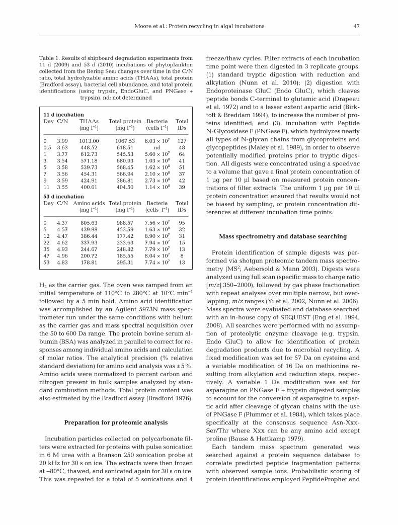
11 d incubationDay C/N THAAs Total protein Bacteria Total
(mg l−1) (mg l−1) (cells l−1) IDs
0 3.99 1013.00 1067.53 6.03 × 10⁷ 1270.5 3.63 448.52 618.51 nd 481 3.77 612.73 545.53 5.60 × 10⁷ 643 3.54 571.18 680.93 1.03 × 10⁸ 415 3.58 539.73 568.45 1.62 × 10⁸ 517 3.56 454.31 566.94 2.10 × 10⁸ 379 3.59 424.91 386.81 2.73 × 10⁸ 4211 3.55 400.61 404.50 1.14 × 10⁸ 39
53 d incubationDay C/N Amino acids Total protein Bacteria Total
(mg l−1) (mg l−1) (cells l−1) IDs
0 4.37 805.63 988.57 7.56 × 10⁷ 955 4.57 439.98 453.59 1.63 × 10⁸ 3212 4.47 386.44 177.42 8.90 × 10⁷ 3122 4.62 337.93 233.63 7.94 × 10⁷ 1535 4.93 244.67 248.82 7.79 × 10⁷ 1347 4.96 200.72 185.55 8.04 × 10⁷ 853 4.83 178.81 295.31 7.74 × 10⁷ 13
Table 1. Results of shipboard degradation experiments from11 d (2009) and 53 d (2010) incubations of phytoplanktoncollected from the Bering Sea: changes over time in the C/Nratio, total hydrolyzable amino acids (THAAs), total protein(Bradford assay), bacterial cell abundance, and total proteinidentifications (using trypsin, EndoGluC, and PNGase +
trypsin). nd: not determined

Mar Ecol Prog Ser 515: 45–59, 2014
ProteinProphet (Keller et al. 2002, Nesvizhskii et al.2003) with thresholds set at 90% confidence levels onPeptideProphet and ProteinProphet for positive pro-tein identifications from SEQUEST search results.Mass spectra from all samples were searched againsta database containing the proteomes of Thalassio -sira pseudonana (marine diatom), Prochlorococcusmarinus (marine cyanobacterium), and CandidatusPelagibacter ubique (marine bacterium belonging tothe SAR11 clade). These proteomes were selected tofollow protein degradation in a diatom dominatedsystem with potential input of bacterial proteins (e.g.Nunn et al. 2010). Results of database comparisonstudies showed functional agreement for over 95%of identified peptides between the T. pseudonana -P. marinus - Ca. P. ubique database versus the largerNCBI non-redundant database containing over 11million protein sequences (Moore et al. 2012b). Tobetter compare shipboard incubations with previousfield observations, non-metric multidimensional scal-ing (NMDS, theory and applications described inBorg & Groenen 2005), was performed using R statis-tical software to group incubation time points withBering Sea water column particles and surface sedi-ments from a previous study (Moore et al. 2012a),based on the distribution of identified proteins ineach sample. Suspended water column particles,sinking sediment trap material, and surface sedi-ments analyzed by Moore et al. (2012a) were col-lected before, during, and after the spring 2009phytoplankton bloom and analyzed using the sameproteomic methods as samples from experimentalincubations.
RESULTS
Algal proteins, identified as originating from dia -toms resembling those of the proteome from Thalas-siosira pseudonana, were detected throughout bothincubations for as long as 53 d (Table 1, Fig. 1A). Inthe 11 d incubation, there was a rapid loss amongidentifiable proteins in the first 0.5 d after the onset ofdarkness, followed by a slower rate of loss after 1 d.In the longer 53 d incubation, there was a rapid lossof identifiable proteins in the first 5 d. The rate of lossslowed thereafter and the number of identified pro-teins remained relatively constant after 22 d. Similartrends were observed in both incubations for THAAs(Fig. 1B) and total protein (Fig. 1C). The THAA distri-bution was fairly consistent among the 11 and 53 dincubations (Appendix 1), with a sharp drop in ala-nine from the start (0 h) to the 12 h time point in the
11 d incubation, and a spike in glutamic acid/gluta-mine at Day 22 of the 53 d incubation. Total bacterialabundance peaked within the first 9 d of both incu-bations and declined thereafter (Fig. 1D). Despite therapid increase in bacterial abundance, only 3 and 2specific prokaryotic proteins were identified over thecourse of the 11 and 53 d incubations respectively.
Among the suite of proteins observed during thedegradation sequence, chloroplast and secretoryproteins were the 2 major cellular compartments rep-resented by identified proteins in both incubations,with smaller contributions of proteins from the mito-chondria, nucleus, ribosome, and unknown compart-ments (Fig. 2). As the incubation progressed, thesequence coverage of identified proteins generallydecreased in tandem with the number of identified
48
B
A140120100
806040200
1000
800
600
400
200
0
# P
rote
ins
Iden
tifie
dµg
TH
AA
s l–1
Protein identifications
THAAs
3.0
2.5
2.0
1.5
1.0
0.5
0
D
C
0 10 20 30 40 50
µg p
rote
in l–1
Day
Cel
ls l–1
(x10
8 )
Bradford protein concentration
Bacteria cells
1000
800
600
400
200
0
Fig. 1. Changes over time during (H) 11 d (2009) and (J) 53 d(2010) incubations of phytoplankton collected from theBering Sea in (A) total protein identifications; (B) total hydrolyzable amino acids (THAAs); (C) total protein
(Bradford assay); (D) bacterial cell abundance

Moore et al.: Protein recycling in algal incubations 49
Days: 0 0.5 1 3 5 7 9 11
Photosynthesis
Binding
BiosynthesisMetabolism
Oxidoreductase
Proteolysis
Translation
TransportGlycolysis
MetabolismTransport
Histone
Biosynthesis
Translation
Binding
Biosynthesis
CatalysisCell Morphogenesis
DehydrogenaseHeat Shock
Glycolysis
Metabolism
ModificationOxidation Reduction
rRNA ProcessingTubulin
Translation
Transport
UnknownTranslation
Unknown
Chloroplast
Mitochondria
Nucleus
Ribosome
Secretory
Unknown
Binding
ATrypsin
PhotosynthesisOxidation Reduction
TranslationBinding
BiosynthesisMetabolism
Transport
EndoGluCChloroplast
MitochondriaRibosomeSecretoryUnknown
Days: 0 0.5 1 3 5 7 9 11
Fig. 2. Proteins identified in incubations at each time point analyzed, organized by compartment and function, showing trypsinand EndoGluC identifications for (A) 11 d and (B, next page) 53 d incubations. Cellular compartments are differentiated by
color, with individual protein sequence coverage (%) represented by shading, as shown in the key

Mar Ecol Prog Ser 515: 45–59, 201450
Binding
BiosynthesisMetabolism
Oxidoreductase
Photosynthesis
Translation
GlycolysisMetabolism
TransportHistone
Translation
Binding
BiosynthesisCell Morphogenesis
OxidoreductaseHeat Shock
GlycolysisMetabolism
ModificationOxidation Reduction
StructureTranslation
Transport
Unknown
Unknown
Chloroplast
MitochondriaNucleus
Ribosome
Secretory
Unknown
Trypsin
B 0 5 12 22 35 47 53Days:
0–10
10–20
20–30
30–40
>40
Ribo-some
Un-known
Secre-tory
NucleusMito-chondria
Chloro-plast
BindingGlycolysisTransport
EndoGluC
% SequenceCoverage
Cellular Compartment
0 5 12 22 35 47 53Days:
Fig. 2 (continued)

Moore et al.: Protein recycling in algal incubations
proteins. Chloroplast proteins were the most persist-ent, increasing in relative abundance from 44% and48% of total proteins at Day 0 to 74 and 67% in thefinal time points of the 11 and 53 d incubationsrespectively. Conversely, the combined proportion ofbiosynthesis, glycolysis, metabolism, and translationproteins dropped from 38% and 27% at Day 0 to15% and 0% percent at the final time points of the 11and 53 d incubations, respectively.
The most dramatic loss of proteins from a specificfunctional group was observed among translationalproteins. Of the 23 translation proteins identified inDay 0 of the 11 d incubation only 8 were identified intime points beyond Day 0, and none were identifiedafter Day 7. Of the 15 translation proteins identifiedat Day 0 in the 53 d incubation only 3 were identifiedin time points after Day 0, and none were identifiedafter Day 12. Furthermore, only 4 secretory proteins,which might be expected to be more available formicrobial recycling than compartmentalized chloro-plast proteins, were identified after Day 22. Of the 11proteins identified at Day 53 of the long incubation, 7were located in the chloroplast.
The use of PNGase F to lyse glycosidic bondsallowed the identification of several modified pep-tides, which appeared during the most rapid loss oftotal identifiable proteins. Two modified peptideswere observed in the 11 d incubation, and 3 modifiedpeptides were observed in the 53 d incubation. Thesepeptides contained the consensus sequence of Asn-Xxx-Ser/Thr (Table 2). Such modified peptides wereonly observed in the first 5 d of both incubations. Theunmodified tryptic version of the peptide from ATPsynthase CF0 B chain subunit I, an important compo-nent in ATP production, was observed in the samePNGase F + trypsin digest as the modified form atDay 5 of the 53 d incubation (Table 2). Inspection ofthe MS2 fragmentation spectra of each peptide
showed the mass change on b-ions that contained thealtered asparagine for the modified peptide(Fig. 3A,B).
Digestion by EndoGluC also resulted in additionalprotein identifications: 8 in the 11 d incubation and 3in the 53 d incubation (Fig. 2A,B). The use ofEndoGluC on extracted protein yielded 4 additionalidentifications at Days 1, 5, 9, and 11 in the 11 d incu-bation. Over the course of the 53 d incubation nomore than one additional protein was identifiedusing EndoGluC at any time point. Secretory pro-teins made up the majority of additional protein IDsusing the EndoGluC treatment. There were no com-mon protein IDs made using both PNGase F andEndoGluC that were not already identified usingtrypsin alone.
In the NMDS analysis, early incubation timepoints (Days 0 to 11) cluster closely together, asdo later incubation time points (Days 22 to 53).Chl-max particles and sinking sediment trap parti-cles are positioned more closely to initial incubationtime points than later time points in the 11 and53 d incubations. The post-bloom shelf surfacesediment is positioned closer to early incubationtime points while post-bloom basin sediment andover wintered shelf sediment are equidistant tointermediate and later incubation time points. Itshould be emphasized that results of NMDS ana -lysis of shipboard incubations and Bering Sea sus-pended particles, sinking sediment trap materialand surface sediments are qualitative in nature.Samples that are ordinated closer together haveprotein distributions that are more similar thansamples that are ordinated farther apart (Fig. 4).Similar ordinations between incubation time pointsand water particles or sediments could allow ap -proximate time frame estimations to be assigned tosamples in the field.
51
Protein Peptide(s) Day
11 d incubationRuBisCO large subunit IHYLGDDVVLQFGGGTIGHPDGIQAGATAN[115]R 1Predicted protein DLAEIWDN[115]SSPVIVQGGSLR 1
53 d incubationRuBisCO large subunit TALDLWKDISFN[115]YTSTDTADFAETATANR 0ATP synthase CF0 B chain subunit I ALIN[115]ETIQKLEGDLL 5Predicted protein QVVELYTEDGLDRPFFAIVETPGSGN[115]VVR 5
Table 2. Peptides identified with the amidase PNGase F + the proteolytic enzyme trypsin during 11 d (2009) and 53 d (2010) in-cubations of phytoplankton collected from the Bering Sea. Each of these individual peptides contain the 1 Da mass increase onasparagine resulting in the 115 Da aspartic acid (indicated by: N[115]), which represents the presence of an N-glycan chainpost translational modification on the peptide. The protein to which the peptides were correlated, and the first day of the
incubation on which the protein was identified are also given

Mar Ecol Prog Ser 515: 45–59, 2014
DISCUSSION
Differential recycling of algal proteins and preservation
The survival of algal proteins over 53 d of microbialrecycling demonstrates that some fraction of intactproteins can survive microbial recycling long enoughto be exported from the marine water column to sedi-
ments. At a sinking rate of up to 100 m d−1 (Smetacek1985), the incubation period of 53 d is more than ade-quate for algal proteins in a diatom-dominated systemto transit the water column to Bering Sea shelf andbasin sediments. The presence of such proteins insediments was previously observed by Moore et al.(2012a), suggesting that other diatom-dominated eco-systems have the potential for export of identifiableprotein from phytoplankton blooms to the sediment.
52
400 600 800 1000 1200 1400 1600 1800 2000
400 600 800 1000 1200 1400 1600 1800 2000
0
10
20
30
40
50
60
70
80
90
100769.88
1253.50
1124.52
817.40 1425.60627.35
915.43546.17 1011.33 1310.41399.14
1523.66
ATP Synthase CF0 B ChainSubunit I Unmodified Peptide:
ALINETIQKLEGDLL
b13+
b12+
b11+
b10+
b9+
883.27b8+
y8+
787.39
y7+
659.37y6+
y5+
417.04
y4+
A
m/z
0
10
20
30
40
50
60
70
80
90
100770.71
713.83
1254.56
1125.53826.89
1426.60
546.221012.41940.38399.15
1524.25 1610.93
ATP Synthase CF0 B ChainSubunit I Modified Peptide:ALIN[115]ETIQKLEGDLL
417.04
y4+
y5+
659.37y6+
787.50y7+
915.51
y8+
743.94
y13++
b9+
b10+
b11+1311.54b12+
b13+
Rel
ativ
e ab
und
ance
B
Fig. 3. MS2 spectra of ATP SynthaseCF0 B chain subunit I peptideALINETIQKLEGDLL showing pep-tide fragment b- and y-ions of the(A) unmodified and (B) modifiedpeptide. The b-ions 9 to 13 are all1 Da greater in the modified pep-tide mass spectra, representing themodified asparagine, (e.g. unmodi-fied b11 = ALINETIQ KLE; modified
b11 = ALIN[115]ETIQKLE)

Moore et al.: Protein recycling in algal incubations
The path of changes to the suite of identifiablealgal proteins seen over time appears to start withproteome changes within the cells, followed quicklyby cell lysis and then microbial recycling. AfterDay 0, in the 11 d incubation, the decrease in thetotal number of identified proteins and in the pro -portion of biosynthetic, glycolysis, metabolism, andtranslation proteins may represent the shutdown ofcellular activity or onset of lysis among algal cells.This is supported by results showing that the biosyn-thetic, metabolism, and translation proteins in thechloroplast were lost much more rapidly than photo -synthetic proteins in the chloroplast, despite thesame level of membrane protection. These changestook place early in the incubation, before the rapidrise in bacterial abundance at Day 9 (of the 11 d incu-bation), which is consistent with the pattern of dia -tom cellular activity and decline observed by Harveyet al. (1995) in laboratory incubations. This initialdrop in total protein IDs, THAAs, and total proteinmay also be due to cell lysis and/or extracellularenzymes, which would release cytoplasmic proteinsand particulate organic matter into the dissolvedphase (Amon & Benner 1996, Vetter & Deming 1999).Particulate sampling, coupled with potential hetero-geneity of carboy material, would then miss thisnewly dissolved material as observed at 0.5 d.
The observed loss of identified translation proteinsat early time points likely indicates the shutdown ofprotein synthesis, as algal cells stop growth undersimulated bloom termination, and undergo internalprotein recycling. The overall loss of identified cyto-plasm proteins was more rapid than the loss of
chloroplast proteins, but identified translation pro-teins located in the chloroplast were lost just as rap-idly as translation proteins located in the ribosome orcytoplasm. In previous studies it has been observedthat proteins encapsulated in organic matter or cellu-lar compartments are preferentially preserved dur-ing microbial recycling (Nguyen et al. 2003, Nunn etal. 2010, Moore et al. 2012a). Cell lysis may haveplayed a role in the loss of cytoplasmic translationproteins, but the rapid loss of identifiable translationproteins with additional membrane protection in cel-lular compartments indicates that some loss was dueto internal recycling within the cell in tandem withmicrobial activity.
After initial cellular recycling, further changes toprotein distribution were observed during microbialrecycling as biosynthesis, glycolysis, metabolism, andtranslation proteins continued to decline until Day 22and were no longer detectable at the conclusion of the53 d incubation. However abundant photo syntheticproteins persisted and remained de tectable after 53 dof degradation (Fig. 2). After 53 d, 5 and 3 peptideswere identified from the RuBisCO large and smallsubunits respectively, contrary to previous findingsthat RuBisCO was degraded rapid ly and not retainedin a lab based degradation of Thalassiosira pseudo-nana (Nunn et al. 2010). Two fucoxanthin chlorophyllbinding proteins (FCPs) were identified throughoutthe 53 d incubation in agreement with findingsthat these proteins re main identifiable for extendedperiods in lab degradation (Nunn et al. 2010), BeringSea shelf sediments (139 m), and Bering Sea basinsediments (3490 m) (Moore et al. 2012a).
53
Fig. 4. Non-metric multidimensional scaling(NMDS) ordination based on the proteindistribution of Bering Sea water columnand sediment samples (Moore et al. 2012a)and the time points of the 11 d and 53 d in-cubations: P4: Chl Max suspended parti-cles; P50: 50 m suspended particles; P100:100 m suspended particles; T40: 40 m sedi-ment trap; T60: 60 m sediment trap; T100:100 m sediment trap; PBS: Post-bloom shelfsediment; PBB: Post-bloom basin sediment;OWS: Overwintered shelf sediment; S0,S0.5, S1, S3, S5, S7, S9, S11: 11 d incubationon Days 0, 0.5, 1, 3, 5, 7, 9 and 11, respec-tively; L0, L5, L12, L22, L35, L47, L53: 53 dincubation on Days 0, 5, 12, 22, 35, 47 and
53, respectively

Mar Ecol Prog Ser 515: 45–59, 2014
Microbial input
At early time points in both incubations, proteinIDs, THAAs, and total protein decreased followed byin creases in bacterial cell numbers. The increase inbacterial abundance was first apparent at Day 3 ofthe 11 d incubation and continued until Day 9, indi-cating that there is a slight lag in cell replication afterthe initial recycling of particulate THAAs, total pro-tein measured spectrophotometrical ly, and identifi-able proteins (Fig. 1). Protein IDs, THAAs, and totalprotein subsequently decrease more slowly afterbacterial cells return to original levels (Fig. 1). Graz-ing by bacterivores, such as flagellates, protists, andviral lysis (Steward et al. 1996, Vaque et al. 2008)may have decreased bacteria cell counts after initialbacterial proliferation. This would limit overall bacte-ria-driven protein recycling in later incubation timepoints. Despite this, bacteria in these incubationsappear to be the primary protein recyclers given thatmacrozooplankton were excluded in these incuba-tions. Using an average bacterial protein content percell of 24 fg cell−1 (Zubkov et al. 1999), the estimatedbacterial fraction of total protein and THAAs washighest at or near the peak bacteria cell abundancein both the 11 and 53 d incubations (Table 1), butnever exceeded 2% of total protein or THAAsthroughout the incubations. This low value for whatis also expected to be a large suite of different bacte-rial protein types may explain why identifiable pro-teins were dominated by diatom sources and fewprokaryotic proteins were observed.
Identifying modified proteins
While difficulties are common in recognizing pro-tein modifications, identification of such modifi cationsby proteomic methods has the potential to more fullycharacterize degraded and modified proteins in envi-ronmental samples. Non-enzymatic glycation is a ma-jor cause of spontaneous damage to cellular and ex-tracellular proteins in physiological systems (Ahmed& Thornalley 2007). Glycation of RuBisCO has beenshown to decrease the enzyme’s activity, and increaseits susceptibility to proteases (Yamauchi et al. 2002).Various mechanisms have been observed in higherplants (Kim & Kim 2003, Sultana et al. 2009) and themicroalga Chlorella zofingiensis (Sun et al. 2011) toprevent and repair protein glycation. Thus, the poten-tial glycation of RuBisCO and other proteins (Table 2)in the early stages of recycling may represent proteinturnover within the cell.
Glycation has also been hypothesized as a mecha-nism for extra-cellular protein preservation and theformation of sedimentary geopolymers (Collins et al.1992, Burdige 2007). Mass spectral analysis withInsPecT (Tanner et al. 2005) was used to identifyrecurring modification masses on precursor ions,revealing potential sugar modifications to proteinsfrom degraded phytoplankton (Nunn et al. 2010). Allmodified peptides in this study were observed duringthe early stages of both incubations while the num-ber of observed biosynthesis, glycolysis, metabolism,and translation proteins decreased. While the 5 dtime period over which modified peptides were seendoes not allow unequivocal evidence that the ob -served peptide modifications and proteome changeswere directly linked to cell death, (e.g. Peters &Thomas 1996), such changes may be the initial stepsthat take place before dormancy and eventually celldeath (Bidle & Falkowski 2004, Nunn et al. 2010).
Identifiable protein correlates to organic matterdegradation proxies
The ratios of THAA/OC and THAA-N/PN can beuseful proxies for degradation status of organic mat-ter (Cowie & Hedges 1994 and references therein).Plotting the number of identified proteins againstthe ratio of THAA/OC and THAA-N/ PN (Fig. 5),reveals that proteins through out the 11 d incubationand the 3 earliest time points of the 53 d incubation(Days 0, 5, 12) have grea ter numbers of protein IDsand higher ratios of THAA/OC and THAA-N/PNthan samples over longer incubation times of the 53 dincubation (Days 22, 35, 47, 53), which cluster to -gether more closely. The shift in the number of iden-tified proteins in the 11 d incubation and early timepoints of the 53 d incubation to the cluster of 4 finaltime points of the 53 d incubation suggests that, afterinitial rapid de gradation, identifiable protein mate-rial degrades much more slowly.
There are significant correlations between thenumber of protein identifications and both the ratiosof THAA/OC and THAA-N/PN (Fig. 5) as observedin suspended particles, sinking material, and surfacesediments in the Bering Sea (Moore et al. 2012a). Incomparing our shipboard incubations to field col-lected material from the same location (Moore et al.2012a), it is striking that the number of identified pro-teins and ratios of THAA/OC and THAA-N/PN forincubations times of 22, 35, 47, and 53 d were verysimilar to those seen in deep basin sediments(3490 m), over wintered shelf sediments (101 m), and
54

Moore et al.: Protein recycling in algal incubations
suspended particles (50 m, 100 m) of the Bering Sea.Although this finding suggests that the rapid proteinlosses seen in shipboard incubations are realistic, italso supports findings that intact proteins contributeto a fraction of marine sedimentary amino acids (Pan-toja & Lee 1999, Nguyen & Harvey 2001, Nunn et al.2010, Moore et al. 2012a). The distribution of remain-ing proteins observed after 22 d also indicates thatabundant chloroplast proteins make up the majorityof identified preserved proteins.
Amino acids are known to be important contribu-tors to particulate and sedimentary marine nitrogenpools (Burdige & Martens 1988, Lee et al. 2000).Analysis by 15N nuclear magnetic resonance (NMR)has also shown that most organic nitrogen present indissolved and particulate organic matter is amidelinked, like the bonds that connect amino acids inproteins (McCarthy et al. 1997, Zang et al. 2001). Thecorrelations between protein IDs, THAA/OC andTHAA-N/PN, and the longevity of identifiable pro-tein observed in this study, and in findings by Mooreet al. (2012a) identifying algal proteins in Bering Seasinking sediment trap material and sediments, sug-gest that diatom dominated systems are importantregions of organic nitrogen and organic carbon ex -port. These studies also show promise for proteomiccharacterization of amide linked nitrogen or protein
material measured as THAAs throughout sedimentsof the global ocean.
Statistical analysis of incubations with environmental samples
Inspection of the identified protein pool amongsediment samples and later incubation time pointswith NMDS, taking into account protein function,shows evidence that the distribution of proteins identified in post-bloom shelf sediment (Moore et al.2012a) matches more closely with incubation timepoints from 0.5 to 12 d than incubation time pointsafter 22 d (Fig. 4). Proteins identified at later timepoints of the 53 d incubation were also identified inpost bloom shelf sediment, but they make up only23% of identifications. The distribution of identifiedproteins at time points after 22 d of the 53 d incuba-tion matches more closely with post-bloom basin sediment and overwintered shelf sediment in whichproteins identified after 22 d make up 38% and 52%of identified proteins, respectively. The distributionof common proteins between later time points, post-bloom basin, and overwintered shelf sediment in -cludes primarily chlorophyll binding proteins, photo-system II proteins, and DNA binding histones. This
55
140
120
100
80
60
40
20
0
140
120
100
80
60
40
20
00 0.05 0.10 0.15 0.20 0.25 0.350.30 0.40
0 0.10 0.20 0.30 0.40 0.50
B
A
Long Incubation Days22, 35, 47, 53
Long Incubation Days22, 35, 47, 53
THAA/OC
THAA/OC vs. IDs
# P
rote
in ID
s
THAA-N/PN
THAA-N/PN vs. IDs
y = 250.02x–14.14R2 = 0.73p ≤ 0.0001
y = 305.29x–17.45R2 = 0.76p ≤ 0.0001
Fig. 5. Plots of total protein identificationsplotted against the organic matter degrada-tion status parameters, for (e) 11 d and (R)53 d incubations, showing fraction of (A) organic carbon made up by total hydro -lyzable amino acids (THAA/OC); and (B)particulate nitrogen made up by total hy-drolyzable amino acid nitrogen (THAA-N/PN). Linear regressions represent thecor relation between identifiable proteins
and organic matter recycling

Mar Ecol Prog Ser 515: 45–59, 2014
provides further evidence that protein materialwhich lasts beyond the apparent 22 d threshold rep-resents material that can be preserved on seasonaltimescales and be available as a food source to thebenthic community, not only during the productivespring bloom, but the ice-covered winter months aswell. The observation that 50 and 100 m suspendedparticles are positioned closer to later time pointsthan post-bloom shelf sediment illustrates the impor-tance of export to sediments towards re moval fromwater column recycling and protein pre servation.
Implications for benthic communities and thenitrogen cycle
Given the reduced impact of microbial recyclingfollowing 22 d of incubation on protein IDs, THAAs,total protein, and the similar ratios seen in THAA/OCand THAA-N/PN after 22 d with Bering Sea sedi-ments, it is reasonable to consider that incubationmaterial after 22 d is representative of primary pro-duction arriving at the sea floor after bloom termina-tion. Approximately 42% of THAAs re main after 22 dof microbial recycling compared to initial amounts inthe 53 d incubation. This is a substantial fraction oforganic nitrogen available to fuel benthic activity.Therefore, the transport of algal proteins from thewater column to sediments indicates that primaryproduction is an important organic nitrogen source tobenthic communities. Recycling by consumers mayreduce the amount of identifiable proteins in sinkingsediment trap material compared to bloom materialas observed by Moore et al. (2012a), but grazingimpact is reduced during periods of high production(Chen et al. 2003, Lovvorn et al. 2005). This agreeswith previous findings that production from thewater column supports high benthic faunal density inthe Bering Sea (Grebmeier et al. 1988).
The contribution of intact protein to marine sedi-ments may also explain why similar δ15N values havebeen observed between water column material andsurface sediments from the Bering Sea shelf duringice covered winter months (Lovvorn et al. 2005), andin time series samples from coastal upwelling regionsof the NE Pacific (Altabet et al. 1999). Furthermore,the longevity of exported phytoplankton proteins tothe continental shelf and basin may be important inproviding a food source to benthic filter feeders dur-ing the ice-covered low production winter monthswhen new material is not being actively exported tosediments. This suggests that protein preservationplays a role in the yearlong ecosystem support of
Bering Sea, and potentially other high latitude coas -tal, benthic communities.
Global calculations of marine nitrogen fixation anddenitrification have estimated a fixed nitrogen deficitof roughly 200 Tg N yr−1 (Mahaffey et al. 2005, Codis-poti 2007). Sedimentary δ15N records however, havesuggested global N cycle homeostasis during theHolocene (Deutsch et al. 2004, Altabet 2007). A morerecent study by Großkopf et al. (2012) has shown thata widely used method for measuring N2 fixation ratesunderestimates the contribution of nitrogen fixingdiazotrophs compared to a newly developed method.This results in a nitrogen fixation increase of 103 to177 ± 8 Tg N yr−1 leaving a remaining fixed nitrogendeficit of 23 to 97 Tg N yr−1. The 2 yr spring summerstudy by Horak et al. (2013) suggested that denitrifi-cation rates in Bering Sea shelf sediments are higherthan previously thought, and that sediment regener-ated NH4
+ is an insignificant contribution to watercolumn dissolved inorganic nitrogen (DIN) and pri-mary production. Coupled nitrification-denitrifica-tion observed by Granger et al. (2011) has also indi-cated that N loss occurring in Bering Sea sediments isgreater than other regions of the ocean and that off-shelf waters potentially contribute nitrate to the outershelf. This study presents evidence suggesting thatexported protein material from the Bering Sea watercolumn to sediments, where it is denitrified, may alsobe an important potential pathway for loss of fixednitrogen transported to the Bering Sea from off shelf,thus contributing to the fixed nitrogen deficit.
CONCLUSIONS
The persistence of algal proteins during both short-term and extended shipboard incubation degrada-tion experiments indicates that some fraction of thismaterial can survive water column recycling in time-frames that are amenable to being transported tomarine sediments. The initial changes to the distribu-tion of identified translation, biosynthesis, glycolysis,and metabolism proteins indicate that internal cellu-lar recycling plays an important role in proteincycling during the early time points of simulatedbloom termination. Glycation modifications wereonly observed during the first 5 d of the 2 incuba-tions, suggesting these modifications may be linkedwith the shutdown of cellular machinery. Continuedrapid loss of secretory proteins during the period ofbacterial cell population growth represents themicro bial recycling of bioavailable proteins that arenot encapsulated in cellular compartments and re -
56

Moore et al.: Protein recycling in algal incubations
leased during cell lysis. The number of identifiableproteins, THAAs, total protein, and the ratios ofTHAA/OC and THAA/PN declined more slowly after22 d. Identifiable proteins that survived after the ob -served 22 d threshold appear to represent materialthat can be preserved over seasonal timescales insediments, and be available as a food source to thebenthic community. The large fraction of remainingTHAAs after initial microbial recycling, and correla-tions between identifiable proteins and the ratios ofTHAA/OC and THAA/PN, suggest that identifiablealgal proteins make up an important fraction of mar-ine sedimentary organic nitrogen. The observedlongevity of identifiable proteins during simulatedbloom termination and microbial recycling suggeststhat diatom dominated systems are important regionsfor organic nitrogen export and potential preserva-tion in sediments.
Acknowledgements. This work was supported by NationalScience Foundation (NSF) grant through the ChemicalOceanography Program to H.R.H. (OCE-0825811), D.R.G.and B.L.N. (OCE-0825790, OCE-1233014) with samples ob -tained as part of the NSF Bering Ecosystem Study Program(BEST) to H.R.H. (ARC-0732667). Computational analysiswas conducted on National Institute of Health sponsoredcomputer cluster awards to D.R.G. (1S10RR023044-01,1U54AI57141-01). We thank Rachel Pleuthner for help con-ducting the degradation experiments and Megan Bernhardtfor preparation of microscope slides. This is MOGEL-OEAScontribution No. 2011-3 of Old Dominion University.
LITERATURE CITED
Aebersold R, Mann M (2003) Mass spectrometry-based pro-teomics. Nature 422: 198−207
Ahmed N, Thornalley PJ (2007) Advanced glycation end-products: What is their relevance to diabetic complica-tions? Diabetes Obes Metab 9: 233−245
Altabet MA (2007) Constraints on oceanic N balance/imbal-ance from sedimentary 15N records. Biogeosciences 4: 75−86
Altabet MA, Pilskaln C, Thunell R, Pride C, Sigman D,Chavez F, Francois R (1999) The nitrogen isotope biogeo-chemistry of sinking particles from the margin of theEastern North Pacific. Deep-Sea Res I 46: 655−679
Amon RMW, Benner R (1996) Bacterial utilization of differ-ent size classes of dissolved organic matter. LimnolOceanogr 41: 41−51
Banahan S, Goering J (1986) The production of biogenic sil-ica and its accumulation on the southeastern Bering Seashelf. Cont Shelf Res 5: 199−213
Bause E, Hettkamp H (1979) Primary structural require-ments for N-glycosylation of peptides in rat liver. FEBSLett 108: 341−344
Bidle KD, Falkowski PG (2004) Cell death in planktonic,photosynthetic, microorganisms. Nat Rev Microbiol 2: 643−655
Birktoft JJ, Breddam K (1994) Proteolytic enzymes: serineand cysteine peptidases. In: Barrett AJ (ed) Methods inenzymology, Vol. 244. Academic Press, San Diego, CA,p 114−126
Borg I, Groenen P (2005) Modern multidimensional scaling: theory and applications, 2nd edn. Springer, New York,NY, p 201−212
Bradford MM (1976) A rapid sensitive method for the quan-titation of microgram quantities of protein utilizing theprinciple of protein-dye binding. Anal Biochem 72: 248−254
Burdige DJ (2007) Preservation of organic matter in marinesediments: controls, mechanisms, and an imbalance insediment organic carbon budgets? Chem Rev 107: 467−485
Burdige DJ, Martens CS (1988) Biogeochemical cycling inan organic-rich coastal marine basin: 10. The role ofamino-acids in sedimentary carbon and nitrogen cycling.Geochim Cosmochim Acta 52: 1571−1584
Chen M, Huang YP, Cai PG, Guo LD (2003) Particulateorganic carbon export fluxes in the Canada Basin andBering Sea as derived from 234Th/238U disequilibria. Arc-tic 56: 32−44
Chivasa S, Tome DFA, Hamilton JM, Slabas AR (2011) Pro-teomic analysis of extracellular ATP-regulated proteinsidentifies ATP synthase beta-subunit as a novel plant celldeath regulator. Mol Cell Proteomics 10: M110.003905
Choi HW, Kim YJ, Hwang BK (2011) The hypersensitiveinduced reaction and leucine-rich repeat proteins regu-late plant cell death associated with disease and plantimmunity. Mol Plant Microbe Interact 24: 68−78
Codispoti LA (2007) An oceanic fixed nitrogen sink exceed-ing 400 Tg N a−1 vs the concept of homeostasis in thefixed-nitrogen inventory. Biogeosciences 4: 233−253
Collins MJ, Westbroek P, Muyzer G, Deleeuw JW (1992)Experimental-evidence for condensation-reactions be -tween sugars and proteins in carbonate skeletons.Geochim Cosmochim Acta 56: 1539−1544
Cowie GL, Hedges JI (1992) Sources and reactivities ofamino acids in a coastal marine environment. LimnolOceanogr 37: 703−724
Cowie GL, Hedges JI (1994) Biochemical indicators of dia-genetic alteration in natural organic matter mixtures.Nature 369: 304−307
Deutsch C, Sigman DM, Thunell RC, Meckler AN, Haug GH(2004) Isotopic constraints on glacial/interglacialchanges in the oceanic nitrogen budget. Global Biogeo -chem Cycles 18: GB4012
Drapeau GR, Boily Y, Houmard J (1972) Purification andproperties of an extracellular protease of Staphylococcusaureus. J Biol Chem 247: 6720−6726
Eng JK, McCormack AL, Yates JR (1994) An approach tocorrelate tandem mass spectral data of peptides withamino-acid-sequences in a protein database. J Am SocMass Spectrom 5: 976−989
Eng JK, Fischer B, Grossmann J, MacCoss MJ (2008) A fastSEQUEST cross correlation algorithm. J Proteome Res 7: 4598−4602
Granger J, Prokopenko MG, Sigman DM, Mordy CW andothers (2011) Coupled nitrification-denitrification in sed-iment of the eastern Bering Sea shelf leads to 15N enrich-ment of fixed N in shelf waters. J Geophys Res 116: C11006, doi: 10.1029/2010JC006751
Grebmeier JM, McRoy CP, Feder HM (1988) Pelagic-ben-thic coupling on the shelf of the Northern Bering and
57
Mar Ecol Prog Ser 515: 45–59, 201458
Chukchi Seas. 1. Food supply source and benthic bio-mass. Mar Ecol Prog Ser 48: 57−67
Großkopf T, Mohr W, Baustian T, Schunck H and others(2012) Doubling of marine dinitrogen-fixation ratesbased on direct measurements. Nature 488: 361−364
Harvey HR, Tuttle JH, Bell JT (1995) Kinetics of phytoplank-ton decay during simulated sedimentation: changes inbiochemical composition and microbial activity underoxic and anoxic conditions. Geochim Cosmochim Acta59: 3367−3377
Horak REA, Whitney H, Shull DH, Mordy CW, Devol AH(2013) The role of sediments on the Bering Sea shelf Ncycle: insights from measurements of benthic denitrifica-tion and benthic DIN fluxes. Deep-Sea Res II 94: 95−105
Jamers A, Blust R, De Coen W (2009) Omics in algae: pavingthe way for a systems biological understanding of algalstress phenomena? Aquat Toxicol 92: 114−121
Keller A, Nesvizhskii AI, Kolker E, Aebersold R (2002)Empirical statistical model to estimate the accuracy ofpeptide identifications made by MS/MS and databasesearch. Anal Chem 74: 5383−5392
Kim HY, Kim K (2003) Protein glycation inhibitory andantioxidative activities of some plant extracts in vitro.J Agric Food Chem 51: 1586−1591
Lee C, Wakeham SG, Hedges JI (2000) Composition andflux of particulate amino acids and chloropigments inequatorial Pacific seawater and sediments. Deep-SeaRes I 47: 1535−1568
Lomas MW, Moran SB, Casey JR, Bell DW and others (2012)Spatial and seasonal variability of primary production onthe Eastern Bering Sea shelf. Deep-Sea Res II 65−70: 126−140
Lovvorn JR, Cooper LW, Brooks ML, De Ruyck CC, BumpJK, Grebmeier JM (2005) Organic matter pathways tozooplankton and benthos under pack ice in late winterand open water in late summer in the north-centralBering Sea. Mar Ecol Prog Ser 291: 135−150
Mahaffey C, Michaels AF, Capone DG (2005) The conun-drum of marine N2 fixation. Am J Sci 305: 546−595
Maley F, Trimble RB, Tarentino AL, Plummer TH Jr (1989)Characterization of glycoproteins and their associatedoligosaccharides through the use of endoglycosidases.Anal Biochem 180: 195−204
McCarthy M, Pratum T, Hedges J, Benner R (1997) Chemi-cal composition of dissolved organic nitrogen in theocean. Nature 390: 150−154
McRoy CP, Hansell DA, Springer AM, Walsh JJ, WhitledgeTE (1987) Global maximum of primary production in theNorth Bering Sea. Eos 68: L1727
Moore EK, Nunn BL, Goodlett DR, Harvey HR (2012a)Identi fying and tracking proteins through the marinewater column: insights into the inputs and preservationmechanisms of protein in sediments. Geochim Cosmo -chim Acta 83: 324−359
Moore EK, Nunn BL, Faux JF, Goodlett DR, Harvey HR(2012b) Evaluation of electrophoretic protein extractionand database-driven protein identification from marinesediments. Limnol Oceanogr Methods 10: 353−366
Moran SB, Lomas MW, Kelly RP, Gradinger R, Iken K,Mathis JT (2012) Seasonal succession of net primary pro-ductivity, particulate organic carbon export, and auto-trophic community composition in the eastern BeringSea. Deep-Sea Res II 65−70: 84−97
Nesvizhskii AI, Keller A, Kolker E, Aebersold R (2003) A sta-tistical model for identifying proteins by tandem mass
spectrometry. Anal Chem 75: 4646−4658Nguyen RT, Harvey HR (2001) Protein preservation in mar-
ine systems: hydrophobic and other non-covalent associ-ations as major stabilizing forces. Geochim CosmochimActa 65: 1467−1480
Nguyen RT, Harvey HR, Zang X, van Heemst JDH, HetenyiM, Hatcher PG (2003) Preservation of algaenan and pro-teinaceous material during the oxic decay of Botryo -coccus braunii as revealed by pyrolysis-gas chromato -graphy/mass spectrometry and 13C NMR spectroscopy.Org Geochem 34: 483−497
Nunn BL, Shaffer SA, Scherl A, Gallis B, Wu M, Miller SI,Goodlett DR (2006) Comparison of a Salmonella typhi -murium proteome defined by shotgun proteomicsdirectly on an LTQ-FT and by proteome pre-fractiona-tion on an LCQ-DUO. Brief Funct Genomics Proteomics5: 154−168
Nunn BL, Ting YS, Malmstroem L, Tsai YS, Squier A,Goodlett DR, Harvey HR (2010) The path to preservation: using proteomics to decipher the fate of diatom proteinsduring microbial degradation. Limnol Oceanogr 55: 1790−1804
Pantoja S, Lee C (1999) Molecular weight distribution ofproteinaceous material in Long Island Sound sediments.Limnol Oceanogr 44: 1323−1330
Peters E, Thomas DN (1996) Prolonged darkness and diatommortality I. Marine Antarctic species. J Exp Mar Biol Ecol207: 25−41
Plummer TH, Elder JH, Alexander S, Phelant AW, TarentinoAL (1984) Demonstration of peptide: N-glycosidase Factivity in endo-beta-N-acetylglucosaminidase F prepa-rations. J Biol Chem 259: 10700−10704
Sambrotto RN, Niebauer HJ, Goering JJ, Iverson RL (1986)Relationships among verticle mixing, nitrate uptake, andphytoplankton growth during spring bloom in the south-east Bering Sea. Cont Shelf Res 5: 161−198
Silvestre F, Gillardin V, Dorts J (2012) Proteomics to assessthe role of phenotypic plasticity in aquatic organismsexposed to pollution and global warming. Integr CompBiol 52: 681−694
Smetacek VS (1985) Role of sinking in diatom life-historycycles: ecological, evolutionary, and geological signifi-cance. Mar Biol 84: 239−251
Springer AM, McRoy CP, Flint MV (1996) The Bering SeaGreen Belt: shelf-edge processes and ecosystem pro -duction. Fish Oceanogr 5: 205−223
Steward GF, Smith DC, Azam F (1996) Abundance and pro-duction of bacteria and viruses in the Bering andChukchi Seas. Mar Ecol Prog Ser 131: 287−300
Sultana N, Choudhary MI, Khan A (2009) Protein glycationinhibitory activities of Lawsonia inermis and its activeprinciples. J Enzyme Inhib Med Chem 24: 257−261
Sun Z, Liu J, Zeng X, Huangfu J, Jiang Y, Wang M, Chen F(2011) Astaxanthin is responsible for antiglycoxidativeproperties of microalga Chlorella zofingiensis. FoodChem 126: 1629−1635
Tanner S, Shu HJ, Frank A, Wang LC and others (2005)InsPecT: identification of posttranslationally modifiedpeptides from tandem mass spectra. Anal Chem 77: 4626−4639
Vaque D, Guadayol O, Peters F, Felipe J and others (2008)Seasonal changes in planktonic bacterivory rates underthe ice-covered coastal Arctic Ocean. Limnol Oceanogr53: 2427−2438
Vetter YA, Deming TW (1999) Growth rates of marine bacte-

Moore et al.: Protein recycling in algal incubations 59
rial isolates on particulate organic substrates solubilizedby freely released extracellular enzymes. Microb Ecol37: 86−94
Waldhier MC, Dettmer K, Gruber MA, Oefner PJ (2010)Comparison of derivatization and chromatographicmethods for GC-MS analysis of amino acid enantiomersin physiological samples. J Chromatogr B 878: 1103−1112
Wiese FK, Wiseman Jr. WJ, Van Pelt TI (2012) Bering Sealinkages. Deep-Sea Res II 65−70: 2−5
Yamauchi Y, Ejiri Y, Tanaka K (2002) Glycation by ascorbicacid causes loss of activity of ribulose-1,5-bisphosphatecarboxylase/oxygenase and its increased susceptibilityto proteases. Plant Cell Physiol 43: 1334−1341
Yi EC, Marelli M, Lee H, Purvine SO, Aebersold R, AitchisonJD, Goodlett DR (2002) Approaching complete per -oxisome characterization by gas-phase fractionation.Electrophoresis 23: 3205−3216
Zang X, Nguyen RT, Harvey HR, Knicker H, Hatcher PG(2001) Preservation of proteinaceous material during thedegradation of the green alga Botryococcus braunii: asolid-state 2D 15N 13C NMR spectroscopy study. GeochimCosmochim Acta 65: 3299−3305
Zubkov MV, Fuchs BM, Eilers H, Burkill PH, Amann R(1999) Determination of total protein content of bacterialcells by SYPRO staining and flow cytometry. Appl Envi-ron Microbiol 65: 3251−3257
11 d incubation Day0 0.5 1 3 5 7 9 11
Alanine 14.92 (0.33) 9.80 (0.18) 9.63 (0.12) 8.55 (0.46) 8.52 (0.17) 9.48 (0.06) 10.08 (0.77) 9.60 (0.16)Glycine 7.37 (0.09) 7.83 (0.05) 8.35 (0.13) 8.42 (0.13) 8.30 (0.19) 8.96 (0.12) 8.86 (0.17) 9.48 (0.11)Valine 6.93 (0.33) 5.42 (0.11) 5.37 (0.08) 5.84 (0.15) 5.69 (0.08) 5.83 (0.10) 5.55 (0.08) 5.74 (0.12)Leucine 6.56 (0.17) 6.32 (0.05) 6.59 (0.01) 6.64 (0.09) 6.48 (0.10) 6.83 (0.04) 6.69 (0.01) 6.62 (0.09)Isoleucine 4.35 (0.16) 4.60 (0.05) 4.49 (0.30) 4.98 (0.10) 4.65 (0.21) 4.35 (0.06) 4.11 (0.16) 4.30 (0.03)Threonine 3.66 (0.25) 7.87 (0.54) 7.29 (0.30) 7.43 (0.35) 8.35 (0.42) 5.93 (0.14) 5.59 (0.32) 6.06 (0.04)Proline 4.42 (0.09) 4.17 (0.03) 4.22 (0.02) 3.81 (0.04) 3.58 (0.10) 4.34 (0.12) 4.21 (0.03) 4.16 (0.07)Aspartic acid/asparagine 5.56 (0.24) 9.53 (0.13) 9.53 (0.10) 10.25 (0.30) 10.07 (0.25) 9.45 (0.05) 9.37 (0.28) 9.37 (0.15)Phenylalanine 2.33 (0.04) 3.13 (0.06) 3.32 (0.01) 3.69 (0.13) 3.49 (0.08) 3.27 (0.06) 3.26 (0.02) 3.25 (0.11)Glutamic acid/glutamine 10.18 (1.40) 12.32 (1.01) 11 (0.16) 10.47 (0.17) 12.22 (0.59) 10.21 (0.87) 11.01 (1.22) 10.31 (0.97)Lysine 3.94 (0.72) 3.18 (0.12) 3.21 (0.11) 3.45 (0.08) 4.53 (0.31) 2.94 (0.38) 3.46 (0.27) 2.81 (0.47)Tyrosine 3.65 (1.92) 1.99 (0.25) 1.22 (0.03) 1.16 (0.13) 2.28 (0.14) 0.68 (0.18) 0.91 (0.13) 0.70 (0.07)
53 d incubation Day0 5 12 22 35 47 53
Alanine 17.93 (1.79) 15.30 (0.51) 15.75 (1.52) 12.87 (0.70) 15.43 (0.27) 15.45 (0.16) 15.30 (0.72)Glycine 13.57 (0.16) 13.24 (0.55) 15.99 (2.47) 11.62 (0.51) 15.37 (0.72) 16.90 (0.29) 17.14 (0.63)Valine 6.18 (0.44) 6.91 (0.39) 5.96 (0.63) 7.72 (0.25) 10.68 (0.68) 11.66 (0.03) 11.26 (0.76)Leucine 9.12 (0.35) 9.26 (0.60) 9.24 (0.92) 7.81 (0.32) 9.21 (0.44) 9.80 (0.18) 9.26 (0.58)Isoleucine 4.37 (0.13) 3.56 (0.23) 4.23 (0.19) 4.28 (0.12) 3.89 (0.03) 3.72 (0.16) 3.53 (0.22)Threonine 3.42 (0.44) 4.18 (0.49) 4.75 (0.67) 7.21 (0.39) 6.70 (0.74) 6.79 (0.48) 6.99 (1.44)Proline 6.04 (0.62) 5.99 (0.20) 7.06 (1.57) 4.51 (0.08) 3.02 (0.49) 2.62 (0.27) 3.13 (0.55)Aspartic acid/asparagine 10.18 (0.88) 10.48 (0.46) 11.39 (0.95) 10.18 (0.59) 4.97 (0.98) 4.88 (0.86) 7.41 (2.08)Phenylalanine 3.69 (0.41) 3.46 (0.28) 3.96 (0.12) 4.16 (0.10) 5.31 (0.21) 5.32 (0.33) 4.98 (0.13)Glutamic acid/glutamine 12.74 (0.57) 14.34 (0.80) 13.63 (3.72) 19.57 (1.41) 12.16 (0.96) 10.61 (0.77) 10.11 (0.28)Lysine 5.43 (0.57) 5.88 (0.44) 4.18 (1.18) 5.11 (0.18) 5.48 (0.71) 4.54 (0.65) 3.63 (0.64)Tyrosine 7.47 (0.35) 7.41 (3.37) 3.87 (1.54) 4.99 (0.38) 7.78 (0.56) 7.69 (1.52) 7.27 (0.95)
Appendix 1. Amino acid distribution (mole %) of phytoplankton derived particulate material from each time point of 11 d and 53 d shipboard incubations of phytoplankton collected from the Bering Sea. Values are means with standard error in parentheses.
Editorial responsibility: Steven Lohrenz, New Bedford, Massachusetts, USA
Submitted: November 11, 2013; Accepted: July 9, 2014Proofs received from author(s): October 27, 2014