protein structure and folding: insights into activity
TRANSCRIPT

Athanasios Yiotakis and Vincent DiveLajeunesse, Sarah Bregant, Robert Thai,Laurent Devel, Fabrice Beau, Evelyne Anne-Sophie Dabert-Gay, Bertrand Czarny, MATRIX METALLOPROTEINASES
OFCONFORMATIONAL VARIABILITY PROBE DEVELOPMENT ANDINSIGHTS INTO ACTIVITY-BASED Modification by a Photoaffinity Probe:Metalloproteinase-12 Covalent Molecular Determinants of MatrixProtein Structure and Folding:
doi: 10.1074/jbc.M805795200 originally published online September 5, 20082008, 283:31058-31067.J. Biol. Chem.
10.1074/jbc.M805795200Access the most updated version of this article at doi:
.JBC Affinity SitesFind articles, minireviews, Reflections and Classics on similar topics on the
Alerts:
When a correction for this article is posted•
When this article is cited•
to choose from all of JBC's e-mail alertsClick here
http://www.jbc.org/content/283/45/31058.full.html#ref-list-1
This article cites 42 references, 12 of which can be accessed free at
by guest on October 16, 2013http://www.jbc.org/Downloaded from by guest on October 16, 2013http://www.jbc.org/Downloaded from by guest on October 16, 2013http://www.jbc.org/Downloaded from by guest on October 16, 2013http://www.jbc.org/Downloaded from by guest on October 16, 2013http://www.jbc.org/Downloaded from by guest on October 16, 2013http://www.jbc.org/Downloaded from by guest on October 16, 2013http://www.jbc.org/Downloaded from by guest on October 16, 2013http://www.jbc.org/Downloaded from by guest on October 16, 2013http://www.jbc.org/Downloaded from by guest on October 16, 2013http://www.jbc.org/Downloaded from by guest on October 16, 2013http://www.jbc.org/Downloaded from

Molecular Determinants of Matrix Metalloproteinase-12Covalent Modification by a Photoaffinity ProbeINSIGHTS INTO ACTIVITY-BASED PROBE DEVELOPMENT AND CONFORMATIONALVARIABILITY OF MATRIX METALLOPROTEINASES*
Received for publication, July 29, 2008, and in revised form, September 2, 2008 Published, JBC Papers in Press, September 5, 2008, DOI 10.1074/jbc.M805795200
Anne-Sophie Dabert-Gay‡, Bertrand Czarny‡, Laurent Devel‡, Fabrice Beau‡, Evelyne Lajeunesse‡, Sarah Bregant‡,Robert Thai‡, Athanasios Yiotakis§, and Vincent Dive‡1
From the ‡CEA, Service d’Ingenierie Moleculaire de Proteines, 152, CE-Saclay, Gif /Yvette 91191, Cedex, France and §Laboratory ofOrganic Chemistry, Department of Organic Chemistry, University of Athens, Panepistimiopolis, Zografou, Athens 15771, Greece
Mass spectroscopy, microsequencing, and site-directedmutagenesis studies have been performed to identify in humanmatrix metalloelastase (hMMP-12) residues covalently modi-fied by a photoaffinity probe, previously shown to be able tocovalently label specifically the active site of matrixmetallopro-teinases (MMPs). Results obtained led us to conclude that pho-toactivation of this probe in complex with hMMP-12 affects asingle residue in human MMP-12, Lys241, through covalentmodification of its side chain � NH2 group. Because x-ray andNMR studies of hMMP-12 indicate that Lys241 side chain ishighly flexible, our data reveal the existence of particular Lys241side-chain conformation in which the � NH2 group pointstoward the photolabile group of the probe, an event explainingthe high levels of cross-linking yield between hMMP-12 and theprobe. Lys241 is not conserved in MMPs, thus differences incross-linking yields observed with this probe between MMPmembers may be linked to the residue variability observed atposition 241 in this family.
Matrix metalloproteases (MMPs)2 belong to a family ofstructurally related extracellular/cell surface-anchored zincendoproteinases able to collectively cleave the protein compo-nent of the extracellular matrix (1, 2). MMPs are considered tobe critical mediators of both normal and pathological tissueremodeling processes (3).MMPs form a group of 23 proteins inhumans containing a catalytic domain belonging to the metz-incin superfamily (4–8). Their overexpression observed in andassociated with various diseases (9), including cancer, arthritis,atherosclerosis, and multiple sclerosis, has stimulated impres-
sive effort over the past 20 years to develop synthetic inhibitorsable to block potently the uncontrolled activity of these poten-tial therapeutic targets (10, 11). Extremely potent inhibitors ofMMPs have been developed, but with the exception of MMP-2(12), MMP-13 (13, 14), and MMP-12 (15), most of these inhib-itors act as broad-spectrum inhibitors of MMPs. Clinical trialsbased on the use of these broad-spectrum inhibitors in patientswith advanced cancers have failed to reach their end points,with severe side effects observed (16). Some reasons for thisfailure, as discussed in recent studies, suggest that some MMPmembers promote tumor progression, but others provide aprotective effect in different stages of cancer progression (17).This highlights the need to use in clinical setting highly selectiveinhibitors able to target only the MMPs responsible for tumorprogression (18–21). Similar trends have been observed in ath-erosclerosis, in which some MMP members have oppositeeffects on plaque stability. In this pathology, MMP-12 has beenreported to promote both lesion and plaque extension (22),justifying our current efforts in developing selective MMP-12inhibitors (15). To further progress on our understanding of thecomplex roles played byMMPs, probes able to detect and iden-tifywhichMMPs are expressed under active forms in a complexbiological environment have recently been developed (23–26).To be informative, such activity-based probes (ABPs) shouldcovalently and exclusively modify the free form of the proteaseactive site, but not the pro-form or the active form in complexwith natural TIMP inhibitors. ABPs for serine and cysteine pro-teinases have been successfully developed by exploiting thepresence of highly conserved nucleophiles in the active site ofthese enzymes and selecting appropriate reactive groups toprovide covalent modification of the targeted enzymes (27, 28).For zinc metalloproteinases, like MMPs, the lack of conservedactive-site nucleophiles requires the use of photolabile groupsto covalently label the enzyme active site (23–26). Followingthis strategy, we recently developed a selectiveMMPprobe thatconsists of a phosphinic peptide core able to block potently andselectively a large set of MMPs, to which an azido photolabilegroup has been grafted onto the inhibitor P1� side chain (probe1, Scheme 1) (26).The choice of the photolabile group and position of its incor-
poration in the inhibitor structure were based on the x-raystructure of phosphinic peptide inhibitor in complex withMMP-9 (29). Based on these structural considerations, the
* This work was supported by funds from the Commissariat a l’Energie Atom-ique and from the European Commission (Grant FP6RDT, CancerDegradome project, LSHC-CT-2003–503297). The costs of publication ofthis article were defrayed in part by the payment of page charges. Thisarticle must therefore be hereby marked “advertisement” in accordancewith 18 U.S.C. Section 1734 solely to indicate this fact.
1 To whom correspondence should be addressed: Tel.: 33-1-6908-2603; Fax:33-1-6908-9071; E-mail: [email protected].
2 The abbreviations used are: MMP, matrix metalloproteinase; hMMP-12,human matrix metalloelastase; Mca, (7-methoxycoumarin-4-yl)acetyl;Dpa, N3-(2,4-dinitrophenyl)-L-2,3-diaminopropionyl; Mca-Mat, Mca-Pro-Leu-Gly-Leu-Dpa-Ala-Arg-NH2; FA, formic acid; ABP, activity-based probe;RP-HPLC, reversed-phase high-performance liquid chromatography;PVDF, polyvinylidene difluoride; ESI, electrospray ionization; MS, massspectrometry; MS/MS, tandem MS.
THE JOURNAL OF BIOLOGICAL CHEMISTRY VOL. 283, NO. 45, pp. 31058 –31067, November 7, 2008© 2008 by The American Society for Biochemistry and Molecular Biology, Inc. Printed in the U.S.A.
31058 JOURNAL OF BIOLOGICAL CHEMISTRY VOLUME 283 • NUMBER 45 • NOVEMBER 7, 2008

azido group of probe 1 is expected to enter into the deep S1�cavity of MMPs, in a position that should favor covalent mod-ification of residues lining this cavity.While probe 1was shownto covalently modify only the active site of MMPs, high varia-tion in cross-linking yield was observed between differentMMPs (26). Indeed, the covalent modification of hMMP-12 byprobe 1 resulted in a cross-linking yield of �45%, whereas thisyield decreased to a few percents for humanMMP-8. To under-stand themolecular determinant of this difference and improvethe design of better ABPs that covalently modified all MMPswith high cross-linking yields, the identification of thehMMP-12 residues covalently modify by probe 1 has been car-ried out.
EXPERIMENTAL PROCEDURES
Chemicals—Commercial reagents were used without addi-tional purification. Buffers and salts were purchased fromSigma. Synthesis and tritium radiolabeling of probe 1were car-ried out as previously reported (26). Mca-Pro-Leu-Gly-Leu-Dpa-Ala-Arg-NH2 (Mca-Mat) was purchased from Novabio-chem and TIMP-1 inhibitor from R&D Systems.Proteins—Synthetic gene encoding the catalytic domain of
the MMP-12 (Met98–Lys266) was obtained from Geneart(Geneart-AG, Germany). This gene was inserted into thepET24a vector, between the NdeI and BamHI site, for expres-sion under the PT7 promoter. A Lys 3 Ala mutant was pro-duced from 5�-ccgtaatgttccccacctacGCatatgttgacatcaacaca-3�and 5�-tgtgttgatgtcaacatatGCGtaggtggggaacattacgg-3� oligo-nucleotides, using the Site-Directed Mutagenesis Kit (Strat-agene). All plasmids were propagated in the Escherichia colistrain XL1-Blue at 37 °C, and all constructions were verified byDNA sequencing using the ABI PRISM 310 Genetic analyzer(Applied Biosystems). Recombinant proteins were expressed inE. coli BL21(DE3 star) cells carrying the MMP-12 catalyticdomain-encoding plasmids. Bacteria were grown at 37 °C in LBmedium supplemented with kanamycin (50 �g/ml). At anabsorbance (600 nm) of 0.6, protein expression was inducedwith 0.5 mM isopropyl-�-thiogalactopyranoside. Five hours
after induction, cells were harvested by centrifugation at5000 � g for 30 min at 4 °C. The pellets were resuspended inbufferA (100mMTris-HCl, pH8.5, 5mMbenzamidinochloride,5 mM 2-mercaptoethanol) and incubated with lysozyme for 1 hat 4 °C. The suspension was then passed through a cell disrup-tion system at 4 °C (Constant Systems Ltd., DaventryNorthants, England) and was then centrifuged at 4 °C for 30min (8000� g). The pelletswerewashed three timeswith bufferB (100 mM Tris-HCl, pH 8.5, 2 M urea, 5 mM 2-mercaptoetha-nol) and dissolved in buffer C (100 mM Tris-HCl, pH 8.5, 8 Murea). Refolding and purification steps were carried out as pre-viously described (30). Refolded proteins were analyzed bySDS-PAGE and found to migrate as a single band. Proteinmolecular weights were determined by electrospray ionization-mass spectrometry and the presence of the Lys3Alamutationwas verified by matrix-assisted laser desorption ionizationtime-of-flight, after tryptic digestion.Enzyme Assays—Enzyme assays and inhibition and titration
experiments were carried out in 50mMTris/HCl buffer, pH 6.8,10 mM CaCl2, at 25 °C, in flat-bottomed 96-well nonbindingsurface plates (Corning-Costar, Schiphol-RijK, The Nether-lands), using the Mca-Pro-Leu-Gly-Leu-Dpa-Ala-Arg-NH2(Mca-Mat) substrate, as described before (15). Fluorescencesignals were monitored using a Fluoroscan Ascent photoncounter spectrophotometer (Thermo-Labsystems) equippedwith a temperature control device and a plate shaker. Titrationexperiments using a calibrated TIMP-1 solution were per-formed to determine the exact enzyme concentration for eachMMP batch: for these experiments, MMP concentrations wereset at �20 nM. Values for kcat/Km were determined from first-order full-time course reaction curves obtained at [S] �� Km([S] � 0.5 mM) and 5 nM final enzyme concentration. Theseprogress curves were monitored by following the increase influorescence at 400 nm (�ex � 340 nm) induced by the cleavageof the Mca-Mat substrate. kcat/Km values were obtained by fit-ting these progress curves (three independent experiments)with the integrated Michaelis-Menten equation by nonlinearregression. The Ki values of probe 1were determined using themethod proposed by Horovitz and Leviski (31).Mass Spectrometry—MS experiments were performed on an
Ion Trap mass spectrometer (Bruker, Esquire-HCT). Ion trapparameters were set as follows: electrospray potential, � 4000V; skimmer voltage, � 40 V; capillary exit, 226 V; and a sourcetemperature of 365 °C. TheMS survey scan wasm/z 400–3000Da with a target mass fixed at m/z 1800 Da and a 5-spectrumscan average.Nano-RP-HPLC—Peptide separation was carried out on an
Ultimate LC Packings/DIONEX UltiMateTM Capillary/Nano-LC System apparatus, using anAcclaim PepMap100 C18,3 �m, 100 Å (150 mm � 75 �m) column at a flow rate of 200nl�min�1 and with detection at 214 nm. Solvent systems were:(A) 95% water, 5% acetonitrile, 0.01% trifluoroacetic acid, and0.04% FA and (B) 5% water, 95% acetonitrile, 0.01% trifluoro-acetic acid, and 0.04% FA. The following gradient was used: t �0min, 100%A; t� 3min, 100%A; t� 73min 70% B; t� 74min100% B; and t � 80 min 100% B.
�-RP-HPLC—Peptide separation was carried out on an Agi-lent 1100 series apparatus and using a X-Bridge (Waters) C18
SCHEME 1
MMP-12 Photoaffinity Probe
NOVEMBER 7, 2008 • VOLUME 283 • NUMBER 45 JOURNAL OF BIOLOGICAL CHEMISTRY 31059
5-�m, 300-Å column (2.1� 150mm, 5�m) at a flow rate of 200�l/min andwith detection at 214 nm. Solvent systemswere: (A)100% water, 0.1% FA and (B) 100% acetonitrile, 0.09% FA. Thefollowing gradient was used: t� 0min, 10%B; t� 5min, 10%B;and t � 45 min 100% B.Photoaffinity Labeling—MMPs (1�M) were incubated with 2
�M of 3H probe 1 in 50 mM Tris/HCl, pH 6.8, 10 mM CaCl2,0.01% Brij35, for 10 min in the dark at room temperature. Thereaction mixture was irradiated using a 1000-watt mercury
lamp (Orsam) for 2 min (10 °C). All irradiation experimentswere performed under inactinic light (sodium light) at 310 nm(50 microwatts�cm�2) on an apparatus equipped with an aper-ture and a series of lenses focusing the polychromatic light to amonochromator (Jobin-Yvon), a series of lenses to focus themonochromatic light to a thermostat support, in which theEppendorf tube containing the mixture to irradiate is inserted.Light intensity was measured with a radiometer IL1700 (Inter-national Light, Newburyport, MA). After irradiation, the reac-
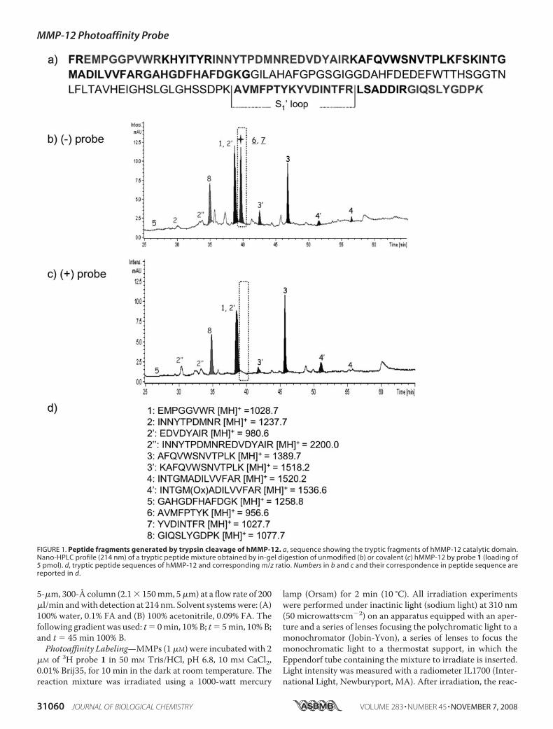
FIGURE 1. Peptide fragments generated by trypsin cleavage of hMMP-12. a, sequence showing the tryptic fragments of hMMP-12 catalytic domain.Nano-HPLC profile (214 nm) of a tryptic peptide mixture obtained by in-gel digestion of unmodified (b) or covalent (c) hMMP-12 by probe 1 (loading of5 pmol). d, tryptic peptide sequences of hMMP-12 and corresponding m/z ratio. Numbers in b and c and their correspondence in peptide sequence arereported in d.
MMP-12 Photoaffinity Probe
31060 JOURNAL OF BIOLOGICAL CHEMISTRY VOLUME 283 • NUMBER 45 • NOVEMBER 7, 2008
tion was quenched by the addition of Laemmli loading bufferfollowed by boiling (5 min, 95 °C). These samples were imme-diately processed for subsequent SDS gel analysis.Electrophoresis—Radiolabeled proteins were diluted in Lae-
mmli loading buffer (final concentration, 0.1% (w/v) bromphe-nol blue, 2% (w/v) SDS, 10% (v/v) glycerol, 50mMTris-HCl, pH6.8, and 100 mM dithiothreitol) and were resolved by SDS-PAGE electrophoresis in a 12% 1 mm-thick SDS gel, using amini-protean III apparatus (Bio-Rad). Silver staining was per-formed as classically described.In-gel Digestion—Gels were stained with Coomassie Blue
R250. Gel pieces containing proteins were excised anddestained by adding 50 �l of 25 mM NH4 HCO3 in 50% aceto-nitrile. After a 10-min incubation with occasional vortexing,the liquid phase was discarded. This procedure was repeatedthree times. Gel peaces were then rinsed (10 min) with aceto-nitrile and dried under vacuum. Gel pieces were reswelled in 25mM NH4 HCO3 buffer containing trypsin (12 ng/�l, modifiedporcine trypsin sequence grade, Promega, Madison, WI). Afterthe trypsin digestion (37 °C, 18 h), the solution was transferredin into an Eppendorf tube, and tryptic peptides were isolated byextraction with 50 �l of 50% acetonitrile in water with 1% FA(2 � 10 min) at room temperature. Peptide extracts werepooled, concentrated under vacuum, and solubilized in 50%acetonitrile in water with 1% trifluoroacetic acid.
Blotting—Transfer of proteins onto a polyvinylidene fluoride(PVDF) membrane was achieved using a semi-dry transfer blotapparatus (Bio-Rad). Gels were rinsed in a 50mMTris/HCl, pH8.5, 20% methanol, 40 mM glycine, 0.0375% SDS in distilledwater (Transfer Buffer). The PVDFmembrane was activated ina bath of methanol, rinsed with water, and then equilibrated inthe transfer buffer. We formed a membrane sandwich for pro-tein transfer between the cathode and the anode: this included,from anode to cathode, a sheet of extra thick blot paper (Bio-Rad), wetted with transfer buffer, then the activated PVDFmembrane, the gel, and finally 2 sheets of extra thick blot paperwettedwith transfer buffer. After transfer, membranes were driedbefore radioactivity analysis or were stained using a CoomassieBlue solution (0.1% R250, 50/49/1 water/ethanol/acetic acid).Radioimaging—Radioactivity imaging and counting of the
PVDF membranes were performed with the beta-ImagerTM2000 from Biospace (Paris, France). This apparatus allows anabsolute counting of the tritium � particles, with a detectionthreshold of 0.007 cpm/mm2 for tritium.Trypsin Digestion of PVDF Membranes—Pieces of PVDF
membranes containing labeled or unlabeled hMMP-12 wereexcised and destained using a 50/50 water/ethanol solution toremove excess of R250. PVDF pieces were incubated in a solu-tion of 50 mM NH4 HCO3, pH 8/acetonitrile (50/50) with tryp-sin (12.5 ng/�l). Digestion was continued for 18–20 h at 50 °C.
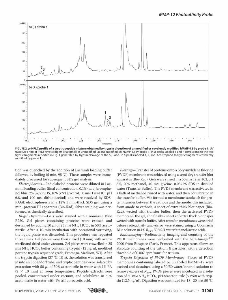
FIGURE 2. �-HPLC profile of a tryptic peptide mixture obtained by trypsin digestion of unmodified or covalently modified hMMP-12 by probe 1. UVtrace (214 nm) of PVDF tryptic digest (100 pmol) of unmodified (a) and modified (b) hMMP-12 by probe 1. In a peaks labeled 6 and 7 correspond to the twotryptic fragments reported in Fig. 1 generated by trypsin cleavage of the S1� loop. In b peaks labeled 1, 2, and 3 correspond to tryptic fragments covalentlymodified by probe 1.
MMP-12 Photoaffinity Probe
NOVEMBER 7, 2008 • VOLUME 283 • NUMBER 45 JOURNAL OF BIOLOGICAL CHEMISTRY 31061

PVDF pieces were then rinsed with a solution of 50% acetoni-trile with 0.1% trifluoroacetic acid.Peptide Mapping—Eluant from the �-HPLC column was
split out into two flows: one at 160 �l/min for UV monitoring(214 nm and 280 nm using a diode array detector) and radioac-tivity measurements; the remaining flow was directed to anelectrospray mass spectrometer for MS. A volume of 0.5 �l ofeach fractionwas spotted onto a glass plate for counting tritiumradioactivity using the beta-ImagerTM 2000. On-line �-HPLC/ESI/MS experiments were performed on an ion trapmass spec-trometer, with the parameters as described above.Edman Sequencing—N-terminal amino acid sequence anal-
ysis was performed by automated Edman degradation using anABI Model 477A/120A Protein-Peptide Sequencing/AnalysisSystem and Analysis Software System, Model 920A (AppliedBiosystems Inc., Foster City, CA). Radioactive fractions elutedfrom the �-HPLC column were pooled and loaded onto a trif-luoroacetic acid-treated cartridge filter, previously conditionedwith BioBrene PlusTM. Prior to each sequence analysis, calibra-tion was performed using a PTH-amino acid standard solution.MS/MS Sequencing of Covalently Modified Peptides—Radio-
active fractions containing covalent adduct peptides were con-centrated by using a reverse phase pipette tip (C18 ZipTip).Off-line nanoelectrospray ionization-mass spectrometry/MSexperiments were performed on a ion trap mass spectrometer.Ion trap parameters for MS were set as follows: nano-electro-spray potential, 1000 V; skimmer voltage, 40 V; capillary exit,226 V; and source temperature, 150 °C. The scan number was
increased to 20 spectra over anm/z of 250–2000 Da, the isolationwidth was set to 1 Da, and the col-lision energy to 0.65 V in theMS/MS-mode. MS/MS spectrawere recorded for double-chargedmolecular peptide ions.Molecular Modeling—Model of
probe 1 interacting with hMMP-12was obtained as previously de-scribed (15). NMR structures in Fig.7 (a and b) where selected from anensemble of 20 NMR structures ofthe hMMP-12 catalytic domaindeposited in Rutgers Collaboratoryfor Structural Bioinformatics PDBunder access code 2POJ.
RESULTS
One-dimensional SDS-PAGE In-gel Digestion—Efficient cross-link-ing experiments were shown to relyon the presence of detergent in sam-ple buffer, a requirement preventingdirect analysis of the photo-adductby MS. In addition, previous workhas demonstrated that light excita-tion of probe 1 in the presence ofhMMP-12 catalytic domain pro-duced two protein species that
could be resolved by one-dimensional SDS-PAGE, the upperband corresponding to the covalently labeled hMMP-12 andthe lower band to unmodified hMMP-12 (26). These consider-ations led us to use first electrophoresis to separate the unmod-ified frommodified forms of hMMP-12 and then proceed to theanalytical characterization of these protein species. After elec-trophoresis, gel pieces containing these protein forms weresubjected to in-gel trypsin digestion, and the correspondingsoluble tryptic fragments were resolved by nano-HPLC andanalyzed online byMS. Nano-HPLC-MS analyses of the trypticfragments from unlabeled (Fig. 1b) hMMP-12 showed a goodoverlapping with its catalytic domain sequence (Fig. 1a).Among the 12 expected tryptic fragments, 8 fragments weredetected in the nano-HPLC profile (Fig. 1, b and d). Based onmass criteria, two peptides co-eluting at the same retentiontime were shown to result from the S1� loop cleavage by trypsin(peak 6: MH � 956.6 and 7: MH � 1027.5; through thisreport, the S1� loop sequence corresponds to the fragmentAla234 to Arg249). The nano-HPLC profile (Fig. 1c) for thecovalently modified form of hMMP-12 (upper band) was char-acterized by an absence of peaks 6 and 7. This suggests that thecovalent modification of hMMP-12 by the probe targets resi-dues of the S1� loop. No additional signals corresponding tomodified peptides were observed in this nano-HPLC profile,thus the corresponding peptides should remain in the gel. Anabsence of radioactivity in the sample buffer (probe 1 incorpo-rates a tritium radioactive atom, Scheme 1) supports this con-
FIGURE 3. �-HPLC ESI mass spectra (negative mode) of peaks 1, 2, and 3 reported in Fig. 2. Molecularweights corresponding to the double-charged ion species contained in these HPLC fractions are reported.
MMP-12 Photoaffinity Probe
31062 JOURNAL OF BIOLOGICAL CHEMISTRY VOLUME 283 • NUMBER 45 • NOVEMBER 7, 2008
clusion. To overcome this drawback, proteins were transferredfrom the gel onto PVDFmembranes, prior to trypsin treatment.Micro Liquid Chromatography-MSAnalysis of Tryptic Digest
Containing Radioactive Species—Analysis of PVDF mem-branes after gel transfer by radioimaging indicated a quantita-tive protein transfer. When peaces of PVDF membrane con-taining hMMP-12 covalently labeled by probe 1were incubatedin buffer, no signal of radioactivity was detected in solution, aresult showing that labeled hMMP-12 remains on/in the mem-
brane. By contrast, when thesemembranes were incubated inbuffer containing trypsin, radioac-tivity was released, suggesting thepresence of hMMP-12 tryptic frag-ment(s) labeled by probe 1 in buffersample. This tryptic fragment mix-ture, including those obtained fromunmodified hMMP-12, were ana-lyzed by �-HPLC, MS, and radioac-tivity counting. Comparison of thecorresponding �-HPLC profiles(Fig. 2a unlabeled hMMP-12 and 2blabeled hMMP-12) revealed thepresence of three additional radio-active peaks in the labeled sample.S1� loop tryptic fragments (peaks 6and 7 in Fig. 2a) were detected in theunlabeled hMMP-12 sample, butwere no longer detected in thelabeled sample, suggesting again acovalent modification of the S1�loop by the photoaffinity probe.MSAnalysis of theThree Radioac-
tive Peptides—Measure of mass-to-charge ratio using a negative detec-tion mode of the above three�-HPLC peaks (F1, F2, and F3) indi-cated that the mass of the fragmentcontained in F3 corresponds app-roximately to the expected mass(observedmass 2712.3 Da, expectedmass 2713.3Da, see “Discussion”) ofthe S1� loop peptide of hMMP-12(residues Ala234 to Arg249, Mw �1964.99) covalently modified byprobe 1 (Mw � 776.27 Da, minus 28Da due to loss of N2 after irradiationof the azide group) (Fig. 3). Thus,the covalent modification ofhMMP-12 by probe 1 preventscleavage of the S1� loop peptide bytrypsin, a result explaining why thecorresponding tryptic fragments areno longer detected after covalentmodification. Furthermore, thisabsence of trypsin cleavage suggeststhat the site of covalent modifica-tion occurs at the Lys241 level or at a
residue near Lys241 (the S1� loop fragment contains only oneinternal tryptic site). Themass-to-charge ratio of the two otherspecies (F1 and F2 in Fig. 3) corresponds to F3-oxidized forms,with one (16, F2) and two (32, F1) degrees of oxidation,respectively. The presence of Met236 in the S1� loop sequencemay explain the observation of oxidized forms (see below).Edman Sequencing—The three purified radioactive fractions
(F1, F2, and F3), containing the covalently modified hMMP-12S1� loop peptide, were pooled and subjected to Edman degra-
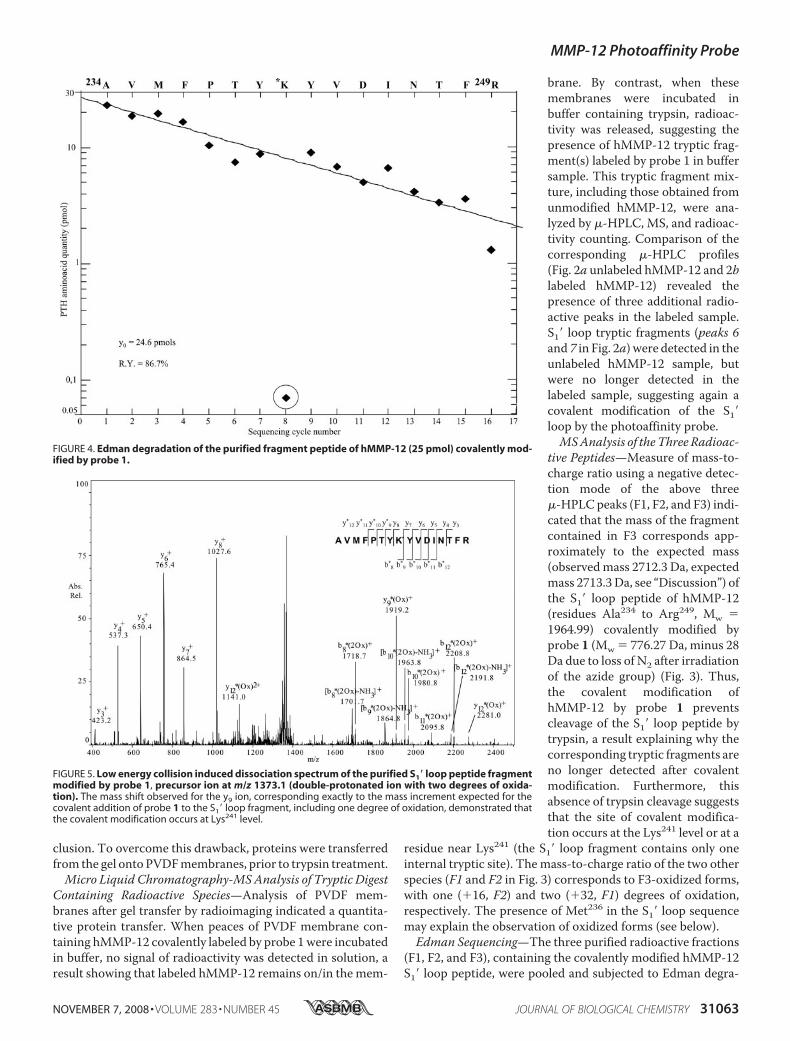
FIGURE 4. Edman degradation of the purified fragment peptide of hMMP-12 (25 pmol) covalently mod-ified by probe 1.
FIGURE 5. Low energy collision induced dissociation spectrum of the purified S1� loop peptide fragmentmodified by probe 1, precursor ion at m/z 1373.1 (double-protonated ion with two degrees of oxida-tion). The mass shift observed for the y9 ion, corresponding exactly to the mass increment expected for thecovalent addition of probe 1 to the S1� loop fragment, including one degree of oxidation, demonstrated thatthe covalent modification occurs at Lys241 level.
MMP-12 Photoaffinity Probe
NOVEMBER 7, 2008 • VOLUME 283 • NUMBER 45 JOURNAL OF BIOLOGICAL CHEMISTRY 31063

dation to further characterize the site of covalent modification.N-terminal sequencing of this peptide started at Ala234 andended at to Tyr240, with no sequencing cycle detected at theLys241 position (Fig. 4). The next sequencing step indicated aTyr residue at position 242, and sequencing identified all sub-sequent S1� loop residues up to Arg249. Therefore, the site ofcovalent modification occurs in the S1� loop, and Lys241 is theonly residue modified by probe 1 in this loop.MS/MS Analysis—The MS/MS analysis of the purified and
photo-cross-linked tryptic fragments corresponding to the S1�loop was carried out for further confirmation and to character-ize the chemical structure of the covalent adduct. MS/MS (col-lision induced dissociation) experiments were performed onthe double-charged precursor ion atm/z [M2H]2 � 1373.1Da, corresponding to the double oxidizedmodified tryptic pep-tide (Mw(F1)� 2744,3 Da, Fig. 3). Fragment ions were assignedaccording to the nomenclature described by Biemann et al.(32). Experimental C-terminal y3 to y8 ionmasses were consist-ent with those predicted, indicating that the C-terminal part ofthe S1� loop from Tyr242 to Arg249 is not modified by probe 1(Fig. 5). All these ions possess the expected theoretical mass,thus no oxidation takes place in theTyr242–Arg249 sequence. Bycontrast, themass shift observed from ion y9 to y12, correspond-ing exactly to the mass increment expected for the covalentaddition of probe 1 to the S1� loop fragment, including onedegree of oxidation, demonstrated that the covalent modifica-tion occurs at the Lys241 level. Because this part of the S1� loopdoes not contain a methionine or amino acids that could be thetarget of oxidation (Pro239–Arg249), the oxidation site in thiscase is expected to occur inside the probe structure. ObservedN-terminal b ion masses (from b8 to b12) are in agreement withthe above conclusion, but these observations alone cannot be
used to determine the site of modification. This identificationwould have required observing the b7 ion, at the least. All b ionsobserved possess two degrees of oxidation, one as a result fromprobe oxidation, as suggested above, and the other probablyresulting from methionine oxidation.Analysis of Lysine 3 Ala Mutant—An hMMP-12 mutant
was produced in which lysine in position 241 of the wild typewas replaced by alanine. Comparison of the catalytic efficiencyof this mutant in cleaving a fluorogenic synthetic substrate spe-cific for MMPs, as well as the Ki value of probe 1 toward themutant, as compared with wild-type hMMP-12, indicates sim-ilar functional properties between the two proteins (Table 1).Photolabeling of this mutant with probe 1, analyzed either bysilver staining or radioactivity counting, revealed significantlyless cross-linking in the mutant (�2% based on radioactivitycounting), than in wild-type hMMP-12 (�45%) (Fig. 6).
DISCUSSION
The various approaches used in this study indicate thatmod-ification of hMMP-12 by probe 1 mostly involved the � aminogroup of Lys241, a residue located on the S1� loop, shaping onepart of the S1� cavity (26). This is consistent with our previoussuggestions that grafting an azide onto a P1� phenylisoxazolineside chain of a phosphinic peptide inhibitor of MMP shouldresult in a photoaffinity probe able tomodify residues of the S1�cavity. However, even by using a molecular model describingthe potential binding mode of 1 within the hMMP-12 activesite, it would be hard to predict the results reported in thisstudy. The main reason for this is the fact that Lys241 is locatedon a loop segment of hMMP-12,which, based onboth x-ray andNMR studies of free hMMP-12 or in complex with syntheticinhibitors, displays high flexibility with the lysine side chainexhibiting high mobility (33–35). Superimposition of twohMMP-12 structuremodels, taken from an ensemble of twentyNMR-derived free hMMP-12 structures (34), provides someclues about the conformational space sampled by the Lys241side chain (Fig. 7). In one of these structures, the position takenby the � amino group of Lys241 is too far away (d 7Å) from thepresumed position of the nitrogen atom of the reactive inter-mediate to predict covalent labeling of the lysine side chain (Fig.
7a). By contrast, the second struc-ture, in which the � amino group ofLys241 points toward the S1� loopcavity, would favor the labeling ofLys241 by probe 1 (Fig. 7b). It isworth noting that, in these NMRstructures, the positions reportedfor the lysine side chain correspondonly to “possible conformations.”Result of the photo-cross-linkexperiments thus leads credence tothese models, in particular to theone in which the lysine side chainpoints toward the S1� cavity. Thebinding of probe 1 to hMMP-12may induce a conformationalchange and stabilize a structuresimilar to that reported in Fig. 7b, in
FIGURE 6. Comparison of the covalent modification of wild-type hMMP-12 and Ala mutant by probe 1.hMMP12 (1 �M) was incubated with probe 1 (2 �M) for 10 min, before UV irradiation (2 min). The MMP12complexes (5 pmol) were resolved by one-dimensional SDS-PAGE electrophoresis and visualized on the gel bysilver staining (left) or the proteins were transferred onto a PVDF membrane that was analyzed with a radioim-ager (right).
TABLE 1Characterization of hMMP-12 Ala mutantkcat/Km and Ki values were determined in Tris/HCl buffer, 50 mM, pH 6.8, CaCl2,10 mM.
hMMP-12 Ala mutantMca-Mat (kcat/Km, M�1�s�1) 1.53.104 (�0.03.104) 1.32.104 (�0.05.104)Probe 1 (Ki, nM) 0.17 (�0.01) 0.35 (�0.04)
MMP-12 Photoaffinity Probe
31064 JOURNAL OF BIOLOGICAL CHEMISTRY VOLUME 283 • NUMBER 45 • NOVEMBER 7, 2008

which the � amino group of Lys241 is pointing in the direction ofand is in close proximity of the probe 1 azide group. Alterna-tively, movements of the lysine side chain may exist on a timescale faster than the lifetime of the nitrene reactive intermedi-ate. The mass observed for the covalently modified S1� loopcorresponds to the theoretical expected mass minus 1. Thisdifference was resolved by considering that the � amino groupof Lys241 is actually labeled in its unprotonated form (NH2). Theequilibrium between NH3
and NH2 forms at pH 7 of the Lys �
amino group could be shifted toward the NH2 form, throughconsumption of the NH2 form by the photochemical reaction.Alternatively, a shift in the lysine conformation toward thenitrene reactive group may change the pKa of that side chain.The exact chemical structure of the covalent adduct formedbetween hMMP-12 and probe 1, after irradiation, has not beenestablished in this study. Based on previous studies of arylazides, irradiation of probe 1 is expected to produce as reactiveintermediates either a dedihydroazepine (Scheme 2a) or a tri-
plet nitrene (Scheme 2b), as reactiveintermediates (36–38). The dedihy-droazepine intermediate is thoughtto react with nucleophilic atomspresent in cysteine and histidineresidues, but less likely with the pro-tonated � amino group of the lysineside chain (Scheme 2, adduct 2a)(38). Triplet nitrene may covalentlymodify the � amino group of a lysineto form the structure reported inScheme 2 (adduct 2b). Simple phe-nylazides produce dedihydroaz-epine upon photolysis in water orbuffer (39). However, the photoge-nerated intermediate was also reli-ant on the nature of the substituentsof the phenyl ring (36–38). How theisoxazoline group in para-position
SCHEME 2
FIGURE 7. Model of probe 1 in complex with hMMP-12 (see “Experimental Procedures”). As comparedwith the standard orientation recommended for metzincins, the structure of MMP-12 catalytic domain hasbeen tilted around the horizontal axis to better bring out the S1� loop. Lys241 is colored in blue, probe 1 in green,and catalytic zinc ion in purple; a and b displayed two possible conformations taken by the Lys241 flexible sidechain, one (a) far away from the azide group and the other (b) nearby this group.
MMP-12 Photoaffinity Probe
NOVEMBER 7, 2008 • VOLUME 283 • NUMBER 45 JOURNAL OF BIOLOGICAL CHEMISTRY 31065

of the phenyl in probe 1 influences which type of reactive inter-mediate is formed is actually unknown. Moreover, in a contextof high affinity, the protein binding site environment mightdetermine the structure of the reactive intermediate that will beformed upon photolysis. Because the two predicted adductshave the samemass (Scheme 2, 2a and 2b), their discriminationby MS and the identification of the reactive intermediateinvolved in the reaction cannot be achieved in the presentstudy. Indeed, given the length of adducts 2a and 2b, ESI/MS/MS mostly leads to peptide bond fragmentation. Thus,characterization of N–C or N–N bonds to discriminatebetween 2a and 2b adducts would require the use of other tech-niques like electronic impact. The fewpercent of covalentmod-ification observed in the Ala mutant prevented further charac-terization of the covalent modification site. Similar weakcovalent modifications occurring in the Ala mutant may alsotake place in wild-type hMMP-12, but this should only be con-sidered as aminor reaction, in comparisonwith themajormod-ification observed at lysine. Inspection of the S1� cavity ofhMMP-12 in the vicinity of the azide of probe 1 indicated thatthe closest atoms to the nitrogen linked to the phenyl (the onethat will form the nitrene) are the H� (d � 3Å) and CH3� (d �3.5Å) of Val235. Labeling of hydrophobic residues is generallyobserved with probes that generate extremely reactive specieslike carbene (38), thus the weak labeling observed in the Alamutantmay be explained by theweaker reactivity of the nitreneformed by probe 1 toward hydrophobic residues, like Val235.Interestingly, in MMP-8 in which the position 241 is occupiedby an alanine, a weak labeling (�2–3%)was previously reportedfor its covalent modification by probe 1 (26). Thus, MMP-8and theAla-MMP-12mutant display a similar reactivity towardprobe1, even though their S1� loop sequences are very different.Thus cross-linking yield between probe 1 and other MMPs ispossibly determined by the chemical nature of the residue inposition 241. This may explain themarked differences in cross-linking yield observed between various MMPs, because thecomposition of the position 241 is highly variable in MMPs(Ala(MMP8), Gln(MMP14), Thr(MMP2, 13, 11), Arg(MMP9), andHis(MMP3)). If true, developing photoaffinity probes with highcross-linkingdedicated to MMPs (23–26), these ABP yieldtoward all MMPs will require the selection of other reactivephoto-activable groups, having lower selectivity toward thechemical nature of the residues surrounding the probe. Thischallenge will have to overcome the various chemical con-straints and the particular shape of theMMPS1� cavity. Probingthe S1� cavity ofMMPs by photoaffinity labeling, as shownhere,provides a unique insight into the conformational variability ofMMPs, a key factor governing their selective recognition ofsubstrates or inhibitors. Despite the development of very smartand different activity-based probes (ABPs) probes have not yetdetected active forms ofMMPs in biological samples. The tightregulation and control of MMP active forms probably explainthis failure, but it also calls for the development of extremelysensitive ABP probes (40). To be achieved, this goal needs totake into account the yield of cross-link achieved by theseprobes toward all MMP members. The data reported in thisstudy should help in the design of better MMP ABP probes,allowing the sensitive detection of their active forms in various
samples, an important objective for both diagnosis and thera-peutic application (41–43).
REFERENCES1. Brinckerhoff, C. E., andMatrisian, L.M. (2002)Nat. Rev.Mol. Cell. Biol. 3,
207–2142. Visse, R., and Nagase, H. (2003) Circ. Res. 92, 827–8393. Page-McCaw, A., Ewald, A. J., and Werb, Z. (2007) Nat. Rev. Mol. Cell.
Biol. 8, 221–2334. Nagase, H., andWoessner, J. F., Jr. (1999) J. Biol. Chem. 274, 21491–214945. Bode, W., Gomis-Ruth, F. X., and Stockler, W. (1993) FEBS Lett. 331,
134–1406. Stocker, W., and Bode, W. (1995) Curr. Opin. Struct. Biol. 5, 383–3907. Massova, I., Lakshmi, P. K., Fridman, R., andMobashery, S. (1998) FASEB
J. 12, 1075–10958. Gomis-Ruth, F. X. (2003)Mol. Biotechnol. 24, 157–2029. Fingleton, B. (2007) Curr. Pharm. Des. 13, 333–34610. Babine, R. E., and Bender, S. L. (1997) Chem. Rev. 97, 1359–147211. Whittaker, M., Floyd, C. D., Brown, P., and Gearing, A. J. (1999) Chem.
Rev. 99, 2735–277612. Rosenblum, G., Meroueh, S. O., Kleifeld, O., Brown, S., Singson, S., Frid-
man, R., Mobashery, S., and Sagi, I. (2003) J. Biol. Chem. 278,27009–27015
13. Engel, C. K., Pirard, B., Schimanski, S., Kirsch, R., Habermann, J., Kingler,O., Schlotte, V., Weithmann, K. U., and Wendt, K. U. (2005) Chem. Biol.12, 181–189
14. Johnson, A. R., Pavlovsky, A. G., Ortwine, D. F., Prior, F., Man, C. F.,Bornemeier, D. A., Banotai, C. A., Mueller, W. T., McConnell, P., Yan, C.,Baragi, V., Lesch, C., Roark, W. H., Wilson, M., Datta, K., Guzman, R.,Han, H. K., and Dyer, R. D. (2007) J. Biol. Chem. 282, 27781–27791
15. Devel, L., Rogakos, V., David, A., Makaritis, A., Beau, F., Cuniasse, P.,Yiotakis, A., and Dive, V. (2006) J. Biol. Chem. 281, 11152–11160
16. Coussens, L. M., Fingleton, B., and Matrisian, L. M. (2002) Science 295,2387–2392
17. Lopez-Otin, C., and Matrisian, L. M. (2007)Nat. Rev. Cancer 7, 800–80818. Overall, C. M., and Lopez-Otin, C. (2002) Nat. Rev. Cancer 2, 657–67219. Egeblad, M., and Werb, Z. (2002) Nat. Rev. Cancer 2, 161–17420. Overall, C. M., and Kleifeld, O. (2006) Nat. Rev. Cancer 6, 227–23921. Fingleton, B. (2006) Front. Biosci. 11, 479–49122. Johnson, J. L., George, S. J., Newby, A. C., and Jackson, C. L. (2005) Proc.
Natl. Acad. Sci. U. S. A. 102, 15575–1558023. Saghatelian, A., Jessani, N., Joseph, A., Humphrey, M., and Cravatt, B. F.
(2004) Proc. Natl. Acad. Sci. U. S. A. 101, 10000–1000524. Chan, E. W., Chattopadhaya, S., Panicker, R. C., Huang, X., and Yao, S. Q.
(2004) J. Am. Chem. Soc. 126, 14435–1444625. Sieber, S. A., Niessen, S., Hoover, H. S., and Cravatt, B. F. (2006) Nat.
Chem. Biol. 2, 274–28126. David, A., Steer, D., Bregant, S., Devel, L., Makaritis, A., Beau, F., Yiotakis,
A., and Dive, V. (2007) Angew. Chem. Int. Ed. Engl. 46, 3275–327727. Cravatt, B. F., Wright, A. T., and Kozarich, J. W. (2008) Annu. Rev. Bio-
chem. 77, 383–41428. Kato,D.,Boatright,K.M.,Berger,A.B.,Nazif,T.,Blum,G.,Ryan,C.,Chehade,
K. A., Salvesen, G. S., and Bogyo, M. (2005)Nat. Chem. Biol. 1, 33–3829. Tochowicz, A., Maskos, K., Huber, R., Oltenfreiter, R., Dive, V., Yiotakis,
A., Zanda,M., Bode,W., andGoettig, P. (2007) J.Mol. Biol. 371, 989–100630. Lang, R., Kocourek, A., Braun,M., Tschesche, H., Huber, R., Bode,W., and
Maskos, K. (2001) J. Mol. Biol. 312, 731–74231. Horovitz, A., and Levitzki, A. (1987) Proc. Natl. Acad. Sci. U. S. A. 84,
6654–665832. Biemann, K. (1990)Methods Enzymol. 193, 886–88733. Nar, H., Werle, K., Bauer, M. M., Dollinger, H., and Jung, B. (2001) J. Mol.
Biol. 312, 743–75134. Bhaskaran, R., Palmier, M. O., Bagegni, N. A., Liang, X., and Van Doren,
S. T. (2007) J. Mol. Biol. 374, 1333–134435. Bertini, I., Calderone, V., Cosenza,M., Fragai, M., Lee, Y.M., Luchinat, C.,
Mangani, S., Terni, B., and Turano, P. (2005) Proc. Natl. Acad. Sci. U. S. A.102, 5334–5339
MMP-12 Photoaffinity Probe
31066 JOURNAL OF BIOLOGICAL CHEMISTRY VOLUME 283 • NUMBER 45 • NOVEMBER 7, 2008

36. Bayley, H., and Staros, J. V. (1984) inAzides andNitrenes, (E. F. V. Scriven,ed) pp. 433–490, Academic Press, New York
37. Fleming, S. A. (1995) Tetrahedron 51, 12479–1252038. Kotzyba-Hibert, F., and Goeldner, M. (1995) Angew Chem. Int. Ed. Engl.
34, 1296–131239. Rizk, M. S., Shi, X., and Platz, M. S. (2005) Biochemistry 45, 543–55140. Hesek, D., Toth, M., Meroueh, S. O., Brown, S., Zhao, H., Sakr, W., Frid-
man, R., and Mobashery, S. (2006) Chem. Biol. 13, 379–38641. Unsal, D., Akyurek, N., Uner, A., Erpolat, O. P., Han, U., Akmansu, M.,
Mentes, B. B., and Dursun, A. (2008) Am. J. Clin. Oncol. 31, 55–6342. Kramer, F., Sandner, P., Klein, M., and Krahn, T. (2008) Biomarkers 13,
270–28143. Rydlova, M., Holubec, L., Jr., Ludvikova, M., Jr., Kalfert, D., Franekova, J.,
Povysil, C., and Ludvikova, M. (2008) Anticancer Res. 28, 1389–1397
MMP-12 Photoaffinity Probe
NOVEMBER 7, 2008 • VOLUME 283 • NUMBER 45 JOURNAL OF BIOLOGICAL CHEMISTRY 31067