proteins are the polymers of α - g.g.u pandey model_ans_d.pharm_… · proteins are the polymers...
TRANSCRIPT

D. Pharm. I Year Main Examinations, 2013 Subject: Biochemistry and Clinical Pathology
Max. Marks: 80 Time: 03 Hours Note: Attempt both the section A and B. Section A is compulsory. Draw neat diagram(s) with proper
labeling wherever necessary. Section A 12X02= 24 (Attempt ALL questions in this section. Each question carries two marks.)
1. a) Define amino acids and proteins with suitable examples?
Ans: Organic acids having a carboxyl group and an amino group bonded to the carbon atom. It
differ from each other in their side chains, or R groups.
Amino acids are building blocks (monomers) of proteins (polymer). Ex. - Glycine, Aspargine,
Glutamic acid etc.
Proteins are the polymers of α -amino acids (Carboxylic and amino group in same carbon) in
which amino acids are joined together by peptide bond. Examples are: gelatin, albumin etc.
b) Define enzyme with examples?
Ans: Enzymes are proteins that are biological catalyst.
It catalyze (accelerate) biochemical reaction by lowering the activation energy. Examples:
Catalase, Urease, pepsin etc.
c) Write short note on Phenylketonuria.
Ans: Phenylketonuria (PKU) is an autosomal recessive metabolic genetic disorder characterized by
a mutation in the gene for the hepatic enzyme phenylalanine hydroxylase (PAH), rendering it
nonfunctional.
This enzyme is necessary to metabolize the amino acid phenylalanine (Phe) to the amino acid
tyrosine.
When PAH activity is reduced, phenylalanine accumulates and is converted into
phenylpyruvate (also known as phenylketone), which can be detected in the urine.
Untreated PKU can lead to mental retardation, seizures, and other serious medical problems.
d) Differentiate between non- essential and essential amino acid with examples?
Ans: Humans are incapable of synthesizing 09 of the 20 common amino acids, and these essential
amino acids must be provided in the diet. Ex. Histidine, Isoleucine, Leucine, Lysine, Tryptophan
etc.
Non-essential amino acids can be synthesized by human cells ex. Aspargine, Alanine, Serine
etc. and not essentially required from diet.

e) What is biological significance of Carbohydrates?
Ans: Functions of Carbohydrates
1. Major metabolic fuel of mammals
2. Storage of energy (e.g., starch and glycogen), and
3. Structural components (e.g., cellulose in plants and chitin in arthropods). The 5-carbon
monosaccharide ribose is an important component of coenzymes (e.g., ATP, FAD, and NAD) and
the backbone of the genetic molecule known as RNA. The related deoxyribose is a component of
DNA.
4. It play key roles in the immune system, fertilization, preventing pathogenesis, blood clotting, and
development
f) Define fatty acid with examples?
Ans: Fatty acids are Aliphatic carboxylic acids occur mainly as esters in natural fats and oils but do
occur in the unesterified form as free fatty acids, a transport form found in the plasma.
Fatty acids that occur in natural fats are usually straight-chain derivatives containing an even
number of carbon atoms. The chain may be saturated (containing no double bonds) or
unsaturated (containing one or more double bonds). Ex. Palmitic acids and stearic acids.
g) What is Lymphocytes? Abnormal high percentage of White blood cells in blood is
called.............................
Ans: Lymphocytes are type of agranular white blood cells of vertebrate immune system.
Abnormal high percentage of White blood cells is is called Leukemia.
h) Vitamin D is .............................(chemical name) and main deficiency disease is……………
Ans: Vitamin D is Calciferol and main deficiency disease is Rickets = poor mineralization of
bone; and/or osteomalacia = bone demineralization
i) Give energetics of aerobic and anaenorobic glycolysis?
Ans: ENERGETICS OF GLYCOLYSIS: Total energy yielded per original molecule of glucose.
A) +2 NADH: Glyceraldehyde-3-Phosphate Dehydrogenase (step 6)
B) +2 ATP net
a. -1 ATP (per original glucose): Hexokinase (step 1)
b. -1 ATP (per original glucose): Phosphofructokinase (step 3)
c. +2 ATP (per original glucose): Phosphoglycerate Kinase (step 7)
d. +2 ATP (per original glucose): Pyruvate Kinase (step 10)

j) Give two examples of glycolipids.
Ans: 1. Galactosyl ceramide ( in brain tissue)
2. Glucosyl ceramide ( in cell membrane)
k) What are functions of erythrocytes?
Ans: Functions of RBCs or erythrocytes:
1) Transportation of oxygen and carbon dioxide.
2) Responsible for homeostatis (pH, Viscosity etc.)
3) Dilate blood vessels during stress/trauma by releasing nitrous oxide
l) Write names of two enzymes used in diagnosis of liver diseases
Ans: 1. Alanine transaminase (ALT)
2. Aspartate transaminase (AST)
Section B 14X4= 56
(Attempt any FOUR questions from this section. Each question carries fourteen marks.)
2. Discuss in details physical and chemical properties of monosaccharides?
Ans. Monosaccharides: single-unit carbohydrates (monomers) o aldoses: carbohydrates with aldehyde group o ketoses: carbohydrates with ketone functionality
Polysaccharides: chains of monosaccharides (polymers) o examples: starch, cellulose, glycogen
Carbohydrates - Stereochemistry
D/L system used for penultimate carbon 1. most oxidized carbon at top 2. continue carbon chain to bottom 3. non-hydrogen substituent on right=D Trivial names indicate all other stereocenters
Monosaccharides of Different Sizes Name # Carbons Formula
Triose 3 C3H6O3 Tetrose 4 C4H8O4 Pentose 5 C5H10O5 Hexose 6 C6H12O6 Common Monosaccharides

Amino Sugars
Mutarotation
Cyclic acetals are in equilibrium with open-chain forms This equilibrium allows a and b forms to interconvert This interconversion can result in a change in the specific rotation
Physical Properties
All monosaccharides have the following physical properties.
(1) After dissolving in water, all monosaccharides give a sweet taste. (2) All monosaccharides can move across the plasma membrane. (3) All monosaccharides are soluble in water. When they are dissolved in water, they would take up
the ring-form. This is the cause for their solubility in water. (4) When monosaccharides are dissolved in water, they would initiate the lowering of water
potential of the solution. (5) The water solutions of monosaccharide are optically active. That means that the water solution of
a monosaccharide can rotate the plane of polarized light. This is called the Optical activity. ll carbohydrates that can be assimilated by living things are dextrorotatory. All amino acids utilized by living things are levorotatory.

Type of sugar Angle of optic rotation D-glucose +52.7 D-fructose -92.4
Chemical Properties A. Glycoside (a type of acetal) Formation B. Methyl ether formation C. Reduction D. Oxidation
1. Cyclic Structures of Monosaccharides (Acetal and Hemiacetal Formation) All monosaccharides with at least five carbon atoms exist predominantly as cyclic hemiacetals and hemiketals. A Haworth structure can be used to depict the a and b anomers of a monosaccharide. Anomers are stereoisomers that differ in the 3-D arrangement of groups at the anomeric carbon of an acetal, ketal, hemiacetal, or hemiketal group. Glycoside Formation: Cyclic monosaccharide hemiacetals and hemiketals react with alcohols to form acetals and ketals, referred to as glycosides.
This equilibrium is acid-catalyzed (both directions)
Oxygen is protonated, making the carbon more reactive The most abundant species at equilibrium is usually the ketone or aldehyde
exception: cyclic acetals Methyl Ether Formation
Given: NaH function -
Carbonyl reduction reactions affect the aldehyde or ketone functionality (NaBH4, H2/Pt...)

Oxidation - I Oxidation by Cu(II) or Ag(I) - a test for reducing sugars
The oxidation of a carbonyl group on a monosaccharide. Since all monosaccharides are in equilibrium with their cyclic form, they are all reducing sugars. •Benedict’s reagent is commonly used to test for the presence of reducing sugars: Reducing sugar + Cu2+ ® oxidized compound + Cu2O (blue) (Orange red ppt) Oxidation - II
Nitric Acid - oxidizes aldehydes and primary alcohols to carboxylic acids
Periodic Acid - cleaves C-C bond of 1,2-diols
Esterfication The –OH groups of monosaccharides can behave as alcohols and react with acids (especially phosphoric acid) to form esters.

3. Give an account of HMP pathway and it significances? Ans: The pentose phosphate pathway (also called the phosphogluconate pathway and the hexose monophosphate shunt) is a process that generates NADPH and pentoses (5-carbon sugars). There are two distinct phases in the pathway. The first is the oxidative phase, in which NADPH is generated, and the second is the non-oxidative synthesis of 5-carbon sugars. This pathway is an alternative to glycolysis. While it does involve oxidation of glucose, its primary role is anabolic rather than catabolic. For most organisms, it takes place in the cytosol; in plants, most steps take place in plastids.

Significance: The primary results of the Pathway are: The generation of reducing equivalents, in the form of NADPH, used in reductive biosynthesis
reactions within cells. (e.g. fatty acid synthesis) Production of ribose-5-phosphate (R5P), used in the synthesis of nucleotides and nucleic acids. Production of erythrose-4-phosphate (E4P), used in the synthesis of aromatic amino acids.
Aromatic amino acids, in turn, are precursors for many biosynthetic pathways, notably including the lignin in wood.
Dietary pentose sugars derived from the digestion of nucleic acids may be metabolized through the pentose phosphate pathway, and the carbon skeletons of dietary carbohydrates may be converted into glycolytic/gluconeogenic intermediates.
In mammals, the PPP occurs exclusively in the cytoplasm, and is found to be most active in the liver, mammary gland and adrenal cortex in the human. The PPP is one of the three main ways the body creates molecules with reducing power, accounting for approximately 60% of NADPH production in humans.
One of the uses of NADPH in the cell is to prevent oxidative stress. It reduces glutathione via glutathione reductase, which converts reactive H2O2 into H2O by glutathione peroxidase. If absent, the H2O2 would be converted to hydroxyl free radicals by Fenton chemistry, which can attack the cell. Erythrocytes, for example, generate a large amount of NADPH through the pentose phosphate pathway to use in the reduction of glutathione.
Hydrogen peroxide is also generated for phagocytes in a process often referred to as a respiratory burst.[2]
4. Classify the protein with suitable example? Give in details about the ammonia formation?
Ans: Proteins have wide structural and functional diversity. It is difficult to classify these on the basis of a single property or a characteristic. However, following are some common basis of classifying them.
Shape and structure Products of hydrolysis Biological functions Classification on the Basis of Shape and Size
On the basis of their composition, proteins get different shapes and size which give an indication of their various functions. These can be broadly put into two classes on the basis of their overall shape. These are as follows: Globular proteins: These contain compactly folded coils of polypeptide chains giving them shape of spheroids or ellipsoids. Examples of this type are albumins, globulins, histones, protamines etc. Fibrous proteins: This class of proteins looks like fibres or threads. These are insoluble in water and aqueous solutions of acids and bases. These have high mechanical strength. Keratins in hair, actins and myosin in muscles and collagen are examples of this type of protein. Classification on the Basis of Products of Hydrolysis On the basis of the products obtained on hydrolysis, proteins can be classified into three categories viz., simple proteins, conjugated proteins and derived proteins. Simple proteins: Simple proteins are those which are made of amino acid units, each joined by a peptide bond. Upon hydrolysis they yield only a mixture of amino acids. Following are some of the types of simple proteins. Types of Simple Proteins with their Examples S. No. Type Examples 1. Albumins Egg albumin, serum albumin, lactalbumin 2. Gliadin Tissue globulin, serum globulin

3. Gliadins Wheat gliadin, hordein (barley) etc. 4. Albuminoids Keratin of hairs, skin, egg shell and bones, elastin, collagen of
tendons, ligaments and bones 5. Histones . Globin of haemoglobin 6. Protamine Salmine from the spermatozoa of salmon fish Conjugated Proteins: Conjugated proteins are composed of simple proteins combined with non protein substances. The non protein substance is called prosthetic group or cofactor. Following are some of the types of conjugated proteins. Types of Conjugated Proteins and their Examples S. No. Type Examples 1. Chromoproteins Haemoglobin, in which the prosthetic group is iron 2. Phosphoproteins Casein in milk and vitellin in egg yolk containing phosphoric acid
as prosthetic group 3. Lipoproteins HDL (high density lipoprotein), LDL (low density
lipoprotein) and VLDL (very low density lipoproteins), have lipids as the prosthetic groups
4. Glycoprotein Ovomucoid of egg white containing a carbohydrate moiety
5. Nucleoproteins Ribosomes and viruses contain nucleic acids 6. Metalloproteins Alcohol dehydrogenase- a Zn containing enzyme 7. Mucoproteins Follicle stimulating hormone, ovomucoid Derived Proteins: These are not naturally occurring proteins and are obtained from simple proteins or conjugated proteins by the action of enzymes and chemical agents, heat, mechanical shaking, UV or X-rays. It includes the following types: 1. Primary e.g., myosin, fibrin 2. Secondary e.g., peptones, peptides, proteoses etc. Classification on the Basis of Biological Functions The involvement of proteins in different functions also makes it a basis for their classification. The different classes along with the functions performed and examples are summaries in Table Types of Proteins on the Basis of Functions Performed S.No. Type Function Example 1. Enzymes Catalytic activity Kinases,
dehydrogenases 2. Storage proteins Store amino acids Myoglobin, ferritin 3. Regulatory proteins Coordinate body activities Insulin, glucagons 4. Structural proteins Give support and structure Keratin, collagen 5. Defensive/Protective
proteins Protect against diseases Immunoglobulins,
antibodies 6. Transport proteins Facilitate import of
nutrients into cells or releases of toxic products into surrounding medium
Haemoglobin
7. Contractile and Mobile proteins
Participate in contractile processes e.g. movement of muscles
Actin, myosin

Ammonia formation
Oxidative Deamination Reaction
Introduction:
Deamination is also an oxidative reaction that occurs under aerobic conditions in all tissues but especially the liver. During oxidative deamination, an amino acid is converted into the corresponding keto acid by the removal of the amine functional group as ammonia and the amine functional group is replaced by the ketone group. The ammonia eventually goes into the urea cycle.
Oxidative deamination occurs primarily on glutamic acid because glutamic acid was the end product of many transamination reactions.
The glutamate dehydrogenase is allosterically controlled by ATP and ADP. ATP acts as an inhibitor whereas ADP is an activator.
Urea Cycle
Urea is the major end product of nitrogen metabolism in humans and mammals. Ammonia, the product of oxidative deamination reactions, is toxic in even small amounts and must be removed from the body. The urea cycle or the ornithine cycle describes the conversion reactions of ammonia into urea. Since these reactions occur in the liver, the urea is then transported to the kidneys where it is excreted. The overall urea formation reaction is:
2 Ammonia + carbon dioxide + 3ATP ---> urea + water + 3 ADP
One amine group comes from oxidative deamination of glutamic acid while the other amine group comes from aspartic acid. Aspartic acid is regenerated from fumaric acid produced by the urea cycle. The fumaric acid first undergoes reactions through a portion of the citric acid cycle to produce oxaloacetic acid which is then changed by transamination into aspartic acid.

High ammonia levels are toxic to humans. A complete block of any step in the urea cycle is fatal since there is no known alternative pathway for the synthesis of urea. Inherited disorders from defective enzymes may cause a partial block in some of the reactions and results in hyperammonemia which can lead to mental retardation. Extensive ammonia accumulation leads to extensive liver damage and death. Liver cirrhosis caused by alcoholism creates an interference in the enzymes which produce carbamyl phosphate in the first step on the cycle.
4. Define essential fatty acids with suitable examples? Discuss the metabolic disorders of lipids?
Ans: Essential Fatty acids
Essential fatty acids, or EFAs, are fatty acids that humans and other animals must ingest because the body requires them for good health but cannot synthesize them. The term "essential fatty acid" refers to fatty acids required for biological processes but does not include the fats that only act as fuel.
Only two fatty acids are known to be essential for humans: alpha-linolenic acid (an omega-3 fatty acid) and linoleic acid (an omega-6 fatty acid). Some other fatty acids are sometimes classified as "conditionally essential," meaning that they can become essential under some developmental or disease conditions; examples include docosahexaenoic acid (an omega-3 fatty acid) and gamma-linolenic acid (an omega-6 fatty acid).
Disorders of Lipid metabolism
S. No. Name of Disorder Causes/Symptoms 1. Obesity Excess fat deposition (20-30% overweight)

Due to imbalance of endocrine system or diet (over eat)
2. Cachexia Due to ill health or malnutrition or hormonal misbalance (Hyperthyroidism/diabetes/ hypopuitatarism) Fats are scanty
3. Familial Hyper lipoproteinemia ( 5 types and type II has to subtype)
Elevation of one or more lipoprotein in blood (chylomicrons/LDL/VLDL etc.) Hereditary deficiency of enzymes some may cause Atherosclerosis
4. Atherosclerosis Gaucher’s Disease Heredity disorder
Increased cerebroside in reticuloendothelial cells Spleen, enlarged liver, skin pigmentation, bone lesions, Glucose replaces Galactose in cerebroside
5. Niemann Pick’s disease Heredity disorder Cells of the liver and spleen become foamy with enlargement of anemia lymphadenopathy and progressive mental and Physcial detoriation onset in early infancy with death Spingomyelin accumulated in these organ
6. Tay Sach’s disease Inherited disorder of Ganglioside breakdown. GM2 Gangliosides accumulate in brain and CNS and due to absence of β-N-acetyl Hexosaminaidase
7 Refsum’s disease Autosomal recessive disorder Abnormal fatty acid Phytanic acid present in vegetable oils could not metabolized in liver by β-oxidation thus accumulate in retina, CNS and liver
8 Dyslipoproteinemia I an V (high Chylomicron) II (high LDL) III (High LDL and VLDL) IV (High VLDL)
Primary- Abnormal lipoprotein metabolism 5 types Skin pigementation (I and IV) II and IV Coronary Heart disease III cataract II and III cause renal disease Type II cause fibrositis, arthritis Secondary- this is acquired due to secondary underlying disease condition that lead to alteration in lipid metabolism. Diabetes mellitus Alcoholism Hypothyroidism Renal failure Nephritic syndrome Liver disease
HDL: High Density Lipoprotein, LDL: Low Density Lipoprotein, VLDL: Very Low Density Lipoprotein

5. Detail mechanism of action of enzymes and their therapeutic uses.
Mechanism of action of enzyme
Introduction - Enzyme Characteristics:
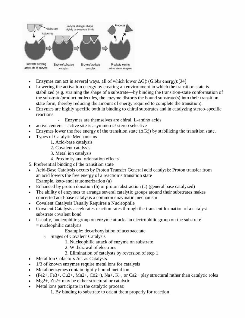
The basic mechanism by which enzymes catalyze chemical reactions begins with the binding of the substrate (or substrates) to the active site on the enzyme. The active site is the specific region of the enzyme which combines with the substrate. The binding of the substrate to the enzyme causes changes in the distribution of electrons in the chemical bonds of the substrate and ultimately causes the reactions that lead to the formation of products. The products are released from the enzyme surface to regenerate the enzyme for another reaction cycle.
The active site has a unique geometric shape that is complementary to the geometric shape of a substrate molecule, similar to the fit of puzzle pieces. This means that enzymes specifically react with only one or a very few similar compounds.
Lock and Key Theory:
The specific action of an enzyme with a single substrate can be explained using a Lock and Keyanalogy first postulated in 1894 by Emil Fischer. In this analogy, the lock is the enzyme and the key is the substrate. Only the correctly sized key (substrate) fits into the key hole (active site) of the lock (enzyme).
Smaller keys, larger keys, or incorrectly positioned teeth on keys (incorrectly shaped or sized substrate molecules) do not fit into the lock (enzyme). Only the correctly shaped key opens a particular lock. This is illustrated in graphic on the left.
Induced Fit Theory:
Not all experimental evidence can be adequately explained by using the so-called rigid enzyme model assumed by the lock and key theory. For this reason, a modification called the induced-fit theory has been proposed by Koshland.
The induced-fit theory assumes that the substrate plays a role in determining the final shape of the enzyme and that the enzyme is partially flexible. This explains why certain compounds can bind to the enzyme but do not react because the enzyme has been distorted too much. Other molecules may be too small to induce the proper alignment and therefore cannot react. Only the proper substrate is capable of inducing the proper alignment of the active site.
Enzymes can act in several ways, all of which lower ΔG‡ (Gibbs energy):[34] Lowering the activation energy by creating an environment in which the transition state is
stabilized (e.g. straining the shape of a substrate—by binding the transition-state conformation of the substrate/product molecules, the enzyme distorts the bound substrate(s) into their transition state form, thereby reducing the amount of energy required to complete the transition).
Enzymes are highly specific both in binding to chiral substrates and in catalyzing stereo-specific reactions
- Enzymes are themselves are chiral, L-amino acids active centers = active site is asymmetric/ stereo selective Enzymes lower the free energy of the transition state (ΔG‡) by stabilizing the transition state. Types of Catalytic Mechanisms
1. Acid-base catalysis 2. Covalent catalysis 3. Metal ion catalysis 4. Proximity and orientation effects
5. Preferential binding of the transition state Acid-Base Catalysis occurs by Proton Transfer General acid catalysis: Proton transfer from
an acid lowers the free energy of a reaction’s transition state Example, keto-enol tautomerization (a)
Enhanced by proton donation (b) or proton abstraction (c) (general base catalyzed) The ability of enzymes to arrange several catalytic groups around their substrates makes
concerted acid-base catalysis a common enzymatic mechanism Covalent Catalysis Usually Requires a Nucleophile Covalent Catalysis accelerates reaction rates through the transient formation of a catalyst-
substrate covalent bond Usually, nucleophilic group on enzyme attacks an electrophilic group on the substrate
= nucleophilic catalysis Example: decarboxylation of acetoacetate
o Stages of Covalent Catalysis 1. Nucleophilic attack of enzyme on substrate 2. Withdrawal of electrons 3. Elimination of catalysts by reversion of step 1
Metal Ion Cofactors Act as Catalysts 1/3 of known enzymes require metal ions for catalysis Metalloenzymes contain tightly bound metal ion (Fe2+, Fe3+, Cu2+, Mn2+, Co2+), Na+, K+, or Ca2+ play structural rather than catalytic roles Mg2+, Zn2+ may be either structural or catalytic Metal ions participate in the catalytic process:
1. By binding to substrate to orient them properly for reaction

2. By mediating oxidation-reduction reactions through reversible changes in the metal ions oxidation state 3. By electrostatically stabilizing or shielding negative charges
Often: Metal ion acts similar to a proton, or polarizes water to generate OH Catalysis can occur through proximity and orientation effects Enzymes are much more efficient catalysts than organic model compounds Due to proximity and orientation effects Reactants come together with proper spatial
relationship Example: p-nitrophenylacetate intramolecular reaction is 24 times faster
Enzymes are usually much bigger than their substrates By oriented binding and immobilization of the substrate, enzymes facilitate catalysis by four
ways 1. bring substrates close to catalytic residues 2. Binding of substrate in proper orientation (up to 102-fold) 3. Stabilization of transition state by electrostatic interactions 4. freezing out of translational and rotational mobility of the substrate (up to 107-fold)
Enzymes catalyze reactions by preferentially binding the transition state An enzyme may binds the transition state of the reaction with greater affinity than its substrate
or products This together with the previously discussed factors accounts for the high rate of catalysis For example, if enzyme binds the transition state
Some important therapeutic enzymes
Enzyme EC number Reaction Use
Asparaginase 3.5.1.1 L-Asparagine H2O L-aspartate + NH3 Leukaemia Collagenase 3.4.24.3 Collagen hydrolysis Skin ulcers Glutaminase 3.5.1.2 L-Glutamine H2O L-glutamate + NH3 Leukaemia Hyaluronidasea 3.2.1.35 Hyaluronate hydrolysis Heart attack Lysozyme 3.2.1.17 Bacterial cell wall hydrolysis Antibiotic
Rhodanaseb 2.8.1.1 S2O32- + CN- SO3
2- + SCN- Cyanide poisoning
Ribonuclease 3.1.26.4 RNA hydrolysis Antiviral
-Lactamase 3.5.2.6 Penicillin penicilloate Penicillin allergy
Streptokinasec 3.4.22.10 Plasminogen plasmin Blood clots Trypsin 3.4.21.4 Protein hydrolysis Inflammation Uricased 1.7.3.3 Urate + O2 allantoin Gout Urokinasee 3.4.21.31 Plasminogen plasmin Blood clots
a Hyaluronoglucosaminidase b thiosulphate sulfurtransferase c streptococcal cysteine proteinase d urate oxidase e plasminogen activator

7. Give an account of chemistry, sources, function and deficiency disease of vitamin C and vitamin A.
Vitamin C
Chemistry: L-ascorbic acid, or vitamin C MW=176.1; mp=193°C [dec]), is a natural compound, whose peculiar antioxidant properties are used in biological systems and for the conservation of several different manufacts.
The ascorbic acid molecule contains four hydroxyl groups in positions 2, 3, 5 and 6; the -OH group in position 3 is acidic (pKa,3=4.2), the hydroxyl in position 2 has pKa,2=11.6, while those in position 5 and 6 behave as a secondary and primary alcoholic residue respectively. The next figure illustrates the tautomeric equilibrium (see Fig. 1) where the C1=O and C3-OH groups interchange with the shift of the double bond.
Fig. 1 - Tautomeric equilibrium in L-ascorbic acid
Vitamin C is very sensitive to even slight heating, to the light, and to the action of oxidizing agents and metal ions.Vitamin C is readily oxidized, especially in aqueous solutions, by reacting with atmospheric oxygen, and behaves as a two-electron donor:
Sources: The richest natural sources are fruits and vegetables, and of those, the Kakadu plum and the camu camu fruit contain the highest concentration of the vitamin. It is also present in some cuts of meat, especially liver.
Functions: 1. Ascorbic acid is well known for its antioxidant activity, 2. Immune modulating activity. It modulate the activities of phagocytes, the production of cytokines and lymphocytes, and the number of cell adhesion molecules in monocytes. 3. Vitamin C is a natural antihistamine. It both prevents histamine release and increases the detoxification of histamine.
Deficiency disease
Scurvy is an avitaminosis resulting from lack of vitamin C, since without this vitamin, the synthesised collagen is too unstable to perform its function. Scurvy leads to the formation of brown spots on the skin, spongy gums, and bleeding from all mucous membranes. The spots are most abundant on the thighs and legs, and a person with the ailment looks pale, feels depressed, and is partially immobilized. In advanced scurvy there are open, suppurating wounds and loss of teeth and, eventually, death. The human body can store only a certain amount of vitamin C, and so the body stores are depleted if fresh supplies are not consumed. It has been shown that smokers who have diets poor in vitamin C are at a higher risk of lung-borne diseases than those smokers who have higher concentrations of vitamin C in the blood.

Vitamin A
Chemistry
In foods of animal origin, the major form of vitamin A is an ester, primarily retinyl palmitate, which is converted to retinol (chemically analcohol) in the small intestine. The retinol form functions as a storage form of the vitamin, and can be converted to and from its visually active aldehyde form, retinal. The associated acid (retinoic acid), a metabolite that can be irreversibly synthesized from vitamin A, has only partial vitamin A activity, and does not function in the retina for the visual cycle. Retinoic acid is used for growth and cellular differentiation.
All forms of vitamin A have a beta-ionone ring to which an isoprenoid chain is attached, called a retinyl group. Both structural features are essential for vitamin activity. The orange pigment of carrots – beta-carotene – can be represented as two connected retinyl groups, which are used in the body to contribute to vitamin A levels. Alpha-carotene and gamma-carotene also have a single retinyl group, which give them some vitamin activity. None of the other carotenes have vitamin activity. The carotenoid beta-cryptoxanthinpossesses an ionone group and has vitamin activity in humans.
Vitamin A can be found in two principal forms in foods:
Retinol, the form of vitamin A absorbed when eating animal food sources, is a yellow, fat-soluble substance. Since the pure alcohol form is unstable, the vitamin is found in tissues in a form of retinyl ester. It is also commercially produced and administered as esters such as retinyl acetate or palmitate.
The carotenes alpha-carotene, beta-carotene, gamma-carotene; and the xanthophyll beta-cryptoxanthin (all of which contain beta-ionone rings), but no other carotenoids, function as provitamin A in herbivores and omnivore animals, which possess the enzyme (15-15'-dioxygenase) which cleaves beta-carotene in the intestinal mucosa and converts it to retinol. In general, carnivores are poor converters of ionine-containing carotenoids, and pure carnivores such as cats and ferrets lack 15-15'-dioxygenase and cannot convert any carotenoids to retinal (resulting in none of the carotenoids being forms of vitamin A for these species).
Sources
Vitamin A is found naturally in many foods: Cod liver oil, liver , dandelion greens , carrot , broccoli leaf, sweet potato, butter, kale, spinach, pumpkin, collard greens, Cheddar cheese, cantaloupe melon, egg, apricot, papaya, mango, pea, broccoli, milk, tomatoes
Deficiency
A deficiency is impaired vision, particularly in reduced light –night blindness. Persistent deficiency gives rise to a series of changes, the most devastating of which occur in the eyes. Some other ocular changes are referred to asxerophthalmia. First there is dryness of the conjunctiva (xerosis) as the normal lacrimal and mucus-secreting epithelium is replaced by a keratinized epithelium. This is followed by the build-up of keratin debris in small opaque plaques (Bitot's spots) and, eventually, erosion of the roughened corneal surface with softening and destruction of the cornea (keratomalacia) and leading to total blindness.

Other changes include impaired immunity (increased risk of ear infections, urinary tract infections, Meningococcal disease), hyperkeratosis (white lumps at hair follicles), keratosis pilaris and squamous metaplasia of the epithelium lining the upper respiratory passages and urinary bladder to a keratinized epithelium. With relations to dentistry, a deficiency in Vitamin A leads to enamel hypoplasia.
Adequate supply, but not excess vitamin A, is especially important for pregnant and breastfeeding women for normal fetal development and in breastmilk. Deficiencies cannot be compensated by postnatal supplementation. Excess vitamin A, which is most common with high dose vitamin supplements, can cause birth defects and therefore should not exceed recommended daily values.
Vitamin A metabolic inhibition as a result of alcohol consumption during pregnancy is the elucidated mechanism for fetal alcohol syndrome and is characterized by teratogenicity closely matching maternal vitamin A deficiency.
Each vitamin is typically used in multiple reactions, and, therefore, most have multiple functions.
Vitamin generic
descriptor name
Vitamer chemical name(s)
Solubility
Recommended dietary allowances
(male, age 19–70)[20]
Deficiency disease
Upper Intake Level
(UL/day)[20]
Overdose disease Food sources
Vitamin A
Retinol, retinal, and four carotenoids including beta carotene
Fat 900 µg
Night-blindness,
Hyperkeratosis, and
Keratomalacia
3,000 µg Hypervitaminosis A
Orange, ripe yellow fruits, leafy vegetables, carrots, pumpkin, squash, spinach, liver, soy milk, milk
Vitamin C Ascorbic acid Water 90.0 mg Scurvy 2,000 mg Vitamin C mega dosage
Many fruits and vegetables, liver