protocol for the fast chromatin immunoprecipitation (chip) method
TRANSCRIPT

Protocol for the fast chromatin immunoprecipitation(ChIP) methodJoel D Nelson1,2, Oleg Denisenko2 & Karol Bomsztyk1,2
1Molecular and Cellular Biology Program and 2UW Medicine at Lake Union, University of Washington, Seattle, Washington 98109, USA. Correspondence shouldbe addressed to K.B. ([email protected]).
Published online 27 June 2006; doi:10.1038/nprot.2006.27
Chromatin and transcriptional processes are among the most intensively studied fields of biology today. The introduction of chromatin
immunoprecipitations (ChIP) represents a major advancement in this area. This powerful method allows researchers to probe specific
protein-DNA interactions in vivo and to estimate the density of proteins at specific sites genome-wide. We have introduced several
improvements to the traditional ChIP assay, which simplify the procedure, greatly reducing the time and labor required to complete
the assay. The simplicity of the method yields highly reproducible results. Our improvements facilitate the probing of multiple
proteins in a single experiment, which allows for the simultaneous monitoring of many genomic events. This method is particularly
useful in kinetic studies where multiple samples are processed at the same time. Starting with sheared chromatin, PCR-ready DNA can
be isolated from 16–24 ChIP samples in 4–6 h using the fast method.
INTRODUCTIONChromatin is composed of DNA, proteins and RNA1–3. Chromatinstructure is dynamic, responds to intra- and extracellular signals,and controls gene expression, DNA replication and repair3–5. ChIPhas proven to be a powerful assay by which to follow the protein-DNA interactions involved in these processes3,6–8. The assay deter-mines whether a protein-DNA interaction is present at a givenlocation within the genome and allows estimation of the density ofa given factor in that region9,10. Along with other techniques11,12,ChIP assays are uncovering an extraordinarily rich and dynamicchromatin environment3,12–16.
In the ChIP assay, protein-DNA complexes are fixed by cross-linking with formaldehyde6,7,17, and the chromatin is sheared,by sonication6,7 or enzymatic18 methods, into DNA fragmentsizes of 200–1,000 base pairs. Complexes containing the factor ofinterest are immunoprecipitated using an antibody specific tothat protein. DNA is purified from the isolated chromatin, andspecific genomic regions are typically detected using PCR (Fig. 1).Appropriate numbers of PCR cycles are used to ensure that thespecific and mock-precipitated DNAs are within the linear rangeof amplification. Enrichment of a specific DNA locus in immuno-precipitated material, as compared to mock precipitation, indi-cates that the factor of interest interacts with this genomic region,and the intensity of the PCR signal is proportional to occupancyat the binding site. Alternatively, an interaction of the factorwith multiple genomic locations can be detected simultaneouslyby applying the isolated DNA to a DNA microarray (ChIPon chip)14,19–21.
In the traditional ChIP assay, the reversal of cross-linking andDNA isolation are the most time-consuming and labor-intensivesteps6,22. The ChIP protocol described here uses a Chelex 100–based DNA isolation procedure that reduces the total time ofreversal of cross-linking and DNA isolation from 1–2 d down toapproximately 1 h (ref. 9). The traditional assay uses a 5-h reversalof cross-linking (though this is typically done overnight), followedby proteinase K digestion, phenol:chloroform extraction andethanol precipitation, requiring several tube transfers. Our protocolyields PCR-ready DNA with two short boiling steps and a short
proteinase K digestion, a procedure done in one tube (Fig. 1and ref. 9).
In addition, we introduce the use of an ultrasonic bath toincrease the rate of protein-antibody binding23 so that for theantibodies that we tested, incubation time was decreased fromZ1 h down to 15 min (refs. 9,10 and Fig. 2).
One of the advantages of the fast ChIP method is that thereduced amount of labor per tube allows for more samples to beprocessed simultaneously. Starting with sheared chromatin, 16–24samples can be easily processed in about 4–6 h (6–8 h whenincluding the time required for real-time PCR with one or twoprimers). The ability to handle multiple samples in one ChIPexperiment greatly facilitates studies in which the binding of severalfactors are profiled at several time points (Fig. 3). Anotherimportant advantage of our protocol is that it saves time whenoptimizing conditions for new studies or antibodies. Finally, themethod is easy to learn even for an inexperienced researcher.
The DNA yield from fast ChIP is as good as or better than thatfrom the traditional method9. We have used this protocol withchromatin from mammalian tissue culture as well as from yeast9,and we believe that it should be easy to adapt for use with otherchromatin sources. A formaldehyde cross-linking and whole-cellextract preparation protocol for budding yeast can be found in ref.6 and a protocol for animal tissues is described in ref. 24.Immunoprecipitation and DNA purification steps (Steps 10–22in PROCEDURE) are identical for all chromatin sources. With anadditional DNA purification (Qiagen MinElute Kit), the fast ChIPmethod has been used by others in combination with microarrays(B. Bernstein, Harvard University, Boston; personal communica-tion). We believe that the fast ChIP protocol detailed below mightimprove most ChIP applications.
To learn the procedure, we suggest beginning with antibodies toRNA pol II and histone H3, because densities of these factors arehigh and these antibodies work well in ChIP (refs. 9,25). For mockIP (background), we recommend either an antibody blocked with aspecific peptide (available for RNA pol II antibody but not for H3),rabbit IgG fraction or no added antibodies. Extracts from cells with
NATURE PROTOCOLS | VOL.1 NO.1 | 2006 | 179
PROTOCOL
© 2
006
Nat
ure
Pub
lishi
ng G
roup
htt
p:/
/ww
w.n
atur
e.co
m/n
atur
epro
toco
ls

the factor of interest knocked out and a specific antibody couldalso be used as a mock control26. Others have also used wholeDNA (input) as a reference to estimate the enrichment of proteinsat a genomic site27, but this method of normalization hasbeen questioned20.
Choose genes for which you can measure mRNA levels (RT-PCR). At silent loci or intergenic untranscribed regions, the signal/
background ratio for the histone H3 should be 80–120, whereas forRNA pol II it should be 1.0 (Fig. 3). At very active genes, the signal/background ratio for RNA pol II could be as high as 50–100,whereas, depending on the gene, for the histone H3 it could be aslow as 20. In general, the level of RNA pol II recruited to a geneshould correspond to the level of the transcript, particularly fortranscripts with short half-lives9,28.
The major limitation of the ChIP assay is the quality of theantibody, as some antibodies work poorly or not at all in ChIP.This limitation could be circumvented by using tagged proteinconstructs (hemagglutinin (HA), Flag or other) expressed fromplasmids26,29.
The ability of some antibodies to recognize their targets maydepend on the phosphorylation state of the target protein. Thus, inour assays we use phosphatase inhibitors during extraction in orderto maintain these phosphorylated states9,30. Phosphatase inhibitorsmay not be necessary for all antibodies.Note: timing considerations for this procedure are listed in Figure 4.
MATERIALSREAGENTS.Chelex 100 (Bio-Rad, cat. no. 142-1253).Proteinase K (Invitrogen, cat. no. 25530-015).Protein A–Sepharose (Amersham, cat. no. 17-5280-01).Formaldehyde (J.T. Baker, cat. no. 2106-02) ! CAUTION Very toxic if inhaled,
ingested or absorbed through skin..PMSF (Sigma, cat. no. P-7626) ! CAUTION Can form flammable gases when
reacting with water. Flush trap well after disposal down a drain. Toxic ifabsorbed through skin or ingested.
.Leupeptin (Sigma, cat. no. L-2884)
.Sodium molybdate dihydrate (Na2MoO4 � 2H2O) (Sigma, cat. no. S-6646)! CAUTION Harmful if inhaled or ingested.
.b-Glycerophosphate (Sigma, cat. no. G-6251)
.Sodium fluoride (NaF) (Sigma, cat. no. S-1504) ! CAUTION Very toxic ifinhaled or ingested.
.Sodium orthovanidate (Na3VO4) (Sigma, cat. no. S-6508)
.p-Nitrophenylphosphate di(tris) salt (Sigma, cat. no. N-3254)
.SYBR Green PCR Master Mix (ABI Biotechnology, cat. no. 4309155)
.Anti–RNA polymerase II (Santa Cruz Biotechnology, cat no. sc899)
Figure 2 | Short
incubation of
chromatin with
antibodies in
ultrasonic bath is
equivalent to overnight
binding. Antibody to
heterogeneous nuclear
ribonucleoprotein K
(hnRNP K) was
preincubated with or
without blocking peptide for 30 min (refs. 9,30). Equal aliquots of sheared
chromatin from rat mesagial cells treated with 12-O-tetradecanoylphorbol-13-
acetate (TPA)10 were incubated either in the ultrasonic bath (15 min, 4 1C) or
rotated overnight (12 h, 4 1C, ‘traditional’). Purified DNA was analyzed by
real-time PCR with primers to the egr-1 gene. The graph shows the ratio of
mean PCR signal from chromatin IP without blocking peptide to that with the
peptide (mock). Error bars represent s.d. of the PCR (done in triplicate).
60
hnR
NP
Kw
ithou
t pep
tide/
with
pep
tide
40
20
0
TPA (h)0 1 2 3
Ultrasonic bathTraditional
4
Figure 1 | General outline of the fast ChIP procedure. After cross-linking
with formaldehyde, the cells are lysed, the fraction containing nuclear
pellets is isolated and chromatin is sheared. Chromatin samples are incubated
with the antibodies in an ultrasonic bath (15 min, 4 1C); after centrifugation,
the precleared samples are mixed with protein A beads (45 min, 4 1C). After
several washes, Chelex 100 suspension is added to the beads; the suspension
is boiled (10 min), the tubes are allowed to cool and then proteinase K is
added. The mix is incubated in a shaker (30 min, 55 1C at 1,400 rpm) and the
beads are then boiled again (10 min). After centrifugation, the PCR-ready
DNA is collected. Samples are stored at –20 1C and can be thawed and
frozen repeatedly.
Cells
Formaldehydecross-linking
(Step 1)
Cell lysis(Step 4)
Sheared chromatin(Step 7)
Incubate in ultrasonicwater bath
15 min, 4 °C(Step 10)
Incubate on rotating platform45 min, 4 °C
(Step 15)
Boil 10 minCoolAdd proteinase KShake 55 °C 30 minBoil 10 min
Chelex 100/H2O
Protein A beads Supernatant
Wash beads
(Step 17–22)Centrifuge
Supernatant
PCR-ready DNAstore –20 °C Discard
Discard
Centrifuge 15,000g5 min, 4 °C
Total DNApurification
Antibody
180 | VOL.1 NO.1 | 2006 | NATURE PROTOCOLS
PROTOCOL
© 2
006
Nat
ure
Pub
lishi
ng G
roup
htt
p:/
/ww
w.n
atur
e.co
m/n
atur
epro
toco
ls
.Anti–histone H3 (Abcam, cat. no. ab1791)
.Nonimmune IgG fraction (Vector Labs, cat. no. I-1000)
EQUIPMENT.Misonix Sonicator 3000 with micro tip (Misonix, cat. no. S3000; brand and
model not critical).Ultrasonic bath (Branson, cat. no. B3510-MT CPN-952-316).Shaking heatblock (Eppendorf, cat. no. 022670000; model not critical).Refrigerated microcentrifuge.Means for quantitative PCR (e.g., real-time PCR ABI 7900 system, ABI
Biotechnology, or thermocycler with means of determining band intensityeither with 32P incorporation or EtBr staining)
.Tube rotator or tumbler at 4 1C
REAGENT SETUP.IP buffer 150 mM NaCl, 50 mM Tris-HCl (pH 7.5), 5 mM EDTA, NP-40
(0.5% vol/vol), Triton X-100 (1.0% vol/vol). For 500 ml, add 4.383 g NaCl,
25 ml of 100 mM EDTA (pH 8.0), 25 ml of 1 M Tris-HCl (pH 7.5), 25 ml of10% (vol/vol) NP-40 and 50 ml of 10% (vol/vol) Triton X-100.m CRITICAL Per 1 ml IP buffer, add the following immediately before use andkeep on ice: 5 ml of 0.1 M PMSF in isopropanol (–20 1C) and 1 ml of 10 mgml–1 leupeptin (aliquots at –20 1C). Add the following phosphatase inhibitorsif necessary: 10 ml of 10 mM Na2MoO4 � 2H2O (4 1C), 10 ml of 1 M b-glycerophosphate (4 1C), 10 ml of 1 M NaF (4 1C), 1 ml of 100 mM Na3VO4
(aliquots at –20 1C) and 13.84 mg of p-nitrophenylphosphate (–20 1C)..1 M glycine Dissolve 18.8 g glycine in ddH2O (may require gentle heating)
and bring up to 250 ml with ddH2O..10% (wt/vol) Chelex 100 Add 1 g Chelex 100 resin to water (MilliQ or
NANOpure) and bring up to a final volume of 10 ml. Store at roomtemperature, 20–25 1C.
.20 lg ml–1 proteinase K Dissolve 100 mg in 5 ml water (MilliQ orNANOpure), aliquot, and store at –20 1C.
PROCEDURECross-linking and harvesting cells1| Add 40 ml of 37% (wt/vol) formaldehyde per 1 ml of overlaying medium to obtain a final concentration of 1.42%; incubatefor 15 min at room temperature.m CRITICAL STEP The cross-linking time and formaldehyde concentration can affect both the efficiency of chromatin shearingand the efficiency of precipitating a specific antigen. Shorter cross-linking times (5–10 min), lower formaldehyde concentrations(1%, wt/vol) or both may improve shearing efficiency; however, for some proteins, especially those that do not directly bindDNA, this might reduce the efficiency of cross-linking and thus the yield of precipitated chromatin.? TROUBLESHOOTING
2| Quench formaldehyde with 125 mM glycine for 5 min at room temperature (141 ml of 1 M glycine per 1 ml of medium).
3| Scrape cells and collect by centrifugation (2,000g for 5 min at 4 1C), then wash twice with cold PBS.’ PAUSE POINT Cell pellets can be stored at –80 1C for at least 1 year.
Lysis m CRITICAL STEP Steps 4–16 must be performed on ice or at 4 1C.
4| Lyse cells from one plate (10–15 cm in diameter) with 1 ml IP buffer containing protease inhibitors (and phosphatase inhi-bitors if needed), by resuspending the pellet and pipetting up and down several times in a microcentrifuge tube (there will be alot of insoluble material).
300
200
100
0
0
10
20
0
5
10
15
0
5
10
15
0
10
20
30
0
50
150
250
0
200
400
600
0
200
400
600
800
00 1 2 3 4 0
TPA (h)
H3-K27m3
H3-K4m3
H3-K4m2
H3
H3-K9Ac
CBP
TBP
hnRNP K
RNA pol II
H3-
K27
m3/
IgG
H3-
K4m
3/Ig
GH
3-K
4m2/
IgG
H3/
IgG
H3-
K9A
c/Ig
G
Sig
nal r
atio
Sig
nal r
atio
Sig
nal r
atio
CB
P/Ig
G
TB
P/Ig
Ghn
RN
P K
/IgG
RN
A p
ol II
/IgG ChIP: ChIP: ChIP:
β-globinegr -1 β-globinegr -1 β-globinegr -1
1 2 3 40 1 2 3 4 0TPA (h)
1 2 3 40 1 2 3 4 0TPA (h)
1 2 3 4
5
10
15
2020
30
40
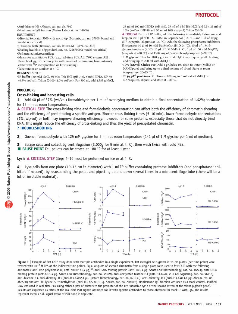
Figure 3 | Example of fast ChIP assay done with multiple antibodies in a single experiment. Rat mesagial cells grown in 15-cm plates (per time point) were
treated with 10�7 M TPA at the indicated time points. Equal aliquots of sheared chromatin from a single plate were used in fast ChIP with the following
antibodies: anti–RNA polymerase II, anti–hnRNP K (4 mg)30, anti–TATA-binding protein (anti-TBP, 4 mg, Santa Cruz Biotechnology, cat. no. sc273), anti–CREB
binding protein (anti-CBP, 4 mg, Santa Cruz Biotechnology, cat. no. sc369), anti–acetylated histone H3 (anti–H3-K9Ac, 2 ml Cell Signaling, cat. no. 9671S),
anti–histone H3, anti–dimethyl H3 (anti–H3-K4m2,1 ml, Upstate Biotechnology, cat. no. 07-030), anti–trimethyl H3 (anti–H3-K4m3,1 mg, Abcam, cat. no.
ab8580) and anti–H3 lysine 27 trimethylation (anti–H3-K27m3,1 mg, Abcam, cat. no. Ab6002). Nonimmune IgG fraction was used as a mock control. Purified
DNA was used in real-time PCR using either a pair of primers to the promoter of the TPA-inducible egr-1 or the second intron of the silent b-globin gene9.
Results are expressed as ratios of the real-time PCR signals obtained for IP with specific antibodies to those obtained for mock IP with IgG. The results
represent mean ± s.d. signal ratios of PCR done in triplicate.
NATURE PROTOCOLS | VOL.1 NO.1 | 2006 | 181
PROTOCOL
© 2
006
Nat
ure
Pub
lishi
ng G
roup
htt
p:/
/ww
w.n
atur
e.co
m/n
atur
epro
toco
ls

5| Centrifuge at 12,000g for 1 min at 4 1C and aspirate the supernatant.
6| Wash the nuclear pellet with 1 ml IP buffer containing inhibitors, by resuspending the pellet, followed by centrifugation.
Sonication7| To shear the chromatin, sonicate the washed pellet resuspended in 1 ml of IP buffer (with inhibitors) per 10-million cells(do not sonicate in volumes above 1 ml as this can decrease sonication efficiency).m CRITICAL STEP Sonication conditions must be determined empirically for each cell or tissue type, and sonicator model;optimal average DNA fragment sizes are 0.5–1 kb. Details of sonication procedure are given in Box 1.
8| Clear the lysate by centrifuging at 12,000g for 10 min at 4 1C. Retain the supernatant.
9| Transfer an aliquot of sheared chromatin (equivalent to 0.2 million cells) to a new microcentrifuge tube; this will be usedfor isolation of total DNA, to determine shearing efficiency and as a control for the amount of input DNA used in precipitations(to extract total DNA from this aliquot, skip to Step 19). The lysate can be aliquoted for use with multiple antibodies at thispoint. For best results, each aliquot should contain chromatin from 2 million cells.’ PAUSE POINT The chromatin can be stored at –80 1C for months.
Immunoprecipitation10| Add antibody to samples and incubate in an ultrasonic water bath for 15 min at 4 1C. For IP, use the desired antibody;for mock IP, use the same antibody preincubated with saturating amounts of its epitope-specific peptide for 30 min at roomtemperature. Alternatively, for mock IP, use the nonimmune IgG fraction from the same species in which the antibodies wereproduced. Incubation with beads without antibodies could also be used as a mock IP. If multiple antibodies are to be used withthe same chromatin preparation, a single mock IP is sufficient as a control for all the antibodies used.m CRITICAL STEP The amount of antibody added should be in excess of the factor being precipitated and thus should be deter-mined empirically for each factor/antibody. For abundant proteins, like histones, we typically used 1–2 mg of affinity-purifiedantibody or 2 ml of whole serum per IP. In addition, whereas the incubation time (15 min) has worked for many of the antibodieswe have used, the kinetics for reaching the equilibrium of antibody binding may differ for each antibody. The incubation timemay need to be increased for some antibodies. If an ultrasonic bath is not available, a long incubation at 4 1C should be used.In the traditional method, the times of incubation range from 1 to 12 h and should be determined empirically for each antibody.? TROUBLESHOOTING
11| Clear the chromatin by centrifugation at 12,000g for 10 min at 4 1C.? TROUBLESHOOTING
12| During Steps 10 and 11, wash protein A–agarose beads (20 ml per IP sample) three times with IP buffer to remove ethanol.A wash consists of resuspending the beads with 1 ml of IP buffer, centrifuging (1,000–2,000g) for a few seconds at 20–25 1C,and aspirating the supernatant.
BOX 1 | ADVICE ON SONICATION
To determine fragment size, extract total DNA from an aliquot of sheared chromatin (Steps 19–22) and run on 1% (wt/vol) agarose gel (stainwith EtBr). Use a sonicator with a microtip, and sonicate in a 1.5-ml tube. When sonicating, do not allow the sample to foam up, as thisdecreases the efficiency of DNA shearing. To avoid foaming, keep the tip of the sonicator probe no more than a few millimeters from thebottom of the tube. If foaming does occur, stop sonication and wait until the bubbles rise to the surface before continuing sonication.Sonication leads to heating of the sample; therefore, hold the sample in an ice-water bath during sonication. There are two main variables totest when determining optimum sonication conditions: the length of sonication and the power output. To avoid excessive heating, the totalsonication time is usually broken up into a number of ‘rounds’ of sonication, with a rest on ice between rounds (for instance, 4 rounds, eachwith 15 s of sonication, with a 2-min rest between rounds). Also, sonication using a series of short pulses is more efficient than that with asingle long pulse (i.e., 15 1-s-long pulses versus 1 15-s-long pulse), as the power output of the sonicator tip decreases from the beginning tothe end of a pulse. To start optimization, set the power output to 50% of the maximum output for the microtip, and sonicate with 10 to 15 1-s-long pulses per round, for two, four, six or eight rounds (with 2-min rest between rounds). Examine the shearing efficiency by means of gelanalysis of the sizes of DNA fragments, and if increasing the sonication time (number of rounds) does not give the desired average fragmentsize, try increasing the power output. When using very high power outputs, the total time for each round of sonication may need to bedecreased if the samples become excessively heated. An example of sonication conditions: we used three rounds of 15 pulses each at50% power output and 90% duty cycle using a Branson Sonifier 200. We also used a Misonix 3000 with 4–6 rounds of 15 1-s pulses at 50%power output.
182 | VOL.1 NO.1 | 2006 | NATURE PROTOCOLS
PROTOCOL
© 2
006
Nat
ure
Pub
lishi
ng G
roup
htt
p:/
/ww
w.n
atur
e.co
m/n
atur
epro
toco
ls

13| Dilute beads 1:1 with IP buffer and aliquot 40 ml of the slurry to clean tubes.m CRITICAL STEP Because agarose beads bind DNA nonspecifically, variation in the amount of added beads can affect the specificsignal/background ratio. Make sure to keep the slurry suspended while distributing and cut off the tips of small-bore pipette tips.? TROUBLESHOOTING
14| Transfer the top 90% of cleared chromatin (Step 11) to a tube with protein A–agarose slurry.m CRITICAL STEP It is very important to avoid carrying over any precipitated material, as this contains aggregated unspecificDNA complexes that can contaminate immunoprecipitated material.? TROUBLESHOOTING
15| Rotate tubes at 4 1C for 45 min on a rotating platform (20–30 rotations per min).
16| Washing the beads. Centrifuge the slurry at 1,000–2,000g for few seconds of 4–25 1C and remove the supernatant.Wash the beads 5–6 times with 1 ml cold IP buffer without inhibitors. A wash consists of resuspending the beads, centrifugingand aspirating the supernatant.
DNA isolationSteps 17–22 are performed at room temperature.17| Add 100 ml 10% (wt/vol) Chelex 100 slurry directly to the washed beads. Keep Chelex beads in suspension while pipetting.Also, make sure to cut off the tips of the fine-bore pipette tips before pipetting the Chelex slurry.
18| Briefly (10 s) vortex samples to mix the slurry, and boil for 10 min.
19| Precipitate the total DNA aliquot taken in Step 9 with 2.5–3 volumes of ethanol, and wash with 70% (vol/vol) ethanol.Then dissolve the dried pellet in 100 ml 10% (wt/vol) Chelex 100 suspension, boil for 10 min and continue processing in thesame way as the IP samples.
20| Optional. Proteinase K treatment. Wait for the samples to cool after boiling and add 1 ml of 20 mg ml–1 proteinase K toeach sample. Vortex, and then shake samples at 55 1C for 30 min on a thermal mixer (1,000 rpm). Boil again for 10 min. Thisboiling step is important to inactivate proteinase K, which may interfere with subsequent PCR.
21| Centrifuge condensate to the bottom of the tube at 12,000g for 1 min of 4 1C. Transfer supernatant (80 ml) to a new tube.Be careful to avoid transferring any Chelex resin as it can lead to a loss of PCR signal.
22| Add 120 ml of water (MilliQ or NANOpure) to beads, vortex for 10 s, centrifuge contents down at 12,000g for 1 min of4 1C, collect 120 ml of supernatant and pool with the previous supernatant. Mix before using.’ PAUSE POINT Store at –20 1C . We have thawed and frozen the samples repeatedly (more than 20 times over months)without loss of PCR signal.
Data analysis23| Purified DNA can be used in PCR at up to 25% of thereaction volume. We used SYBR Green Master Mix in a 10 mlreaction (2.5 ml DNA template, 0.3 ml primer pair (10 mMeach), 5 ml Master Mix and 2.2 ml H2O) in 384-well plates onan ABI 7900 (default three-step method, 40 cycles). Use ROXdye to correct for loading. Acquire data using the SDS 2.2.1program (ABI Biotechnology). For each primer pair, set thereadout in the middle of the linear range of amplificationsignals. The data can be exported to Excel spreadsheets.The relative occupancy of the immunoprecipitated factorat a locus is estimated using the following equation:2^(Ctmock – Ctspecific), where Ctmock and Ctspecific are meanthreshold cycles of PCR done in triplicate on DNA samples frommock and specific immunoprecipitations (ref. 9 and Figs. 2and 3). If gel electrophoresis is used to estimate theintensities of PCR products, then the relative occupancy of afactor at a locus is estimated as the ratio of the intensity ofthe specific IP band to that of the mock IP band10.? TROUBLESHOOTING
TIMELINESteps 1–3 Crosslink and harvest cells/tissue
(Store at –80 °C)
(Store at –80 °C)
(Store at –20 °C)
Steps 4–6 Lyse cells and wash pellet (30 min)
Steps 7–9 Shear chromatin and centrifuge (60 min)
Step 10 Add antibody and incubate in ultrasonic bath (15 min)
Step 11 Centrifuge to clear aggregates (10 min)
Steps 14–15 Apply chromatin to protein A beads (45 min)
Step 16 Wash beads (30–60 min)
Steps 17–18 Add Chelex 100 and boil beads (10 min)
Step 21 Centrifuge and collect supernatant (1 min)
Step 22 Add 120 µl PCR-grade water to beads, vortex and pool with previous supernatant (1 min)
Step 23 Run PCR
Step 20 Add proteinase K (optional) and incubate at 55 °C (30 min)
Step 20 Boil again (10 min)
Figure 4 | Timeline of the fast ChIP protocol.
NATURE PROTOCOLS | VOL.1 NO.1 | 2006 | 183
PROTOCOL
© 2
006
Nat
ure
Pub
lishi
ng G
roup
htt
p:/
/ww
w.n
atur
e.co
m/n
atur
epro
toco
ls

� TIMINGSee Figure 4.
? TROUBLESHOOTINGSee Table 1.
ANTICIPATED RESULTSWe found that four 15-cm plates of primary rat mesangial cells (80% confluence, approximately 2.5–3.0 � 107 cells) yield 2 mlof sheared chromatin, of which 150 ml is sufficient for ChIP with one antibody. This allowed us to probe 12 different factorsfrom one chromatin preparation (Fig. 3). Each chromatin IP yields sufficient amounts of DNA for 80 10-ml real-time PCRs, whichallowed us to monitor as many as 20–25 genomic sites.
ACKNOWLEDGMENTS We thank members of the K.B. lab for valuable discussions ofthe method. This work was supported by the US National Institutes of Health(DK45978 and GM45134) and the Juvenile Diabetes Research Foundation (K.B.).
COMPETING INTERESTS STATEMENT The authors declare that they have nocompeting financial interests.
Published online at http://www.natureprotocols.comReprints and permissions information is available online at http://npg.nature.com/reprintsandpermissions
1. Bernstein, E. & Allis, C.D. RNA meets chromatin. Genes Dev. 19, 1635–1655(2005).
2. Schubeler, D. & Elgin, S.C. Defining epigenetic states through chromatin and RNA.Nat. Genet. 37, 917–918 (2005).
3. Felsenfeld, G. & Groudine, M. Controlling the double helix. Nature 421, 448–453(2003).
4. Sims, R.J. III, Mandal, S.S. & Reinberg, D. Recent highlights of RNA-polymerase-II-mediated transcription. Curr. Opin. Cell Biol. 16, 263–271 (2004).
5. Thiriet, C. & Hayes, J.J. Chromatin in need of a fix: phosphorylation of H2AXconnects chromatin to DNA repair. Mol. Cell 18, 617–622 (2005).
TABLE 1 | Troubleshooting table
STEP PROBLEM REASON SOLUTION
1, 10, 16 Lower than expectedspecific IP/mock IPsignal ratio
Antibody not suitable for ChIP. Step 10. Check to make sure that the antibody is suggested for usewith ChIP. Try running an IP with uncrosslinked extracts, followedby a western blot to see if the antibody can precipitate the targetprotein. Also, try running a positive control antibody to make surethat all other aspects of the protocol are working. ChIP-gradeantibody to RNA pol II is a good positive control as it works well andChIP results can be compared to gene expression (transcript) levels.
Insufficient time for antibody bindingto the chromatin.
Step 10. Try increasing the time that the chromatin is incubatedwith the antibody.
Cross-linking time is too long, maskingthe epitope.
Step 1. Try decreasing cross-linking time (or formaldehyde con-centration).
Insufficient amount of chromatin used.If the PCR signal is equally low for bothspecific and mock IPs, the input may betoo low.
Step 10. Increase the amount of starting material until thespecific/mock IP ratio stops improving. A good starting point: use1–4 million cells per sample.
Too few washes leaving high levels ofnonspecific binding on the beads.
Step 16. Increase the number of washes, or add NaCl or LiCl (to afinal concentration of 0.5 M or 250 mM, respectively) to the washbuffer during the IP, and see if the ratio increases.
10, 11,13, 14
Variation in specific/mock IP ratiosbetween experiments
Insoluble chromatin complexes were notcleared from soluble chromatin beforeincubation with protein A beads.
Steps 11 and 14. Centrifuge chromatin after incubation withantibody and only apply the top 80–90% to the protein A beads.
Too little input leading to high Ct valuesfor real-time PCR or high cycle numbersfor regular PCR.
Step 10. Increase the amount of input chromatin until results forrepeated PCR experiments are reproducible.
Large variation in the amount of proteinA–agarose beads incubated with thechromatin, as the beads bind chromatinnonspecifically and contribute to thebackground.
Step 13. Keep protein A–agarose beads suspended and use pipettetip with cut-off tip when distributing to tubes.
23 No PCR signal PCR reaction did not work. Step 23. Run a PCR with genomic DNA from the same organism, as apositive control for primers and master mix.
Insufficient number of PCR cycles used(if not using real-time PCR).
Step 23. Run a few different reactions with increasing numbers ofcycles until a band is obtained.
184 | VOL.1 NO.1 | 2006 | NATURE PROTOCOLS
PROTOCOL
© 2
006
Nat
ure
Pub
lishi
ng G
roup
htt
p:/
/ww
w.n
atur
e.co
m/n
atur
epro
toco
ls
6. Kuo, M.H. & Allis, C.D. In vivo cross-linking and immunoprecipitation for studyingdynamic Protein: DNA associations in a chromatin environment. Methods 19,425–433 (1999).
7. Orlando, V., Strutt, H. & Paro, R. Analysis of chromatin structure by in vivoformaldehyde cross-linking. Methods 11, 205–214 (1997).
8. Impey, S. et al. Defining the CREB regulon: a genome-wide analysis oftranscription factor regulatory regions. Cell 119, 1041–1054 (2004).
9. Nelson, J.D., Denisenko, O., Sova, P. & Bomsztyk, K. Fast chromatinimmunoprecipitation assay. Nucleic Acids Res. 34, e2 (2006).
10. Ostrowski, J., Kawata, Y., Schullery, D.S., Denisenko, O.N. & Bomsztyk, K.Transient recruitment of the hnRNP K protein to inducibly transcribed gene loci.Nucleic Acids Res. 31, 3954–3962 (2003).
11. Dundr, M. et al. A kinetic framework for a mammalian RNA polymerase in vivo.Science 298, 1623–1626 (2002).
12. Cheutin, T. et al.Maintenance of stable heterochromatin domains by dynamic HP1binding. Science 299, 721–725 (2003).
13. Cosma, M.P., Tanaka, T. & Nasmyth, K. Ordered recruitment of transcription andchromatin remodeling factors to a cell cycle- and developmentally regulatedpromoter. Cell 97, 299–311 (1999).
14. Liu, C.L. et al. Single-nucleosome mapping of histone modifications inS. cerevisiae.. PLoS Biol. 3, e328 (2005).
15. Metivier, R. et al. Transcriptional complexes engaged by apo-estrogen receptor-alpha isoforms have divergent outcomes. EMBO J. 23, 3653–3666 (2004).
16. Koyanagi, M. et al. EZH2 and histone 3 trimethyl lysine 27 associated with Il4 andIl13 gene silencing in Th1 cells. J. Biol. Chem. 280, 31470–31477 (2005).
17. Solomon, M.J. & Varshavsky, A. Formaldehyde-mediated DNA-proteincrosslinking: a probe for in vivo chromatin structures. Proc. Natl. Acad. Sci. USA82, 6470–6474 (1985).
18. Thorne, A.W., Myers, F.A. & Hebbes, T.R. Native chromatin immunoprecipitation.Methods Mol. Biol. 287, 21–44 (2004).
19. Bernstein, B.E. et al. Genomic maps and comparative analysis of histonemodifications in human and mouse. Cell 120, 169–181 (2005).
20. Pokholok, D.K. et al. Genome-wide map of nucleosome acetylation andmethylation in yeast. Cell 122, 517–527 (2005).
21. Cawley, S. et al. Unbiased mapping of transcription factor binding sites alonghuman chromosomes 21 and 22 points to widespread regulation of noncodingRNAs. Cell 116, 499–509 (2004).
22. Orlando, V. Mapping chromosomal proteins in vivo by formaldehyde-crosslinked-chromatin immunoprecipitation. Trends Biochem. Sci. 25, 99–104 (2000).
23. Chen, R. et al. Ultrasound-accelerated immunoassay, as exemplified by enzymeimmunoassay of choriogonadotropin. Clin. Chem. 30, 1446–1451 (1984).
24. Chaya, D. & Zaret, K.S. Sequential chromatin immunoprecipitation from animaltissues. Methods Enzymol. 376, 361–372 (2004).
25. Schwabish, M.A. & Struhl, K. Evidence for eviction and rapid deposition ofhistones upon transcriptional elongation by RNA polymerase II. Mol. Cell. Biol.24, 10111–10117 (2004).
26. Denisenko, O. & Bomsztyk, K. Yeast hnRNP K-like genes are involved in regulationof the telomeric position effect and telomere length. Mol. Cell. Biol. 22, 286–297(2002).
27. Kurdistani, S.K., Tavazoie, S. & Grunstein, M. Mapping global histone acetylationpatterns to gene expression. Cell 117, 721–733 (2004).
28. Sandoval, J. et al. RNAPol-ChIP: a novel application of chromatinimmunoprecipitation to the analysis of real-time gene transcription. Nucleic AcidsRes. 32, e88 (2004).
29. Waugh, D.S. Making the most of affinity tags. Trends Biotechnol. 23, 316–320(2005).
30. Van Seuningen, I., Ostrowski, J. & Bomsztyk, K. Description of an IL-1-responsivekinase that phosphorylates the K protein. Enhancement of phosphorylationby sequence-selective DNA and RNA motifs. Biochemistry 34, 5644–5650(1995).
NATURE PROTOCOLS | VOL.1 NO.1 | 2006 | 185
PROTOCOL
© 2
006
Nat
ure
Pub
lishi
ng G
roup
htt
p:/
/ww
w.n
atur
e.co
m/n
atur
epro
toco
ls