protocols for isolation and evaluation of muse cells · protocols for isolation and evaluation of...
TRANSCRIPT

Protocols for isolation and evaluation of Muse cells
Table of Contents
Be sure to read the following comments because these are important issues influencing the Muse cell yield.
1) Materials: 1-1) Commercial culture cells as a source for Muse cells 1-2) Reagents, instruments and equipment 2) Cell culture: 2-1) Culture medium 2-2) Serum lot check 2-3) Thawing frozen cells 2-4) Subculture of mesenchymal stem cells (example of a 10-cm dish scale culture) 2-5) Cryopreservation of the cells for making stocks 3) Procedures for labeling Muse cells with SSEA-3: 3-1) Preparation of secondary antibodies 3-2) Preparation of FACS buffer 3-3) Preparation of mesenchymal stem cells for Muse cell collection 3-4) Staining with primary antibodies 3-5) Staining with secondary antibodies 4) Analysis of SSEA-3-positivity and the procedure for collecting Muse cells 4-1) Gate setting and data acquisition 4-2) Muse cell sorting 5) Poly-HEMA coating of wells and dishes 6) Bulk production of Muse cell derived-clusters by using methylcellulose gel 7) Generation of Muse cell-clusters in single cell-suspension culture 8) Evaluation of pluripotency of Muse cell-clusters: Alkaline phosphatase reaction 9) Evaluation of pluripotency of Muse cell-clusters: Gelatin culture for trilineage differentiation

[Flowchart of Muse cell collection]
: Muse cell (SSEA-3 positive)
1. Preparation of cell suspension
2. Muse cell staining with anti-SSEA-3 antibody (primary antibody)
Anti-SSEA-3 (Rat, IgM)
Staining of cell suspensions
SSEA-3
Adhesion culture
Detachment
Cell suspension
Intracellular
Figure: Staining image of SSEA-3
Anti-SSEA-3
3. Staining with fluorescent probe-labeled anti-Rat IgM (secondary antibody)
Figure: Staining image of secondary antibody Staining of cell suspensions
Anti-SSEA-3 Secondary antibody
Intracellular
Laser
Fluorescence Scattering light
Polarizer
Analysis of Muse cells with anti-SSEA-3 antibody + labeled secondary antibody
Muse cells (SSEA-3 positive)
Isolated Muse cells
4. Analysis and isolation of Muse cells by FACS
Mixture of cells and buffer
Laser irradiation, droplet formation and electric charge
Collection of target cells
Secondary antibody
Mesenchymal stem cell
SSEA-3
Plasma membrane
Plasma membrane

1) Materials
1-1) Commercial culture cells as a source for Muse cells (cells used in our laboratory)
Cultured mesenchymal stem cells are the source of Muse cells. Mesenchymal stem cells can be obtained commercially or by primary culture of tissue sources. The below four mesenchymal stem cells are commercially obtainable cells confirmed in Dezawa’s lab to yield a reasonable proportion of Muse cells.
Human Bone Marrow Mesenchymal Stem Cells; BM-MSCs (Cat#PT-2501, Lonza)
Normal Human Dermal Fibroblasts-adult skin; NHDF (Cat#CC-2511, Lonza)
Human Dermal Fibroblasts-adult; HDFa (Cat#2320, ScienCell research laboratories)
Human Adipose Derived Stem Cells; ADSCs (Cat#PT-5006, Lonza)
1-2) Reagents, instruments and equipment
Reagents
[Important] Human-FGF-2, premium grade (for culture of BM-MSCs, Cat#130-093-840, Miltenyi)
Change of lot number sometimes associates with change of activity. The latest information is announced in the home page of Dezawa’s lab, ‘Protocol’.
[Important] Rat anti-SSEA-3 antibody (Cat#330302, BioLegend or Cat#MA1-020, Thermo Fisher Scientific)
Change of lot number sometimes associates with change of Muse cell activity. The latest information for human FGF-2 and anti-SSEA-3 antibody is announced in the Dezawa’s lab home page. Please go to ‘Protocol’ of the home page and check the information.
Rat IgM Isotype control (Cat#400801, BioLegend)
Goat anti-Rat IgM antibody (FITC-labeled) (Cat#112-095-075, Jackson ImmunoResearch)
Fetal bovine serum (FBS) Selection of FBS is the key point because Muse cell activity and proportion are largely dependent on the quality of FBS. Please be sure to read "Lot check for FBS" in the next section for selection of FBS.
FBS (for inactivation of trypsin, no manufacturer specified)
[Important] Low-glucose DMEM+GlutaMAX (Cat#10567, Thermo Fisher Scientific) →for culturing BM-MSCs, NHDF and HDFa
[Important] High-glucose DMEM+GlutaMAX (Cat#11965, Thermo Fisher Scientific) →for culturing ADSCs
Kanamycin Sulfate (100 x) (Use at 1x dilution in media, Cat#15160-054, Thermo Fisher Scientific)
PBS (10 x) (Cat#27575-31, Nacalai tesque)
Sterile water (1x, for PBS preparation) (Cat#06442-95, Nacalai tesque)
Trypsin (0.25 %)/EDTA (Cat#25200-072, Thermo Fisher Scientific)
FluoroBrite DMEM (Cat#A18967-01, Thermo Fisher Scientific)
BSA (Bovine serum albumin) (Cat#01860-65, Nacalai tesque)
EDTA (Cat#15111-45, Nacalai tesque)
Gelatin (Cat#G-1890, Sigma)
Poly-HEMA [poly(2-hydroxyethyl methacrylate)] (Cat#P3932, Sigma)
MethoCult H4100 (Cat#04100, StemCell Technologies)
Instruments and equipment
Cell counter plate (disposable) (Cat# WC2-100S, Waken BTech Co, Ltd)
Cellbanker 1 plus, cryopreservation solution for culture cells (Cat#CB021, ZENOAQ)
CryoTube vials (Cat#377267, Thermo Fisher Scientific)
BiCell, CryoTube container (for preserving culture cells in deep freezer) BICELL/SANO910/ ask [email protected]
[Recommended] 10-cm dish (Cat#150464, Thermo Fisher Scientific)
1.5-mL tube (Cat#BM-15, BMBio)
15-mL tube (Cat#352096, Corning)
50-mL tube (Cat#352070, Corning)
Cell strainer (40 µm) (Cat#352340, Falcon)
0.22 µm filter (Cat#SLGV033RS, Merck Millipore)
Centrifuge with swing rotor (15 mL, 50 mL) (no manufacturer specified)
Centrifuge with swing rotor and cooler (1.5 mL) (no manufacturer specified)
Cell sorter (BD FACS Aria II) (used in Dezawa’s lab)
FACS analysis software (BD FACSDiva) (used in Dezawa’s lab)

2) Cell culture 2-1) Culture medium
BM-MSCs: Low-glucose DMEM, 10%FBS, 1 ng/mL FGF-2, 0.1 mg/mL Kanamycin
NHDF and HDFa: Low-glucose DMEM, 10%FBS, 0.1 mg/mL Kanamycin
ADSCs: High-glucose DMEM, 15%FBS, 0.1 mg/mL Kanamycin
ATTENTION! Use low-glucose DMEM or high-glucose DMEM depending on the cell type. For cultures of BM-MSCs, NHDF and HDFa, always use 10% FBS (HyClone) / low-glucose DMEM (Thermo Fisher Scientific), and NOT high-glucose DMEM. Use of high-glucose DMEM induces hypoproliferative capacity and decreases Muse cell yield. On the other hand, for culture of ADSCs, use 15% FBS (HyClone) / high-glucose DMEM (Thermo Fisher Scientific).
ATTENTION! Use FGF-2 (bFGF) for culture of bone marrow-derived mesenchymal stem cells. For culture of BM-MSCs, add FGF-2 (concentration: 1 ng/mL, Cat#130-093-840, Miltenyi). Be aware that Muse cell yields change markedly by using products from several manufacturers (refer to the following figure). For the latest information, please contact Mari Dezawa (mdezawa*med.tohoku.ac.jp, please convent “*” into “@”) because the SSEA-3-positive rate depends on lots and manufacturers (Fig. 1)
Figure 1. Quality of FGF-2 produces a difference in SSEA-3-positive cell ratio

ATTENTION! The cells listed in 1-1) Commercial culture cells as a source for Muse cells are examples of sources that can be used to reproduce our data. Other mesenchymal stem cells may also work as a source for Muse cells. However, the outcome cannot be guaranteed unless the above cell types are used.
ATTENTION! When purchasing cells, culture media specialized for the cells should also be bought from the same company to maintain the cells according to the manufacturer’s instructions.
ATTENTION! The cells listed in 1-1) Commercial culture cells as a source for Muse cells have a number of different lots. Because the cell growth rate and Muse cell ratio may differ among lots, we recommend purchasing a couple of lots and then selecting the best lot for the experiment
2-2) Serum lot check Mesenchymal stem cells are used for the lot check. Expanded mesenchymal stem cells are collected by
trypsin incubation. Count the number of cells and suspend cells at a concentration of 1×10⁴ cells/500 μL in 10% FBS (for serum
lot check) in DMEM (+Kanamycin) and plate them individually in a 24-well plate. Incubate cells at 37°C, 5% CO2. Change medium the next day. Culture medium is exchanged every 2 to 3 days. Perform subculture when cells reach at 90% confluence,. Cells should be expanded to 1:2 (one 90%
confluent plate is subcultured to two plates). Never exceed 1:3. Incubate cells at 37°C, 5% CO2. Cells should be subcultured at least for 2 to 3 times under the same FBS lot before evaluation. Do not use the
initial culture for the quality check of the FBS lot because the effect of the serum from the past medium may be remained.
The quality of the FBS lot will be determined based on cell growth, morphology, and spontaneous formation of specific cell clusters. Cell growth; cells are strongly recommended to subculture when they reach 90% confluent (see Figure 2). When cells are in a good condition, the frequency of subculture (namely the timing the cells reach 90% confluent) is every 2 to 3 days. However, when cells are in a bad condition, the timing of subculture requires more than 4 days, suggesting that cells are in the low proliferative activity. Morphology: an example of the morphology of cells in good and bad conditions is provided below.
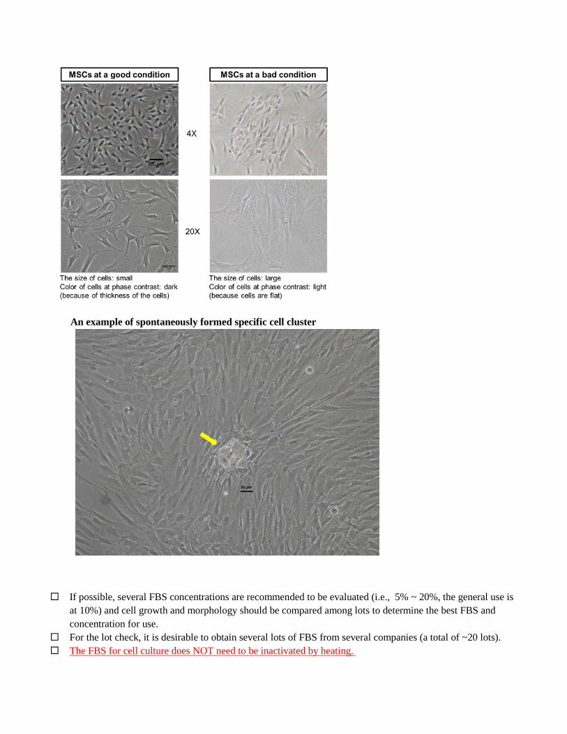
An example of spontaneously formed specific cell cluster
If possible, several FBS concentrations are recommended to be evaluated (i.e., 5% ~ 20%, the general use is at 10%) and cell growth and morphology should be compared among lots to determine the best FBS and concentration for use.
For the lot check, it is desirable to obtain several lots of FBS from several companies (a total of ~20 lots). The FBS for cell culture does NOT need to be inactivated by heating.

2-3) Thawing frozen cells
ATTENTION! Carefully read the manufacturer’s instructions prior to thawing purchased cells. Purchased cells usually arrive frozen in a vial. Transfer the vial into liquid N2 as soon as it arrives, and keep it there until use. Wash and clean hands using 70% ethanol (EtOH). Use sterilized gloves if necessary. Set a water bath to 37°C. Clean the bench by using 70% EtOH. Remove the frozen vial from the liquid N2, and quickly transfer to the 37°C water bath to thaw the cells.
Take care not to touch the vial cap to the water, otherwise the cells will be easily contaminated by bacteria.
ATTENTION! To avoid cell death, remove the vial from the water bath before it is completely thawed. The best time to remove the vial from the water bath is the point at which the solution still contains a piece of ice. Clean the vial by 70% EtOH, and then bring it to the clean bench. Carefully open the cap, and melt the piece of ice with gentle pipetting. Transfer cells to a 15-mL Falcon tube. Slowly add culture medium to the tube to make a final volume 10 mL. Centrifuge the 15-mL tube at 300 g for 5 min. Remove the supernatant, add 1 mL culture medium and loosen the cell pellet with gentle pipetting, add 9 mL
culture medium, and count the total number of the cells by cell counter plate. Then plate the cells at the density indicated as follows: 1) cells just after the purchase for BM-MSCs, NHDF, HDFa, ADSC; 1.5 x 10⁴ cells/cm2 , 2) cells that were once expanded after purchase and were stored in liquid N2; 2 x 10⁴ cells/cm2.
ATTENTION! The cells should be plated homogenously. Culture the cells overnight at 37°C, 5% CO2. At next day of plating cells, remove the culture media and add 10 mL fresh culture medium for medium
exchange. If floating dead cells are still visible, wash a couple of times with culture media to remove all the dead cells.
ATTENTION! In the case of cells purchased from a company, thawing the frozen cells and culturing for the first time is counted as passage 1 (P=1) in this protocol. ATTENTION! If the cells were cryopreserved at P=4 and then thawed, the cells are counted as P=4 at this point and passage number continues after following subculture (i.e., P=5, P=6, ...). When the cells reach 90% confluence, subculture them as described in 2-4) Subculture of mesenchymal
stem cells (example of a 10-cm dish scale culture)
Vial
37℃

2-4) Subculture of mesenchymal stem cells (example of a 10-cm dish scale culture)
ATTENTION! The best timing for subculture of mesenchymal stem cells (NHDFs, HDFa, BM-MSCs, and ADSCs) is at when cells reach 90% confluence. Please see how NHDF/HDFa and BM-MSCs look like at 90% confluence in Figure 2 and 3.
ATTENTION! The growth of mesenchymal stem cells is strongly suppressed by contact inhibition. Please watch the cells and perform subculture before reaching 100% confluence.
ATTENTION! Subculture the cells at 1:2 ratio. Never exceed 1:3 ratio; one ~90% confluent dish should be split into two dishes and never into more than 3 dishes. If Muse cells are expanded at 1:3 or more, then the percent of Muse cells will substantially decrease.
ATTENTION! Please use the specified culture medium recommended by the company you purchase the cells until P=2. After P=3, use 10% FBS in DMEM (+Kanamycin) for culture.
Figure 2. An example of 70%, 80%, 90% and over confluent in NHDF/HDFa
Bad timing for subculture (over confluent: cells are piling up)
Best timing for subculture (~90% confluent; some spaces are remaining between cells)
70% confluent 80% confluent 90% confluent Over confluent

Figure 3. An example of 70%, 80%, 90% and over confluent in BM-MSCs
Protocol for subculture: When the mesenchymal stem cells (NHDFs, HDFa, BM-MSCs, and ADSCs) reach 90% confluence, subject
the cells to subculture. Remove the culture medium, wash the cells with 10 mL serum-free DMEM for several times.
Add 2 ml trypsin per 10-cm dish. Rotate the dish to distribute the trypsin uniformly, and then incubate the cells at 37°C in 5% CO2 for 5 min.
After 5 min incubation, check under a phase contrast microscope whether the cells have detached from the dish.
ATTENTION! If all the cells are not detached, incubate for 5 more minutes at 37°C in 5% CO2 or add 1 mL trypsin and incubate for a couple of minutes. If these treatments do not work, the trypsin itself might be deactivated. Please prepare a fresh trypsin and redo.
If cell detachment is confirmed, add 1 mL FBS to inactivate the trypsin reaction. Gently pipette the cells in the dish using a P-1000 with a blue tip for dissociation of the cells.
ATTENTION! DO NOT use the P-200 yellow tip for pipetting. The opening of the yellow tip is not wide enough so that the cells will be seriously damaged by pipetting.
70% confluent 80% confluent 90% confluent Over confluent

Prepare a new 50-mL tube, and transfer the cells and reagents. Add 7 mL serum-free DMEM to the dish to collect the remaining cells, and transfer all the cells and reagents to the 50-mL tube.
Centrifuge the 50-mL tube at 300 g for 5 min. Discard the supernatant, add 1mL of culture medium and loosen the cell pellet by gentle pipetting. Add 19 mL culture medium to the cells and plate into two 10-cm dishes.
ATTENTION! Always subculture the mesenchymal stem cells at a ratio at 1:2. Never expand cells to 1:3 or more. Otherwise, cell growth or ratio of Muse cells will decrease. In case of a 10-cm dish, for example, expand one 10-cm dish to two 10-cm dishes.
Incubate the cells overnight at 37°C, 5% CO2. Exchange the medium the next day. Cells are maintained by exchanging medium every 2 to 3 days.
ATTENTION! Mesenchymal stem cells can be kept expanded and then used for Muse cell collection. We usually use P=4~P=10 cultured mesenchymal stem cells for collecting Muse cells for analytical and transplantation experiments, while cells over P=11 are not used for experiments, since the activity of Muse cells may not be fully guaranteed. We strongly recommend you to preserve mesenchymal stem cells in liquid N2 if Muse cells are not in use in the near-term rather than keeping subculture of the cells for a longer period.

2-5) Cryopreservation of the cells for making stocks At P=3 or P=4 of cultured mesenchymal stem cells (NHDFs, HDFa, BM-MSCs, and ADSCs), we usually dispense the cells and make a stock by cryopreservation When mesenchymal stem cells reach 90% confluence, subject the cells to subculture. Remove the medium,
and wash the cells several times with 10 mL serum-free DMEM. Add 2 ml trypsin per 10-cm dish. Rotate the dish to uniformly distribute the trypsin, and then incubate at
37°C, 5% CO2 for 5 min. After 5 min incubation, check under a phase contrast microscope to examine whether the cells have detached
from the dish. Once cell detachment is confirmed, add 1 mL serum to inactivate the trypsin reaction. Gently pipette the cells and solution to dissociate cells using a P-1000 with a blue tip. DO NOT use yellow
tip. Prepare a new 15-mL tube, and transfer all the cells and reagents. Add another 7 mL serum free-DMEM to
the dish to collect the remaining cells, and transfer the cells and reagents to the 15-mL tube. Centrifuge the 15-mL tube at 300 g for 5 min. Discard the supernatant. Add Cellbanker 1 plus (1 mL) to the cells, and gently mix by pipetting. ATTENTION! The correct volume of Cellbanker 1 plus is 2~3 x 10⁶ cells/mL/tube. At least 1 mL of cells + Cellbanker 1 plus should be placed into one cryotube. Transfer the cells in 1 mL Cellbanker 1 plus to a cryotube and screw the lid to form a seal. Place the tube into the BiCell and gradually freeze at -80°C for 24 h. After freezing at -80°C for 24 h, transfer the tube into liquid N2 and store the cells.
ATTENTION! The cells can be stored at -80°C for 2~3 days without losing activity, but for a longer storage period, the cells must be stored in liquid N2.

3) Procedures for labeling Muse cells with SSEA-3 ATTENTION! If the purpose of Muse cell collection is to form Muse cell-derived clusters either by single cell suspension or by methylcellulose gel, please prepare poly-HEMA-coated plates in advance of making clusters. For details, refer to the section 5) Poly-HEMA coating of wells and dishes. Please bear in mind that poly-HEMA coating requires overnight treatment. 3-1) Preparation of secondary antibodies Dissolve commercially obtained FITC-labeled anti-rat IgM antibody (Cat#112-095-075, Jackson
ImmunoResearch) at 1.0 mg/mL in sterile water, dispense, and store at −30°C before use. 3-2) Preparation of FACS buffer Freshly prepare FACS buffer just before use and cool on ice constantly after preparation. Discard remainder
and do not re-use. FACS buffer 5% BSA 5 mL 100 mM EDTA 1 mL PBS or FluoroBrite DMEM 44 mL Total 50 mL/50-mL tube ATTENTION! 5% BSA solution: dissolve BSA in PBS or FluoroBrite DMEM, sterilize with 0.22 µm filter and store at 4°C. 100 mM EDTA solution: dissolve in PBS or FluoroBrite DMEM, sterilize with 0.22 µm filter and store at 4°C. If cell viability is decreased by using PBS, please use FluoroBrite DMEM instead. 3-3) Preparation of mesenchymal stem cells for Muse cell collection ATTENTION! Different from the subculture of mesenchymal stem cells described above, the mesenchymal stem cells must reach 100% confluence just before collecting Muse cells by FACS. If mesenchymal stem cells were under or over 100% confluence, the yield of Muse cells will be substantially decreased.

Figure 4. An example of the best timing for FACS in NHDF in 100 % confluent. Yellow arrows are locus that started pile up.
ATTENTION! For FACS analysis, mesenchymal stem cells must be completely dissociated into single cells. Because mesenchymal stem cells are sticky and the cells are at 100% confluent before trypsin incubation, incubation with trypsin should be longer than the usual subculture to obtain completely dissociated cells. ATTENTION! When obtaining Muse cells from frozen mesenchymal stem cells, the cells must be subcultured at least once before being subjected to FACS analysis. Mesenchymal stem cells soon after thawing and plating are weak and unstable, and provide a lower ratio of SSEA-3+ Muse cells. After thawing, the mesenchymal stem cells should be cultured to reach 90% confluence, subcultured at least once until they reach 100% confluence, and then subjected to FACS analysis. ATTENTION! When primary cultured mesenchymal stem cells are to be analyzed by FACS, the cells should be at P=4 to P=10. If the cells are earlier than P=3, collected Muse cells will be weak and unstable, and if they are later than P=10, the activity of Muse cells will be low. Using cells cultured in a 10-cm dish (the following procedures are for a 10-cm dish). Remove culture media and wash the cells several times with PBS to completely remove serum. Add trypsin (0.25%)/EDTA (2 mL) and incubate cells at 37°C in incubator for 5-10 min until cells are
detached from the dish. Stop trypsin reaction by adding FBS (1 mL) and transfer cells and all the medium into a 15-mL tube. Collect remaining cells in the dish with serum free-DMEM (7 mL) and transfer them into the same 15-mL
tube (total: ~10 mL). Centrifuge the tube with swing rotor (400 g, 5 min, room temperature) ATTENTION! Use centrifuge with swing rotor.

Remove supernatant and resuspend cell pellet in 10 mL of PBS or FluoroBrite DMEM. Centrifuge the tube with swing rotor (400 g, 5 min, room temperature) Remove supernatant, resuspend cell pellets with FACS buffer (1 mL), and sample a part of the cells for
counting. Add FACS buffer (9 mL) (total: 10 mL). Centrifuge with swing rotor (400 g, 5 min, room temperature). Count the number of cells during the above
processes. Remove supernatant and resuspend cell pellets in FACS buffer to establish cell density of 1×10⁶ cells/100 µL. ATTENTION! Be sure that cell density does not exceed 1×10⁶ cells/100 µL, otherwise cell will make aggregation. The upper limit of a cell suspension stainable in a 1.5-mL tube is 1×10⁷ cells/1000 µL. If the number of cells exceeds the upper limit, split the cells into another tubes. Sample dispensing and examples of cell numbers Dispense cell suspension of 1×10⁶ cells/100 µL into a 1.5-mL tube.
Table 3-1. Dispensing and cell number examples
3-4) Staining with primary antibodies ATTENTION! Be aware that anti-SSEA-3 antibodies released by several manufacturers do not work for labeling Muse cells. Recommended manufacturers of anti-SSEA-3 (anti-stage-specific embryonic antigen-3, clone MC-631, Rat IgM) antibodies are BioLegend (Cat#330302) and Thermo Fisher Scientific (Cat#MA1-020). Anti-SSEA-3 antibody of clone MC-631 is also available from several other manufacturers; however, those do not work for labeling Muse cells (see figure below). In order to receive the latest information, please contact Prof. Dezawa (mdezawa*med.tohoku.ac.jp) because the SSEA-3-positive rate markedly depends on company and lot number. Please convent “*” into “@”.
# Sample Dispensing volume
Number of cells
#1 Without staining 100 µL 1×10⁶ cells #2 Secondary antibodies only (anti-
Rat IgM, FITC) 100 µL 1×10⁶ cells
#3 Rat IgM Isotype Control + Secondary antibodies only
100 µL 1×10⁶ cells
#4 Rat anti-SSEA-3 + Secondary antibodies only
100 µL 1×10⁶ cells

Figure 5. The SSEA-3+ ratio in BM-MSCs by different company anti-SSEA-3 antibody (as of 2017).
While antibody from company D had a high positive rate, the negative population showed high background staining. D is not recommended.
ATTENTION! When using cells other than BM-MSCs, NHDF, HDFa and ADSC listed above, blocking with FcR is strongly recommended to be performed in the SSEA-3 antibody staining. Please incubate cells with 10% normal human serum at 4°C for 20 min before staining with primary antibodies.
ATTENTION! Prepare samples for isotype control. This is necessary for the accurate estimation of SSEA-3-positivity in FACS. Our laboratory use Rat IgM Isotype Control (Cat#400801, BioLegend) and evaluate the SSEA-3-positive cell rate based on the gating of Isotype Control in FACS analysis.
Place samples #1, 2, 3 and 4 in Table 3-1 on ice (samples should be kept on ice until analysis). Add rat IgM isotype control at a concentration of 0.5 µg/100 µL into Sample #3, stir slowly with a pipette,
and incubate on ice for 1 h (stir slowly every 10 min with a pipette). Add manufacturer-recommended rat anti-SSEA-3 antibody at the concentration of 0.5 µg/100 µL in Sample
#4, stir slowly with a pipette, and incubate on ice for 1 h (stir slowly every 10 min with a pipette). ATTENTION! After adding antibodies, stir slowly with a pipette to ensure a thorough reaction. A sample video is provided for demonstrating the correct pipetting → http://www.stemcells.med.tohoku.ac.jp/protocol/movie/suspension_good.mp4 When adding antibodies (both primary and secondary) to cell suspension for reaction, stir slowly with a pipette. Please pay attention not to leave any small pellet at the bottom of the tube. The pellet should be completely dispersed by pipetting. For larger volume suspensions (for example, 700-1000 µL/tube), disperse the pellet by inverting the tube, not by pipetting. Please prohibit harsh pipetting or vortexing. These may seriously damage cells and result in increase of cell death.

Table 3-2 Primary antibody list # Sample Rat IgM Isotype Control Rat anti-SSEA-3 #1 Without staining − − #2 Secondary antibodies only − − #3 Rat IgM Isotype Control + Secondary antibodies + (0.5 µg/100 µL) − #4 Rat anti-SSEA-3 + Secondary antibodies − + (0.5 µg/100 µL)
Start washing 1 h after the incubation with antibody. Add FACS buffer to make the total volume 1 mL. Centrifuge (400 g, 5 min, 4°C) ATTENTION! For 1.5-mL tubes, use a swing rotor for centrifugation. Remove supernatant and leave ~100 μL solution in the 1.5-mL tube (FACS buffer and cell pellet at 0.1 mL
total volume remains). After resuspending the pellet with slow stirring with a pipette, add 900 μL FACS buffer. Centrifuge (400 g, 5 min, 4°C) Remove supernatant and leave ~100 μL solution in the 1.5-mL tube. After resuspending the pellet with slow stirring with a pipette, add 900 μL FACS buffer. Centrifuge (400 g, 5 min, 4°C) Remove supernatant and leave ~100 μL solution in the 1.5-mL tube. After resuspending the pellet with slow stirring with a pipette, add 900 μL FACS buffer. Centrifuge (400 g, 5 min, 4°C) Remove supernatant and leave ~100 μL solution in the 1.5-mL tube. Resuspend the pellet with slow stirring using a pipette. 3-5) Staining with secondary antibodies ATTENTION! Non-specific staining may occur due to impurities in antibody solutions. To prevent such conditions, centrifuge secondary antibodies immediately before use (10,000 g, 3 min, 4°C) to precipitate impurities, and use supernatants only to eliminate impurities. Store remaining secondary antibodies at 4°C. Expiration date is within 2 weeks. Our lab uses anti-rat IgM antibody, FITC conjugates (Cat#112-095-075, Jackson Immunoresearch); however, anti-rat IgM antibody, APC conjugates (Cat#112-136-075, Jackson Immunoresearch) is also usable. Please select either FITC or APC conjugated rat IgM depending on the situation. Thaw on ice the secondary antibody (FITC or APC-labeled anti-rat IgM antibody) that were stored at −30°C. Mix the thawed secondary antibody solution with a pipette, centrifuge (10,000 g, 3 min, 4°C), and use the
supernatant for cell staining. Store the remaining secondary antibody at 4°C. Expiration date is within 2 weeks after thawing.
Add secondary antibody at a concentration of 1 µg/100 µL to Samples #2, 3 and 4, stir slowly with a pipette and incubate on ice for 1 h (stir slowly every 10 min with a pipette)

Table 3-3 Secondary antibody list # Sample FITC-labeled anti-rat IgM antibody #1 Without staining − #2 Secondary antibody only + (1 µg/100 µL) #3 Rat IgM Isotype Control + Secondary antibody + (1 µg/100 µL) #4 Rat anti-SSEA-3 + Secondary antibody + (1 µg/100 µL)
Start washing 1 h after the incubation with antibody. Add FACS buffer to make the total volume 1 mL Centrifuge (400 g, 5 min, 4°C) Remove supernatant and leave ~100 μL solution in the 1.5-mL tube (FACS buffer and cell pellet at ~100 μL
total volume remains). After resuspending the pellet with slow stirring with a pipette, add 900 μL FACS buffer. Centrifuge (400 g, 5 min, 4°C) Remove supernatant and leave ~100 μL solution in the 1.5-mL tube. After resuspending the pellet with slow stirring with a pipette, add 900 μL FACS buffer. Centrifuge (400 g, 5 min, 4°C) Remove supernatant and leave ~100 μL solution in the 1.5-mL tube. Resuspend the pellet in FACS buffer to make the cell concentration 1 x 10⁶ cells/ 0.1 mL and prepare for
analyses. Option: If the number of cells is larger than 1×10⁷ cells and cell aggregates are observed in suspensions before FACS Resuspend the pellet in an appropriate amount of FACS buffer and filter the cells with a cell strainer (40 µm). Centrifuge (400 g, 5 min, 4°C) Remove supernatant and leave ~100 μL solution in the 1.5-mL tube. Resuspend the pellet in FACS buffer to make the cell concentration 1 x 10⁶ cells/ 0.1 mL and prepare for
analyses.

4) Analysis of SSEA-3-positivity and the procedure for collecting Muse cells ATTENTION! A sample with a lot of dead cells is inappropriate for analysis and collection. Non-specific staining is likely to occur for dead cells, resulting in an inaccurate measurement of the Muse cell rate. Even if the sampling is forced for the further procedure, cell survival will be largely decreased. If the FSC (frontal scatting light)-area (A) vs. SSC (side scattering light)-A plot demonstrated a dead cell population in FACS (see the figure below), preparation of another new fresh sample is strongly recommended. 4-1) Gate setting and data acquisition (The following data was obtained by BD FACS Aria II) Load unstained sample #1 first. Adjust sensitivity for SSC and FSC and perform Area Scaling. Refer to the following figure.
Develop a plot of SSC-A vs. FSC-A. Refer to the following figure.
Successful example A sample with a lot of dead cells
(Inappropriate for analysis and collection)
One large population is visible Two populations are visible
Area scaling (before setting) Area scaling (after setting)
* Place dots on a diagonal line to upper right.
Draw a Gate P1.
Set SSC and FSC sensitivities to make the P1 % population more than 95%
Dead cell population
Normal cell population
Normal cell population

Develop a histogram of Count vs FITC-A. Refer to the following figure.
Develop a plot of SSC-A vs. FITC (screen to determine SSEA-3-positive rate). Refer to the following figure.
After setting all conditions, obtain data for an unstained sample loaded without change (Sample #1).
Load the stained sample #2 reacted with secondary antibody only.
Load the stained sample #3 reacted with Rat IgM Isotype Control + secondary antibody.
Adjust FITC voltage to make the whole histogram curve included in the graph.
Reflect the dots in Gate P1 to SSC-A vs. FITC, and newly form Gate P2. Set Gate P2 not to include any dots.
Adjust the position of Gate P2 that may not include any dots.
Gate P2 should be 0% for SSEA-3.
Adjust the position of Gate P2 that may not include any dots.
Gate P2 should be 0% for SSEA-3.

Load the stained sample #4 stained with Rat anti-SSEA-3 + secondary antibodies.
4-2) Muse cell sorting
To eliminate doublets, develop the plot for FSC-W vs FSC-H and SSC-W vs SSC. See the figure below.
Prepare a tube containing 10% FBS/DMEM (choose low-glucose or high-glucose type according to the cell type) and set it at a sorting site for receiving sorted cells.
Start sorting Muse cells by collecting the populations of Gate P4
Reflect the dots in Gate P2 to FSC-W vs FSC-H, and newly form Gate P3. This corresponds to Gate P2 of sample #4.
Set Gate P3 not to include doublets.
Reflect the dots in Gate P3 to SSC-W vs SSC-H, and newly form Gate P4. This corresponds to Gate P3 of sample #4.
Set Gate P4 not to include doublets.
Do not change the position of Gate P2 set at Isotype control sample #3.
Gate P2: this case showed 5.2% of SSEA-3-positive cells in BM-MSC.

5) Poly-HEMA coating of wells and dishes One of the characters of Muse cells is that they form clusters similar to ES cell-derived embryoid body in suspension culture. The formation of clusters is often used for evaluation of Muse cells. There are two methods to make Muse cell-derived clusters; one is single cell suspension culture and the other is suspension culture in Methylcellulose gel. However, Muse cells easily adhere to the bottom of culture dish/well unless the dish/well is not coated with poly-HEMA, the material that completely blocks the adherence of Muse cells. Therefore, pretreatment of dish/well with poly-HEMA before suspension culture is strongly required. 5-1) Add 1.2 g poly-HEMA (poly 2-hydroxyethyl methacrylate, Cat#P3932, Sigma) into 40 mL 95% EtOH (38 mL 99.5% EtOH + 2 mL MilliQ). . ATTENTION! Poly-HEMA is highly insoluble. It will not dissolve in 100% EtOH, either. Therefore, please keep in mind to fill the tube first with 95% EtOH solution and then add the poly-HEMA. 5-2) Shake the solution for several hours at 37°C to dissolve the poly-HEMA completely. Do not tilt the tube. The poly-HEMA at the bottom of the tube will not dissolve. 1. Add the dissolved poly-HEMA to the dish/well, and rotate the dish to homogenously cover the bottom of the dish/well.
Dish Volume 10 cm 3.2 mL 6 cm 1.3 mL
3.5 cm 500 μL 12 well 200 μL 24 well 100 μL 48 well 70 μL 96 well 25 μL
2. Leave the cover off the dish, and allow the dish to completely dry out in a clean cabinet overnight. The door of the cabinet should be left open 10 to 20 cm. Do not turn on a UV light. ATTENTION! When evaporating the poly-HEMA solution in dish/well in a clean cabinet, EtOH will be saturated in the cabinet. In this situation, dish/well will not dry completely, even when allowed to be placed in the cabinet overnight. Do not to make poly-HEMA coated dishes more than 20 dishes/plates at one time. ATTENTION! Dried dishes can be stored in the dark, at room temperature for a couple of months. 3. Wash the poly-HEMA-coated dish at least 3 times by PBS before use.
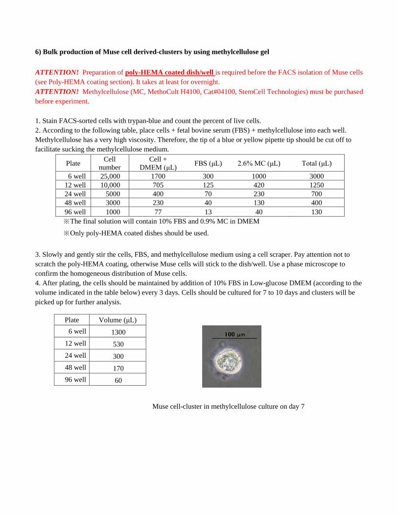
6) Bulk production of Muse cell derived-clusters by using methylcellulose gel
ATTENTION! Preparation of poly-HEMA coated dish/well is required before the FACS isolation of Muse cells (see Poly-HEMA coating section). It takes at least for overnight. ATTENTION! Methylcellulose (MC, MethoCult H4100, Cat#04100, StemCell Technologies) must be purchased before experiment. 1. Stain FACS-sorted cells with trypan-blue and count the percent of live cells. 2. According to the following table, place cells + fetal bovine serum (FBS) + methylcellulose into each well. Methylcellulose has a very high viscosity. Therefore, the tip of a blue or yellow pipette tip should be cut off to facilitate sucking the methylcellulose medium.
Plate Cell number
Cell + DMEM (μL) FBS (μL) 2.6% MC (μL) Total (μL)
6 well 25,000 1700 300 1000 3000 12 well 10,000 705 125 420 1250 24 well 5000 400 70 230 700 48 well 3000 230 40 130 400 96 well 1000 77 13 40 130 ※The final solution will contain 10% FBS and 0.9% MC in DMEM ※Only poly-HEMA coated dishes should be used.
3. Slowly and gently stir the cells, FBS, and methylcellulose medium using a cell scraper. Pay attention not to scratch the poly-HEMA coating, otherwise Muse cells will stick to the dish/well. Use a phase microscope to confirm the homogeneous distribution of Muse cells. 4. After plating, the cells should be maintained by addition of 10% FBS in Low-glucose DMEM (according to the volume indicated in the table below) every 3 days. Cells should be cultured for 7 to 10 days and clusters will be picked up for further analysis.
Plate Volume (μL) 6 well 1300
12 well 530 24 well 300 48 well 170 96 well 60
Muse cell-cluster in methylcellulose culture on day 7

7) Generation of Muse cell-clusters in single cell-suspension culture ATTENTION! 96 well poly-HEMA coated dishes need to be prepared before FACS isolation. It takes over night for preparation. (see Poly-HEMA coating above) ATTENTION! Use 10% FBS in low-glucose DMEM for culturing BM-MSCs, NHDFs and HDFs, and 15% FBS in high-glucose DMEM for ADSCs. Furthermore, be sure to add 1 ng/mL FGF-2 (bFGF) to culture BM-MSCs. 1. Stain FACS sorted cells by trypan-blue and count the number of live cells. 2. For limiting dilution, calculate cell number and adjust cell solution in medium. For example: each well needs 100 μL medium. For 96 well-one plate, 96 cells are suspended in 9600 μL medium. However, we routinely adjust the medium to make ~3 cells per well. Therefore, the cell solution will be ~288 cells in 9600 μL medium. The cell solution will be gently mixed and then plated 100 μL to each well. This will properly make 1 cell in each well after plating. Logical calculation is usually too strict to make single cell in each well. 3. Next day, observe each well under phase microscope and check vacant well and well with multiple number of cells from counting. Those wells should be eliminated from counting the cluster formation ratio. 4. Add 30 μL of medium for each well every 3 days. Culture for 7~10 days and pick up Muse cell-derived clusters for analysis.
Clusters formed in single cell-suspension culture at day 7.

8) Evaluation of pluripotency of Muse cell-clusters: Alkaline phosphatase reaction
ATTENTION! Purchase Leukocyte Alkaline Phosphatase Kit (Cat#86R-1KT, Sigma). ATTENTION! Do not use PBS for alkaline phosphatase (ALP) reaction. PBS is used for stopping the reaction. 1. Prepare ALP solution according to the manufacture's protocol. In brief,
Mix 10 μL of Sodium Nitrite Solution and 10 μL of FRV-Alkaline Solution. Both solutions are provided in the kit. Leave for 2 min in room temperature, and then add 450 μL saline.
↓ Add 10 μL Naphthol AS-BI Alkaline Solution to the above solution (this is also provided in the kit).
2. Collect Muse cell-clusters in 1.5-mL tube, add 1 mL saline to suspend the clusters. Do not use PBS!! 3. Centrifuge at 400 g in room temperature for 5 min. Remove the supernatant. 4. Add 1 mL saline to suspend the clusters. 5. Centrifuge at 400 g in room temperature for 5 min. Remove the supernatant. 6. Add 1 mL saline to suspend the clusters. 7. Centrifuge at 400 g in room temperature for 5 min. Remove the supernatant as much as possible. 8. Fix the clusters by 4% paraformaldehyde. → This is optional. Reaction would be stronger without fixation. We usually skip this procedure. 9. Add 200 μL ALP solution to the clusters. Incubate in 37°C incubator for 15 min. → The manufacture’s protocol instructs us to incubate in room temperature, but in our experience, 37°C gives better reaction. 10. Add 800 μL PBS to stop the ALP reaction. 11. Centrifuge at 400 g in room temperature for 5 min. Remove the supernatant. 12. Add 1 mL PBS to suspend the clusters. 13. Centrifuge at 400 g in room temperature for 5 min. Remove the supernatant. 14. Transfer clusters to slide class and observe under light microscope.

9) Evaluation of pluripotency of Muse cell-clusters: Gelatin culture for trilineage differentiation
ATTENTION! Prepare gelatin coated dish or cover slip before doing following experiment. 1. Preparing Gelatin coated dish or cover slip. Gelatin ( Cat#G-1890, Sigma) → Stock solution is 0.1% gelatin in PBS. Sterilize by autoclaving and use. For coating, load plentiful amount of 0.1% gelatin solution in plastic wells or wells placed cover slips (we usually use 18 mm diameter round cover slip for 24 well plate) in the bottom, incubate at 37 °C at least for 30 min.
For use, aspirate gelatin solution and directly use for experiment without washing.
After coating, pay attention NOT TO DRY the coated wells or cover slips. 2. Pick up Muse cell-derived clusters. Use glass capillary or pipetman (P20 scale) for pick up the clusters.
ATTENTION! In the case of clusters formed in methylcellulose, wash clusters by Low-glucose DMEM for a couple of times because methylcellulose clings to cluster disturb its adherence to dish or cover slip. In brief, supply 200 μL Low-glucose DMEM into each well of 4 well plate, transfer clusters into Low-glucose DMEM and wash several times by pipetting.
3. Remove gelatin solution after incubation in 37 °C, and quickly supply 10% FBS in Low-glucose DMEM into each well. Pay attention not to dry the dish or cover slip.
Initially, the volume of medium should be a bit lesser than usual. Lesser volume makes transferred clusters easier to adhere to the bottom of dish or to the set cover slip.
For example, 250 μL for 24 well scale, ~800 μL for 12 well. 4. Transfer clusters into above well. 5. After a couple of hours, add the 10% FBS in Low-glucose DMEM for volume-up. For 24 well, 300~400 μL and
for 12 well, 1 mL solution is preferable for the final volume.
6. Clusters will adhere to the bottom of the well or cover slip by next day or latest by 3 days. The cells gradually
expand out of the cluster (see the picture below).

Expansion of cells from the cluster. 10 days after culture.
7. Culture for 1~2 weeks, and then subject the samples to RT-PCR or immunocytochemistry. <For RT-PCR> Use following small scale kits for isolation of mRNA and for reverse transcription. NucleoSpin RNA XS: Cat#740 902.10, Macherey-Nagel SuperScript VILO cDNA Synthesis Kit: Cat#11754050, Thermo Fisher Scientific TaKaRa Ex Taq: Cat#RR001A , TaKaRa
human RT-PCR primer β-actin F: 5’-GGCGGACTATGACTTAGTTGCGTTACACC-3’
R: 5’-AAGTCCTCGGCCACATTGTGAACTTTG-3’ Nkx2.5 F: 5’-GGGACTTGAATGCGGTTCAG-3’
R: 5’-CTCCACAGTTGGGTTCATCTGTAA-3’ α-fetoprotein F: 5’-CCACTTGTTGCCAACTCAGTGA-3’
R: 5’-TGCAGGAGGGACATATGTTTCA-3’ MAP-2 F: 5’-ACTACCAGTTTCACACCCCCTTT-3’
R: 5’-AAGGGTGCAGGAGACACAGATAC-3’ GATA6 F: 5’-CCTGCGGGCTCTACAGCAAGATGAAC-3’
R: 5’-CGCCCCTGAGGCTGTAGGTTGTGTT-3’
<For immunocytochemistry> Fix the sample with 4% (v/v) paraformaldehyde / 0.01M PBS
Antibodies for use anti-SMA (Cat#MS-113-P0, Thermo Fisher Scientific, 1:100) anti-Neurofilament-M (Cat#AB1987, Merck Millipore, 1:200) anti-α-fetoprotein (Cat#N1501, DAKO, 1:100) anti-desmin (Cat#550626, BD Biosciences, 1:100) anti-cytokeratin 7 (Cat#MAB3226, Merck Millipore, 1:100)
Blocking solution: 20% (vol/vol) BlockAce / 5% (wt/vol) BSA / 0.3% (vol/vol) Triton X-100 / 0.02M D-PBS Antibody diluent: 5% (vol/vol) BlockAce / 1% (wt/vol) BSA / 0.3% (vol/vol) Triton X-100 / 0.02M D-PBS