proton radiotherapy for skull base and para-spinal … documents... · carmen ares, center for...
TRANSCRIPT

Carmen Ares, Center for Proton Therapy, Winter School, January 10-13, 2010
Proton Radiotherapy for Skull Base and
Para-spinal Tumors
Carmen Ares

Carmen Ares, Center for Proton Therapy, Winter School, January 10-13, 2010
• Primary tumors-
Chordomas, Chondrosarcomas
• Secondary infiltration or involvement by intracranial tumors-
Meningiomas
• Secondary infiltration by primary H&N tumors-
Adenoid Cystic Ca
-
Nasopharynx
Ca-
Paranasal
Sinus Ca
Tumors of the Skull Base

Carmen Ares, Center for Proton Therapy, Winter School, January 10-13, 2010
Skull Base Primary Tumors:
Chordomas and Chondrosarcomas

Carmen Ares, Center for Proton Therapy, Winter School, January 10-13, 2010
Primary tumors: Chordomas
– rare primary bone tumor
# incidence rate <0.1 per 100,000 per year
# accounts for 1 -
4%
of all primary malignant bone tumors
– arise from embryonic remnants of notochord
– show a dual epithelial-mesenchymal
differentiation

Carmen Ares, Center for Proton Therapy, Winter School, January 10-13, 2010
–
microscopic foci of notochord remain in the vertebral bodies of the embryo
– malignant transformation
typically occurs in:
# 3rd
- 4th
decades of life for spheno-occipital
lesions
# 5th
- 6th
decades for the sacro-coccygeal
type
Primary tumors: Chordomas

Carmen Ares, Center for Proton Therapy, Winter School, January 10-13, 2010
- as have ectodermal
origin are technically not sarcomas
-
however traditionally classified and approached as sarcomas
on the basis of being a primary bone tumor
Primary
tumors: Chordomas

Carmen Ares, Center for Proton Therapy, Winter School, January 10-13, 2010
– Usually relatively slow-growing, low grade malignancies
– Localization
– Sacrum
50% -
60%
– Skull base region 25% -
35%
– Cervical spine 10%
– Thoraco-lumbar spine 5%
Primary
tumors: Chordomas

Carmen Ares, Center for Proton Therapy, Winter School, January 10-13, 2010
– Have low metastatic potential
–
Control of primary disease remains the major therapeutic challenge
– Metastatis
usually occur after the local failure of the disease (to
lung, bone, soft tissue, lymph node, liver, and skin)
Primary
tumors: Chordomas

Carmen Ares, Center for Proton Therapy, Winter School, January 10-13, 2010
• Develop in any bone performed by cartilage
•
Primary chondrosarcomas
of the cranial base arise from the
chondrocranium
• Association with
# Ollier’s
disease (enchondromatosis)
# Maffucci’s
syndrome (enchondromatosis
+ hemangiomatosis)
Primary tumors: Chondroscarcomas

Carmen Ares, Center for Proton Therapy, Winter School, January 10-13, 2010
Primary chondrosarcomas
of the cranial base arise from the chondrocranium
ChondrocraniumPart of the skull base that undergoes enchondral
ossification during fetal life
Primary tumors: Chondroscarcomas
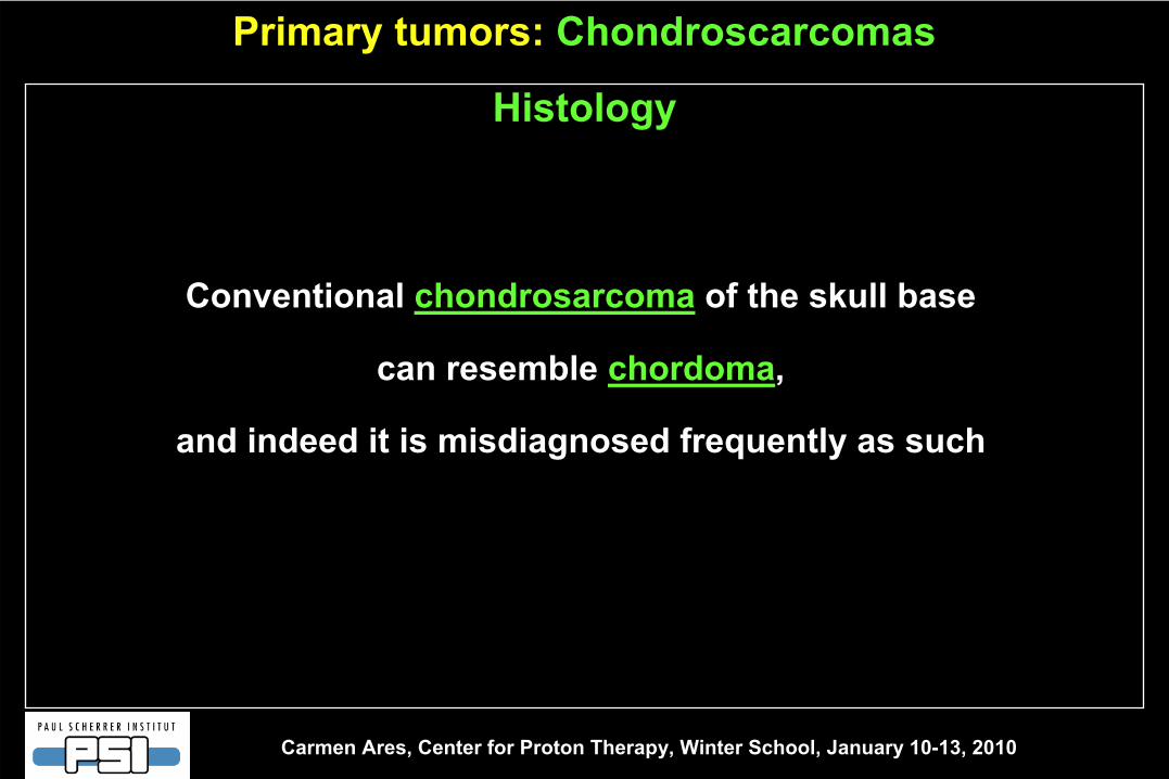
Carmen Ares, Center for Proton Therapy, Winter School, January 10-13, 2010
Histology
Conventional chondrosarcoma
of the skull base
can resemble chordoma,
and indeed it is misdiagnosed frequently as such
Primary tumors: Chondroscarcomas

Carmen Ares, Center for Proton Therapy, Winter School, January 10-13, 2010
IMMUNOHISTOCHEMISTRY: Chondrosarcoma (ChSa) vs. Chordoma (Ch)
ChSa
Ch
Epithelial Markers(cytokeratin, EMA)
S-100
- +
+ +

Carmen Ares, Center for Proton Therapy, Winter School, January 10-13, 2010
MGH*
In 37 %
of the 200 patients treated with PT for low-grade ChSa, the referral diagnosis was Ch and changed to ChSa
upon
review
of the tumor material at MGH utilizing:
- H & E Stains
- Immunohistochemistry
* Rosenberg AE et al, Am J Surg
Pathol
1999;23(11):1370
IMMUNOHISTOCHEMISTRY: Chondrosarcoma (ChSa) vs. Chordoma (Ch)

Carmen Ares, Center for Proton Therapy, Winter School, January 10-13, 2010
Chordomas:
Midline, soft, gelatinous
Chondrosarcomas:
Midline
or lateral, can be
calcified, hard
Primary Skull Base Tumors

Carmen Ares, Center for Proton Therapy, Winter School, January 10-13, 2010
• Chordomas and chondrosarcomas
located in the skull base
are uncommon tumors and challenging to manage
• The ability to obtain a
complete surgical resection remains
elusive for many patients
• Collaboration between surgeon and radiation oncologist critical
- Surgical goal:
accomplish maximally safe tumor reduction
- Improve geometry for Proton RT (decompression of brainstem, optic chiasm etc.)
Primary Skull Base Tumors

Carmen Ares, Center for Proton Therapy, Winter School, January 10-13, 2010
Primary Tumors of the Skull base
Results

Carmen Ares, Center for Proton Therapy, Winter School, January 10-13, 2010
Primary skull base tumors: Surgery series
• Tzortzidis
et al (#) in 74 patients with chordomas who underwent
surgery aimed at performing complete resection, accomplished gross total removal in 53 patients (71.6%)
• The 10-year recurrence-free survival was 31%
indicating that
even after a gross total removal the likelihood of tumor control
is low with surgery alone
#
Neurosurgery 2006;59:230-7

Carmen Ares, Center for Proton Therapy, Winter School, January 10-13, 2010
•
Local control rates after total doses <60 Gy is disappointing and most patients died of locally progressive disease
•
Recurrences rates as high as 70 to 100%
even for small lesions have been reported after conventional RT
- Catton, R&O1996 5 y LC 20%
- Rich, Cancer 1985
5 y LC 28%
- Zorlu, Neurol
Sci
2000
5 y LC 23%
Primary skull base tumors: Photon RT series

Carmen Ares, Center for Proton Therapy, Winter School, January 10-13, 2010
• Debus, Heidelberg, 2000
IJROBP 2000;47(3):591-596
- - - - - - - - - - - -
• Krishnan, Mayo Clinic, 2005 Neurosurgery 2005;56(4):777
• Hasegawa, Japan, 2007
J Neurosurg 2007;107(4):752
• Martin, Pittsburgh, 2007
J Neurosurg 2007;107(4):758
• Liu, Beijing, 2008
Neurol
Res 2008;30(6):557
• Dassoulas, U. Virginia, 2009 J Neurooncol
2009;94:243
Primary skull base tumors: Stereotactic RT and Radiosurgery series

Carmen Ares, Center for Proton Therapy, Winter School, January 10-13, 2010
Debus 2000, Heidelberg - IJROBP 2000;47(3):591-596
Chordomas Chondrosarcomas
n 37 8 Median dose 66.6 Gy 64.9 Gy
mean follow-up
27 months
19 months
5-y LC
50 %
100 %
Primary skull base tumors: Stereotactic RT series

Carmen Ares, Center for Proton Therapy, Winter School, January 10-13, 2010
Chordomas
Chondrosarcomas
n 5-y LC
n 5-y LC
Krishan, 05*
25
32 %
4
100 %
Martin,07#
18
53 %
10
80 %
Hasegawa, 07
30
7 76 % (for Ch + ChSa)
Liu, 08
28
21 %
-
Dassoulas, 09
15 50% (3 pat. 2nd
RS for out of field recurrence)
* 19 patients external RT previously
or in conjunction with RS (median dose 50.4 Gy)#
22 patients external RT previously
Primary skull base tumors: Radiosurgery series

Carmen Ares, Center for Proton Therapy, Winter School, January 10-13, 2010
•
Stereotactic RT and Radiosurgery can be a good option
for the treatment of selected patients with small chondrosarcomas
of
the skull base
•
Local control of selected patients with small chordomas
of the skull base is inferior to proton series
Primary skull base tumors: Stereotactic RT and RS series

Carmen Ares, Center for Proton Therapy, Winter School, January 10-13, 2010
PSI experience
Skull base chordomas and chondrosarcomas
Experience 1998 -
2005

Carmen Ares, Center for Proton Therapy, Winter School, January 10-13, 2010
Fraction
Dose: 2.0 Gy (RBE), 5 frcts. per week
CTV = 54 Gy (RBE)
GTV = 74 Gy (RBE)
OAR constraints: OPTIC Chiasm
and Nerves: 60 Gy(RBE); Brainstem
surface
64 Gy(RBE), BS-Center: 53 Gy(RBE), BS max. volume: 60 Gy(RBE) < 1.0 cc.
Fractionated
Proton Therapy
at Paul Scherrer Institute

Carmen Ares, Center for Proton Therapy, Winter School, January 10-13, 2010
• N = 64 patients (Oct-98 Nov-05)– Chordoma 42 (65%)(3/42 chondroid
features)
– Chondrosarcoma 22 (34%)(low grade 5 G2, 17 G1)
• Mean age 44.5 years
• Minimum follow-up 14 months
• Mean follow-up 38 months (14 -
92 months)
Material & Methods
Primary skull base tumors: PSI experience

Carmen Ares, Center for Proton Therapy, Winter School, January 10-13, 2010
•
Prescription dose (mean) (at 2 Gy(RBE)
per frct., 4 fractions per week)
– Chordoma (Ch)
74 Gy(RBE) (range 67 -
74)– Chondrosarcoma(ChSa)
68 Gy(RBE) (range 63 -
74)
• GTV volume (mean) 25.8 cc (1.5 -
100 cc)– Ch 27 cc– ChSa
23 cc
Material and Methods
Primary skull base tumors: PSI experience

Carmen Ares, Center for Proton Therapy, Winter School, January 10-13, 2010
Local control defined as radiological control by MRI ±
CT
Local control definition
Skull base tumors: PSI experience
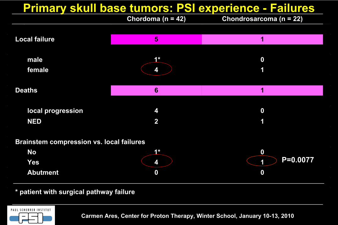
Carmen Ares, Center for Proton Therapy, Winter School, January 10-13, 2010
P=0.0077
* patient with surgical pathway failure
00Abutment14Yes01*No
Brainstem compression vs. local failures
12NED04local progression
16Deaths
14female01*male
15Local failure
Chondrosarcoma (n = 22)Chordoma (n = 42)Primary skull base tumors: PSI experience -
Failures
P=0.0077

Carmen Ares, Center for Proton Therapy, Winter School, January 10-13, 2010
Time to local failure
months
0 20 40 60 800.
00.
20.
40.
60.
81.
0
ChordomaChondrosarcoma
P=0.25
Actuarial Local Control
3
years 5 years
Chordomas
87 % 81 % Chondrosarcomas 94 % 94 %
Local controlPrimary skull base tumors: PSI experience

Carmen Ares, Center for Proton Therapy, Winter School, January 10-13, 2010
Disease Specific Survival
3 years 5 yearsChordomas 90%
81%
Chondrosarcomas 100 %
100 %
Disease Specific Survival
months
0 20 40 60 80
0.0
0.2
0.4
0.6
0.8
1.0
Chordoma, N=42Chondrosarcoma, N=22
P=0.09
Primary skull base tumors: PSI experience

Carmen Ares, Center for Proton Therapy, Winter School, January 10-13, 2010
Example of chondrosarcoma with subsequent local relapse
Pre-Proton-RT
GTV
V95 48%
Brainstem
compression
Primary skull base tumors: PSI experience

Carmen Ares, Center for Proton Therapy, Winter School, January 10-13, 2010
Prognostic factors for LC in chordoma:• Brainstem compression
yes / no
p= 0.0077
• Residual tumor volume
≤
/ > 25 cc p= 0.03
• Gender
n.s.
• Age
n.s.
• PT for
primary / recurrence
n.s.
• GTV V95
n.s.
• GTV max, mean or min dose
n.s.
Primary skull base tumors: PSI experience
cccc

Carmen Ares, Center for Proton Therapy, Winter School, January 10-13, 2010
• Asymptomatic MRI white matter changes:
5 patients(= G1 neurologic toxicity)
• High grade late toxicity (all Ch):
4 patients– optic pathway G 4 1 patient (unilateral blindness)
G 3 1 patient (unilateral visual deficit, steroid dependent)
– neurologic G 3 2 patients (symptomatic brain necrosis)
• Any patient presented brainstem toxicity
Radiation induced late toxicity (CTCAE v3.0)
Primary skull base tumors: PSI experience

Carmen Ares, Center for Proton Therapy, Winter School, January 10-13, 2010
Actuarial 5-year freedom for high grade late toxicity 94%
Due to the small number of events no risk factors predictive of high grade toxicity were identified
Radiation induced toxicity (CTCAE v3.0)
Skull base tumors: PSI experience

Carmen Ares, Center for Proton Therapy, Winter School, January 10-13, 2010
Skull Base Chordomas: Comparison of Literature
* at 3.0 Gy (RBE) per fraction** 5y LC = 100% for 19 patients to 60.8 GyE
n Radiation Meandose
LC LC LC
3 -yr 5 -yr 10 -yr
Munzenrider, 1999 169 PT, RT 76 73 54Terahara, 1999 115 PT, RT 69 59 44Hug, 1999 33 PT, RT 71 67 59Noel, 2005 100 PT, RT 67 86 @2y 53 @4y
Igaki, 2004 13 PT, RT 72 67 46
Schulz-Ertner, 2007 96 Carbon, RT 60 * 81 70Mizoe, 2009** 33 Carbon 57 * 85 64Ares, (PSI) 2009 42 PT 74 87 81

Carmen Ares, Center for Proton Therapy, Winter School, January 10-13, 2010
Skull Base Chondrosarcomas: Comparison of Literature
* at 3.0 CGE per fraction** IJROBP
3 -yr
949468PT22Ares, (PSI) 2009**
60*
75@4y91PT, RT26Noel, 2003
91
94
71PT, RTJohson, 2002
94
949872PT, RT229Munzenrider, 199910 -yr5 -yrLC LC LC Mean
doseRadiationn
58
96Carbon, RT54 89@4ySchulz-Ertner, 2007
75Hug, 1999 25 PT, RT 71

Carmen Ares, Center for Proton Therapy, Winter School, January 10-13, 2010
5-ye
ar L
ocal
Con
trol
rate
s(%
)
20
40
60
80
100
20 40 60 80 100
Chordomas of the Base of Skull
Dose [ Gy (RBE)]
MGH 1999PSI 2009LLUMC 1999
GSI
Romero 1993Zorlu
2000
SRT –
Heidelb. 2000
C-Ions
Photons
Protons
Small Chordomas Chondrosarcomas

Carmen Ares, Center for Proton Therapy, Winter School, January 10-13, 2010
Chordoma
and Chondrosacoma
of Skull Base and C-Spine (Phase I/II-protocol: selective result reporting)
ASTRO 2009, J. Munzenrider (IJROBP 72(1), suppl.)
105 patients with skull base or cervical spine Chondrosarcoma or ChordomaFrom 1987 to 1993, either “70.2 (LD) vs. 76 CGE (HD)”
F/U: median 16.7 y (4.5-20 y)
Results 5-yr 10-yr 15-yr
OS all pts. (LD -
HD) 81 vs. 81 %
61 vs. 55 %
57 vs. 45 %
LC for CSA (LD -
HD) 94 vs. 85 %
89 vs. 67 %
89 vs. 58 % (p=0.045)
Median time to failure: 11.9 yNo statement on complications in abstract

Carmen Ares, Center for Proton Therapy, Winter School, January 10-13, 2010
ASTRO 2009, J. Munzenrider (IJROBP 72(1), suppl.)
Results:
Late failures occur (MEDIAN time 11 y) – long term f/u essential
High dose arm with inferior local control – difficult to explain and not consistent with other reported data
Long term LC for Chondrosarcomas at MGH lower than previously reported. At 10 years likely 80%-90 % compared to >95% ? – conflicting data from the same institution
Further results of PROG trial needed
Chordoma
and Chondrosacoma
of Skull Base and C-Spine
(Phase I/II-protocol: selective
result
reporting)

Carmen Ares, Center for Proton Therapy, Winter School, January 10-13, 2010
Proton-RT for Skull Base Chordomas
Improved LC for “smaller”
size
• < 70 ml vs. > 70 ml (MGH)
• < 20cc vs. 20 -
35cc vs. > 35 cc (LBL) (80% vs. 33%)
• < 25 ml vs. > 25 ml (LLUMC) (100% vs. 56%)
Prognostic Factors:
Influence of (residual) tumor size on the ability to achieve local control:
0
20
40
60
80
100
0 10 20 30 40 50 60 70
p = 0.03 ≥
25ml GTV
< 25ml GTV
Hug, et al. J Neurosurg
1999;91:432
Loma Linda UMC Analysis

Carmen Ares, Center for Proton Therapy, Winter School, January 10-13, 2010
RT for Skull Base ChordomasPrognostic Factors:Influence of ability to deliver dose vs. limitations of dose delivery 2nd
to normal structure constraints:
Hug, et al. J Neurosurg
1999;91:432
0
20
40
60
80
100
0 10 20 30 40 50 60 70
No brainstem compression
brainstem compression
Loca
l con
trol
p = 0.04
Noel, et al. Acta
Oncol
2005;44:700
Factors
predicting
Local
Control: • 95% GTV encompassed
by 95% Isodose
(p=0.01)
• Minimal dose < 56 Gy to GTV (p=0.04)
Carmen Ares, Center for Proton Therapy, Winter School, January 10-13, 2010
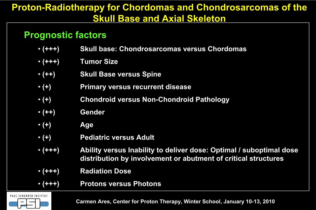
Proton-Radiotherapy for Chordomas and Chondrosarcomas of the Skull Base and Axial Skeleton
Prognostic factors• (+++)
Skull base: Chondrosarcomas versus Chordomas
• (+++)
Tumor Size
• (++)
Skull Base versus
Spine
• (+)
Primary versus recurrent disease
• (+)
Chondroid versus Non-Chondroid Pathology
• (++)
Gender
• (+)
Age
• (+)
Pediatric versus Adult
•
(+++)
Ability versus Inability to deliver dose: Optimal / suboptimal dose distribution by involvement or abutment of critical structures
• (+++)
Radiation Dose
• (+++)
Protons versus Photons

Carmen Ares, Center for Proton Therapy, Winter School, January 10-13, 2010
Future directions for primary skull base tumors

Carmen Ares, Center for Proton Therapy, Winter School, January 10-13, 2010
RT for Skull Base Chordomas
GOAL
• Develop a risk-classification
low -
intermediate -
high
to correlate with recommendations for adjuvant Tx
observation -
aggressive Tx -
palliative Tx
•
Rather than stating “
all skull base chordoma patients should / should not undergo adjuvant Tx”
the question should be
“
WHICH patient will likely benefit from adjuvant Tx”

Carmen Ares, Center for Proton Therapy, Winter School, January 10-13, 2010
• Dose escalation Biological GTV definition
– PET uptake– functional MRI [measure of angiogenesis]
Simultaneous Integrated Boost
Hypofractionation
With Gantry 2 possibility of dose escalation with:-
smaller pencil beam
-
collimators: dose escalation using inhomogeneous coverage of the GTV
•
Concomitant use of biologic agents for high-risk patients not suited for dose escalation
Futute
directions

Carmen Ares, Center for Proton Therapy, Winter School, January 10-13, 2010
Secondary infiltration from intracranial tumors:
Meningiomas

Carmen Ares, Center for Proton Therapy, Winter School, January 10-13, 2010
Secondary infiltration from intracranial tumors: Meningiomas
• extra-axial, slow-growing tumors that arise from the arachnoid
cap cells of the
central nervous system
• 13 to 26% of primary intracranial tumors
• most common non-glial
brain tumors
• The majority are benign (WHO Grade I)
• atypical (WHO Grade II) or anaplastic
(malignant, WHO Grade III) are
uncommon, accounting for 4.7–7.2% and 1.0–2.8% of all resected meningiomas
• If the tumor is resectable, complete surgical excision is the standard therapy and results in excellent (68–92%) long term tumor control for benign meningiomas

Carmen Ares, Center for Proton Therapy, Winter School, January 10-13, 2010
Indications for RT
• Subtotal excision
Local recurrence rates can be decreased
from 50 -
60% to 12 -
23% at 8 -
10
years
• Not resectable
tumor
or contraindication to surgery
5-y local tumor control rates of 80–86%
• Atypical and malignant meningiomas at high risk for local failure after surgery
RT is recommended to decrease the probability of local recurrence
Secondary infiltration from intracranial tumors: Meningiomas

Carmen Ares, Center for Proton Therapy, Winter School, January 10-13, 2010
Adjuvant treatment or Radical treatment by:
• conventional external beam photon radiotherapy
• 3D conformal radiation therapy
• radiosurgery
• stereotactic fractionated radiotherapy
• intensity modulated radiotherapy (IGRT-IMRT)
• tomotherapy
• etc.etc.
Secondary infiltration from intracranial tumors: Meningiomas
Carmen Ares, Center for Proton Therapy, Winter School, January 10-13, 2010
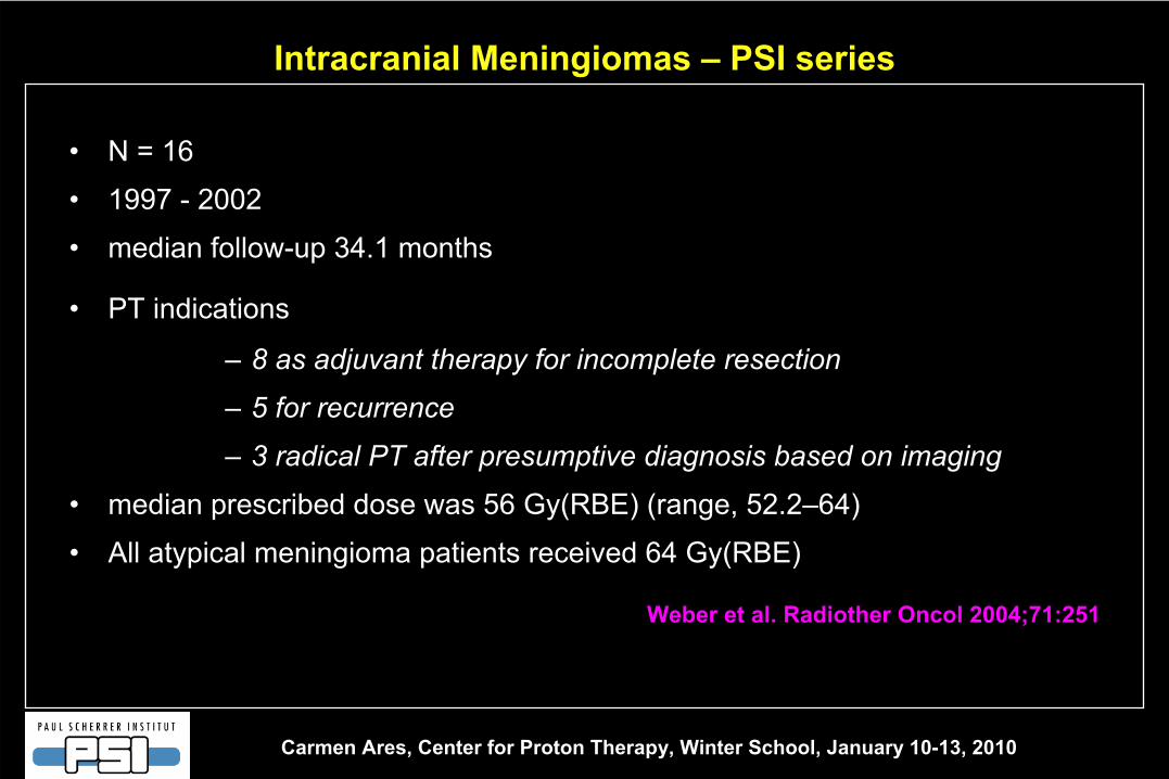
Intracranial Meningiomas
–
PSI series
• N = 16
• 1997 -
2002
• median follow-up 34.1 months
• PT indications
–
8 as adjuvant therapy for incomplete resection
–
5 for recurrence
–
3 radical PT after presumptive diagnosis based on imaging
• median prescribed dose was 56 Gy(RBE) (range, 52.2–64)
• All atypical meningioma
patients received 64 Gy(RBE)
Weber et al. Radiother
Oncol
2004;71:251

Carmen Ares, Center for Proton Therapy, Winter School, January 10-13, 2010
• 3-year local control 91.7%
• 3-year progression-free survival 91.7 %
• 3-year overall survival were
92.7%
• No patient died from recurrent meningioma
• Cumulative 3-year toxicity free survival was 76.2%.
• One patient with radiation induced optic neuropathy (SOMA Grade 3)
• One patient with retinopathy (SOMA Grade 2)
• One patient developed a symptomatic brain necrosis (CTCAE Grade 4)
Weber et al. Radiother
Oncol
2004;71:251
Intracranial Meningiomas
–
PSI series

Carmen Ares, Center for Proton Therapy, Winter School, January 10-13, 2010
Proton –
RT indications at PSI
• Complex benign meningiomas
• Anaplastic
meningiomas
• Malignant meningiomas
Intracranial Meningiomas

Carmen Ares, Center for Proton Therapy, Winter School, January 10-13, 2010
Secondary infiltration by primary H&N tumors
Adenoid Cystic Ca

Carmen Ares, Center for Proton Therapy, Winter School, January 10-13, 2010
Primary site: tongue
Skull base recurrence at 6 y.
Adenoid Cystic Carcinoma of the H&N
Patient A Patient B

Carmen Ares, Center for Proton Therapy, Winter School, January 10-13, 2010
Adenoid Cystic Carcinoma of the Lacrimal
gland
(treated at Massachusetts General Hospital)
“Sculpting”
of the dose distribution by protons

Carmen Ares, Center for Proton Therapy, Winter School, January 10-13, 2010
Adenoid Cystic Carcinomas with infiltration of the skull base

Carmen Ares, Center for Proton Therapy, Winter School, January 10-13, 2010
Adenoid Cystic Carcinomas with infiltration of the skull base

Carmen Ares, Center for Proton Therapy, Winter School, January 10-13, 2010
Adenoid Cystic Carcinomas with infiltration of the skull base

Carmen Ares, Center for Proton Therapy, Winter School, January 10-13, 2010
Adenoid Cystic Carcinomas with infiltration of the skull base

Carmen Ares, Center for Proton Therapy, Winter School, January 10-13, 2010
Secondary infiltration by primary H&N tumors
Nasopharynx Ca
Paranasal Sinus Ca

Carmen Ares, Center for Proton Therapy, Winter School, January 10-13, 2010
Proton Therapy for Re-irradiation of recurrent Nasopharynx
Ca.
Lin R et al for LLUMC, Radiology1999;213:489
Optimal DVH
>90 % of Prescription Dose to >90% of GTV ( 8 patients)
Suboptimal DVH
(8 patients)
• Patients: 16• Dx: recurrent Ca of Nasopharynx after full course photon RT• Re-treatment: Protons to 59.4 –
70.2 CGE at 1.8-2.0 CGE per day
• F/U: mean 23.7 months (range 4 –
47 months)

Carmen Ares, Center for Proton Therapy, Winter School, January 10-13, 2010
Increased LC translates into increased Overall Survival rates: p=0.006
Lin R et al. for LLUMC -
Radiology 1999; 213:489-494
Local Control probability is dependent on ability to deliver intended dose
P= 0.05
Optimal DVH

Carmen Ares, Center for Proton Therapy, Winter School, January 10-13, 2010
PT for primary sphenoid sinus malignancies Truong MT et al. MGH. Head &Neck 2009
• 20 patients (10 SCC, 7 ACC, 2 Neuroendocrine
tumor, 1 AdenoCa)
• 1991 –
2005
• PT –
median dose 76 Gy
(RBE)
• Median follow-up 27 months
• 2 year
- LC
86%
- Regional control 86%
- Freedom from metastasis 50%

Carmen Ares, Center for Proton Therapy, Winter School, January 10-13, 2010
PT for primary sphenoid sinus malignancies Truong MT et al. MGH. Head &Neck 2009
• 2 years DFS 31%
• 2 years OS 53%
• Negative predictive factors−
Oropharyngeal
involvement (p=0.005)
−
Anterior cranial fossa
invasion (p=.02)−
Brain invasion (p=.05)

Carmen Ares, Center for Proton Therapy, Winter School, January 10-13, 2010
• Referral Centers for rare diseases
• Accumulation of large series of patients treated homogeneously
• Add to understanding of natural history of disease
• Foster multidisciplinary approach
• Accomplishes previously unknown CURE in some patients/tumors
• Understand Prognostic Factors for others
• Develop new treatment algorithms
Proton-Radiotherapy for Skull Base Tumors
Conclusions

Carmen Ares, Center for Proton Therapy, Winter School, January 10-13, 2010
Spot Scanning Proton Radiation Therapy
for Para-spinal Tumors

Carmen Ares, Center for Proton Therapy, Winter School, January 10-13, 2010
Para-spinal Tumors:
Introduction
• Primary malignant tumors of the vertebral column are relatively rare with prevalence of
2.5 to 8.5 cases per 100.000 persons per
year
• In adults−
Plasmocytoma
30%
− Chondrosarcoma
10%
− Chordoma
< 5%
− Osteosarcoma
< 5%
• In children−
Ewing’s sarcoma
4 –
10%

Carmen Ares, Center for Proton Therapy, Winter School, January 10-13, 2010
Para-vertebral Tumors:
Treatment generalities
• Difficult treatment paradigm because of the complexities of
tumor resection (en-bloc resection) and significant resistance to
chemotherapy and radiotherapy of some of these tumors
• Novel uses and improvements in advanced radiation techniques
improve local control

Carmen Ares, Center for Proton Therapy, Winter School, January 10-13, 2010
• High-dose RT can provide local control to these “radioresistant”
tumors
• Doses > 70 Gy
have demonstrated the benefit
• These dose are greater than OAR tolerance (i.e. spinal cord)
Due to the dose restrictions for the spinal cord and other
surrounding structures (esophagus, bowel, kidney) the results
of conventional RT have been disappointing
Para-vertebral Tumors: Treatment generalities
Carmen Ares, Center for Proton Therapy, Winter School, January 10-13, 2010
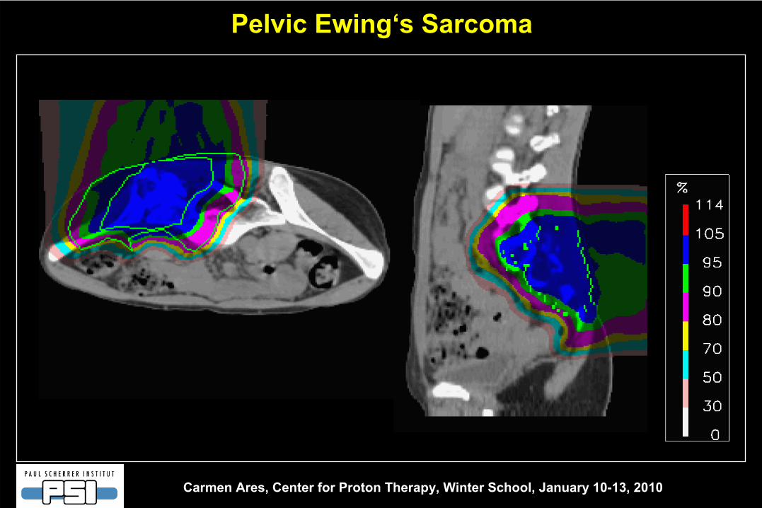
Pelvic
Ewing‘s
Sarcoma

Carmen Ares, Center for Proton Therapy, Winter School, January 10-13, 2010
Extracranial
Chordomas
of the Axial Skeleton treated with Spot Scanning Proton Therapy
at PSI

Carmen Ares, Center for Proton Therapy, Winter School, January 10-13, 2010
16
94 11
• Update of the initial publication (Rutz HP et al. IJROBP 2007:67(2):512)Updated manuscript in progress.
• N = 40
• 1999 –
2005
• Location:
Extracranial
Chordomas
of the Axial Skeleton treated with Spot Scanning Proton Therapy
at PSI
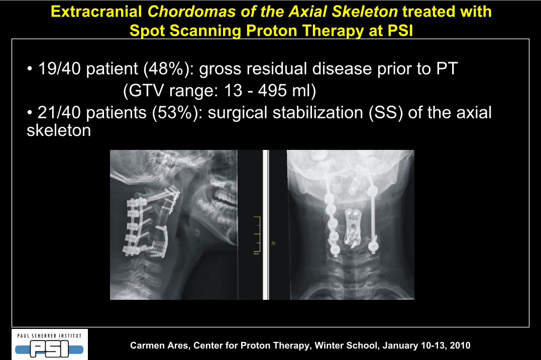
Carmen Ares, Center for Proton Therapy, Winter School, January 10-13, 2010
• 19/40 patient (48%): gross residual disease prior to PT(GTV range: 13 -
495 ml)
•
21/40 patients (53%): surgical stabilization (SS) of the axial skeleton
Extracranial
Chordomas
of the Axial Skeleton
treated with Spot Scanning Proton Therapy
at PSI

Carmen Ares, Center for Proton Therapy, Winter School, January 10-13, 2010
• Median total dose: 72 Gy
(RBE) (range: 59.4 –
75.2 Gy
(RBE))
• Follow-up period:
- Median
43 months (range 24 –
91 months)
Extracranial
Chordomas
of the Axial Skeleton treated with Spot Scanning Proton Therapy
at PSI

Carmen Ares, Center for Proton Therapy, Winter School, January 10-13, 2010
5-year LC: 62 %
Local control probability13 / 40 patients with local failure
Chordomas
of the Axial Skeleton
at PSI: 5-year outcome
data

Carmen Ares, Center for Proton Therapy, Winter School, January 10-13, 2010
Prognostic factors analyzed for LC / OS :
• surgical stabilization (SS)
• residual disease
Chordomas
of the Axial Skeleton
at PSI: 5-year outcome
data

Carmen Ares, Center for Proton Therapy, Winter School, January 10-13, 2010
No SS-R:
• only
1 LF in 19 pts•
resulting in actuarial
LC rate at 5 years of 100%
With
SS-R:
• 12 LF in 21 pts.•
yielding a 5 year LC
rate of 30% (p=0.0003)
with SS-R
no SS-R
P=0.003
Chordomas
of the Axial Skeleton
at PSI: 5-year outcomes
data
5 year LC: 100%
5 year LC: 30%
p=0.0003
Local Control Probability (LC): Surgical Stabilization

Carmen Ares, Center for Proton Therapy, Winter School, January 10-13, 2010
Local Control Probability (LC): residual disease
The overall LC-rate: Significantly reduced in patients with residual disease
5-year LC rate: 66%
Chordomas
of the Axial Skeleton
at PSI: 5-year outcomes
data
P=0.04825-year LC rate: 47%
5-year LC rate: 66%
P=0.0482
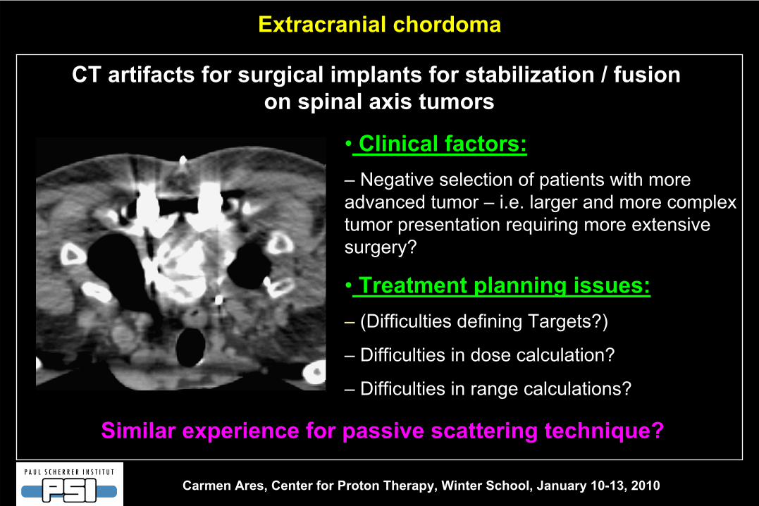
Carmen Ares, Center for Proton Therapy, Winter School, January 10-13, 2010
• Clinical factors:–
Negative selection of patients with more
advanced tumor –
i.e. larger and more complex tumor presentation requiring more extensive surgery?
• Treatment planning issues:– (Difficulties defining Targets?)
– Difficulties in dose calculation?
– Difficulties in range calculations?
CT artifacts for surgical implants for stabilization / fusionon spinal axis tumors
Extracranial chordoma
Similar
experience
for passive scattering
technique?

Carmen Ares, Center for Proton Therapy, Winter School, January 10-13, 2010
Chordomas
of the Axial Skeleton
at PSI: 5-year outcomes
data
Late Toxicity (CTCAE)
• Neurotoxicity:
none
• GI or GU-toxicity: none
•
2 Grade III toxicities: 1 soft tissue necrosis and 1 osteonecrosis
in patients following extensive surgeries and reconstruction
Carmen Ares, Center for Proton Therapy, Winter School, January 10-13, 2010
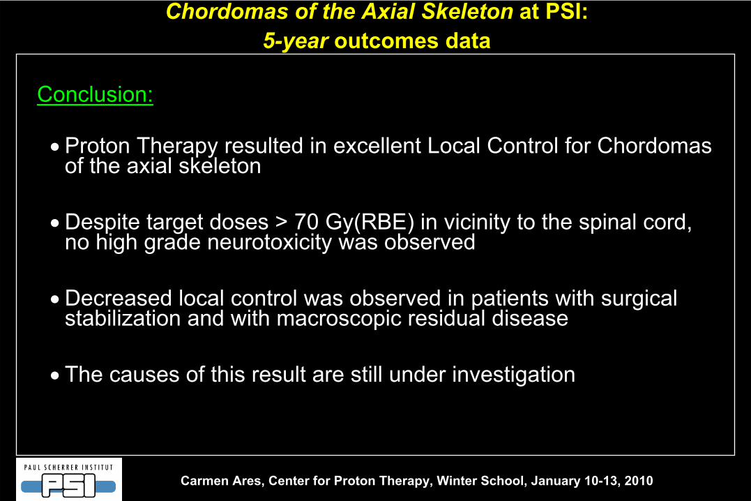
Conclusion:
•
Proton Therapy resulted in excellent Local Control for Chordomas of the axial skeleton
•
Despite target doses > 70 Gy(RBE) in vicinity to the spinal cord, no high grade neurotoxicity was observed
•
Decreased local control was observed in patients with surgical stabilization and with macroscopic residual disease
•
The causes of this result are still under investigation
Chordomas
of the Axial Skeleton
at PSI: 5-year outcomes data

Carmen Ares, Center for Proton Therapy, Winter School, January 10-13, 2010
Mixed photon/proton
RT for Sacral
Chordomas: long-term
MGH experience
Park, Delaney et al. MGH, IJROBP, 65:1514, 2006- 27 patients
- Tx: 1982-2002
- S+RT 21 pts, RT alone
6 pts
- 16 primary, 11 recurrent
Chordomas
-
Mean
dose 71 Gy(RBE) primary, 77 Gy (RBE) recurrent
Chordoma
- Minimum F/U 3 years
- Factors:
- Primary
vs. rec.
- Positive vs. negative margins
- S+RT vs. RT only
Local
Control
(27 pts.):
72% at 5 years
57% at 10 years

Carmen Ares, Center for Proton Therapy, Winter School, January 10-13, 2010
Phase II Study
of high-dose
Photon/Proton RT of Spine
SarcomasDeLaney et al. MGH, IJROBP 2009;74:732-9
- n = 50
- 29 Chordomas, 14 ChSa, 7 other
- 50 % gross disease
- 77.4 Gy(RBE) gross disease
- 70.2 microsc., 1.8 Gy/fract.
- Some
patients dural
plaque
(Y90)
- Median F/U 48 months
- Factors: Primary
vs. Recurrence
(p=signif.)
- Spine
stabilization:
5/16 (31%) LF versus
4/34 (12%) without (p=0.103)
- No myelopathy, 3 sacral
nerve injuries
at 77 Gy
Local
Control
(50 pts.):
78% at 5 years

Carmen Ares, Center for Proton Therapy, Winter School, January 10-13, 2010
IGRT-IMRT Photons for Paraspinal
Chordomas and SarcomasTerekakis, Bilsky
et al. MSKCC, IJROBP, 69:1502-08, 2007
27 patients
Subtotal resection or unresect. disease
5/27 pts. prior RT
Standard fractionated XRT
Median prescr. Dose: 66 Gy
(54-71 Gy)
Median F/U 17 months
( 2.1-
47.3)
act. 2-year
Local Control: 65%

Carmen Ares, Center for Proton Therapy, Winter School, January 10-13, 2010
Carbon
ions
for
sacral
chordomaImai
R, Kamada
T, et al. NIRS –
Chiba
–Japan, IJROBP 2009 (in press)
• N =38 unresectable
sacral chordoma
(1996 -
2003)
- 30 no previous treatment
- 8 recurrence after previous resection
• Carbon ion median dose 70,4 GyE
(52.8 –
73.6) in 16f in 4weeks
• Median follow-up 80 months
• 5-y OS 86%
• 5-y LC 89%
• 27/30 with primary tumor remained ambulatory with or without supportive devices
• 2 patients G3 skin reaction
• 2 patients G4 skin reaction
• 1 patient sacral fracture
• No G3 or G4 urinary or rectal toxicity
• 3 severe and permanent neurological impairment

Carmen Ares, Center for Proton Therapy, Winter School, January 10-13, 2010
Increasing dose levels with protons
(long-term data: eyes, skull base, paraspinal
tumors, unresectable
sarcomas)
=
Increasing tumor control
Proton Radiotherapy for Skull Base and Para-spinal Tumors

Carmen Ares, Center for Proton Therapy, Winter School, January 10-13, 2010
Thank you for your attention!