pseudomonas aeruginosa genotyping kit 2 · contents of the test report ... assignment of unknown p....
TRANSCRIPT

Pseudomonas aeruginosa
Genotyping Kit 2
Array Hybridisation Kit for epidemiological typing of Pseudomonas aeruginosa
isolates.
Kit order number: 245600096
96 reactions (ArrayStrip format)
For Research Use Only. Not for Use in Diagnostic Procedures.

www.clondiag.com www.alere.com
P. aeruginosa Genotyping Kit 2
2 05-16-04-004-V03_Manual_ P. aeruginosa.pdf
Content
BACKGROUND ................................................................................................................................. 1
GENERAL INSTRUCTIONS FOR USE ................................................................................................. 2
Intended Use ................................................................................................................................ 2
Specifications ............................................................................................................................... 2
Technical Support ........................................................................................................................ 2
Safety Precautions ....................................................................................................................... 3
Material Safety Data Sheets (MSDS) ........................................................................................... 3
Shipping Precautions ................................................................................................................... 3
REAGENTS AND DEVICES ................................................................................................................. 4
Assay Components, Storage and Stability ................................................................................... 4
DNA Labelling and Amplification .............................................................................................. 4
Hybridisation and Detection .................................................................................................... 4
Instrumentation & Software .................................................................................................... 5
Components required but not provided .................................................................................. 5
SOFTWARE INSTALLATION .............................................................................................................. 7
Assay Plugin and SDK for the ArrayMate ..................................................................................... 7
Experiment String Collector ......................................................................................................... 9
Test the Assay Plugin ................................................................................................................. 10
PROTOCOL ..................................................................................................................................... 11
Culturing and Harvesting Bacterial Cells.................................................................................... 11
Extraction of DNA ...................................................................................................................... 11
Via Spin Columns (e.g. Qiagen DNeasy Blood&Tissue) .......................................................... 12
Linear Amplification and Internal Biotin Labelling .................................................................... 14
Hybridisation .............................................................................................................................. 14
General Remarks - Handling of Arrays ................................................................................... 14
General Remarks - Handling of Liquids .................................................................................. 15
General Remarks – the Substrate (Precipitating Dye) D1 ...................................................... 16
General Remarks - Thermoshakers ........................................................................................ 16
Protocol for Quantifoil’s BioShake iQ and Eppendorf’s Thermomixer Comfort with Microtitre Plate Adapter ........................................................................................................ 17
Data Analysis .............................................................................................................................. 18
Starting the ArrayMate Reader .............................................................................................. 18
Worklist .................................................................................................................................. 20
Data Acquisition in the ArrayMate Reader ............................................................................ 21
Results ........................................................................................................................................ 23
Export of Test Reports ............................................................................................................ 24
Contents of the Test Report ................................................................................................... 26
Kinship Analysis: Markers of the Core Genome (“KinCode” / “SNP Pattern”) ...................... 26
Marker Controls ..................................................................................................................... 26
Plausibility Controls ................................................................................................................ 27
Accessory Genome and Microevolution (“Features of the Isolate”) ..................................... 27
Recommendation for a Project Specific Database .................................................................... 28
String Collector ....................................................................................................................... 28

www.clondiag.com www.alere.com
Excel-Based Data Mining Tool ................................................................................................ 29
TROUBLESHOOTING ...................................................................................................................... 30
LITERATURE ................................................................................................................................... 33
ADDITIONAL INFORMATION ......................................................................................................... 34
Warranty .................................................................................................................................... 34
Quality Control ........................................................................................................................... 34
List of Components for Separate Order ..................................................................................... 34
CONTACT ....................................................................................................................................... 35
APPENDIX 1 – Flow Chart .............................................................................................................. 36
APPENDIX 2 – Probes and Primers for KinCode Genotyping and the Accessory Genome ........... 37


P. aeruginosa Genotyping Kit 05-16-04-004-V03_Manual_ P. aeruginosa.pdf 1
BACKGROUND
The ALERE P. aeruginosa Genotyping Kit 2 is a quick and simple method for the DNA-based
assignment of unknown P. aeruginosa isolates to phylogenetic lineages.
RNA-free, unfragmented genomic DNA from pure and monoclonal P. aeruginosa colony
material is amplified approximately 50-fold and internally labelled with biotin-11-dUTP using a
linear amplification protocol. In contrast to standard PCR, a multiplex primer extension reaction
is performed with two nested primers per target in each cycle. Two-versus-one primer for each
target increase and synchronize the yield of biotin labelled single stranded (ss) DNA product for
all markers. This allows a simultaneous sequence specific labelling and amplification of an
essentially unlimited number of targets. However, sensitivity is lower than in a standard PCR
(whereas contamination with amplicons is nearly impossible) and for that reason the method is
restricted to clonal colony material and cannot be performed on samples such as swabs or
other patient samples. Resulting biotin labelled ssDNA is transferred and hybridised to DNA
oligonucleotide microarrays with 99 probes for different genetic markers plus controls. All of
them are printed in five spots each.
The array contains three different kinds of markers and each marker is represented by one or
more probes:
- 16 markers for kinship analysis (represented in total by 36 different probes)
- 39 markers of the so called accessory genome (represented in total by 39 different
probes) which may be used for the analysis of microevolutionary events
- 24 probes for internal control of the analytic process. These probes test for proper DNA
preparation (using conserved genes of P. aeruginosa), PCR (using a synthetic template),
hybridisation (using control spots with biotinylated antisense partners) and staining
reaction (using biotinylated control spots), respectively.
Oligonucleotide probes are directly spotted onto the array, primers and control
oligonucleotides are present in the MasterMix B1P2.
The resulting typing data provide information for kinship analysis. Strain assignment using this
information was described in detail by Wiehlmann et al. 2007, PNAS vol. 104 no. 19, p. 8101–
8106.

www.clondiag.com www.alere.com
P. aeruginosa Genotyping Kit 2
2 05-16-04-004-V03_Manual_ P. aeruginosa.pdf
GENERAL INSTRUCTIONS FOR USE
Intended Use
For Research Use Only. Not for Use in Diagnostic Procedures.
This assay allows genotypic characterisation of P. aeruginosa isolates for research and
epidemiological applications. It cannot be used for other bacteria than P. aeruginosa.
Specifications
Upon receipt the assay components need to be stored at different temperatures as specified on
the package insert. The assay is to be performed at an ambient temperature of 18°C to 28°C.
Technical Support
If you require any further information on this product please contact:
Alere Technologies GmbH
Löbstedter Straße 103 -105
D-07749 Jena - Germany
phone: +49 (0) 36 41 / 3111-0
fax: + 49 (0) 36 41 / 3111-120
www.clondiag.com www.alere.com
P. aeruginosa Genotyping Kit 2 05-16-04-004-V03_Manual_ P. aeruginosa.pdf 3
Safety Precautions
The assay is intended for use by personnel that are trained in microbiological and
molecular methods. Preparation of DNA from pure P. aeruginosa colonies (clones) requires
expertise in microbiology and the local regulations for handling of pathogenic
microorganisms (biosafety level 2) are to be obeyed.
Isolated, cell-free P. aeruginosa DNA may be processed without further biosafety
precautions, although contamination with P. aeruginosa or other bacteria needs to be
ruled out.
Always wear protective clothes as required for laboratory work by your local regulations.
Material Safety Data Sheets (MSDS)
According to OSHA 29CFR1910.1200, Commonwealth of Australia [NOHSC: 1005, 1008(1999)]
and the latest amendments to the European Union Directives 67/548/EC and 1999/45/EC, the
enclosed reagents do not require a Material Safety Data Sheet (MSDS). They do not contain
more than 1% of a component classified as hazardous and do not contain more than 0.1 % of a
component classified as carcinogenic. MSDS therefore are not provided. Nevertheless, the
buffers may cause irritation if they come into contact with eyes or skin, and may cause harm if
swallowed. The regular precautions associated with laboratory work should be obeyed (e.g.,
wear protective goggles, gloves and lab coat and avoid contact with the reagents). In case, any
liquids are spilled, clean with disinfectant and/or laboratory detergent and water.
Alere assumes no liability for damage resulting from handling or contact with these products. If
you have any questions please contact our Technical Support (see above).
Shipping Precautions
RID/ADR: “Kein Gefahrgut”/ No dangerous goods
IMDG: No dangerous goods
IATA: No dangerous goods

www.clondiag.com www.alere.com
P. aeruginosa Genotyping Kit 2
4 05-16-04-004-V03_Manual_ P. aeruginosa.pdf
REAGENTS AND DEVICES
Assay Components, Storage and Stability
All reagents are provided in a certain surplus amount (see below). In case of need, all
components may also be ordered separately; please refer to the order numbers at the end of
this user guide. For pricing please contact your local representative or our customer service,
respectively.
The expiry date can be found on each bottle and on the outer package. All components have
been tested for stability for short term shipment (<1 week) at ambient temperature (< 37 °C).
The assay components with a rather limited stability are D1 and C3. The other components
have proven to be stable for up to six months after the assay expiry date has passed.
DNA Labelling and Amplification
B1P2: 2x Labelling Buffer.
Store at 2-8 °C. Surplus: 25%.
B2: Labelling Enzyme.
Store at 2-8 °C. Surplus: 50%.
Hybridisation and Detection
ArrayStrips P.aeruginosa-4 (12 x 8 samples); protected against light and sealed under
inert gas. Store at 15°C to 28°C. After opening to be used within two weeks. Close the
unused wells with caps, protect them against humidity and dust and store them at a
dark place.
Please note: Avoid any touching or scratching of the surface of the microarray at the
bottom of the well. Do not store or handle unused wells at an air humidity
of more than 60% since this may irreversibly corrode the spots.
ArrayStrip Cover(24 strips)
C1: Hybridisation Buffer.
www.clondiag.com www.alere.com
P. aeruginosa Genotyping Kit 2 05-16-04-004-V03_Manual_ P. aeruginosa.pdf 5
Store at 18-28 °C, protect against sunlight. Surplus: 100%.
C2: Washing Buffer 1.
Store at 18-28 °C, protect against direct sunlight. Surplus: 100%.
C3: HRP Conjugate 100x.
Store at 2-8 °C, protect against direct sunlight. Surplus: 100%.
C4: Conjugate Buffer. Store at 18-28 °C, protect against direct sunlight. Surplus: 200%
C5: Washing Buffer 2. Store at 18-28 °C, protect against direct sunlight. Surplus: 200%.
D1: Horseradish Peroxidase Substrate. Store at 2-8 °C, protect against direct sunlight.
Surplus: 50%.
Instrumentation & Software
ArrayMate Reader (to be ordered separately, for details see below)
The P. aeruginosa Genotyping assay may be used on the ArrayMate reader only. The
alternative devices ATR01/03 are not suitable for reading ArrayStrip based assays. In case
of any questions please contact us.
Iconoclust software (provided with the reader)
Test specific software plug-in (can be downloaded from Alere/CLONDIAG´s website, check
periodically for updates, for details see below). Information (such as spot names, marker
names, location of the spots on the array, size of the image taken by the reader’s specific
camera) is delivered with the reader or can be downloaded from our website. These test-
specific plug-ins will occasionally be updated. Please check the NEWS section of our
website www.alere-technologies.com. Support is available under [email protected].
Components required but not provided
Growth media for the cultivation of P. aeruginosa
The test should be performed with colonies harvested from Columbia blood agar. Other
nutrient-rich media (e.g. 2xTY or LB) may also suffice, but have not systematically been
tested. Liquid media should not been used because contaminations or mixed cultures
cannot easily be ruled out.

www.clondiag.com www.alere.com
P. aeruginosa Genotyping Kit 2
6 05-16-04-004-V03_Manual_ P. aeruginosa.pdf
Equipment and consumables needed for the cultivation of P. aeruginosa (incubator,
inoculation loops, Petri dishes)
DNA preparation kits:
The assay has been tested with the DNeasy Blood & Tissue Kit from Qiagen (cat# 69504)
and High Pure DNA Isolations Kit from Roche (Cat. No. 11796828001).
Please note: The DNA specimen needs to be free of RNA. Recommendation: a pre-
treatment with RNase A while DNA preparation.
1x PBS
RNAse A (we recommend Qiagen’s RNase A solution, 100 mg/ml, Qiagen order# 19101)
Equipment needed for DNA isolation, e.g. pipettes, centrifuge, thermoshaker or automated
device (see above)
Photometer (OD 260 nm) for measuring the concentration of DNA
Equipment for non denaturing agarose DNA gel electrophoresis for quality control of DNA
Thermocycler for PCR
Thermoshaker
We strongly recommend the BioShake iQ by Quantifoil Instruments
(http://www.qinstruments.com/) equipped with a customised heating block designed to fit
ArrayStrips. Alternatively, you may use Eppendorf’s Thermomixer Comfort, equipped a
heating block for microtitre plates.
Pipettes: suitable for 1µL-5µL volumes, 90µL, 100µL, 200µL, 1000µL
Multichannel Pipettes for 100-200 µL
Reaction vials suitable for PCR
Ultrapure (PCR grade) water

www.clondiag.com www.alere.com
P. aeruginosa Genotyping Kit 2 05-16-04-004-V03_Manual_ P. aeruginosa.pdf 7
SOFTWARE INSTALLATION
For analysis of the final image of the DNA microarray on the ArrayMate a specific software plug-
in is required. This software plugin can be downloaded from our website Hwww.alere-
technologies.com under “Downloads” (plug-ins). Please install it on your reader according to
the following instructions. Further analysis can be made after transferring the data from the
ArrayMate to your computer by using the “Experiment String Collector” and the “Excel-Based
Data Mining Tool” (see page 29).
Assay Plugin and SDK for the ArrayMate
The following instruction describes the installation of the AssayPlugin and ArrayMate
installation software (SDK).
1. Download the AssayPlugin and the ArrayMate SDK from www.alere-technologies.com.
2. Copy downloaded files (Plugin and SDK setup) to an USB Memory Stick and connect it to
the ArrayMate
3. Log on as user admin to the ArrayMate (default password: 12345)
4. Open the Windows Explorer and navigate to the downloaded setup files.

www.clondiag.com www.alere.com
P. aeruginosa Genotyping Kit 2
8 05-16-04-004-V03_Manual_ P. aeruginosa.pdf
5. Start installation: double click on setup file of the AssayPlugin
6. The welcome screen of the setup appears
7. Follow the instructions and press Finish to complete the installation
8. Repeat this process for the SDK Setup
9. Log off and log in again as User R&D (password: abcde)

www.clondiag.com www.alere.com
P. aeruginosa Genotyping Kit 2 05-16-04-004-V03_Manual_ P. aeruginosa.pdf 9
Experiment String Collector
The Experiment String Collector is a tool to collect the main experiment information from a set
of experiments from the ArrayMate. The use of this tool is described below.
1. Download the Experiment String Collector from www.alere-technologies.com to your
computer
2. Start the Setup executable (a welcome screen will appear)
3. Follow the instructions and press Finish to complete. The software will start
automatically after the installation.
Please Note: The Experiment String Collector is designed to work on Windows 7.

www.clondiag.com www.alere.com
P. aeruginosa Genotyping Kit 2
10 05-16-04-004-V03_Manual_ P. aeruginosa.pdf
Test the AssayPlugin
The Software installation can be tested with the unprocessed Array by the following steps:
1. Log on to the ArrayMate in User R&D Mode (default password: abcde) and start a New
Run
2. Choose automatic detection in Experiment Infos and press Next. Place the strip rack
with the unprocessed ArrayStrip into the ArrayMate than press Next
3. After the Experiment Run the ArrayMate automatically enters the Archive mode and
displays the results of the last experiment.
4. Each cell of the columns image, raw data and results must contain an “X”. Otherwise,
please retry the installation process of the AssayPlugin and the installation software
(SDK).

www.clondiag.com www.alere.com
P. aeruginosa Genotyping Kit 2 05-16-04-004-V03_Manual_ P. aeruginosa.pdf 11
PROTOCOL
Please note: All protocol steps described here in detail are also available as a short flow chart
protocol (see Appendix 1).
Culturing and Harvesting Bacterial Cells
P. aeruginosa strains are potential pathogens. All procedures for cultivation of the bacterium
and DNA preparation need to be performed by properly trained staff in a biosafety level 2
facility.
Grow P. aeruginosa on Columbia blood agar (overnight at 37°C or 48 hrs at room temperature).
Make sure that you have a pure, monoclonal culture of P. aeruginosa. Contamination with
other bacteria, especially with other non-fermenting Gram-negative rods needs to be strictly
avoided. If necessary, sub-clone, and incubate again.
Extraction of DNA
The required sample type for the P. aeruginosa genotyping assay is 0.5-2 µg
(cDNA = 0.1-0.4 µg/µl) of intact genomic DNA from a single clone. The DNA specimen needs to be
free of RNA and it should not be fragmented. This can be determined by agarose gel
electrophoresis.
DNA should not be prepared by disrupting P. aeruginosa cells using bead beaters,
ultrasonication or aggressive chemicals such as in alkaline lysis protocols. Most performance
problems with the P. aeruginosa genotyping assay are due to insufficient amounts or quality of
DNA preparation. We therefore strongly recommend following the protocols outlined below.
The use of automated systems for DNA preparation (EZ1, Qiacube, Magnapure etc.) has not yet
been systematically evaluated with this assay. While there are positive experiences with some
of our other assays, we recommend testing some known strains for evaluation prior to routine
use of these systems. Lysis steps and addition of RNase should be performed as described
below before loading the samples in an automated system for DNA preparation.

www.clondiag.com www.alere.com
P. aeruginosa Genotyping Kit 2
12 05-16-04-004-V03_Manual_ P. aeruginosa.pdf
Via Spin Columns (e.g. Qiagen DNeasy Blood&Tissue)
Add an inoculating loop full of monoclonal colony material of the P. aeruginosa isolate to
0.2 ml 1xPBS and vortex thoroughly.
Loop empty
Loop full
It is important to harvest enough bacteria; this is a prerequisite
for extraction of a sufficient amount of DNA.
Take an inoculating loop of 1mm diameter filled with bacteria as
shown in the left picture.
Proceed with the DNA preparation protocol of the DNA preparation kit. For the Qiagen
DNeasy Blood&Tissue Kit that is as follows:
Add 20 µL proteinase K (Qiagen Kit, or equivalent) and add 200 µL buffer AL (Qiagen Kit)
Vortex shortly or shake vigorously.
Incubate for 30-60 min at 56°C and 550 rpm in the thermoshaker.
Add 4 μl RNase A (100 mg/ml), mix by vortexing, and incubate for 2 min at room
temperature before continuing
Add 200 µl ethanol (96-100 %)
Vortex the sample and centrifuge shortly.
Transfer the complete content of the tube (including any precipitate) into a spin column
that is placed in a 2 ml collection tube.
Centrifuge at room temperature, time and speed need to be determined depending on
viscosity of the sample and type of centrifuge used (e.g., 1 min at 8000 rpm). All liquid
should be collected in the collection tube afterwards.
Discard collection tube with liquids.
Place the spin column in a new 2 ml collection tube (provided with the Qiagen kit).
Add 500 µl Buffer AW1.

www.clondiag.com www.alere.com
P. aeruginosa Genotyping Kit 2 05-16-04-004-V03_Manual_ P. aeruginosa.pdf 13
Centrifuge at room temperature (e.g., 1 min at 8000 rpm).
Discard collection tube with liquids.
Place the spin column in a new 2 ml collection tube (provided with the Qiagen kit).
Add 500 µl Buffer AW2.
Centrifuge at room temperature, the membrane of the spin column should be dry, and all
liquid should be in the collection tube (e.g., 3 min at 14000 rpm).
Discard collection tube with liquids.
Place the spin column in a clean 1.5 ml tube (not provided with the Qiagen kit).
Add 50 µl Buffer AE (or PCR grade distilled water) directly onto the membrane of the spin
column.
Incubate at room temperature for 5 min to elute DNA.
Centrifuge (e.g., 1 min at 8000 rpm).
Optional: add another 50 µl Buffer AE (or PCR grade distilled water) directly onto the
membrane, incubate at room temperature for 1-5 min and centrifuge again.
Discard the spin column.
Please note : Ethanol from Washing Buffers strongly inhibits the enzymes used in the assay.
Such contamination might occur during elution of prepared DNA by drops
adhering to the funnel of the spin columns. Thus these funnels should be gently
touched and tried with sterile filter paper or wipes prior to the elution step.
Alternatively, prepared DNA can shortly be heated to evaporate ethanol (e.g., 10
min at 70 °C with open lid).
Check for DNA integrity and absence of RNA (e.g., agarose gel). If necessary, you might
perform another digestion step with additional RNase A (not provided). Measure DNA
concentration (A260 method), it shouldn´t be less than 0.1 µg/µl. The concentration might
be increased by heating and evaporation of water, or by using a speed vac centrifuge.
www.clondiag.com www.alere.com
P. aeruginosa Genotyping Kit 2
14 05-16-04-004-V03_Manual_ P. aeruginosa.pdf
Linear Amplification and Internal Biotin Labelling
Please keep in mind the limited surplus of reagents whilst pipetting. The surplus of B1P2
labelling reagent is 25 %.
Prepare a Master Mix by combining 4.9 µL of B1P2 labelling reagent and 0.1 µL of B2 (DNA
polymerase) per sample.
Add 5 µl of P. aeruginosa DNA (cDNA = 0.1-0.4 µg/µl) prepared as described above to 5 µl of
the Master Mix (B1P2/B2). Do not forget to label the vial!
Perform amplification in a pre-programmed thermocycler (e.g., Eppendorf Mastercycler
gradient with heated lid, VWR, cat No. 460-0108) according to following protocol:
Pre-heat cover/lid to 105°C
300 sec at 96°C
50 cycles with
20 sec at 62°C
40 sec at 72°C
60 sec at 96°C
Cool down to 4°C, hold
The amplification products can be stored frozen until usage
Please note: When using another device, some adaptations might be necessary. Before
starting routine use, please test the protocol with a few known reference strains.
Hybridisation
General Remarks - Handling of Arrays
Never touch the array surface!
Avoid complete drying of the array surface during processing!
Do not allow it to stay without liquid for more than two minutes!
Never rinse the wells with distilled water after the hybridisation step, use only C2
Washing Buffer!

www.clondiag.com www.alere.com
P. aeruginosa Genotyping Kit 2 05-16-04-004-V03_Manual_ P. aeruginosa.pdf 15
Unused wells should be capped during the whole procedure. The strips may be processed up to
three times without a loss of quality of properly capped unused arrays. Close all wells that will
not be used with a cap und leave it there until you use these wells (for storage conditions after
use: see section “Assay components, storage and stability/Hybridisation and Detection”).
Always label your array strips with a laboratory marker at the recommended position. Never
label them on the bottom or across the data matrix barcode! This may cause an error.
Avoid contact of data matrix barcode with organic solvents! The ArrayMate needs the
information encoded in the data matrix to perform the assay and the analysis afterwards.
Avoid touching the bottom of the microarray strip and keep it clean.
General Remarks - Handling of Liquids
We recommend the use of a multichannel pipette and reagent reservoirs. Please keep in mind
the limited surplus of C1 (100 %).
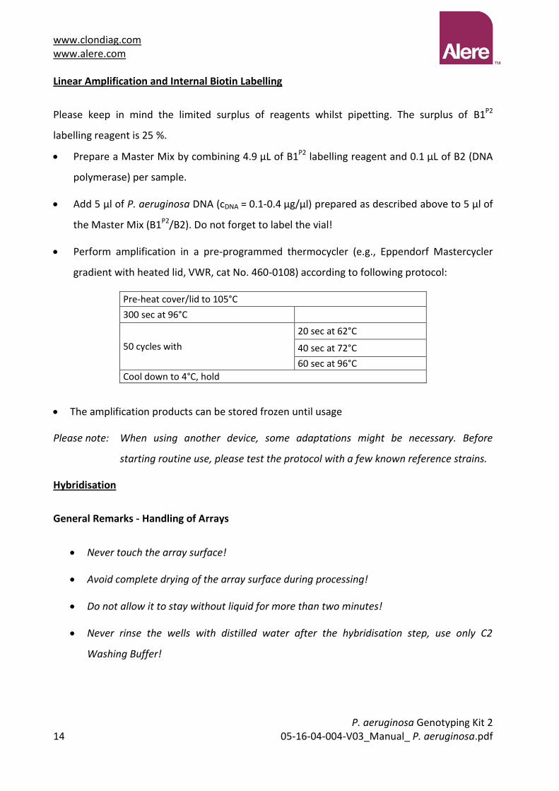
We strongly recommend that the liquid is removed by pipetting rather than by inverting the
strips and flicking the liquids out. Fine tipped soft, disposable Pasteur pipettes are suited best
(such as BRANDT / Cat. No. 612-2856). Always place the pipette tip at the cavity between the
array and the wall of the reagent well. If you touch the array surface, probes may be scratched
off and this may cause an error.
www.clondiag.com www.alere.com
P. aeruginosa Genotyping Kit 2
16 05-16-04-004-V03_Manual_ P. aeruginosa.pdf
General Remarks – the Substrate (Precipitating Dye) D1
An appropriate amount of substrate (precipitating dye) should be filled into an Eppendorf tube
and taken out of the refrigerator when starting the procedure allowing it to pre-warm to room
temperature / 25 °C. Cold D1 may yield weak signals. D1 should shortly be centrifuged prior to
use to remove bubbles as well as possible precipitates.
Triggered by peroxidase, in case of positive reactions, the dye precipitates but it is not
covalently bound. The precipitate can be dissolved by vigorous shaking. Thus the arrays must
not be shaken, dropped or moved abruptly during the staining procedure and afterwards.
After completion of staining, remove and discard reagent D1 as completely as possible and scan
immediately. The dye precipitate fades slowly in presence of liquids.
General Remarks - Thermoshakers
The correct temperature within the vessels is essential; therefore always use the appropriate
equipment for heating. Because of a possibly inhomogeneous distribution of the temperature
within the heating block and because of possible differences between displayed and actual
temperatures, the use of different brands of thermoshakers might affect test performance. We
tested the assay with BioShake iQ by Quantifoil Instruments (http://www.qinstruments.com/)
equipped with a customised heating block designed to fit ArrayStrips and Eppendorf’s
Thermomixer Comfort, equipped with a heating block for microtitre plates. When using other
devices, some modifications to the protocol might be necessary. Before starting routine use,
please test the protocol with a few known reference strains or with the control DNA (P.
aeruginosa PAO1).

www.clondiag.com www.alere.com
P. aeruginosa Genotyping Kit 2 05-16-04-004-V03_Manual_ P. aeruginosa.pdf 17
Protocol for Quantifoil’s BioShake iQ and Eppendorf’s Thermomixer Comfort with Microtitre
Plate Adapter
Switch on the thermoshaker and let it pre-heat to 60 °C.
Remove the ArrayStrip(s) from the pouch.
Insert the ArrayStrip(s) into the white frame. Assure the correct orientation (data matrix
barcode close to row A) and proper fit.
Pre-wash the array in two steps:
First, PCR-grade distilled water, 200 µl per well, just pipette 4x up and down, remove and
discard.
Second, C1 Hybridisation Buffer, 200 µl per well, at 60 °C, 2 min and 550 rpm in the
thermoshaker.
Add 90 µL of buffer C1 to each tube with (10 µL) labelled amplification product, mix gently.
Remove the buffer from the well with the array and add the mixture of C1 and labelled
amplification product.
Incubate at 60 °C, 60 min and 550 rpm in the thermoshaker.
Meanwhile, prepare conjugate: for each experiment add 1 µl conjugate 100x HRP to 100 µl
C4 Conjugation Buffer. This mixture is stable for around one working day at room
temperature; C3 is delivered with a surplus of 100%, C4 is delivered with a surplus of 200%.
Suggested pipetting scheme:
1 well
2-3 wells
4-6 wells
7-10 wells
11-15 wells
16-20 wells
21-30 wells
31-40 wells
C3 1.5 µl 3.5 µl 7 µl 11 µl 16 µl 21 µl 32 µl 42 µl
C4 150 µl 350 µl 700 µl 1100 µl 1600 µl 2100 µl 3200 µl 4200 µl
Remove liquid from the well and add 200 µl C2 Washing Buffer, discard C2.
Repeat this step twice.

www.clondiag.com www.alere.com
P. aeruginosa Genotyping Kit 2
18 05-16-04-004-V03_Manual_ P. aeruginosa.pdf
The thermoshaker needs some minutes to cool down from 60 °C to 30 °C. To speed up use
the cooled cover (for Quantifoil’s BioShake iQ, can be ordered from Quantifoil) or an ice
pack (Eppendorf’s Thermomixer Comfort).
After removal of C2 Washing Buffer add 100 µl diluted conjugate to each well, incubate at
30 °C, 10 min and 550 rpm in the thermoshaker.
Remove liquid and wash with 200 µl C5 Washing Buffer, pipette 4x up and down, remove
and discard.
Repeat this step.
Add 100 µl of D1 substrate (precipitating dye, at 25 °C, see above) per well.
Incubate at 30 °C, 5 min in the thermoshaker, but do not shake!
Remove liquid completely.
The bottom of the ArrayStrips may be cautiously be cleaned with wipes, bubbles may be
removed by removing and adding D1.
Scan and process (see below).
Data Analysis
Starting the ArrayMate Reader
We recommend starting the ArrayMate Reader after having started the hybridisation; this
allows you to conveniently start the device and to import the worklist file (see below)
Please note that this is a short instruction only. For more detailed information please refer to
the ArrayMate User Manual.
Switch on the ArrayMate (main switch on the rear below the electric cable plug, operating
switch on the bottom/left corner of the front side).
Switch on the screen (switch right hand below the screen).
www.clondiag.com www.alere.com
P. aeruginosa Genotyping Kit 2 05-16-04-004-V03_Manual_ P. aeruginosa.pdf 19
Log on as R&D User (Research and Development User) for full access to test specific
software (a default password will be provided together with the ArrayMate device). If you
log on as User, you will obtain only raw values, but no interpretation as positives/negatives
and no strain assignment. Administrator log-in will allow manipulation of file folders and
software; and this should be done only upon direct advice of Alere’s IT team.
The user interface will be loaded, ArrayMate performs internal testing. This will require less
than a minute.
Click on the icon New Run (left upper edge of the screen). A suggestion for a run name /
folder name for the new run appears in the top line of the screen. You may modify or
change the experiment name at your convenience.
Type in your operator ID (optional).
You may enter a comment into the memo field (optional).
www.clondiag.com www.alere.com
P. aeruginosa Genotyping Kit 2
20 05-16-04-004-V03_Manual_ P. aeruginosa.pdf
Worklist
A Worklist file allows linking an identifier such as a laboratory/sample number to a position of
an array within the ArrayStrip. For privacy reasons, arrays should not be identified by patient
names. Worklists can be generated using spreadsheet software such as EXCEL (see below) but
must be saved in the *.txt file format that can be imported into the test specific ArrayMate
software. Do not use special characters (such as : ; ()[] / \ ä ü etc.).
Create a list with at least three columns with obligatory headers in the order: position /
sample ID / assay ID (Table 1).
Positions are continuously numbered from 1 to a maximum of 96. Position 1 would
correspond to A1, 8 to H1, 9 to A2 and 96 to H12 (Table 2). Do not leave empty lines in the
worklist. If you use EXCEL, position numbers should be typed into column A.
Sample ID is strain / sample / laboratory number such as exported from your LIMS (or
assigned in any different way). Patient name should not be used as Sample ID.
The Assay ID enables the system to identify the current test and to correctly use
information on layout, spot number, and identity etc.. P. aeruginosa Genotyping 2 Assay
has the Assay ID: 10418. Assay ID numbers must not be confused as this could lead to errors
or loss of data.
You may add further columns and headers with notes and comments at your convenience.
Information from these columns will not appear on the result screen or in the Test Report.
We recommend using a printout of the worklist as template for pipetting.
Safe the worklist as tab separated *.txt file on the memory stick provided together with the
ArrayMate.
To avoid confusion, make sure that worklists are named unambiguously or that worklists
from earlier experiments are deleted.

www.clondiag.com www.alere.com
P. aeruginosa Genotyping Kit 2 05-16-04-004-V03_Manual_ P. aeruginosa.pdf 21
Table 1: Example worklist:
Position sampleID assayID comment
1 2013-12345 10418
2 2013-12346 10418
3 2013-12347 10418
4 2013-12348 10418
5 2013-12349 10418
6 2013-12350 10418
7 987654 10418 Isolate referred from Dr. J. Doe.
8 P. aeruginosa PAO1 10418 Control strain
Table 2: Positions in the 96 well format
Data Acquisition in the ArrayMate Reader
Insert your memory stick containing the worklist into any of the USB ports down to the
right side of the ArrayMate.
Press the button ; a folder selection dialog will open.
Select your worklist (path: “My Computer/Removable Disk”).
Open your selected worklist with Enter or the button Open.
Press the button (your imported worklist opens in a separate window). Proofread. If
the new window is empty, or if it was the wrong worklist, repeat the import.
Press the button OK; the worklist window will close.
1 2 3 4 5 6 7 8 9 10 11 12
A 1 9 17 25 33 41 49 57 65 73 81 89
B 2 10 18 26 34 42 50 58 66 74 82 90
C 3 11 19 27 35 43 51 59 67 75 83 91
D 4 12 20 28 36 44 52 60 68 76 84 92
E 5 13 21 29 37 45 53 61 69 77 85 93
H 6 14 22 30 38 46 54 62 70 78 86 94
G 7 15 23 31 39 47 55 63 71 79 87 95
H 8 16 24 32 40 48 56 64 72 80 88 96

www.clondiag.com www.alere.com
P. aeruginosa Genotyping Kit 2
22 05-16-04-004-V03_Manual_ P. aeruginosa.pdf
Leave the memory stick attached to the ArrayMate if you intend to export Test Reports
afterwards.
Press the button Next (bottom / right on the screen; reader opens).
Carefully insert the appropriate metallic adapter / frame into the ArrayMate. Do not apply
any strong force. Assure proper fit, otherwise the images may be out of focus.
Carefully insert the white frame with the array strips into the metallic adapter. Assure the
correct orientation (Position A1 in the frame next to the data matrix barcode on the
adapter) and proper fit, otherwise the images may be out of focus.
ArrayStrip frame with Strips
inserted in accordance with the
worklist.
Please note: ArrayStrips must be clean. They should not contain any liquids during analysis.
Barcodes must be clean. There must be no ArrayStrip Covers on the wells to be
analysed (however, unused wells should remain covered).
Press the button Next (bottom / right on the screen; reader closes, analysis program starts,
reading takes 2-10 min dependent on the number of strips; reader takes images, and
automatically analyses the data). The progress of the reading is indicated by the following
symbols:
photographed:
in analysis:
ready:
The reader indicates the end of the entire process with an acoustic signal (beep).
Press the button Next (bottom / right on the screen; reader opens).
Remove the white frame with the ArrayStrip(s).
Press the button Next (bottom / right on the screen; reader closes).

www.clondiag.com www.alere.com
P. aeruginosa Genotyping Kit 2 05-16-04-004-V03_Manual_ P. aeruginosa.pdf 23
Results
On the left hand side of the screen you will see a list showing all runs stored on the ArrayMate´s
hard disk. A run contains the results from all arrays analysed together within one frame. If this
list is not displayed:
Press the button Archive (left hand) and activate the flag Browse (top left).
The runs are organised like folders in “Windows Explorer”, and named by default according to
the date of acquisition.
Example: There is one experiment run in this archive:
If you click on the plus symbol left on the run name, the folder opens and you will see a list of
the individual arrays ordered by Sample ID.

www.clondiag.com www.alere.com
P. aeruginosa Genotyping Kit 2
24 05-16-04-004-V03_Manual_ P. aeruginosa.pdf
Click on a Sample ID and the P.aeruginosa 2 Genotyping Report for this array is shown in the
window on the right:
Export of Test Reports
Two result files in html-format will be generated. The shorter report gives a summary on
genotyping information. A longer html result sheet (“result_B.res.html”) provides information
on all probes. Possible error messages in these reports are explained below (see
Troubleshooting).
Other files that are generated and can be exported include
A *.csv file containing the experimental result as a string which can be imported into an
Excel-based list (for data mining; see chapter 3 below),
A *.txt file with the raw measurements,
An image file (*.bmp) showing the actual photo of the array,
A second image file (*.png) in which the coordinate grid is superimposed and the
recognised spots are circled, and
A *.xml file providing the same information as the html result sheet for future export
into databases etc.,
A *.log file containing specific data of the evaluation of spots and substances,
An *.out file containing output log data which help our service to trace image evaluation
errors.

www.clondiag.com www.alere.com
P. aeruginosa Genotyping Kit 2 05-16-04-004-V03_Manual_ P. aeruginosa.pdf 25
Please note: Only complete runs can be exported. The export of individual Test Reports is not
possible.
Right-click on the selected run (a menu appears with the option Export Run).
Right-click on Export Run (a file browser opens).
Click on My Computer, subsequently on Removable Disk, and choose the folder to save or
click on the button Make New Folder (on the bottom; a new folder icon appears).
Rename the new folder (e.g. with the experiment run name or date).
Click on the OK button (data are exported into the new folder on your memory stick).
Do NOT remove the memory stick as long as the hourglass symbol is visible.
Switch off the device by clicking on the Power-button (left / down on the screen):
Switch off the Screen. There is no need to physically switch off the ArrayMate.

www.clondiag.com www.alere.com
P. aeruginosa Genotyping Kit 2
26 05-16-04-004-V03_Manual_ P. aeruginosa.pdf
Contents of the Test Report
The ArrayMate creates a test report that lists all markers analysed along with an individual test
result for each marker. In addition, the test report provides a rating of the data quality along
with identification of a possibly erroneous step of the experimental process.
The html file (*.html) may be viewed with any browser software (e.g. Internet Explorer, Firefox
etc.) and may be printed or converted into a *.pdf file (for details please refer to the
ArrayMate’s user manual). The contents of the test report are described in more detail in the
following section.
Kinship Analysis: Markers of the Core Genome (“KinCode” / “SNP Pattern”)
Sixteen different markers of the core genome were selected for genetic identification of P.
aeruginosa isolates. In the test report, these markers are named “SNP Pattern” and are
formatted as a string of 16 digits. Each digit may adopt the value “0”, “1”, “2”, “X”, respectively.
Strings containing “0” and “1” digits are used for deriving the four-digit alphanumeric
“KinCode” which contains all information of the 16 digit code, but is easier to comprehend.
However, if the test result of some digit is unclear, it is rated “2” in the 16 digit string. If the test
result of a marker is invalid, it is rated “X” in the 16 digit string, and then the four-digit KinCode
remains incomplete. In these cases, the respective alphanumeric digit of the KinCode is
replaced by “X“. For each complete KinCode, background information regarding the frequency
of the respective code is given, based on a data base established by L. Wiehlmann
(unpublished).
SNP pattern and KinCode were described by Wiehlmann et al. 2007 (see literature).
Marker Controls
These controls are important for the rating of data quality. Furthermore, in case of handling
mistakes they provide important information regarding the experimental step that may have
failed.

www.clondiag.com www.alere.com
P. aeruginosa Genotyping Kit 2 05-16-04-004-V03_Manual_ P. aeruginosa.pdf 27
If the Staining Control failed (and along with it all other controls) this indicates failure
of the Horseradish Peroxidase-mediated conversion of D1, that means a failure of one
of the steps after hybridisation.
If all controls (including the Hybridisation Control) failed, but not the Staining Control,
this indicates a failure of the Hybridisation step.
If the Labelling, Genus/Species and DNA Stringency Control failed, but not the
Hybridisation Control and the Staining Control, this indicates a failure of PCR.
If only the Genus/Species and DNA Stringency Control failed, this indicates poor quality
of DNA.
Failure of Negative Controls indicates a high nonspecific signal background.
Please refer to the troubleshooting section below for further information.
Plausibility Controls
Some combinations of markers in nature are impossible to occur for biological reasons. The
software checks for those combinations and returns a warning if they are diagnosed. In these
cases the test result is implausible. Please refer to the troubleshooting section below for further
information.
Accessory Genome and Microevolution (“Features of the Isolate”)
In addition to the markers that characterise a clone, additional markers of the accessory
genome were added to the array. Originally (Wiehlmann et al. 2007), 41 markers of the
accessory genome had been selected, but meanwhile two of them (TB-C47-1 and TB-C47-2)
have been removed because of faulty results.
The remaining 39 markers are listed in the test report as “Features of the Isolate” and they may
be exported along with the KinCode as a 39 digit code in the “0”, “1”, “2” or “X” format as
described above. This information may be used to characterise microevolutionary events within
a clone that is characterised by a uniform core genome pattern. The markers of the accessory
genome have been described in detail by Wiehlmann et al. 2007 (see literature).

www.clondiag.com www.alere.com
P. aeruginosa Genotyping Kit 2
28 05-16-04-004-V03_Manual_ P. aeruginosa.pdf
Recommendation for a Project Specific Database
The CSV file (*.csv), which contains the experimental result as a string. For comparison of the
results and for data mining each of these strings may be imported into an “Excel Based Data
Mining Tool” (see 3.1) as suggested by Wiehlmann et al. (2007). Alere Technologies provides a
drawing of this Excel sheet that has been developed from the sheet suggested by Wiehlmann et
al. Further, Alere Technologies provides a tool (the “Experiment String Collector” see 3.2) that
enables the user to collect the strings from all *csv files within one folder in one single step.
Both, the Excel datasheet and the string collector, are described in the following section.
String Collector
The tool called “Experiment String Collector” enables the user to collect all data (strings) from
*.csv files found within one folder into one output *.csv file (CommaSeparatedValue files being
generic files for export into spreadsheet programs).
As outlined above and in the ArrayMate manual, the results of each experimental run can be
exported from the ArrayMate to an USB stick into a folder. In order to collect all data strings
found within this folder into a single *.csv file, proceed as follows:
Download the string collector from our website HUwww.alere-technologies.comUH and
install it on your office computer according to the instructions you will find in the
Software and Reader section of this manual.
Open the string collector by double clicking its icon on your desktop; the main window
appears:
Select the path of the source folder (the folder of the ArrayMate Data export) by clicking
1
2

www.clondiag.com www.alere.com
P. aeruginosa Genotyping Kit 2 05-16-04-004-V03_Manual_ P. aeruginosa.pdf 29
into area 1 (indicated by the first red circle on the image above)
Select the path of the target folder (the folder to save the *.csv file containing the
collected data strings) by clicking into area 2 (indicated by the second red circle on the
image above)
Single click the generate button; all *.csv files in the source folder will be scanned, and
the content will be collected within one *.csv file named Experiments_YYYY_MM_DD-
HH_MM_SS.csv
Click on the button open File; the Experiments.csv file created opens in the notepad text
editor
The content of the new *.csv file may now be transferred into an Excel-based Data
Mining Tool by copy / paste.
Excel-Based Data Mining Tool
An Excel-Based Data Mining tool was developed together with the group of Lutz Wiehlmann.
The template for a user specific data sheet can be downloaded from our website: www.alere-
technologies.com.
The Excel sheet draft is separated into two areas:
The first area (columns A to M) is meant for entering data like source of the specimen
manually.
The second area (columns N to BS) is meant for pasting experiment results provided by
the ArrayMate in the *.csv files in a copy / paste manner. These data consist of the
Identifier of the result, the date of test, the KinCode and the strings for SNP pattern
and Features of the Isolate as a 16 + 39 digit string that may be used for Kinship
Analysis.
In order to copy the data from an individual *.csv file into the data mining list proceed as
follows:

www.clondiag.com www.alere.com
P. aeruginosa Genotyping Kit 2
30 05-16-04-004-V03_Manual_ P. aeruginosa.pdf
Open the *.csv-file of the experiment with any editor program (Editor, Notepad, etc.),
select and copy “all“
paste the string into the data mining list in the “Pseudomonas test results” table,
column N “Identifier of result”
Enter the appropriate additional data into columns A to M manually
In case it is intended to enter the result of more than one array into the database we
recommend the use of the String Collector (see below).
TROUBLESHOOTING
For training and qualification purposes we recommend our Control-L Kit (Alere order number
205500010). In case of doubt, this kit is the perfect tool to make sure that your laboratory
processes are working correctly. It may also be used for the functional testing of individual kit
components.
Poor DNA Quality: Only the DNA Extraction Control failed. In these cases we recommend to re-
check DNA quantity and quality by loading leftover DNA on an agarose gel.
The amount of DNA is crucial. A260 readings will cover RNA and other contaminants as
well. Therefore pure DNA preparation without bulk amounts of RNA is prerequisite for
proper DNA concentration measurement. RNAse treatment prior to A260 reading
therefore is necessary (component A2 contains RNAse).

www.clondiag.com www.alere.com
P. aeruginosa Genotyping Kit 2 05-16-04-004-V03_Manual_ P. aeruginosa.pdf 31
Some P. aeruginosa colonies protect themselves with thick layers of slime. It may help to
wash the bacterial pellet 3x in 1 ml of 5 mM EDTA, pH 7.0 (re-suspend and centrifuge at
3,000 × g for 6 min at room temperature, discard the supernatant).
DNA must not be fragmented, as fragmentation reduces the efficiency of amplification and
labelling due to the distance between primer and probe binding sites.
DNA should be free of RNA, as free RNA reduces the efficiency of amplification and
labelling by effectively removing primer from the reaction mix due to competitive
hybridisation.
DNA must be free of any traces of ethanol, as ethanol strongly influences the amplification.
This is an important issue as, e.g., in the top of QIAGEN tubes a drop of ethanol-containing
washing buffer might get trapped. The tubes really need to be centrifuged on high speed.
Alternatively, it is possible to heat the sample prior to adding it to the labelling mix (some 5
minutes at 70 °C). Some problems with samples from the Qiagen EZ1 device for example
were resolved after heating the samples.
All reagents for DNA extraction need to be within the recommended shelf-life and stored in
the appropriate way.
We have good experience with the manual QIAGEN DNeasy Kit and the automated device EZ1.
Contrarily, alkaline lysis, mechanical bead-beater extraction or ultrasonication in our hands
resulted in poor DNA recovery, poor DNA quality or both.
An alternative method that is fast and simple but that did work in the hands of several
customers was described by Wiehlmann et al. 2007 (see literature)
PCR failed: Labelling and DNA Extraction Controls failed, but not the Hybridisation Control and
the Staining Control. In this case, please carefully review the PCR process for correctness. In
some cases, inhibitors of PCR are isolated along with DNA. In these cases a rerun of DNA
purification may help.
Hybridisation failed: All controls (including the Hybridisation Control) failed, but not the
Staining Control. In this case please carefully review the hybridisation step. Was the buffer (C1)
used correct? Was the hybridisation temperature used correctly?

www.clondiag.com www.alere.com
P. aeruginosa Genotyping Kit 2
32 05-16-04-004-V03_Manual_ P. aeruginosa.pdf
Staining failed: All controls failed. This indicates a failure of one of the steps after hybridisation.
The most likely reasons are:
The component D1 was too cold when administered. Please carefully follow the steps that
are outlined in section 8.
Horseradish Peroxidase Conjugate (C3) or its substrate D1 may have degraded during
storage. If this may be an option, we recommend testing C3 and D1 with our Control-L kit.
Enzymatic reaction is inhibited by carryover of buffer C1. Ensure proper washing of the
wells to remove all of Buffer C1 prior to adding Horseradish Peroxidase Conjugate (C3/C4).
Horseradish Peroxidase may have degraded because of an incubation temperature >>30 °C.
Therefore, please review the incubation step with C3/C4 for correctness. Make sure that
the shaker indeed has cooled down to 30 °C before incubation.
Negative control failed. In this case a high nonspecific background may result in false-positive
markers. In this case please carefully check the hybridisation step and the subsequent washing
steps for correctness. Did you use the correct buffers? Did you incubate at the correct
temperature? Is your thermoshaker working correctly i.e. at the temperature it is supposed to
work? Alternatively, these options may be checked:
The C3/C4 may not have been washed away correctly. In this case, the D1 turns dark
green whilst incubating and this may result in nonspecific background.
The array may have dried out during hybridisation if the cap had not been closed
properly.
Plausibility controls failed. This may either indicate a failure of the test protocol; in this case
the additional failure of other controls (see above) is expected. In any case we recommend to
review the conditions of hybridisation (especially the real hybridisation temperature) and the
washing steps after hybridisation.
Alternatively, failure of the plausibility controls may indicate the mixture of different P.
aeruginosa clones; in this case we recommend subcloning and repeat of the experiment.

www.clondiag.com www.alere.com
P. aeruginosa Genotyping Kit 2 05-16-04-004-V03_Manual_ P. aeruginosa.pdf 33
Physical damage to the array. Scratching of the array surface with a pipette tip can lead to
damage of an array spot that prohibits the acquisition of a valid signal. In this case the
respective spot is set invalid (=”1”), the corresponding spot may be positive and valid (=”0”).
We recommend reviewing section 4 for general precautions in array handling.
LITERATURE
Wiehlmann et al. 2004, JOURNAL OF BACTERIOLOGY Vol. 186, No. 13, p. 4228–4237.
Wiehlmann et al. 2007, PNAS vol. 104 no. 19, p. 8101–8106.
www.clondiag.com www.alere.com
P. aeruginosa Genotyping Kit 2
34 05-16-04-004-V03_Manual_ P. aeruginosa.pdf
ADDITIONAL INFORMATION
Warranty
Alere guarantees the performance as described in this manual. Usage of the Kit was
successfully tested at ambient temperatures up to 37 °C, a guarantee is limited to ambient
temperature in the laboratory between 18 °C and 28 °C. Kit components comprise the
ArrayStrips, the reagents for DNA labelling and for detection of labelled DNA products on the
array, the ArrayMate reader and its software. In case one of these components fails within the
expiry date due to other reason than misuse, contact Alere for replacement or refund. Terms
and conditions apply.
If you have any problem or question, please contact our technical service.
Quality Control
Each batch is stringently tested with the use of standard DNA preparations for good
performance and correctness of results.
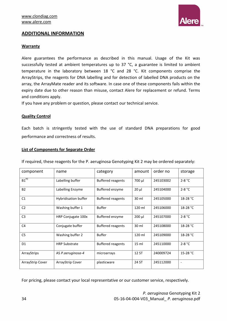
List of Components for Separate Order
If required, these reagents for the P. aeruginosa Genotyping Kit 2 may be ordered separately:
component name category amount order no storage
B1P2
Labelling buffer Buffered reagents 700 µl 245103002 2-8 °C
B2 Labelling Enzyme Buffered enzyme 20 µl 245104000 2-8 °C
C1 Hybridisation buffer Buffered reagents 30 ml 245105000 18-28 °C
C2 Washing buffer 1 Buffer 120 ml 245106000 18-28 °C
C3 HRP Conjugate 100x Buffered enzyme 200 µl 245107000 2-8 °C
C4 Conjugate buffer Buffered reagents 30 ml 245108000 18-28 °C
C5 Washing buffer 2 Buffer 120 ml 245109000 18-28 °C
D1 HRP Substrate Buffered reagents 15 ml 245110000 2-8 °C
ArrayStrips AS P.aeruginosa-4 microarrays 12 ST 240009724 15-28 °C
ArrayStrip Cover ArrayStrip Cover plasticware 24 ST 245112000 -
For pricing, please contact your local representative or our customer service, respectively.

www.clondiag.com www.alere.com
P. aeruginosa Genotyping Kit 2 05-16-04-004-V03_Manual_ P. aeruginosa.pdf 35
LEGAL MANUFACTURER
Alere Technologies GmbH
Loebstedter Str. 103-105
07749 Jena, Germany
CONTACT
www.clondiag.com www.alere.com
P. aeruginosa Genotyping Kit 2
36 05-16-04-004-V03_Manual_ P. aeruginosa.pdf
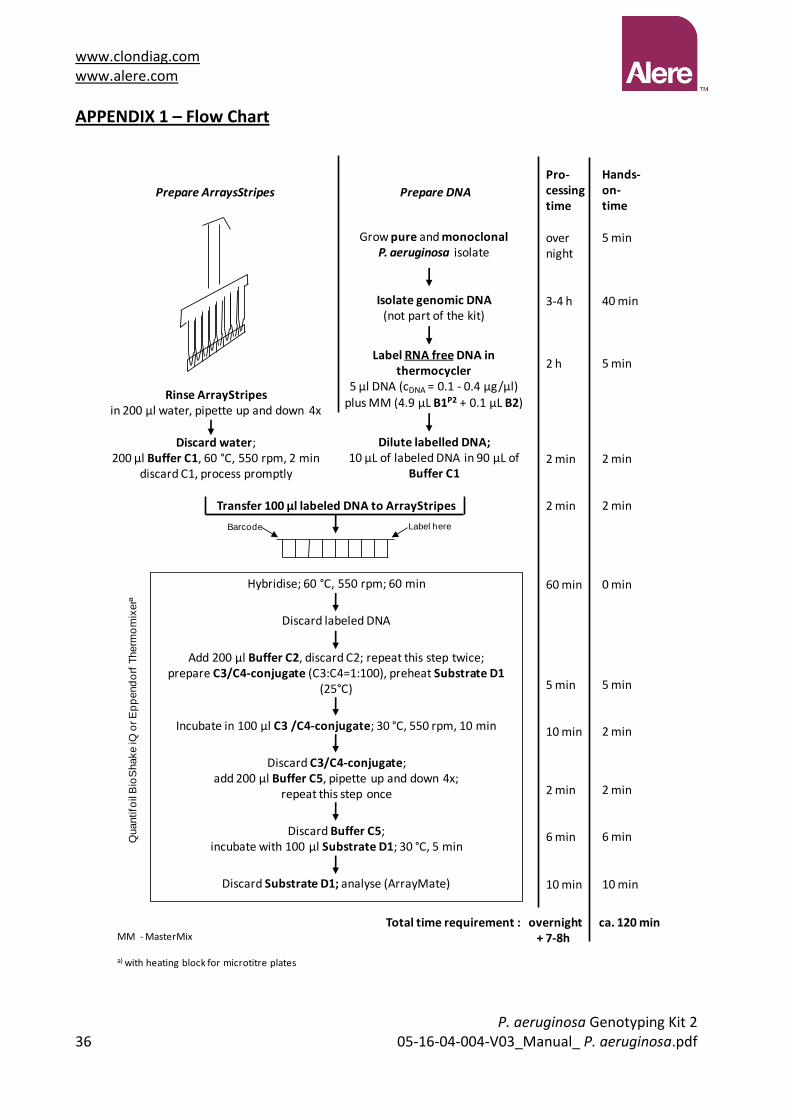
APPENDIX 1 – Flow Chart
Transfer 100 µl labeled DNA to ArrayStripes
Hands-on-time
5 min
40 min
5 min
2 min
2 min
0 min
5 min
2 min
2 min
6 min
10 min
Pro-cessingtime
overnight
3-4 h
2 h
2 min
2 min
60 min
5 min
10 min
2 min
6 min
10 min
Hybridise; 60 °C, 550 rpm; 60 min
Discard labeled DNA
Add 200 µl Buffer C2, discard C2; repeat this step twice;prepare C3/C4-conjugate (C3:C4=1:100), preheat Substrate D1
(25°C)
Incubate in 100 µl C3 /C4-conjugate; 30 °C, 550 rpm, 10 min
Discard C3/C4-conjugate;add 200 µl Buffer C5, pipette up and down 4x;
repeat this step once
Discard Buffer C5; incubate with 100 µl Substrate D1; 30 °C, 5 min
Discard Substrate D1; analyse (ArrayMate)
Total time requirement : overnight ca. 120 min+ 7-8h
Prepare DNAPrepare ArraysStripes
Rinse ArrayStripesin 200 µl water, pipette up and down 4x
Discard water;200 µl Buffer C1, 60 °C, 550 rpm, 2 min
discard C1, process promptly
Grow pure and monoclonal P. aeruginosa isolate
Isolate genomic DNA(not part of the kit)
Label RNA free DNA in thermocycler
5 µl DNA (cDNA = 0.1 - 0.4 µg/µl)plus MM (4.9 µL B1P2 + 0.1 µL B2)
Dilute labelled DNA; 10 µL of labeled DNA in 90 µL of
Buffer C1
MM - MasterMix
a) with heating block for microtitre plates
Quantifo
il B
ioS
hake iQ
or E
pp
end
orf
Therm
om
ixera
Barcode Label here

www.clondiag.com www.alere.com
P. aeruginosa Genotyping Kit 2 05-16-04-004-V03_Manual_ P. aeruginosa.pdf 37
APPENDIX 2 – Probes and Primers for KinCode Genotyping and the Accessory Genome
Probes Reference Probes Antisense primer 1
Antisense primer 2
oriC PAO PAO1- Sequence, Stover et al. (updated 2006) GAAGCCCAGCAATTGCGTGTTTC
AGCCTCGACACCGGTTCTCG
ACCATCTCGTTCATCCCCAGG
oriC non-PAO UCBPP-PA14, complete genome, Lee et al. (2006) GAAGCCCAGCAACTGCGTGTTTC
oprL (1) PAO PAO1- Sequence, Stover et al. (updated 2006) GGTGCTGCAGGGTGTTTCGCCGG
TTCTGAGCCCAGGACTGCTCG
TCGACGCGACGGTTCTGAGCC
oprL (1) non-PAO UCBPP-PA14, complete genome, Lee et al. (2006) GGTGCTGCAGGGCGTTTCGCCGG
oprL (2) PAO PAO1- Sequence, Stover et al. (updated 2006) GTGCTGCAGGGTGTTTCGCCG
oprL (2) non-PAO UCBPP-PA14, complete genome, Lee et al. (2006) GCTGCAGGGCGTTTCGCCG
fliCa (1) PAK PAK, Totten and Lory (1990), flagellin type a2, Giske et al. (2006)
CAAGATCGCCGCAGCGGTCAAC
AGCTGATGGTATCGCCGTCGC
CTAGTGATCGCACCGGAGCC
fliCa (1) non-PAK ATCC15691, Spangenberg et al. (1998), flagellin type a1, Giske et al. (2006)
CAAGATCGCCGCTGCGGTCAAC
fliCa (2) PAK PAK, Totten and Lory (1990), flagellin type a2, Giske et al. (2006)
CAAGATCGCCGCAGCGGTCAACGAC
fliCa (2) non-PAK ATCC15691, Spangenberg et al. (1998), flagellin type a1, Giske et al. (2006)
CAAGATCGCCGCTGCGGTCAACGAC
alkB2 PAO PAO1- Sequence, Stover et al. (updated 2006) CCTCGCCCTGTTCCCACCGCTCTGG
TTCCTCGCCGGCATAGTAGGC
alkB2 non-PAO ATCC 15691, Morales et al. (2004) CTCGCCCTGTTCCCGCCGCTCTGG
citS-1 PAO PAO1- Sequence, Stover et al. (updated 2006) TCGAGCAACTGGCAGAGAAATCCG
GCAGGTAGCAGGTTTCCAGG
AACTGTTCCTTCTGCGCGGCG
citS-1 non-PAO UCBPP-PA14, complete genome, Lee et al. (2006) CGAGCAACTGGCGGAGAAATCCG
citS-2 PAO PAO1- Sequence, Stover et al. (updated 2006) GCGGAAAACTTCCTGCACATGATGTT
TGATCGGCTTGGTCTCGCAGG
GCTGATCGGCTTGGTCTCGC
citS-2 non-PAO Kiewitz and Tummler. J Bacteriol. (2000) 182:3125-35. GCGGAAAACTTCCTCCACATGATGTT
oprI (1) PAO PAO1- Sequence, Stover et al. (updated 2006) AGCTCAGCAGACTGCTGACGAGG
GCTGGCTTTTTCCAGCATGCG
TTGCGGCTGGCTTTTTCCAGC
oprI (1) non-PAO UCBPP-PA14, complete genome, Lee et al. (2006) AGCTCAGCAGACCGCTGACGAG
oprI (2) PAO PAO1- Sequence, Stover et al. (updated 2006) GCTCAGCAGACTGCTGACGAGGCTAACG
oprI (2) non-PAO UCBPP-PA14, complete genome, Lee et al. (2006) GCTCAGCAGACCGCTGACGAGGCTAAC
ampC-1 PAO PAO1- Sequence, Stover et al. (updated 2006) ACGGCCGCCGGGTGACGCC CGCATCTTGTCCTGGGTCAGG
TCGTCGAGGCGCATCTTGTCC
ampC-1 non-PAO De Champs et al. (2002), Kiewitz and Tummler. J Bacteriol. (2000) 182:3125-35.
ACGGCCGCCAGGTGACGCCG
ampC-3 PAO PAO1- Sequence, Stover et al. (updated 2006) CGACCTACGCGCCGGGCAG GGCGAGATAGCCGAACAGGC
CACTTGCTGCTCCATGAGCC
ampC-3 non-PAO De Champs et al. (2002), Kiewitz and Tummler. J Bacteriol. (2000) 182:3125-35.
CGACCTATGCGCCGGGCAGC
ampC-4 PAO PAO1- Sequence, Stover et al. (updated 2006) CGTTCGAACGGCTCATGGAGCAG
ACGTCGAGGTGGGTCTGTTCG
GTAGCCTTCGGCATCCAGCG
ampC-4 non-PAO De Champs et al. (2002), Kiewitz and Tummler. J Bacteriol. (2000) 182:3125-35.
CGTTCGAACGACTCATGGAGCAGC
ampC-5 PAO PAO1- Sequence, Stover et al. (updated 2006) TGGAGCAGCAAGTGTTCCCGGC
amplified with primers of ampC-4
ampC-5 non-PAO De Champs et al. (2002), Kiewitz and Tummler. J Bacteriol. (2000) 182:3125-35.
TGGAGCAGCAACTGTTCCCGGC
ampC-6 PAO PAO1- Sequence, Stover et al. (updated 2006) GAACAAGACCGGTTCCACCAACGG
TCGGCATTGGGATAGTTGCGG
ampC-6 non-PAO UCBPP-PA14, complete genome, Lee et al. (2006) AACAAGACCGGCTCCACCAACGG
ampC-7 PAO PAO1- Sequence, Stover et al. (updated 2006) CGACCTGGGCCTGGTGATCCT
TTGGGATAGTTGCGGTTGGC
TGGCGTAGGCGATCTTCACCC
ampC-7 non-PAO De Champs et al. (2002), Kiewitz and Tummler. J Bacteriol. (2000) 182:3125-35.
GCGACCTGGGACTGGTGATCCTGG
fliC a ATCC15691, Spangenberg et al. (1998) GTCGCTGAACGGCACCTACTTCA
CGATCGCGATGTCGACGGTGC
TGCCGATCGCGATGTCGACG
fliC b PAO1- Sequence, Stover et al. (updated 2006) GCCGACCAACTGAACTCCAACTCG
TGACGTTCTCGCCGGTAGCG
CAGTAGCGGTACCGGTCTGC
exoS PAO1- Sequence, Stover et al. (updated 2006) CAGCCCAGTCAGGACGCGCA
CAGGGTCGCCAGCTCGCTCGCC
AGGGTCGCCAGCTCGCTCGC
exoU UCBPP-PA14, complete genome, Lee et al. (2006) CGCCAGTTTGAGAACGGAGTCACC
AGTGATCTGCCGCGGCCCTGCC
GTGATCTGCCGCGGCCCTGC
fpvA type I PAO1- Sequence, Stover et al. (updated 2006) CCTGAATCCGACCATTCGCGAGTC
CGTTCAGGTCGTAGACCGCGC
GCGATACCAACTGTCCTGCGGC
fpvA type IIa de Chial et al. (2003) TCGGACTGTACTCCTACGAAGCAGC
TGCCGAAGGTGAATGGCTTGCC
CCTGATGGTCCGATCCCAGC

www.clondiag.com www.alere.com
P. aeruginosa Genotyping Kit 2
38 05-16-04-004-V03_Manual_ P. aeruginosa.pdf
Probes Reference Probes Antisense primer 1
Antisense primer 2
fpvA type IIb Spencer et al. (2003) CCAATCCCTATCGCTGGAACCGTACC
GCCGAGGGTCAAGAACCACTGG
TCTTGGCCCAGTCATAGCGGC
fpvA type III de Chial et al. (2003) GCTCGGGACTCGCATTTCGTCC
TAACCCCAAGGCCCATTGGAGG
GCCACCGCCTTCGAATAACCCC
fpvB PAO1- Sequence, Stover et al. (updated 2006) GCGTTATTGCTCGGTCTCTCCTCG
AATTGCTCGAGGGATGCGGC
GGTCGAAACGGATGCGCAGG
LES LES400 (personal communication C. Winstanley) TGCATAGGAGTCATGCCGACAGCA
GCCCCGCGTCATTTTCACGTCG
AATGCTCTGGGCAACGAGCC
PA0636 PAO1- Sequence, Stover et al. (updated 2006) GCCAATTGGGTCAGCAAGCAACG
ATGCCATCGTTGAAGGCACCGC
TGCCATCGTTGAAGGCACCG
PA0722 PAO1- Sequence, Stover et al. (updated 2006) CGTGTCGCGAACTCGCATGGC
TCTGGCGGAATCAGGTAGGCC
CTTCCGGGGAGAAACCACCG
PA0728 PAO1- Sequence, Stover et al. (updated 2006) CTGGAGCCTGCGAAAGTGGCTC
AGCCAAGACGGTTGTTCGCGG
TCAATGACGCCGAGTTGGCGC
PA2185 PAO1- Sequence, Stover et al. (updated 2006) ACGAGGGTGATGGCTGGGAATACG
CTCGGACAGGTTCACGCTGG
GCCATTCGCTGCAACACCTCC
PA2221 PAO1- Sequence, Stover et al. (updated 2006) CAGTTGTCGCCAGGTCTGGAGAATCC
TTCCTGGGCCAGAGTTGGACC
AGCTTAAGGCCGTGGCACTCG
PA3835 PAO1- Sequence, Stover et al. (updated 2006) CACATCAATGTCAGCCCACGCCA
CCGGAGAATTCGCGTCCACC
TGCTGACGATGAAGCCCCAGC
fla-island Arora et al. (2001) ACCTGTGTCGCTGGAGGGTATGTT
CCCGTGTTTCCGTAGACCTTGC
orfA Arora et al. (2001) CGCTGGAGGGTATGTTCCGCAAGG
GTTCCACAGGCGCTGCGGCGC
GTTCCACAGGCGCTGCGGCG
orfI Arora et al. (2001) CCTGGACCTCTCCAAGGTTCGCCT
AAACTGCCCCGCCCCCCATCC
GGAAAAACTGCCCCGCCCCCC
orfJ Arora et al. (2001) GCCATTCCGACGACCAAACAAGGC
ACGCTCGCAGCGCCTCACGCG
GGCCTGGCTGCGAACGCTCGC
PA0980 PAO1- Sequence, Stover et al. (updated 2006) CGGTATGAAGATGGGTGGTTGGGTCG
ACCTCCAGCACCGACACACC
ATCCGATCCACCTCCAGCACC
XF1753 UCBPP-PA14, complete genome, Lee et al. (2006) TGCGAGGACCAGAAACCTTGATGG
GCGCGCGTTCGAGAAACAGG
CGGAGGTTGAAAAGCTGGCCC
acetyltransferase UCBPP-PA14, complete genome, Lee et al. (2006) CGAAGCGTAGGGTCTTCGTAGCC
ACGACGTCACCGTCGAGACCG
ACCGCCTTTCTGGTGAGCTGG
pKL-1 Klockgether et al. (2004) CACCATGCAAATGCTCGATGGACTGC
ATCTGAACCGAGGGGATCCGC
CCCGGGAGTCATTGGTCTGG
pKL-3 Klockgether et al. (2004) TCTGAACTGCGGCTATCACCTGGA
GACCTACACTCCAACCGCTGG
TTCCCTTGCTGCCGAGAAGC
TB-C47-1 P.aeruginosa TB, pKLC102 related gene island integrated in tRNA(Lys) PA4541.1
GCAGGCGTCCAAGTTGGAGCTCTCC
GCCTGTTGGACCCCTTTGACC
TACTCCTGCCTGTTGGACCCC
TB-C47-2 P.aeruginosa TB, pKLC102 related gene island integrated in tRNA(Lys) PA4541.1
TCCAACAGGCAGGAGTACAGGGTG
TCTGTCAATCCCCTTTGGGG
AGCCCCTTTCTGTCAATCCCC
PAPI-1 pili chaperone UCBPP-PA14, complete genome, Lee et al. (2006) GGAACACAACGTGGGGCGTGAC
CGCTCAAGCGCTATCCCACC
CGCCATCGGCCTGTACAACG
PAPI-1 luminal binding protein
UCBPP-PA14, complete genome, Lee et al. (2006) CCAGTTGGCACCACCATGCTTGC
CGGTAGAGAGCTGGGTTGGC
AACCTGGAGCTAGGGCAGAGC
pKLC conserved hypothetical
Klockgether et al. (2004) GCCTGCCTACTTGTTCCCAACGC
CTACCCAGCTTGGGCGTAGC
AAGCGATAGCCGTGCTCCTGC
pKLC adhesin Klockgether et al. (2004) GGCTGTATTGCCCGCCATTCTCC
CCGGCTATATCCGCGGCTACC
ATTGGCGCTGCTGTTTACGCCC
pKLC fatty acid synthase
Klockgether et al. (2004) CGACAGACAGAAAGGGTTCTTGCGC
GGTGGCGTCGGGTTTTTCTGC
AGGTCGTAGCGGAAGGTGGTGG
PAGI-2/3-4 Larbig et al. (2002) GCGCCTTCTCCTCTTTGCAGATGT
TGTCCCGGCTCAGTTCAACG
GCAACACCTTGGCGTTTGTCC
PAGI-2/3-5 Larbig et al. (2002) CAGTATGGTACGGACACGAAGCGC
TCAAGCTCGTTGTGGACCGC
GTTACGACGGCGTGCTGTCGG
PAGI-2/3-6 Larbig et al. (2002) CCATGGTCGGAACAGGCACGATATGC
CAACACGCGACTGGCGATCC
TACATCATCCGCAACGGCGGC
C-45 Larbig et al. (2002) CGAGGAGTTTCGGACCCGCTTTGA
TCATCCAGCAAGCCATTGCGC
TGGAGTCGCTTTCCGCCATCG
C-46 Larbig et al. (2002) CGAAGTCTGAGGTGTGGACCCGC
CGCGGTGCTGGTTGCGCTGC
CGCTGGCAGTTCCGCTGGCC
C-47 Larbig et al. (2002) CCACTCGATCATGTTGAGCATCGGCTCC
TATTGACGACCTACCGCGCGCC
CACCAAGAACCCGCTGCTCG
PAGI-2 Larbig et al. (2002) GCATCATTGCGCGTCACATCTGGT
ACGCAACGTATTCGGCGACCC
CGCAACGTATTCGGCGACCC
PAGI-2/3-1 Larbig et al. (2002) GACCGCAAGCAGAAACGGCATGC
GGTGCTCGACCCAAGCATCG
TCCTTGAGTTCCTTGGCGCGG
PAGI-3-1 Larbig et al. (2002) CCCGTTGCTCATAACCCGTTCCTG
GACGAATACCCAGCTGCGTGG
GCAGACGAATACCCAGCTGCG
PAGI-3-8 Larbig et al. (2002) GGTTAGTCCCTTCTGCCCGCATCG
ATCGTGGCAGGATGTCCACCG
TAGGCGGGCCTTTTGAAGGTGC
tRNA(Pro)- island 1 P.aeruginosa TB, gene island integrated into tRNA(Pro) PA2736.1
GTGTCACGGCCCATGTCTAGCAGC
TCCACGCCGAGGGACGTGCC
GCTCCACGCCGAGGGACGTGCC
tRNA(Pro)- island 2 P.aeruginosa TB, gene island integrated into tRNA(Pro) PA2736.1
AGGCCATGGGCTAGCCGGATGC
AGGAGGCCGATGACAACACCC
TGCCGATTCCATGCTCACGCC
PAGI-1 Liang et al. (2001) TTCTCGGTGTCGAGGGATTCTCGG
GCATTCGCCACGGAAGGAAGG
GAAGGCATCATGGCATTCGCC