psilocybin induces rapid and persistent growth of
TRANSCRIPT

Report
Psilocybin induces rapid a
nd persistent growth ofdendritic spines in frontal cortex in vivoGraphical abstract
Psilocybin
Longitudinal 2p imaging Learned helplessness
Saline
Psilocybin
mEPSC recording
Increase spine density for >1 month
Ameliorate stress-related behavioral deficit
Promote excitatory neurotransmission
Cg1/M2
Psil
Highlights
d Psilocybin ameliorates stress-related behavioral deficit
in mice
d Psilocybin increases spine density and spine size in frontal
cortical pyramidal cells
d Psilocybin-evoked structural remodeling is persistent for at
least 1 month
d The dendritic rewiring is accompanied by elevated excitatory
neurotransmission
Shao et al., 2021, Neuron 109, 1–10August 18, 2021 ª 2021 Elsevier Inc.https://doi.org/10.1016/j.neuron.2021.06.008
Authors
Ling-Xiao Shao, Clara Liao, Ian Gregg,
Pasha A. Davoudian, Neil K. Savalia,
Kristina Delagarza, Alex C. Kwan
In brief
Psilocybin is a classical psychedelic that
shows promise as a treatment for
depression. Shao et al. show that
psilocybin administration leads to long-
lasting modifications to the neural
architecture in mice. The increases in the
density and strength of neuronal
connections may underlie the enduring
behavioral effects of the compound.
ll

Report
Psilocybin induces rapid and persistent growthof dendritic spines in frontal cortex in vivoLing-Xiao Shao,1 Clara Liao,2 Ian Gregg,1 Pasha A. Davoudian,2,3 Neil K. Savalia,2,3 Kristina Delagarza,1
and Alex C. Kwan1,2,4,5,*1Department of Psychiatry, Yale University School of Medicine, New Haven, CT 06511, USA2Interdepartmental Neuroscience Program, Yale University School of Medicine, New Haven, CT 06511, USA3Medical Scientist Training Program, Yale University School of Medicine, New Haven, CT 06511, USA4Department of Neuroscience, Yale University School of Medicine, New Haven, CT 06511, USA5Lead contact
*Correspondence: [email protected]
https://doi.org/10.1016/j.neuron.2021.06.008
SUMMARY
Psilocybin is a serotonergic psychedelic with untapped therapeutic potential. There are hints that the use ofpsychedelics can produce neural adaptations, although the extent and timescale of the impact in a mamma-lian brain are unknown. In this study, we used chronic two-photonmicroscopy to image longitudinally the api-cal dendritic spines of layer 5 pyramidal neurons in the mouse medial frontal cortex. We found that a singledose of psilocybin led to �10% increases in spine size and density, driven by an elevated spine formationrate. The structural remodeling occurred quickly within 24 h and was persistent 1 month later. Psilocybinalso ameliorated stress-related behavioral deficit and elevated excitatory neurotransmission. Overall, the re-sults demonstrate that psilocybin-evoked synaptic rewiring in the cortex is fast and enduring, potentiallyproviding a structural trace for long-term integration of experiences and lasting beneficial actions.
INTRODUCTION
Serotonergic psychedelics are compounds that produce an
atypical state of consciousness characterized by altered percep-
tion, cognition, andmood. It has long been recognized that these
compounds may have therapeutic potential for neuropsychiatric
disorders, including depression, obsessive-compulsive disor-
der, and addiction (Nichols, 2016; Vollenweider and Preller,
2020). Among serotonergic psychedelics, psilocybin is recently
shown to relieve depression symptoms rapidly and with sus-
tained benefits for several months (Carhart-Harris et al., 2016;
Davis et al., 2021; Griffiths et al., 2016; Ross et al., 2016). This
progress led to a ‘‘Breakthrough Therapy’’ designation by the
United States Food and Drug Administration (FDA) in 2019 and
the initiation of multi-site clinical trials to test psilocybin as a
treatment for major depressive disorder.
It is well established that structural neuroplasticity in the frontal
cortex is key to the action of antidepressants. Synaptic atrophy
is found in the prefrontal cortex of patients with depression (Dre-
vets et al., 1997; Holmes et al., 2019). Likewise, synaptic defi-
ciencies, including loss of dendritic arborization, reduced spine
density, and damped neurotransmission, are present in the fron-
tal cortex of rodent chronic stress models (Cook and Wellman,
2004; Liston et al., 2006; Radley et al., 2004; Yuen et al., 2012).
By contrast, compounds with fast-acting antidepressant effects
promote structural plasticity to reverse the synaptic deficits
caused by chronic stress (Duman and Aghajanian, 2012). For
instance, a single dose of ketamine leads to higher spine density
in the medial frontal cortex of rodents (Li et al., 2010), which is
due to an increase in spine formation rate (Moda-Sava et al.,
2019; Phoumthipphavong et al., 2016), likely involving elevated
calcium signaling in the dendritic compartment (Ali et al., 2020a).
What is the current evidence that serotonergic psychedelics
such as psilocybin can alter synaptic architecture? A few studies
have shown that the expression of genes involved in synaptic
plasticity is elevated after administration of serotonergic psyche-
delics in rats (Nichols and Sanders-Bush, 2002; Vaidya et al.,
1997). In neuronal cultures, bath application of serotonergic psy-
chedelics induces transient increases in spine size (Jones et al.,
2009) and proliferation of dendritic branches (Ly et al., 2018; Ly
et al., 2020). A recent study showed that an analog of ibogaine,
a psychedelic with different molecular targets than psilocybin,
increases spine formation rate in mice (Cameron et al., 2021).
Finally, in the pig, psilocybin administration was associated
with higher binding of a presynaptic protein tracer in positron
emission tomography (Raval et al., 2021). Although these studies
provided clues linking serotonergic psychedelics to structural
and functional neuroplasticity, significant gaps remain. In partic-
ular, there has been no direct demonstration of psilocybin-
induced structural plasticity at cellular resolution in amammalian
brain. Importantly, the timescale in which such synaptic rewiring
may occur in vivo is unknown.
Neuron 109, 1–10, August 18, 2021 ª 2021 Elsevier Inc. 1
ll
Please cite this article in press as: Shao et al., Psilocybin induces rapid and persistent growth of dendritic spines in frontal cortex in vivo, Neuron (2021),https://doi.org/10.1016/j.neuron.2021.06.008
Thy1-GFP-M mouse
200 μm
Cg1/M2
PrL
A B
H
10 μm
Eliminated spine
Stable spineNew spine
Day -3
Day -1
Day 34
Day 1
Day 3
Day 5
Day 7
I
5 μm
10 μm
Psilocy
bin (1
mg/k
g)
or sa
line
Surgery
-14 -3 1-1 0 3 345 7
2p im
aging
Day
2p im
aging
2p im
aging
2p im
aging
2p im
aging
2p im
aging
2p im
aging
J K LSpine density
Spine head width
Spine density Spine density
M N OSpine head width Spine head width
0 0.25 0.5 1 2Dose (mg/kg)
5
10
15
20
Hea
d-tw
itch
resp
onse
sin
10
min
utes
MaleFemale
0
F G
0 50 100 150Time (min)
0
1
2
3
Hea
d-tw
itch
resp
onse
s (m
in-1)
-20
-10
0
10
20
30 Saline Psilocybin
Day -3
Day -1
Day 1
Day 3
Day 5
Day 7
Day 34
Fold
cha
nge
(%)
Day -3
Day -1
Day 1
Day 3
Day 5
Day 7
Day 34
Saline Psilocybin
Saline Psilocybin
-5
0
5
10
15
20 Saline Psilocybin
Day -3
Day -1
Day 1
Day 3
Day 5
Day 7
Day 34
Fold
cha
nge
(%)
Saline Psilocybin
Day -3
Day -1
Day 1
Day 3
Day 5
Day 7
Day 34
Saline Psilocybin
Treatment: P = 0.01
Treatment: P = 0.01
Day -3
Day -1
Day 1
Day 3
Day 5
Day 7
Day 34
-5
0
5
10
15
20
Fold
chan
ge(%
)
-5
0
5
10
15
20
Fold
cha
nge
(%)
Day -3
Day -1
Day 1
Day 3
Day 5
Day 7
Day 34
-20
-10
0
10
20
30
Fold
cha
nge
(%)
-20
-10
0
10
20
30
Fold
cha
nge
(%)
Saline
Psilocy
bin
Ketamine
-80
-60
-40
-20
0
20
Cha
nge
in e
scap
e fa
ilure
(%)
Susceptible Susceptible
Test
1Te
st 2
0
20
40
60
80
100
Esca
pe fa
ilure
(%)
Susceptible Susceptible
Resilient Resilient
Test
1Te
st 2
Test
1Te
st 2
Saline Psilocybin Ketamine
360 shocks0.15 mA
360 shocks0.15 mA
30 shocks0.15 mA
30 shocks0.15 mA
Inescapable Escapable EscapableInescapable1 2 4 53Day
Psilocy
bin (1
mg/k
g),
ketam
ine (1
0 mg/k
g),
or sa
line
Test
1Te
st 2
Induc
tion
Induc
tion
EC D
P = 0.004 Treatment: P = 0.09
(legend on next page)
llReport
2 Neuron 109, 1–10, August 18, 2021
Please cite this article in press as: Shao et al., Psilocybin induces rapid and persistent growth of dendritic spines in frontal cortex in vivo, Neuron (2021),https://doi.org/10.1016/j.neuron.2021.06.008
RESULTS
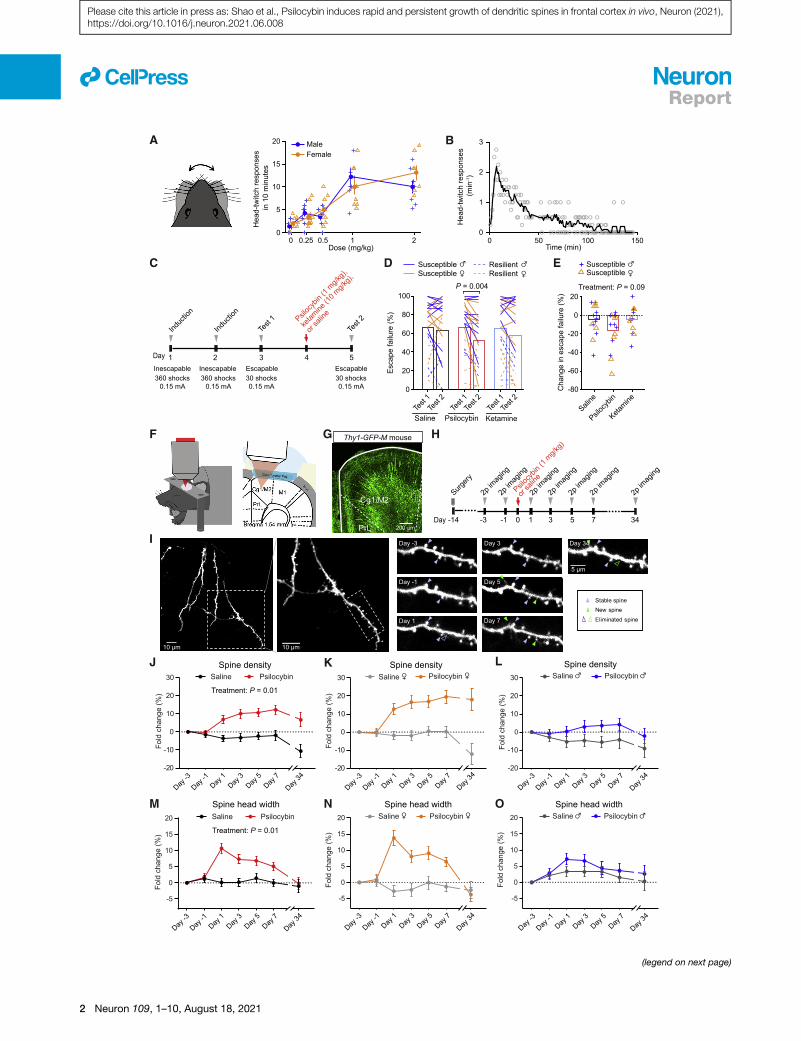
A single dose of psilocybin leads to long-lastingincreases in spine density and spine head width in themouse medial frontal cortexTo test the potency and dose dependence of psilocybin in mice,
we measured the head-twitch response, a classic assay for
characterizing psychedelic compounds in rodents. We observed
that mice would exhibit high-frequency headshakes intermit-
tently after administration of psilocybin (Video S1). We charac-
terized 82 C57BL/6J mice (40 males and 42 females) with five
doses of psilocybin (0, 0.25, 0.5, 1, and 2mg/kg, intraperitoneally
[i.p.]; range, 7–10 per sex per dose). A sharp rise of elicited head-
twitch responses occurred at 1 mg/kg (Figure 1A), consistent
with prior reports (Halberstadt et al., 2011; Sherwood et al.,
2020). Thus, we chose to use 1 mg/kg (the inflection point of
the dose-dependence curve) to assess psilocybin’s effect on
structural plasticity. At this dose, the rate of head-twitch re-
sponses peaked at 6–8 min after administration and then gradu-
ally declined until they ceased at �2 h (Figure 1B).
Next, to determine if this dose is associated with mitigation of
stress-related phenotypes in mice, we tested the effect of 1 mg/
kg psilocybin in a learned helplessness paradigm (Chourbaji
et al., 2005). Mice received prolonged stress in the form of
repeated, inescapable footshocks over two induction sessions,
and then were tested for active avoidance behavior with escap-
able footshocks1daybeforeand1dayafter treatment (Figure1C).
Susceptible animals are mice in a learned-helpless state charac-
terized by reduced attempts to escape from the footshocks. We
used 68 C57BL/6J mice to compare psilocybin (1 mg/kg, i.p.)
against saline and ketamine (10 mg/kg, i.p.), which served as
negative and positive controls (Wu et al., 2021).Within individuals,
psilocybin reduced the proportion of escape failures (p = 0.004,
post hoc Bonferroni-corrected t test; Figure 1D). For susceptible
animals, the psilocybin group had a decrease or no change in
escape failures in all but 1 of the 16 mice tested (94%; Figures
1E and S1A). Across individuals, comparison among saline, keta-
mine, and psilocybin did not reveal a main effect of treatment (p =
0.09, two-way ANOVA; Figure 1E), presumably because of the
across-subject variability in the behavioral responses. Neverthe-
less, overall, these results indicate that psilocybin can ameliorate
maladaptive behavior induced by uncontrollable stress in mice.
In the body, psilocybin is dephosphorylated to psilocin, an
agonist of 5-HT2A receptors that are densely expressed in apical
dendrites of layer 5 pyramidal neurons in the medial frontal cor-
tex of primates and rodents (Aghajanian andMarek, 1997; Jakab
and Goldman-Rakic, 1998; Willins et al., 1997). We therefore hy-
pothesize that psilocybin may modify the dendritic architecture
in the medial frontal cortex. We used chronic two-photon micro-
scopy to track apical dendritic spines in the cingulate/premotor
(Cg1/M2) region of the medial frontal cortex of Thy1GFP mice
(line M), in which a sparse subset of infragranular (layer 5 and
6) pyramidal neurons express GFP (Feng et al., 2000) (Figures
1F and 1G).We imaged before and after administering psilocybin
(1 mg/kg, i.p.) or saline at 2-day intervals and then again
�1 month later for a total of seven imaging sessions (Figures
1H and 1I). In total, we tracked 1,820 dendritic spines on 161
branches from 12 animals (6 males and 6 females). Spine
morphology was analyzed blind to experimental conditions us-
ing standardized procedures (Holtmaat et al., 2009). We took
advantage of the longitudinal data to normalize the change in
spine density as fold change in individual dendritic segments.
For statistical analyses, we used a mixed-effects model
including treatment, sex, and days as factors, as well as all inter-
action terms. Variation within mouse and dendrite across days
was accounted by including random effects terms for dendrites
nested by mice. Our results indicate that a single dose of psilo-
cybin induces a significant elevation in spine density (+7% ±
2% on day 1 and +12% ± 3% on day 7; main effect of treatment,
p = 0.011, mixed-effects model; Figures 1J–1L), an increase in
the width of spine heads (+11% ± 2% on day 1 and +5% ± 1%
on day 7; main effect of treatment, p = 0.013; Figures 1M–1O),
and higher spine protrusion lengths (Figures S1B–S1D). Details
for all statistical tests, including sample sizes, are provided in Ta-
ble S1.
Psilocybin elevates the formation rate of dendriticspines in vivo
Increased spine density could be due to a higher formation rate,
a lower elimination rate, or both. To distinguish between the pos-
sibilities, we analyzed the same dendritic segments across adja-
cent imaging sessions to determine the turnover of dendritic
spines. In females, the spine formation rate increased by 8% ±
2% after psilocybin (absolute values for the formation rate:
Figure 1. Psilocybin increases the density and size of dendritic spines in the mouse medial frontal cortex
(A) Head-twitch responses as a function of dose, tested on 82 C57BL/6J mice.
(B) Time course of head-twitch responses after administrating psilocybin (1 mg/kg, i.p.), averaged from two males and two female C57BL/6J mice. Line, moving
average.
(C) Timeline for the learned helplessness assay.
(D) The proportion of escape failure for all animals in test 1 and test 2, i.e., before and after psilocybin (1 mg/kg, i.p.), ketamine (10 mg/kg, i.p.), and saline
administration.
(E) The change in escape failure, from test 1 to test 2, for susceptible animals for psilocybin, ketamine, and saline treatments.
(F) Imaging setup.
(G) Fixed coronal section from Thy1GFP mice.
(H) Timeline for the longitudinal imaging study.
(I) Example field of view.
(J) Effects of psilocybin or saline treatment on spine density, plotted as fold-change from baseline value on day �3 (mean ± SEM).
(K and L) Similar to (J), plotted separately for females and males.
(M–O) Similar to (J)–(L) for spine head width.
Sample sizes and details of the statistical analyses are provided in Table S1. See also Figure S1.
llReport
Neuron 109, 1–10, August 18, 2021 3
Please cite this article in press as: Shao et al., Psilocybin induces rapid and persistent growth of dendritic spines in frontal cortex in vivo, Neuron (2021),https://doi.org/10.1016/j.neuron.2021.06.008

7%±1%onday�1 and 15%±2%onday 1;main effect of treat-
ment, p = 0.034, mixed-effects model; Figures 2A and 2B). Like-
wise, the spine formation rate was higher by 4% ± 2% in males
after psilocybin (absolute values for the formation rate: 6% ± 1%
on day �1 and 10% ± 2% on day 1). By contrast, there was no
change in the elimination rate of spines (Figure 2C). The increase
in spine formation rate was highest shortly after psilocybin
administration and then diminished in subsequent days to return
to baseline level and in equilibrium with the elimination rate.
These data therefore support the view that the long-term in-
crease in spine density is due to an initial boost of enhanced
spine formation.
A fraction of the psilocybin-induced spines is persistentfor at least 1 monthA key question waswhether the new spines formed after psilocy-
bin administration would persist, because nascent dendritic
spines can take 4 days to mature into functional synapses (Knott
et al., 2006). For this reason, we tracked the new spines formed
after psilocybin on day 1 and found that approximately half of
them remained stable on day 7 (49% ± 10% for females and
52% ± 12% for males; Figure 2D). This suggests that a portion
of the new dendritic spines induced by psilocybin would become
functional synapses. Furthermore, because clinical trials indi-
cated that psilocybin may provide long-term benefits for up to
several months, for a subset of four mice, we imaged yet again
at a further time point at 34 days after administration to find
B C
D Saline Psilocybin
Saline Psilocybin
Formation rate Elimination rate
Day -1
Day 1
Day 3
Day 5
Day 7
Day -1
Day 1
Day 3
Day 5
Day 7
Day 7
Day 34
Day 7
Day 34
0
Frac
tion
ofne
w s
pine
s pe
rsis
ts (%
)
7 7 13 13
Saline Psilocybin
Day 7
Day 34
Day 7
Day 34
Frac
tion
ofne
w s
pine
s pe
r sis
ts (%
)
11 11 13 13
-5
0
5
10
15
Day -1
Day 1
Day 3
Day 5
Day 7
Saline Psilocybin
Day -1
Day 1
Day 3
Day 5
Day 7
Treatment: P = 0.03
50
100
150
0
50
100
150
-5
0
5
10
15
10 μm
A
10 μm
Day -3
Day -1
Day 1
Day 3
Day 5
Day 7
5 μm
Diff
eren
ce (%
)
Diff
eren
ce (%
)
Figure 2. Psilocybin elevates the formation
rate of dendritic spines
(A) Example field of view. Purple arrowhead, sta-
ble spine. Green arrowhead, new spine.
(B) Effects of psilocybin or saline treatment on the
formation rates of dendritic spines for female and
male mice, plotted as difference from baseline
value on day �1 (mean ± SEM).
(C) Similar to (B) for elimination rates.
(D) Fraction of spines newly formed on day 1 that
remained stable on day 7 and day 34 for female
and male mice. Filled circles, individual dendritic
segments.
Sample sizes and details of the statistical ana-
lyses are provided in Table S1. See also Figure S2.
that a fraction of the psilocybin-evoked
new spines remained persistent (34% ±
10% for females and 37% ± 12% for
males; Figures 2D and S2). Psilocybin-
induced spines were not significantly
different and therefore were no less sta-
ble than spines formed in control condi-
tions (main effect of treatment, p = 0.9,
two-way repeated-measures ANOVA).
Intriguingly, select individual dendritic
branches appeared to retain all of the
new spines, while other branches lost
them almost completely, suggesting het-
erogeneity and potentially responsive
and nonresponsive subpopulations of
pyramidal neurons. Altogether, these results demonstrate that
a single dose of psilocybin induces rapid and long-lasting den-
dritic remodeling in layer 5 pyramidal neurons in the mouse
medial frontal cortex.
Ketanserin pretreatment, sufficient to abolish head-twitch responses, does not block psilocybin-inducedstructural plasticityMultiple lines of evidence demonstrated that 5-HT2A receptors
are essential for serotonergic psychedelics’ psychotomimetic
effects in humans (Vollenweider et al., 1998) and head-twitch re-
sponses in mice (Gonzalez-Maeso et al., 2007; Keiser et al.,
2009). To study whether the effects of psilocybin on structural
plasticity may involve 5-HT2A receptors, we reduced the number
of available 5-HT2A receptors in the brain by pretreating animals
with the 5-HT2A receptor antagonist ketanserin (1 mg/kg, i.p.),
10 min prior to the administration of psilocybin (1 mg/kg, i.p.)
or saline. Behaviorally, the ketanserin pretreatment abolished
completely the psilocybin-induced head-twitch responses (Fig-
ure 3A). Next, we repeated the two-photon imaging experiments
in ketanserin-pretreatedmice and tracked 1,443 dendritic spines
on 120 branches from 8 animals (4 males and 4 females) (Fig-
ure 3B). We found that although the enhancing effect of psilocy-
bin on spine density was no longer statistically significant (+5%±
2% on day 1 and +8% ± 2% on day 7; main effect of treatment,
p = 0.09, mixed-effects model; Figures 3C, S3A, and S3B), there
were still detectable increases in spine head width (+12% ± 1%
llReport
4 Neuron 109, 1–10, August 18, 2021
Please cite this article in press as: Shao et al., Psilocybin induces rapid and persistent growth of dendritic spines in frontal cortex in vivo, Neuron (2021),https://doi.org/10.1016/j.neuron.2021.06.008

on day 1 and +12% ± 1% on day 7; main effect of treatment, p =
0.01; Figures 3D, S3C, and S3D), spine protrusion length (Fig-
ures S3E–S3G), and spine formation rate (absolute values for
the formation rate: 5% ± 1% on day �1 and 10% ± 2% on day
1 for female mice; 8% ± 1% on day �1 and 14% ± 2% on day
1 for male mice; Figures 3E and S3H). It was previously deter-
mined that 1 mg/kg ketanserin led to only an �30% blockade
of 5-HT2A receptors in the rat neocortex (Smith et al., 1995), likely
due to limited transport into the brain for rodents (Syv€anen et al.,
2009). Therefore, in agreement with a recent study in the
Day-3
Day-1
Day1
Day3
Day5
Day7
-5
0
5
10
15
20
Fold
chan
ge( %
)
Ketanserin + SalineKetanserin + Psilocybin
Spine head widthKetanserin + SalineKetanserin + Psilocybin
Fold
cha
nge
(%)
Day-3
Day-1
Day1
Day3
Day5
Day7
Spine density
Surgery
-14 -3 1-1 0 3 5 7
2p im
aging
Day
2p im
aging
2p im
aging
2p im
aging
2p im
aging
2p im
aging
Hea
d-tw
itch
resp
onse
sin
10
min
utes
-5
0
5
10
15
Diff
eren
ce (%
)
Formation rate
Ketanserin + Saline Ketanserin + Psilocybin
Ketanserin + Saline Ketanserin + Psilocybin
Ketans
erin p
re-tre
atmen
t +
psilo
cybin
(1 m
g/kg)
or sa
line
Treatment: P = 0.09 Treatment: P = 0.01 Treatment: P = 0.03
A B
C D E
0
5
10
15
Ketanserin + SalineKetanserin + PsilocybinSaline + Psilocybin
0
10
20
-10
30
P = 0.0156P = 0.0257
0
2
4
6
mEP
SC fr
eque
ncy
(Hz)
-30
-20
-10
0
mEP
SC a
mpi
ltude
(pA)
Treatment: P = 0.0002
Treatment: P = 0.06
0.5 s
25 pA
Psilocybin
Saline
Saline
Psilocybin
F G
H
Cum
ulat
ive
prob
abilit
y
Psilocybin Saline
Saline Psilocybin
0 2000 4000 60000
20
40
60
80
100
Inter-event intervals (ms)
60mEPSC magnitude (pA)
0 20 40
Cum
ulat
ive
prob
abilit
y
Psilocybin Saline
Saline Psilocybin
0
20
40
60
80
100
Day -1
Day 1
Day 3
Day 5
Day 7
Day -1
Day 1
Day 3
Day 5
Day 7
Figure 3. Mechanistic details revealed by ketanserin pretreatment and electrophysiological characterizations
(A) Head-twitch responses after administrating psilocybin without saline pretreatment (10 min prior; n = 3 mice), psilocybin (1 mg/kg, i.p.) with ketanserin pre-
treatment (1 mg/kg, 10 min prior; n = 3), and saline with ketanserin pretreatment (n = 4).
(B) Timeline for the experiment.
(C) Effects of psilocybin or saline treatment on spine density in animals pretreated with ketanserin, plotted as fold change from baseline value on day �3
(mean ± SEM).
(D) Similar to (C) for spine head width.
(E) Effects of psilocybin or saline treatment on the formation rates of dendritic spines for female and male mice pretreated with ketanserin, plotted as difference
from baseline value on day �1.
(F) Representative traces of mEPSCs recorded from putative layer 5 pyramidal neurons of Cg1/M2 in brain slices.
(G) Grouped and cumulative distribution plots of mEPSC frequency for animals that received psilocybin or saline 24 h before recording. Each open circle denotes
a cell (n = 25 cells from four females for saline; 24 cells from four females for psilocybin; 19 cells from five males for saline; 23 cells from four males for psilocybin).
(H) Similar to (G) for mEPSC amplitude.
Sample sizes and details of the statistical analyses are provided in Table S1. See also Figure S3.
llReport
Neuron 109, 1–10, August 18, 2021 5
Please cite this article in press as: Shao et al., Psilocybin induces rapid and persistent growth of dendritic spines in frontal cortex in vivo, Neuron (2021),https://doi.org/10.1016/j.neuron.2021.06.008

hippocampus (Hesselgrave et al., 2021), our results demonstrate
that while a moderate knockdown of 5-HT2A receptor function
eliminates head-twitch responses, it is not sufficient to abolish
the psilocybin-induced structural remodeling in mice.
Psilocybin elevates excitatory neurotransmission inmedial frontal cortexMost, but not all, dendritic spines are functional glutamatergic
synapses. To elaborate on the effects of psilocybin on synaptic
function, we performed whole-cell recordings in brain slices to
measure miniature excitatory postsynaptic currents (mEPSCs)
from putative layer 5 pyramidal neurons, identified based on
morphology, in Cg1/M2 (Figure 3F). The results showed that
24 h after treatment, we could detect an increase in mEPSC fre-
quency in psilocybin-treated animals compared to saline
controls (main effect of treatment, p = 0.0002, two-way
ANOVA; Figure 3G). We also report a moderate effect of psilocy-
bin onmEPSC amplitude (main effect of treatment, p = 0.06, two-
way ANOVA; Figure 3H). Because mEPSC frequency and ampli-
tude reflect the number and strength of synapses, these results
demonstrate that the psilocybin-induced structural remodeling
is accompanied by enhanced excitatory neurotransmission.
Dependence of psilocybin-induced structuralremodeling on brain region and dendrite typeTo further support the conclusions, we tried to replicate the find-
ings in a completely separate cohort of animals using a different
approach. We administered Thy1GFPmice with psilocybin (1 mg/
kg, i.p.) or saline, sacrificed them 24 h later, and imaged coronal
brain sections using confocal microscopy. We expanded ana-
lyses to six areas of the brain, including two zones that encom-
pass apical and basal dendrites and three regions of the frontal
cortex: Cg1/M2, prelimbic/infralimbic (PrL/IL), and primary mo-
tor cortex (M1) (Figures 4A–4C). The results, consisting of
23,226 dendritic spines counted on 1,885 branches from 12 an-
imals (6 males and 6 females), reaffirmed the ability of psilocybin
to promote the growth of new dendritic spines in Cg1/M2 in fe-
male mice (spine density: 0.46 ± 0.02 versus 0.50 ± 0.01 mm�1;
Figure 4D). Effects of psilocybin on spine density were more pro-
nounced in female animals than in male animals (treatment 3
sex, p = 0.013, two-way ANOVA; Figure 4D). We did not detect
differences in spine protrusion length and spine head width (Fig-
ures 4E and 4F), which may be due to the across-subjects
design, as we could not normalize the changes to the same den-
dritic branch, and therefore, this approach had less power than
the within-subjects design of the chronic imaging experiment.
We detected select morphological differences in PrL/IL and
M1, including increases in spine protrusion length in PrL/IL
(main effect of treatment, p = 0.026, two-way ANOVA), spine
density in M1 in females (treatment 3 sex, p = 0.021, two-way
ANOVA), and spine head width in M1 in females (treatment 3
sex, p = 0.008, two-way ANOVA), suggesting that the plas-
ticity-promoting impact may not be unique to Cg1/M2 (Figures
4G–4N). Furthermore, psilocybin had significant impact on basal
dendrites in Cg1/M2, leading to higher spine density and spine
protrusion length (spine density: main effect of treatment, p =
0.0004; spine protrusion length: main effect of treatment, p =
0.012, two-way ANOVA; Figures 4O–4R and S4). Overall, the
two sets of data converge to show that psilocybin promotes
the growth of dendritic spines in layer 5 pyramidal neurons in
the medial frontal cortex.
DISCUSSION
This study demonstrates that a single dose of psilocybin evokes
growth of dendritic spines in the medial frontal cortex of the
mouse. The persistence of the neural modifications is notable
andmay relate to the compound’s therapeutic effects for at least
two reasons. First, depression is associated with a loss of synap-
ses in the frontal cortex (Holmes et al., 2019). Restoring the num-
ber of neuronal connectionsmay correct such deficit, providing a
biological mechanism for alleviating symptoms of depression.
Second, structural remodeling is integral to learning and facili-
tates the storage of lifelong memories (Xu et al., 2009; Yang
et al., 2009). Psilocybin-induced neural plasticity could prime
the brain for integrating new psychological experiences. Regard-
less of the relative importance of these mechanisms, which are
not mutually exclusive, our results indicate that the underlying
structural trace in the brain is enduring and can be observed a
long time after the initial drug exposure.
There is an ongoing debate over whether the hallucinogenic
effects of serotonergic psychedelics are dissociable from the
therapeutic effects (Olson, 2020; Yaden and Griffiths, 2020).
Consistent with another new study (Hesselgrave et al., 2021),
our results indicate that structural remodeling in the medial fron-
tal cortex is undeterred by a moderate knockdown of 5-HT2A re-
ceptor availability. The possibility to disrupt psilocybin’s acute
behavioral effects without abolishing structural plasticity actions
has clear implications for treatment in the clinic. However, it is
not yet clear if the results will extrapolate to humans, because
5-HT2A receptors have species-dependent differences in disso-
ciation kinetics with serotonergic psychedelics (Kim et al., 2020).
Moreover, our results do not rule out the involvement of 5-HT2Areceptors, because this dose of ketanserin only blocks �30%
of 5-HT2A receptors in rodents (Smith et al., 1995), and the unaf-
fected receptors might be enough to drive the dendritic remod-
eling. This number may be compared to the 50%–70% 5-HT2Areceptor occupancy level required for the more intense psilocy-
bin-induced psychological experience in humans (Madsen et al.,
2019). Future studies with region- and cell-type-specific
knockout of serotonin receptor subtypes are needed to produce
more decisive evidence on the role of 5-HT2A and other recep-
tors in mediating the effects of psilocybin on dendritic plasticity.
By showing that the time course for psilocybin-induced struc-
tural remodeling is rapid and persistent in vivo, our study suggests
that synaptic rewiring may be a mechanism shared by com-
pounds with rapid antidepressant effects. Of note, the timing of
psilocybin’s effect on the neural architecture is reminiscent of ke-
tamine, which at subanesthetic dose causes similar rapid in-
crease in spine density and elevation of spine formation rate in
themedial frontal cortex (Moda-Sava et al., 2019; Phoumthippha-
vong et al., 2016). However, still unknown is how drugs with
disparate molecular targets may yield comparable modifications
on neural architecture and behavior (Kadriu et al., 2021; Savalia
et al., 2021). Elucidating the mechanisms will be crucial toward
unraveling the neurobiology of rapid-acting antidepressants.
llReport
6 Neuron 109, 1–10, August 18, 2021
Please cite this article in press as: Shao et al., Psilocybin induces rapid and persistent growth of dendritic spines in frontal cortex in vivo, Neuron (2021),https://doi.org/10.1016/j.neuron.2021.06.008

c
e f
b
a
db
a c
d
f
A
300 μm 1 mm
M2M1
IL
PrL
Cg1
Bregma 1.78 mm B
a
0.0
0.4
0.8
1.2Basal dendrites
Saline
Psilocybin (1 mg/kg)
O
5 μm
Spin
e de
nsity
(spi
nes
/μm
)
Spin
e pr
otru
sion
leng
th ( μ
m)
Spin
e he
ad w
idth
(μm
)
Treatment P = 0.0004
Treatment P = 0.01
Cg1/M2 Cg1/M2 Cg1/M2
Cg1
/M2
P Q R
PsilocybinSaline Saline
PsilocybinD E
H I J
Apical tuft dendritesSaline
Psilocybin (1 mg/kg)
M1
PrL/
IL
C
Spin
e de
nsity
(spi
nes
/μm
)
Treatment х SexP = 0.01
Spin
e pr
otru
sion
leng
th (μ
m)
Spin
e he
ad w
idth
(μm
)
0.0
0.5
1.0
1.5
0.0
0.5
1.0
1.5
2.0
0.0
0.4
0.8
1.2Cg1/M2
0
1
2
3
Cg1/M2 Cg1/M2
0.0
0.4
0.8
1.2
0.0
0.5
1.0
1.5
2.0
PrL/IL
Spin
e de
nsity
(spi
nes
/μm
)
S pin
e pr
otru
sion
leng
th (μ
m)
Spi n
e he
ad w
idth
(μm
)
TreatmentP = 0.03
PrL/ILPrL/IL
0.0
0.5
1.0
1.5
0.0
0.4
0.8
1.2
0.0
0.5
1.0
1.5
2.0
Spin
e de
nsity
(spi
nes
/μm
)
S pin
e pr
otru
sion
leng
th (μ
m)
Spin
e he
ad w
idth
(μm
)
M1Treatment х Sex
P = 0.02TreatmentP = 0.008
M1M1L M N
5 μm
5 μm
5 μm
Cg1
/M2
Saline
Psilocybin (1 mg/kg)
Apical tuft dendrites
Saline
Psilocybin (1 mg/kg)
Apical tuft dendrites
G
F
K
20 μm
b
ba
Tuft Basal
5 μm
100 μm
0.0
0.5
1.0
1.5
2.0
0.0
0.5
1.0
1.5
2.0
Figure 4. Region-specific effects of psilocybin
(A) Stitched confocal image of a coronal brain section from a Thy1GFP mouse.
(B) Magnified images showing apical and basal dendritic segments.
(C) Images of apical dendrites in Cg1/M2.
(D) Effects of psilocybin and saline on spine density for apical dendrites in Cg1/M2. Open circles, individual dendritic segments. Gray line, mean ± SEM.
(E) Similar to (D) for spine protrusion length.
(legend continued on next page)
llReport
Neuron 109, 1–10, August 18, 2021 7
Please cite this article in press as: Shao et al., Psilocybin induces rapid and persistent growth of dendritic spines in frontal cortex in vivo, Neuron (2021),https://doi.org/10.1016/j.neuron.2021.06.008

STAR+METHODS
Detailed methods are provided in the online version of this paper
and include the following:
d KEY RESOURCES TABLE
d RESOURCE AVAILABILITY
B Lead contact
B Materials availability
B Data and code availability
d EXPERIMENTAL MODEL AND SUBJECT DETAILS
d METHOD DETAILS
B Psilocybin
B Head-twitch response
B Learned helplessness
B Surgery
B Two-photon imaging
B Confocal imaging
B Brain slice preparation
B Whole-cell recording
d QUANTIFICATION AND STATISTICAL ANALYSIS
B Analysis of the imaging data
B Statistics
SUPPLEMENTAL INFORMATION
Supplemental information can be found online at https://doi.org/10.1016/j.
neuron.2021.06.008.
ACKNOWLEDGMENTS
We thank Ben Kelmendi, Chris Pittenger, and Mikael Palner for discussions;
Jane Taylor for use of open-field activity boxes; Huriye Atilgan and Heather Or-
tega for assistance on setting up video recording; and Adam Halberstadt for
advice on scoring head-twitch response. Psilocybin was generously provided
by Usona Institute’s Investigational Drug & Material Supply Program; the
Usona Institute IDMSP is supported by Alexander Sherwood, Robert Kargbo,
and Kristi Kaylo in Madison, WI. This work was supported by the Yale Center
for Psychedelic Science, NIH/NIMH grant R01MH121848 (A.C.K.), NIH/NINDS
training grant T32NS041228 (C.L.), and NIH/NIGMS medical scientist training
grant T32GM007205 (N.K.S. and P.A.D.). We thank the Yale Center for
Advanced Light Microscopy Facility for their assistance with confocal imaging,
supported in part via NIH grant S10OD023598.
AUTHOR CONTRIBUTIONS
L.-X.S. and A.C.K. designed the research. L.-X.S. performed the two-photon
and confocal imaging experiments and analyzed the two-photon imaging
data. N.K.S. blinded and I.G. analyzed the confocal imaging data. C.L. per-
formed and analyzed the behavioral experiments. I.G. and K.D. assisted with
analyzing the behavioral data. P.A.D. performed the electrophysiological re-
cordings. L.-X.S. blinded and P.A.D. analyzed the electrophysiological data.
N.K.S. assisted with the statistical analyses. L.-X.S. and A.C.K. wrote the pa-
per with input from all other authors.
DECLARATION OF INTERESTS
A.C.K. received psilocybin from the investigational drug supply program at
Usona Institute, a non-profit organization. The authors declare no other
competing interests.
INCLUSION AND DIVERSITY
We worked to ensure sex balance in the selection of non-human subjects.
While citing references scientifically relevant for this work, we also actively
worked to promote gender balance in our reference list.
Received: February 20, 2021
Revised: May 16, 2021
Accepted: June 7, 2021
Published: July 5, 2021
REFERENCES
Aghajanian, G.K., and Marek, G.J. (1997). Serotonin induces excitatory post-
synaptic potentials in apical dendrites of neocortical pyramidal cells.
Neuropharmacology 36, 589–599.
Ali, F., Gerhard, D.M., Sweasy, K., Pothula, S., Pittenger, C., Duman, R.S., and
Kwan, A.C. (2020a). Ketamine disinhibits dendrites and enhances calcium sig-
nals in prefrontal dendritic spines. Nat. Commun. 11, 72.
Ali, F., Shao, L.X., Gerhard, D.M., Sweasy, K., Pothula, S., Pittenger, C.,
Duman, R.S., and Kwan, A.C. (2020b). Inhibitory regulation of calcium tran-
sients in prefrontal dendritic spines is compromised by a nonsense Shank3
mutation. Mol. Psychiatry. Published online March 11, 2020. https://doi.org/
10.1038/s41380-020-0708-6.
Cameron, L.P., Tombari, R.J., Lu, J., Pell, A.J., Hurley, Z.Q., Ehinger, Y.,
Vargas, M.V., McCarroll, M.N., Taylor, J.C., Myers-Turnbull, D., et al. (2021).
A non-hallucinogenic psychedelic analogue with therapeutic potential.
Nature 589, 474–479.
Carhart-Harris, R.L., Bolstridge, M., Rucker, J., Day, C.M.J., Erritzoe, D.,
Kaelen, M., Bloomfield, M., Rickard, J.A., Forbes, B., Feilding, A., et al.
(2016). Psilocybin with psychological support for treatment-resistant depres-
sion: an open-label feasibility study. Lancet Psychiatry 3, 619–627.
Chourbaji, S., Zacher, C., Sanchis-Segura, C., Dormann, C., Vollmayr, B., and
Gass, P. (2005). Learned helplessness: validity and reliability of depressive-like
states in mice. Brain Res. Brain Res. Protoc. 16, 70–78.
Cook, S.C., and Wellman, C.L. (2004). Chronic stress alters dendritic
morphology in rat medial prefrontal cortex. J. Neurobiol. 60, 236–248.
Davis, A.K., Barrett, F.S., May, D.G., Cosimano, M.P., Sepeda, N.D., Johnson,
M.W., Finan, P.H., and Griffiths, R.R. (2021). Effects of Psilocybin-Assisted
Therapy on Major Depressive Disorder: A Randomized Clinical Trial. JAMA
Psychiatry 78, 481–489.
Drevets, W.C., Price, J.L., Simpson, J.R., Jr., Todd, R.D., Reich, T., Vannier,
M., and Raichle, M.E. (1997). Subgenual prefrontal cortex abnormalities in
mood disorders. Nature 386, 824–827.
Duman, R.S., and Aghajanian, G.K. (2012). Synaptic dysfunction in depres-
sion: potential therapeutic targets. Science 338, 68–72.
Feng, G., Mellor, R.H., Bernstein, M., Keller-Peck, C., Nguyen, Q.T., Wallace,
M., Nerbonne, J.M., Lichtman, J.W., and Sanes, J.R. (2000). Imaging neuronal
subsets in transgenic mice expressing multiple spectral variants of GFP.
Neuron 28, 41–51.
Gonzalez-Maeso, J., Weisstaub, N.V., Zhou, M., Chan, P., Ivic, L., Ang, R.,
Lira, A., Bradley-Moore, M., Ge, Y., Zhou, Q., et al. (2007). Hallucinogens
(F) Similar to (D) for spine head width.
(G–J) Similar to (C)–(F) for PrL/IL.
(K–N) Similar to (C)–(F) for M1.
(O–R) Similar to (C)–(F) for basal dendrites in Cg1/M2.
Sample sizes and details of the ANOVA models are provided in Table S1. See also Figure S4.
llReport
8 Neuron 109, 1–10, August 18, 2021
Please cite this article in press as: Shao et al., Psilocybin induces rapid and persistent growth of dendritic spines in frontal cortex in vivo, Neuron (2021),https://doi.org/10.1016/j.neuron.2021.06.008

recruit specific cortical 5-HT(2A) receptor-mediated signaling pathways to
affect behavior. Neuron 53, 439–452.
Griffiths, R.R., Johnson, M.W., Carducci, M.A., Umbricht, A., Richards, W.A.,
Richards, B.D., Cosimano, M.P., and Klinedinst, M.A. (2016). Psilocybin pro-
duces substantial and sustained decreases in depression and anxiety in pa-
tients with life-threatening cancer: A randomized double-blind trial.
J. Psychopharmacol. 30, 1181–1197.
Grutzendler, J., Kasthuri, N., and Gan, W.B. (2002). Long-term dendritic spine
stability in the adult cortex. Nature 420, 812–816.
Halberstadt, A.L., Koedood, L., Powell, S.B., and Geyer, M.A. (2011).
Differential contributions of serotonin receptors to the behavioral effects of in-
doleamine hallucinogens in mice. J. Psychopharmacol. 25, 1548–1561.
Hesselgrave, N., Troppoli, T.A., Wulff, A.B., Cole, A.B., and Thompson, S.M.
(2021). Harnessing psilocybin: antidepressant-like behavioral and synaptic ac-
tions of psilocybin are independent of 5-HT2R activation in mice. Proc. Natl.
Acad. Sci. USA 118, e2022489118.
Holmes, S.E., Scheinost, D., Finnema, S.J., Naganawa, M., Davis, M.T.,
DellaGioia, N., Nabulsi, N., Matuskey, D., Angarita, G.A., Pietrzak, R.H.,
et al. (2019). Lower synaptic density is associated with depression severity
and network alterations. Nat. Commun. 10, 1529.
Holtmaat, A., Bonhoeffer, T., Chow, D.K., Chuckowree, J., De Paola, V., Hofer,
S.B., H€ubener, M., Keck, T., Knott, G., Lee, W.C., et al. (2009). Long-term,
high-resolution imaging in the mouse neocortex through a chronic cranial win-
dow. Nat. Protoc. 4, 1128–1144.
Jakab, R.L., and Goldman-Rakic, P.S. (1998). 5-Hydroxytryptamine2A seroto-
nin receptors in the primate cerebral cortex: possible site of action of halluci-
nogenic and antipsychotic drugs in pyramidal cell apical dendrites. Proc. Natl.
Acad. Sci. USA 95, 735–740.
Jones, K.A., Srivastava, D.P., Allen, J.A., Strachan, R.T., Roth, B.L., and
Penzes, P. (2009). Rapid modulation of spine morphology by the 5-HT2A se-
rotonin receptor through kalirin-7 signaling. Proc. Natl. Acad. Sci. USA 106,
19575–19580.
Kadriu, B., Greenwald, M., Henter, I.D., Gilbert, J.R., Kraus, C., Park, L.T., and
Zarate, C.A. (2021). Ketamine and Serotonergic Psychedelics: Common
Mechanisms Underlying the Effects of Rapid-Acting Antidepressants. Int. J.
Neuropsychopharmacol. 24, 8–21.
Keiser, M.J., Setola, V., Irwin, J.J., Laggner, C., Abbas, A.I., Hufeisen, S.J.,
Jensen, N.H., Kuijer, M.B., Matos, R.C., Tran, T.B., et al. (2009). Predicting
new molecular targets for known drugs. Nature 462, 175–181.
Kim, K., Che, T., Panova, O., DiBerto, J.F., Lyu, J., Krumm, B.E., Wacker, D.,
Robertson, M.J., Seven, A.B., Nichols, D.E., et al. (2020). Structure of a
Hallucinogen-Activated Gq-Coupled 5-HT2A Serotonin Receptor. Cell 182,
1574–1588.e1519.
Knott, G.W., Holtmaat, A., Wilbrecht, L., Welker, E., and Svoboda, K. (2006).
Spine growth precedes synapse formation in the adult neocortex in vivo.
Nat. Neurosci. 9, 1117–1124.
Li, N., Lee, B., Liu, R.J., Banasr, M., Dwyer, J.M., Iwata, M., Li, X.Y.,
Aghajanian, G., and Duman, R.S. (2010). mTOR-dependent synapse formation
underlies the rapid antidepressant effects of NMDA antagonists. Science 329,
959–964.
Liston, C., Miller, M.M., Goldwater, D.S., Radley, J.J., Rocher, A.B., Hof, P.R.,
Morrison, J.H., and McEwen, B.S. (2006). Stress-induced alterations in pre-
frontal cortical dendritic morphology predict selective impairments in percep-
tual attentional set-shifting. J. Neurosci. 26, 7870–7874.
Ly, C., Greb, A.C., Cameron, L.P., Wong, J.M., Barragan, E.V., Wilson, P.C.,
Burbach, K.F., Soltanzadeh Zarandi, S., Sood, A., Paddy, M.R., et al. (2018).
Psychedelics Promote Structural and Functional Neural Plasticity. Cell Rep.
23, 3170–3182.
Ly, C., Greb, A.C., Vargas, M.V., Duim, W.C., Grodzki, A.C.G., Lein, P.J., and
Olson, D.E. (2020). Transient Stimulation with Psychoplastogens Is Sufficient
to Initiate Neuronal Growth. ACS Pharmacol. Transl. Sci. 4, 452–460.
Madsen, M.K., Fisher, P.M., Burmester, D., Dyssegaard, A., Stenbæk, D.S.,
Kristiansen, S., Johansen, S.S., Lehel, S., Linnet, K., Svarer, C., et al. (2019).
Psychedelic effects of psilocybin correlate with serotonin 2A receptor occu-
pancy and plasma psilocin levels. Neuropsychopharmacology 44, 1328–1334.
Moda-Sava, R.N., Murdock, M.H., Parekh, P.K., Fetcho, R.N., Huang, B.S.,
Huynh, T.N., Witztum, J., Shaver, D.C., Rosenthal, D.L., Alway, E.J., et al.
(2019). Sustained rescue of prefrontal circuit dysfunction by antidepressant-
induced spine formation. Science 364, eaat8078.
Nichols, D.E. (2016). Psychedelics. Pharmacol. Rev. 68, 264–355.
Nichols, C.D., and Sanders-Bush, E. (2002). A single dose of lysergic acid di-
ethylamide influences gene expression patterns within the mammalian brain.
Neuropsychopharmacology 26, 634–642.
Olson, D.E. (2020). The Subjective Effects of Psychedelics May Not Be
Necessary for Their Enduring Therapeutic Effects. ACS Pharmacol. Transl.
Sci. 4, 563–567.
Phoumthipphavong, V., Barthas, F., Hassett, S., and Kwan, A.C. (2016).
Longitudinal Effects of Ketamine on Dendritic Architecture In Vivo in the
Mouse Medial Frontal Cortex. eNeuro 3, ENEURO.0133-0115.2016.
Pologruto, T.A., Sabatini, B.L., and Svoboda, K. (2003). ScanImage: flexible
software for operating laser scanningmicroscopes. Biomed. Eng. Online 2, 13.
Radley, J.J., Sisti, H.M., Hao, J., Rocher, A.B., McCall, T., Hof, P.R., McEwen,
B.S., and Morrison, J.H. (2004). Chronic behavioral stress induces apical den-
dritic reorganization in pyramidal neurons of the medial prefrontal cortex.
Neuroscience 125, 1–6.
Raval, N.R., Johansen, A., Donovan, L.L., Ros, N.F., Ozenne, B., Hansen, H.D.,
and Knudsen, G.M. (2021). A Single Dose of Psilocybin Increases Synaptic
Density and Decreases 5-HT2A Receptor Density in the Pig Brain. Int. J. Mol.
Sci. 22, 835.
Ross, S., Bossis, A., Guss, J., Agin-Liebes, G., Malone, T., Cohen, B.,
Mennenga, S.E., Belser, A., Kalliontzi, K., Babb, J., et al. (2016). Rapid and sus-
tained symptom reduction following psilocybin treatment for anxiety and
depression in patients with life-threatening cancer: a randomized controlled
trial. J. Psychopharmacol. 30, 1165–1180.
Savalia, N.K., Shao, L.X., and Kwan, A.C. (2021). A Dendrite-Focused
Framework for Understanding the Actions of Ketamine and Psychedelics.
Trends Neurosci. 44, 260–275.
Schneider, C.A., Rasband, W.S., and Eliceiri, K.W. (2012). NIH Image to
ImageJ: 25 years of image analysis. Nat. Methods 9, 671–675.
Sherwood, A.M., Halberstadt, A.L., Klein, A.K., McCorvy, J.D., Kaylo, K.W.,
Kargbo, R.B., and Meisenheimer, P. (2020). Synthesis and Biological
Evaluation of Tryptamines Found in Hallucinogenic Mushrooms:
Norbaeocystin, Baeocystin, Norpsilocin, and Aeruginascin. J. Nat. Prod. 83,
461–467.
Smith, R.L., Barrett, R.J., and Sanders-Bush, E. (1995). Neurochemical and
behavioral evidence that quipazine-ketanserin discrimination is mediated by
serotonin2A receptor. J. Pharmacol. Exp. Ther. 275, 1050–1057.
Syv€anen, S., Lindhe, O., Palner, M., Kornum, B.R., Rahman, O., Langstrom, B.,
Knudsen, G.M., and Hammarlund-Udenaes, M. (2009). Species differences in
blood-brain barrier transport of three positron emission tomography radioli-
gands with emphasis on P-glycoprotein transport. Drug Metab. Dispos. 37,
635–643.
Thevenaz, P., Ruttimann, U.E., and Unser, M. (1998). A pyramid approach to
subpixel registration based on intensity. IEEE Trans. Image Process. 7, 27–41.
Vaidya, V.A., Marek, G.J., Aghajanian, G.K., and Duman, R.S. (1997). 5-HT2A
receptor-mediated regulation of brain-derived neurotrophic factor mRNA in
the hippocampus and the neocortex. J. Neurosci. 17, 2785–2795.
Vollenweider, F.X., and Preller, K.H. (2020). Psychedelic drugs: neurobiology
and potential for treatment of psychiatric disorders. Nat. Rev. Neurosci. 21,
611–624.
Vollenweider, F.X., Vollenweider-Scherpenhuyzen, M.F., B€abler, A., Vogel, H.,
andHell, D. (1998). Psilocybin induces schizophrenia-like psychosis in humans
via a serotonin-2 agonist action. Neuroreport 9, 3897–3902.
Willins, D.L., Deutch, A.Y., and Roth, B.L. (1997). Serotonin 5-HT2A receptors
are expressed on pyramidal cells and interneurons in the rat cortex. Synapse
27, 79–82.
llReport
Neuron 109, 1–10, August 18, 2021 9
Please cite this article in press as: Shao et al., Psilocybin induces rapid and persistent growth of dendritic spines in frontal cortex in vivo, Neuron (2021),https://doi.org/10.1016/j.neuron.2021.06.008

Wu, M., Minkowicz, S., Dumrongprechachan, V., Hamilton, P., and
Kozorovitskiy, Y. (2021). Ketamine rapidly enhances glutamate-evoked den-
dritic spinogenesis in medial prefrontal cortex through dopaminergic mecha-
nisms. Biol. Psychiatry 89, 1096–1105.
Xu, T., Yu, X., Perlik, A.J., Tobin, W.F., Zweig, J.A., Tennant, K., Jones, T., and
Zuo, Y. (2009). Rapid formation and selective stabilization of synapses for
enduring motor memories. Nature 462, 915–919.
Yaden, D.B., and Griffiths, R.R. (2020). The Subjective Effects of Psychedelics
Are Necessary for Their Enduring Therapeutic Effects. ACS Pharmacol. Transl.
Sci. 4, 568–572.
Yang, G., Pan, F., andGan,W.B. (2009). Stablymaintained dendritic spines are
associated with lifelong memories. Nature 462, 920–924.
Yuen, E.Y., Wei, J., Liu, W., Zhong, P., Li, X., and Yan, Z. (2012). Repeated
stress causes cognitive impairment by suppressing glutamate receptor
expression and function in prefrontal cortex. Neuron 73, 962–977.
llReport
10 Neuron 109, 1–10, August 18, 2021
Please cite this article in press as: Shao et al., Psilocybin induces rapid and persistent growth of dendritic spines in frontal cortex in vivo, Neuron (2021),https://doi.org/10.1016/j.neuron.2021.06.008

STAR+METHODS
KEY RESOURCES TABLE
RESOURCE AVAILABILITY
Lead contactFurther information and requests for resources and reagents should be directed to and will be fulfilled by the Lead Contact Alex C.
Kwan ([email protected]).
Materials availabilityThis study did not generate new unique reagents.
Data and code availabilityThe data that support the findings and the code used to analyze the data in this study will bemade publicly available at https://github.
com/Kwan-Lab.
EXPERIMENTAL MODEL AND SUBJECT DETAILS
All experiments were performed on males and females. Animals were randomly assigned in the saline and psilocybin groups. No an-
imals were excluded from data analysis. Thy1GFP line M (Tg(Thy1-EGFP)MJrs/J, Stock No.007788) transgenic mice and C57BL/6J
(Stock No. 000664) mice were obtained from Jackson Laboratory. For head-twitch response and learned helplessness, 6 to 10-
week-old C57BL/6J mice were used. For electrophysiology, 6 to 8-week-old C57BL/6J mice were used. For two-photon imaging,
Thy1GFP mice underwent surgery when they were 6 to 8-week-old and then were used for imaging �2 weeks later. For confocal im-
aging, 8 to 12-week-old Thy1GFPmice were used. Mice were group housed (2 – 5mice per cage) under controlled temperature in a 12
hr light–dark cycle (7:00 AM to 7:00 PM)with free access to food andwater. Animal care and experimental procedures were approved
by the Institutional Animal Care & Use Committee (IACUC) at Yale University.
REAGENT or RESOURCE SOURCE IDENTIFIER
Chemicals, peptides, and recombinant proteins
Psilocybin Usona Institute NA
Ketanserin Sigma-Aldrich S006
Ketamine Henry Schein 055853
Picrotoxin Sigma-Aldrich P1675
Tetrodotoxin (TTX) Abcam ab120055
QX 314 bromide Tocris 1014
Deposited data
Dataset and R and MATLAB analysis code This paper https://github.com/Kwan-Lab.
Experimental models: organisms/strains
Mouse: Thy1-GFP M: Tg(Thy1-EGFP)MJrs/J The Jackson Laboratory RRID: IMSR_JAX:007788
Mouse: C57BL/6J The Jackson Laboratory RRID: IMSR_JAX:000664
Software and algorithms
ScanImage Vidrio Technologies http://scanimage.vidriotechnologies.com/display/
SIH/ScanImage+Home; RRID:SCR_014307
ImageJ National Institutes of Health https://imagej.nih.gov/ij/; RRID:SCR_003070
Prism8 GraphPad Prism https://www.graphpad.com/scientific-
software/prism/; RRID:SCR_002798
MATLAB Mathworks https://www.mathworks.com/; RRID:SCR_001622
R Project for Statistical Computing Bell Laboratories https://cran.rstudio.com/; RRID:SCR_001905
Easy Electrophysiology Easy Electrophysiology https://www.easyelectrophysiology.com/;
RRID:SCR_021190
llReport
Neuron 109, 1–10.e1–e4, August 18, 2021 e1
Please cite this article in press as: Shao et al., Psilocybin induces rapid and persistent growth of dendritic spines in frontal cortex in vivo, Neuron (2021),https://doi.org/10.1016/j.neuron.2021.06.008

METHOD DETAILS
PsilocybinPsilocybin was obtained from Usona Institute’s Investigational Drug & Material Supply Program. The chemical composition of psi-
locybin was confirmed by high performance liquid chromatography.
Head-twitch responseHead-twitch response was evaluated using 40 male and 42 female C57BL/6J mice. Upon arrival, animals habituated at the housing
facility for > 2 weeks before behavioral testing. Behavioral testing took place between 10:00 AM and 4:00 PM. Animals were weighed
and injected intraperitoneally with saline or psilocybin (0.25, 0.5, 1, or 2 mg/kg). For ketanserin pretreated groups, animals received
ketanserin (1mg/kg, i.p.; S006, Sigma-Aldrich) 10min before administration of saline or psilocybin (1mg/kg, i.p.). Meanwhile, a group
of animals received saline (10 mL/kg, i.p.) 10 min before administration of psilocybin (1 mg/kg, i.p.) as positive controls. We tried a
higher dose of ketanserin (4 mg/kg, i.p.; n = 8 mice), however animals became visibly ill. We measured head-twitch response in
groups of two animals: after injections, the two animals were immediately placed into separate chambers, made by inserting a plastic
divider to halve an open-field-activity box (12’’ W x 6’’ H x 10’’ D). The box was within a sound attenuating cubicle with a built-in near-
infrared light source and a white light source (interior: 28’’ W x 34’’ H x 22’’ D, Med Associates Inc.). Videos were recorded by a high-
speed (213 fps), near-infrared camera (Genie Nano M1280, Teledyne Dalsa) mounted overhead above the open-field-activity box.
Typical recordings were 30 minutes long and, for a subset of mice (2 males and 2 females), extended to > 150 minutes. Between
each measurement, the open-field activity box was thoroughly cleaned with 70% ethanol. The videos were scored for head twitches
by an experienced observer blind to the experimental conditions.
Learned helplessnessLearned helplessness was evaluated using 34 male and 34 female C57BL/6J mice. Upon arrival, animals habituated at the housing
facility for > 2 weeks before behavioral testing. Behavioral testing took place between 7:00 AM and 4:00 PM. All animals underwent a
5-day protocol, adapted frompreviously described procedures formice (Chourbaji et al., 2005). An animal was placed in a shuttle box
separated by a guillotine door which, when open, allowed the animal to shuttle between two compartments (16’’ x 6.5’’ x 8.5,’’ Med
Associates Inc.). On Day 1, the mouse received an induction session which involved 360 inescapable footshocks (0.15 mA) with var-
iable duration (1–3 s) and variable inter-shock interval (1–15 s). The guillotine door was open throughout the induction session. OnDay
2, the animal received another induction session with the same parameters. On Day 3, the animal underwent Test 1. The test session
began with the guillotine door opening. Each test session involved a series of 30 footshocks (0.15 mA). The animal would receive a
footshock from the grid floor of the compartment it was presently in. Footshock was terminated if the animal shuttled to the other
compartment (‘‘escape’’) or at the end of the 10 s if it did not shuttle (‘‘escape failure’’), whichever occurred earlier. Each footshock
was followed by an inter-shock interval (30 s), during which the guillotine door was closed. Escape latency was defined as the time
elapsed from onset of footshock to time crossing the guillotine door, measured by infrared photobeams, for escape trials. Escape
latency was set to 10 s for escape failure trials. On Day 4, 24 hr after Test 1, the animal was weighed and injected with saline
(10 mL/kg, i.p.), ketamine (10 mg/kg, i.p.), or psilocybin (1 mg/kg, i.p.) and then immediately returned to their home cages. On
Day 5, 24 hr after treatment, the animal underwent Test 2 which followed the same procedures as Test 1. At the end, data from
all animals were collated, and mice were classified as ‘‘resilient / non-learned helpless’’ or ‘‘susceptible / learned helpless’’ based
on their performance in Test 1. Escape failures and escape latencies were used as indicators of learned helplessness, and a k-means
(k = 2) clustering algorithm was applied for classification.
SurgeryPrior to surgery, the mouse was injected with carprofen (5 mg/kg, s.c.; 024751, Henry Schein Animal Health,) and dexamethasone
(3 mg/kg, i.m.; 002459, Henry Schein Animal Health). During surgery, the mouse was anesthetized with isoflurane (3 – 4% for induc-
tion and 1 – 1.5% for the remainder of surgery) and fixed in a stereotaxic apparatus (David Kopf Instruments). The body of the mouse
rested on a water-circulating heating pad (Stryker Corp) set to 38�C. Petrolatum ophthalmic ointment (Dechra) was used to cover the
animal’s eyes. The hair on the head was shaved, and the scalp was wiped and disinfected with ethanol pad and betadine. An incision
wasmade to remove the skin and the connective tissue above the skull was removed. Subsequently, a dental drill was used tomake a
�3-mm-diameter circular craniotomy above the right medial frontal cortex (center position: +1.5 mmanterior-posterior, AP; +0.4 mm
medial-lateral, ML; relative to bregma). Artificial cerebrospinal fluid (ACSF, containing (in mM): 135 NaCl, 5 HEPES, 5 KCl, 1.8 CaCl2,
1 MgCl2; pH 7.3) was used to irrigate the exposed dura above brain. A two-layer glass window was made from two round 3-mm-
diameter, #1 thickness glass coverslip (64-0720 (CS-3R), Warner Instruments), bonded by UV-curing optical adhesive (NOA 61,
Norland Products). The glass window was carefully placed over the craniotomy and, while maintaining a slight pressure, adhesive
(Henkel Loctite 454) was used to secure the glass window to the surrounding skull. A stainless steel headplate was affixed on the
skull with C&BMetabond (Parkell) centered on the glass window. Carprofen (5 mg/kg, s.c.) was given to themouse immediately after
surgery and on each of the following 3 days. Themousewould recover for at least 10 days after the surgery before the start of imaging
experiments.
llReport
e2 Neuron 109, 1–10.e1–e4, August 18, 2021
Please cite this article in press as: Shao et al., Psilocybin induces rapid and persistent growth of dendritic spines in frontal cortex in vivo, Neuron (2021),https://doi.org/10.1016/j.neuron.2021.06.008

Two-photon imagingThe two-photon microscope (Movable Objective Microscope, Sutter Instrument) was controlled by ScanImage 2019 software (Po-
logruto et al., 2003). The laser excitation was provided by a tunable Ti:Sapphire femtosecond laser (Chameleon Ultra II, Coherent) and
focused onto the mouse brain with a water-immersion 20X objective (XLUMPLFLN, 20x/0.95 N.A., Olympus). The laser power
measured at the objective was % 40 mW. To image GFP-expressing dendrites, the laser excitation wavelength was set at
920 nm, and a 475 – 550 nm bandpass filter was used to collect the fluorescence emission. During an imaging session, the mouse
was head fixed and anesthetized with 1 – 1.5% isoflurane. Body temperature was controlled using a heating pad and DC Temper-
ature Controller (40-90-8D, FHC) with rectal thermistor probe feedback. Each imaging session did not exceed 2 hr. We imaged apical
tuft dendrites at 0 – 200 mm below the dura. To target Cg1/M2 region, we imaged within 0 – 400 mm of the midline as demarcated by
the sagittal sinus. Multiple fields of view were imaged in the samemouse. For each field of view, 10 – 40-mm-thick image stacks were
collected at 1 mmsteps and at 10243 1024 pixels at 0.11 mmper pixel resolution.We kept the same set of imaging parameters for the
different imaging sessions.
For longitudinal imaging, we would return to the same fields of view across imaging sessions by locating and triangulating from a
landmark on the left edge of the glass window. Eachmouse was imaged on days�3,�1, 1, 3, 5 and 7 relative to the day of treatment.
A subset of mice (2 males and 2 females) was imaged additionally on day 34. On the day of treatment (day 0), there was no imaging,
and the mouse was injected with either psilocybin (1 mg/kg, i.p.) or saline (10 mL/kg, i.p.). For ketanserin pretreated groups, animals
received ketanserin (1 mg/kg, i.p.) 10 min before administration of psilocybin (1 mg/kg, i.p.) or saline (10 mL/kg, i.p.). After injection,
themousewas placed in a clean cage under normal room lighting to visually inspect for head-twitch responses for 10minutes, before
returning the mouse to its home cage.
Confocal imagingEachmouse was injected with either psilocybin (1 mg/kg, i.p.) or saline (10mL/kg, i.p.). At 24 hr after injection, the mouse was deeply
anesthetized with isoflurane and transcardially perfused with phosphate buffered saline (PBS, P4417, Sigma-Aldrich) followed by
paraformaldehyde (PFA, 4% in PBS). The brains were fixed in 4% PFA for 24 hr at 4�C, and then 50-mm-thick coronal brain slices
were sectioned using a vibratome (VT1000S, Leica) and placed on slides with coverslip with mounting medium. The brain slices
were imaged with a confocal microscope (LSM 880, Zeiss) equipped with a Plan-Apochromat 63x/1.40 N.A. oil objective for dendritic
spine imaging and a Plan-Apochromat 20x/0.8 N.A. objective for stitching images of an entire brain slice.
Brain slice preparationFemale and male mice were randomly selected to receive either psilocybin (1 mg/kg, i.p.) or saline (10 mL/kg, i.p.) 24 hr before the
experiment. The experimenter performing the electrophysiological recordings and analysis was blinded to the treatment condition.
Coronal brain slices containing Cg1/M2 were prepared following procedures in a prior study (Ali et al., 2020b). Briefly, mice were
deeply anesthetized with isoflurane and rapidly decapitated. The brain was quickly isolated into ice-cold slicing solution containing
(in mM): 110 choline, 25 NaHCO3, 11.6 sodium ascorbate, 7 MgCl2, 3.1 sodium pyruvate, 2.5 KCl, 1.25 NaH2PO4, 0.5 CaCl2, and 20
glucose. Acute coronal slices (300 mm thick) were cut with a vibratome (VT1000 S, Leica Biosystems). The vibratome chamber was
surrounded by ice and filled with oxygenated slicing solution. Slices were incubated in artificial cerebral spinal fluid (aCSF) containing
(in mM): 127 NaCl, 25 NaHCO3, 2.5 KCl, 2 CaCl2, 1.25 NaH2PO4, 1 MgCl2, and 20 glucose for 30 min at 34�C. The slices were then
maintained at room temperature for a minimum of 30 min before recording. The slicing solution and aCSF were prepared with de-
ionized water (18.2 MU-cm), filtered (0.22 mm), and bubbled with 95%O2 and 5%CO2 for at least 15 min prior to use and throughout
the slice preparation and recording.
Whole-cell recordingSlices were placed into an open bath chamber and perfused constantly with aCSF (2-3 mL/min) supplemented with tetrodotoxin
(0.5 mM; Abcam) and picrotoxin (50 mM) to block Na+ currents and GABAA receptors for isolating miniature excitatory post-synaptic
currents (mEPSCs). aCSF was warmed and maintained at 34�C via an inline heater with closed-loop feedback control. Recording
pipettes were pulled from borosilicate glass (BF-150-86-10, Sutter Instruments) to a resistance of 2-4 MU with a puller (P97, Sutter
Instruments) and filled with double-filtered (0.22 mm) internal solution containing (in mM): 100 CsMeSO4, 25.5 CsCl, 10 Glucose, 10
HEPES, 8 NaCl, 4Mg-ATP, 0.3 Na3-GTP, and 0.25 EGTA (pH 7.3, adjustedwith 1MCsOH). Liquid junction potential was calculated to
be 12.1mV and was not corrected for in recordings. Slices were visualized using differential interference contrast in a microscope
(BX51W, Olympus) with a CCD camera (Retiga Electro, QImaging). Putative layer 5 pyramidal neurons were targeted for recording
based on morphological features including large cell body, prominent apical dendrite, and distance from the pia. Cells were targeted
with a depth of at least 30 mm below the surface of the slice. Electrophysiological recordings were performed on neurons that initially
formed a stable seal and subsequently broke in successfully to the whole-cell configuration. Recordings were amplified (MultiClamp
700B,Molecular Devices) and digitized at 20 kHz (Digidata 1550,Molecular Devices). Neuronswere held at�70mVduring recording.
Recordings were excluded if the holding current > 200 pA when held at�70 mV or if the access resistance increased by > 10% from
baseline during the recording o if the access resistance exceeds 25 MU at any point of the recording. Analysis of mEPSCs was con-
ducted offline using the Easy Electrophysiology software (Easy Electrophysiology Ltd), with a template search algorithm. All drugs
and regents were obtained from Sigma-Aldrich or Tocris unless otherwise noted.
llReport
Neuron 109, 1–10.e1–e4, August 18, 2021 e3
Please cite this article in press as: Shao et al., Psilocybin induces rapid and persistent growth of dendritic spines in frontal cortex in vivo, Neuron (2021),https://doi.org/10.1016/j.neuron.2021.06.008
QUANTIFICATION AND STATISTICAL ANALYSIS
Analysis of the imaging dataAnalysesof the two-photon andconfocal imaging dataweremostly similar, with an additional pre-processing step formotion correction
of the two-photon imaging data using the StackReg plug-in Thevenaz et al. (1998) in ImageJ (Schneider et al., 2012). Structural param-
eters such as spine head width and spine protrusion length were quantified based on a standardized protocol (Holtmaat et al., 2009;
Phoumthipphavong et al., 2016). Briefly, if a protrusion extended for > 0.4 mm from the dendritic shaft, a dendritic spine was counted.
The head width of a dendritic spine was measured as the width at the widest part of the head of the spine. The protrusion length of a
dendritic spine referred to the distance from its root at the shaft to the tip of the head. The line segment tool in ImageJwas used tomea-
sure the distances. Change in spine density, spine headwidth and spine protrusion length across imaging sessionswere shown as fold-
change from the value measured on the first imaging session (day�3) for each dendritic segment. The spine formation rate was calcu-
latedas thenumberofdendritic spinesnewly formedbetween twoconsecutive imagingsessionsdividedby the total numberofdendritic
spines observed in the first imaging session. The spine elimination rate was calculated as the number of dendritic spines lost between
two consecutive imaging sessions divided by the total number of dendritic spines observed in the first imaging session. To assess the
long-termdynamicsof the spine formationandelimination rates across imaging sessions,wecalculated thedifference from thebaseline
rate,whichwas the spine formationorelimination rateof the samedendritic segment beforepsilocybin andsaline injection (i.e., fromday
�3 to day�1). To quantify the persistence of newly formed spines, we calculated the number of dendritic spines newly formed on day 1
that are still present on day 7 and day 34, and divided by the total number of newly formed dendritic spines on day 1.
StatisticsSample sizes and statistical analyses for each experiment are listed in Table S1. Sample sizes were selected based on previous ex-
periments reported in related publications (Grutzendler et al., 2002; Phoumthipphavong et al., 2016). GraphPad Prism 8 and R were
used for statistical analysis. In the figures, data are presented as the mean ± SEM per dendritic branch.
For learnedhelplessness,weusedamixedeffectsmodel to test howproportionof escape failures (dependent variable)was impacted
by fixedeffects of treatment (saline versus ketamine versuspsilocybin), test number (Test 1 versusTest 2), and sex (female versusmale),
includingall secondandhigher-order interaction terms.Within-mousevariationwas includedasa randomeffects term.Posthocpaired-
samples t testswereused toanalyze thechange inDay1andDay2proportionofescape failures for the three treatmentconditions,using
Bonferroni correction for multiple comparisons. For ketanserin pretreatment experiments, a Kruskal-Wallis test (non-parametric one-
way ANOVA) was used to test the difference in 10-minute head twitch responses across treatment groups (Saline + Psilocybin versus
Ketanserin + Psilocybin versus Ketanserin + Saline), followed by Dunn’s multiple comparisons test for post hoc pairwise comparisons.
For in vivo two-photon imaging, dendritic spine scoringwas performedwhile blind to treatment and time. Longitudinalmeasurements
of dendrite structure were analyzed with mixed effects models for repeated-measures using the lme4 package in R. Linear mixed ef-
fects models were preferred to the commonly used repeated-measures analysis of variance (ANOVA) due to fewer assumptions being
made about the underlying data (e.g., balanced sampling, compound symmetry). Separatemixed effectsmodelswere created for each
of five dependent variables: fold-change in spine density, fold-change in average spine head width, fold-change in average spine pro-
trusion length, spine formation rate, and spine elimination rate. Each model included fixed effects for treatment (psilocybin versus sa-
line), sex (female versus male), and time (Day 1, 3, 5, and 7) as factors, including all second and higher-order interactions between
terms. Importantly, variation within mouse and dendrite across days was accounted by including random effects terms for dendrites
nestedbymice. Visual inspection of residual plots revealed nodeviations fromhomoscedasticity or normality.P-valueswere calculated
by likelihood ratio tests of the fullmodel with the effect in question against themodel without the effect in question.Post hoc t-testswere
used to contrast psilocybin and saline groups per day, with and without splitting the sample by sex, applying Bonferroni correction for
multiple comparisons. Spine persistence from two-photon imaging was analyzed with separate repeated-measures ANOVAs for male
and female mice, using fixed effects of treatment (psilocybin versus saline), time (day 7 versus day 34), and their interaction as inde-
pendent predictorswithin dendrite. The same statistical analysiswas applied to two-photon imaging data following ketanserin pretreat-
ment, where the treatment groups were ketanserin + psilocybin versus ketanserin + saline.
For electrophysiology data, blinding procedures involved one person inject psilocybin or saline, another person performing
recording and measurements blind to treatment. Data were unblinded after all the measurements were completed. Two-way
ANOVAs were used for mEPSC frequency and amplitude statistics. Treatment (psilocybin versus saline), sex (female versus
male), and their interaction were included as independent predictors. Post hoc t-tests were used to contrast psilocybin and saline
groups within sex, applying Bonferroni correction for multiple comparisons.
For confocal imaging data, blinding procedures involved one person performing imaging, another person scrambling the image file
names, and a third person performing dendritic structural measurements blind to sex, treatment, and brain region. Data were unblinded
after all of themeasurements were completed. For each brain region in the confocal dataset (Cg1/M2, PrL/IL, andM1), separate two-way
ANOVAs were constructed for apical and basal dendrites using spine density, spine head width, or spine protrusion length as the depen-
dentvariable.Treatment (psilocybinversussaline), sex (femaleversusmale),and their interactionwere includedas independentpredictors.
Post hoc t-tests were used to contrast psilocybin and saline groups within sex, applying Bonferroni correction for multiple comparisons.
llReport
e4 Neuron 109, 1–10.e1–e4, August 18, 2021
Please cite this article in press as: Shao et al., Psilocybin induces rapid and persistent growth of dendritic spines in frontal cortex in vivo, Neuron (2021),https://doi.org/10.1016/j.neuron.2021.06.008