pulmonary atelectasis in general anaesthesia
TRANSCRIPT
ACTAUNIVERSITATIS
UPSALIENSISUPPSALA
2019
Digital Comprehensive Summaries of Uppsala Dissertationsfrom the Faculty of Medicine 1566
Pulmonary Atelectasis in GeneralAnaesthesia
Clinical Studies on the Counteracting Effects ofPositive End-Expiratory Pressure
ERLAND ÖSTBERG
ISSN 1651-6206ISBN 978-91-513-0635-3urn:nbn:se:uu:diva-380317

Dissertation presented at Uppsala University to be publicly examined in Vårdskolans aula, ingång 21, Västmanlands sjukhus Västerås, Friday, 31 May 2019 at 13:15 for the degree of Doctor of Philosophy (Faculty of Medicine). The examination will be conducted in Swedish. Faculty examiner: Docent Sophie Lindgren (Sahlgrenska akademin, Göteborgs universitet).
AbstractÖstberg, E. 2019. Pulmonary Atelectasis in General Anaesthesia. Clinical Studies onthe Counteracting Effects of Positive End-Expiratory Pressure. Digital Comprehensive Summaries of Uppsala Dissertations from the Faculty of Medicine 1566. 63 pp. Uppsala: Acta Universitatis Upsaliensis. ISBN 978-91-513-0635-3.
Partial lung collapse, i.e., pulmonary atelectasis, is common during general anaesthesia. The main causal mechanism is reduced lung volume with airway closure and subsequent gas absorption from preoxygenated alveoli. Atelectasis impairs oxygenation and forms the pathophysiological basis for postoperative pulmonary complications. Positive end-expiratory pressure (PEEP) counteracts the loss in lung volume, but its role in preventing atelectasis during anaesthesia is not clear.
All studies included in this thesis were prospective randomized clinical trials. In the first study, oxygenation was used as a surrogate measure of atelectasis in obese patients undergoing laparoscopic gastric bypass. The subsequent studies used single-slice computed tomography (CT) to evaluate atelectasis in healthy patients undergoing non-abdominal surgery.
Paper I: We studied the use of continuous positive airway pressure (CPAP) and PEEP during induction of anaesthesia and a reduced inspired oxygen fraction (FiO2) during emergence. Oxygenation was maintained in the group that received CPAP during induction, followed by a PEEP of 10 cmH2O. Postoperative oxygenation was impaired in the group that received a high FiO2 during emergence.
Paper II: An early oxygen washout manoeuvre to quickly restore nitrogen levels and thus stabilize the alveoli, had no effect on atelectasis at the end of surgery. Both study groups exhibited small atelectasis after being ventilated with a moderate PEEP of 6-8 cmH2O during anaesthesia.
Paper III: The effect of PEEP versus zero PEEP on atelectasis formation and oxygenation at the end of surgery was compared. The PEEP group maintained oxygenation better and exhibited less atelectasis than the zero-PEEP group, with atelectasis involving a median 1.8% of total lung area compared with 4.6% in the zero-PEEP group (P = 0.002).
Paper IV: Postoperative atelectasis was compared between a group in which PEEP was maintained during emergence preoxygenation with FiO2 1.0 and a group in which PEEP was withdrawn just before the start of emergence preoxygenation with FiO2 1.0. The two groups had small atelectasis when fully awake at 30 min after extubation, with no statistically significant difference between them.
In conclusion, preserved end-expiratory lung volume is the key to avoiding atelectasis, in particular when an increased oxygen reserve is required during airway manipulation. PEEP is both necessary and sufficient to minimize atelectasis in healthy patients undergoing non-abdominal surgery.
Keywords: General anaesthesia, pulmonary atelectasis, positive end-expiratory pressure, oxygen, computed tomography, continuous positive airway pressure, mechanical ventilation
Erland Östberg, Uppsala University, Centre for Clinical Research, County of Västmanland, Västmanlands sjukhus Västerås, SE-72189 Västerås, Sweden.
© Erland Östberg 2019
ISSN 1651-6206ISBN 978-91-513-0635-3urn:nbn:se:uu:diva-380317 (http://urn.kb.se/resolve?urn=urn:nbn:se:uu:diva-380317)

Till min mamma Kristinaoch pappa Kjell


Do, or do not. Thereis no try.
– Master Yoda

List of Papers
This thesis is based on the following papers, which are referred to in the text by their Roman numerals.
I. Edmark L, Östberg E, Scheer H, Wallquist W, Hedenstierna G, Zetterström H: Preserved oxygenation in obese patients receiving protective ventilation during laparoscopic surgery: a randomized controlled study. Acta Anaesthesiol Scand 2016; 60:26–35
II. Östberg E, Auner U, Enlund M, Zetterström H, Edmark L: Mini-mizing atelectasis formation during general anaesthesia – oxygen washout is a non-essential supplement to PEEP. Ups J Med Sci 2017; 122:92–8
III. Östberg E, Thorisson A, Enlund M, Zetterström H, Hedenstierna G, Edmark L: Positive end-expiratory pressure alone minimizes atelectasis formation in nonabdominal surgery: A randomized con-trolled trial. Anesthesiology 2018; 128:1117–24
IV. Östberg E, Thorisson A, Enlund M, Zetterström H, Hedenstierna G, Edmark L: Positive end-expiratory pressure and postoperative atelectasis: A randomized controlled trial. In press, Anesthesiology 2019
Reprints were made with permission from the respective publishers.

Contents
Introduction ................................................................................................... 13 Pulmonary atelectasis ............................................................................... 13 Protective ventilation and positive end-expiratory pressure .................... 13
Aims of the studies........................................................................................ 17
Materials and methods .................................................................................. 18 Paper I ...................................................................................................... 18
Study population .................................................................................. 18 Anaesthesia and monitoring ................................................................ 18 Arterial blood gases and estimation of venous admixture ................... 19
Paper II ..................................................................................................... 19 Study population .................................................................................. 19 Anaesthesia and monitoring ................................................................ 19 Intervention .......................................................................................... 20 Computed tomography ........................................................................ 20 Arterial blood gases and estimation of venous admixture ................... 22
Paper III .................................................................................................... 22 Study population .................................................................................. 22 Anaesthesia and monitoring ................................................................ 22 Computed tomography ........................................................................ 23 Arterial blood gases and oxygenation .................................................. 23
Paper IV ................................................................................................... 23 Study population .................................................................................. 23 Anaesthesia and monitoring ................................................................ 23 Computed tomography and emergence from anaesthesia ................... 24 Arterial blood gases and oxygenation .................................................. 24
Statistical analysis ......................................................................................... 25
Ethics ............................................................................................................ 26
Results and discussion .................................................................................. 27 Paper I ...................................................................................................... 27 Paper II ..................................................................................................... 29 Paper III .................................................................................................... 31 Paper IV ................................................................................................... 33
Merged results from studies II–IV ........................................................... 34
General discussion ........................................................................................ 36 Other factors affecting atelectasis formation ............................................ 39 Methodological aspects and study limitations .......................................... 42
Conclusions ................................................................................................... 45
Strategy for minimizing atelectasis formation in perioperative care ............ 46
Future perspectives ....................................................................................... 47
Sammanfattning på svenska (Summary in Swedish) .................................... 49
Acknowledgements ....................................................................................... 52
References ..................................................................................................... 54

Abbreviations
ARDS Acute respiratory distress syndrome
ASA American Society of Anesthesiologists physical status
BMI Body mass index
CI Confidence interval
CPAP Continuous positive airway pressure
CT Computed tomography
EELV End-expiratory lung volume
ETCO2 End-tidal carbon dioxide concentration
ETO2 End-tidal oxygen concentration
EVA Estimated venous admixture
FIO2 Inspired oxygen fraction
FRC Functional residual capacity
HPV Hypoxic pulmonary vasoconstriction
HU Hounsfield units
IBW Ideal body weight
IQR Interquartile range
PaCO2 Arterial carbon dioxide partial pressure PaO2 Arterial oxygen partial pressure
PaO2/FIO2 Ratio of the arterial oxygen partial pressure to the inspired oxygen fraction
PACU Post-anaesthesia care unit
PEEP Positive end-expiratory pressure
PETCO2 End-tidal carbon dioxide pressure
PPC Postoperative pulmonary complication
RM Recruitment manoeuvre

SaO2 Arterial oxygen saturation
SD Standard deviation
SpO2 Peripheral oxygen saturation
TCI Target-controlled infusion
V̇A/Q̇ Alveolar ventilation to lung perfusion ratio
VILI Ventilator-induced lung injury
ZEEP Zero end-expiratory pressure
13
Introduction
Pulmonary atelectasis During induction of anaesthesia, patients routinely breathe 100% oxygen as a safety measure in case of unexpected airway or ventilation difficulties.1–3 The supine position, together with a loss in respiratory muscle tone caused by the anaesthesia, results in a reduction in the pulmonary resting volume (end-ex-piratory lung volume, EELV)4 and closure of small airways.5–8 Behind these closed airways, oxygen-rich alveolar gas is continuously absorbed by the pul-monary circulation, until the alveoli collapse.9,10 This phenomenon is termed absorption atelectasis, and is the mechanism behind the observation that 90% of patients develop atelectasis in dependent lung regions shortly after induc-tion of anaesthesia.9,11–14 Atelectasis contributes to impaired oxygenation,15,16 and plays an important role in the pathophysiology behind the development of other postoperative pulmonary complications (PPCs).17–20 Such complica-tions, including oxygenation difficulties and pneumonia, are more common than postoperative cardiac complications and are associated with a prolonged hospital stay and increased health-care costs as well as with increased morbid-ity and mortality.21–27 Although PPCs occur most frequently in high-risk pa-tients undergoing thoracic or major abdominal surgery, the risks cannot be ignored in the large population of healthy patients undergoing other types of surgery. In a recent large international observational study, the risk of devel-oping a severe PPC for low-risk patients undergoing various surgical proce-dures was 1.6%.25 With an estimated number of over 230 million surgeries being performed globally every year,28 and a large proportion of these being performed on relatively healthy patients undergoing non-major surgery, even a small reduction in the overall complications for this group will involve a large number of individuals.
Protective ventilation and positive end-expiratory pressure Alongside atelectasis, mechanical ventilation per se can cause injury to the lungs.29 During spontaneous breathing, downward movement of the dia-phragm combined with contraction of the rib muscles results in a negative
14
intrathoracic pressure that sucks air into the lungs. In contrast, during mechan-ical ventilation, gas is being forced into the lungs by a positive pressure. Alt-hough the result, i.e., air entering the lungs, might seem to be the same, from a physiological point of view these two modes of ventilation are entirely dif-ferent entities.30 Ever since the polio epidemic in the 1950s resulted in the dawn of widely used ventilation support, it has been known that positive pres-sure ventilation by any of several mechanisms can be harmful.31,32 Paradoxi-cally, along with enabling essential life support, mechanical ventilation can thus induce or worsen lung injury. A modern term for this is ventilator-in-duced lung injury (VILI), which refers to the damage to the lungs caused by high inflation pressures (barotrauma) and over-inflation (volutrauma).33,34 A third mechanism, termed atelectrauma has also been identified, which is the result of the cyclic opening and closing of minor airways.35–38 All three mech-anisms cause injury to alveolar epithelial cells and vascular endothelial cells and may induce inflammation.39,40
In 1963, Bendixen et al. demonstrated improved oxygenation in anaesthetized patients after periodic hyperinflation of the lungs.41 They concluded that pul-monary atelectasis is common during anaesthesia, and that it leads to venous admixture by shunting. Their recommendation to use large tidal volumes dur-ing mechanical ventilation was adopted to prevent this. Of note, the authors also mentioned an alternative way of counteracting atelectasis by “increased resistance to expiration”, although this was only used experimentally at that time.42,43 The routine use of large tidal volumes was later questioned, and prac-tice started to change after two studies showing that ventilation with lower tidal volumes and positive end-expiratory pressure (PEEP) increased survival among patients with acute respiratory distress syndrome (ARDS).44,45 Subse-quent studies indicated that critically ill patients without ARDS also benefitted from so-called lung-protective strategies.46–50 The term ‘protective ventilation’ was later attached to a strategy of combining low tidal volumes, PEEP, and recruitment manoeuvres (RMs).
Following these findings in the intensive-care setting, lung-protective strate-gies started to influence the nature of routine mechanical ventilation during anaesthesia.51,52 In contrast to the lungs of patients being treated in the inten-sive-care unit, in the operating theatre and during general anaesthesia, it is usually healthy lungs that are being ventilated, and for a shorter period. There-fore, efforts to deliver a protective ventilation strategy might be less important. However, evidence is accumulating that these patients may also benefit from a protective approach, especially the use of low tidal volumes.53–58 Unfortu-nately, large studies on protective ventilation in this setting have produced conflicting results.59–62 This may in part be explained by different definitions of PPCs and differences in study populations, together with differences re-
15
garding levels of PEEP, tidal volumes, and timing of RMs. Another explana-tion may be that in studies where an intervention group receives a ventilation concept, that includes several interventions, it might be difficult to draw con-clusions about which, if any, of the different interventions have an effect on the chosen outcome.63,64
RMs, i.e., hyperinflation of the lungs, have been used as one component of lung-protective ventilation during anaesthesia.65,66 However, high peak inspir-atory pressures are needed to fully expand collapsed lung tissue, also in healthy lungs.67 Hemodynamic comprise is a common adverse effect68,69 and pulmonary barotrauma has been reported.70 It is not clarified which patients that benefit from RMs, although some authors have advocated for their routine use during anaesthesia.71,72
Thus, although mechanical ventilation remains a cornerstone of the anaes-thetic care of most patients undergoing surgery, robust evidence-based recom-mendations on how to perform it are still lacking. Furthermore, the diversity among patients needing anaesthesia is a particular challenge to clinicians. It is possible to identify distinct patient groups with characteristics who might ex-hibit specific risks/needs; for example, obese patients undergoing laparo-scopic gastric bypass and patients undergoing major open abdominal sur-gery.73,74 Another large subgroup is healthy patients undergoing low-risk sur-gery procedures. A protective ventilation profile with, for example, repeated RMs might be beneficial in the first two groups, but useless or even harmful in the last group.
The same uncertainty applies to the specific use of PEEP.75–77 It is has been shown that PEEP can improve oxygenation78 by counteracting airway closure and restoring or increasing EELV in anaesthetized patients.79 A few studies have also demonstrated that PEEP might prevent atelectasis formation.80,81 However, although PEEP has been used for decades, it has predominantly been in the intensive-care setting during mechanical ventilation in a variety of pathological situations, most importantly ARDS.82 In intensive care, it is now considered an integral part of a protective ventilation strategy. Nonetheless, its precise role during anaesthesia, and more specifically its effect on atelec-tasis prevention in healthy patients undergoing elective surgery, remains un-certain. Thus, the recommended level of PEEP, or even whether it should be used at all during general anaesthesia, remains controversial.18,75,83
This thesis has its focus on PEEP. The alveolar oxygen concentration and hence the inspired oxygen fraction (FIO2) is invariably a crucial factor in ate-lectasis formation. Therefore, the overall aim was to improve knowledge

16
about the effects of PEEP and its interaction with FIO2 in influencing atelec-tasis formation and venous admixture during general anaesthesia in adult pa-tients.
17
Aims of the studies
Paper I – to study the effects of continuous positive airway pressure (CPAP) followed by PEEP during preoxygenation and induction of anaesthesia on perioperative oxygenation in obese patients undergoing laparoscopic gastric bypass. A secondary aim was to study the effects of low oxygen concentration during emergence on postoperative oxygenation in the same patient group.
Paper II – to investigate whether an immediate restoration of a low oxygen content after preoxygenation and intubation would limit atelectasis formation as analysed by computed tomography (CT) at the end of surgery.
Paper III – to test the hypothesis that PEEP as a single intervention would be sufficient to minimize atelectasis formation and thus maintain oxygenation in healthy lungs during non-abdominal surgery.
Paper IV – to study emergence from anaesthesia and test the hypothesis that withdrawing PEEP prior to emergence preoxygenation would reduce postop-erative atelectasis formation.
18
Materials and methods
Paper I This was a randomized controlled trial undertaken at Västerås Hospital from March 2012 until March 2014. The primary outcome measure was oxygena-tion calculated as the estimated venous admixture (EVA).
Study population Forty patients scheduled for laparoscopic gastric bypass under general anaes-thesia were included. Inclusion criteria were body mass index (BMI) greater than 35 kg/m2, American Society of Anesthesiologists (ASA) physical status I or II, and age 24–49 years. We excluded patients with an anticipated difficult airway, smokers, and patients with BMI greater than or equal to 50 kg/m2 or any history of significant ischaemic heart disease or respiratory disease (chronic obstructive pulmonary disease or non-optimally treated asthma).
Anaesthesia and monitoring We used standard anaesthesia monitoring in addition to an indwelling arterial catheter for invasive blood pressure monitoring and arterial blood gas sam-pling. The Datex-Ohmeda S/5 Avance (GE Healthcare, USA) was used for ventilation together with a side-stream adapter for registration of ventilatory parameters and gas concentrations.
All patients were preoxygenated supine and in a 20 to 30-degree head-up tilt with an FIO2 of 1.0 for 3 min. For patients in the intervention group (A), we used a CPAP of 10 cmH2O during preoxygenation and induction. Target-con-trolled infusions (TCIs) of propofol and remifentanil were used for induction of anaesthesia. Tracheal intubation was facilitated by rocuronium. Thereafter, all patients were ventilated with an FIO2 of 0.4, a tidal volume of 7 ml/kg ideal body weight (IBW), and a PEEP of 10 cmH2O. End-tidal carbon dioxide con-centration (ETCO2) was kept between 4.5 and 5.5 kPa by adjusting the respir-atory frequency. Anaesthesia was maintained with remifentanil (TCI) and sevoflurane. During emergence, the intervention group was divided into two groups with different FIO2; patients in group A1 were given an FIO2 of 0.31
19
whereas patients in group A2 were ventilated with a standard FIO2 of 1.0, sim-ilar to the control group (C).
Arterial blood gases and estimation of venous admixture All samples were obtained with the patients breathing air and in a 20 to 30-degree head-up tilt. The first blood sample, T1, was drawn before induction. Samples T2 and T3 were taken during anaesthesia, and before and after cap-noperitoneum, respectively. Sample T4 was obtained one hour after extuba-tion.
Since mixed venous blood was not collected, we calculated an estimated ve-nous admixture (EVA), using the assumption that the arterio-venous oxygen content difference was 40 ml/l.
Paper II This randomized controlled, evaluator-blinded study was undertaken between September 2014 and May 2015 at Köping Hospital. The primary outcome measure was atelectasis area at the end of surgery.
Study population We included 24 patients scheduled for orthopaedic day surgery. The inclusion criteria were ASA physical status I and II, age 40–75 years, and BMI less than 30 kg/m2. Patients with any significant respiratory or ischaemic heart disease were excluded, as were those with an anticipated difficult airway. Active smokers and ex-smokers with a history of more than six pack-years were also excluded, because a previous study indicated increased atelectasis among this group.84
Anaesthesia and monitoring In addition to standard anaesthesia monitoring, we measured blood pressure invasively using an arterial catheter placed in the radial artery. All patients were preoxygenated with FIO2 1.0 for 3 min. TCIs with propofol and remifen-tanil were used to induce and maintain anaesthesia, and a single dose of rocu-ronium was administered during induction to facilitate tracheal intubation. The patients were ventilated using the Datex-Ohmeda S/5 Avance (GE Healthcare) with an FIO2 of 0.30–0.35. Tidal volumes were set to 7 ml/kg IBW and PEEP to 6 cmH2O, or 8 cmH2O in the case of BMI greater than or equal to 25 kg/m2. The respiratory rate was set to 9–11 per min to maintain an ETCO2
of approximately 5 kPa. Mean arterial blood pressure was maintained above
20
60 mmHg throughout anaesthesia, using small doses of phenylephrine or ephedrine as required.
Intervention As soon as correct positioning of the endotracheal tube was confirmed, the control group was ventilated using the settings described above. The interven-tion group first received an oxygen washout manoeuvre consisting of a pre-set volume-controlled ventilation. We used (1) a high fresh gas flow of 10 l/min; (2) a low oxygen content, i.e., air; (3) large tidal volumes of 15 ml/kg IBW; (4) PEEP 10 cmH2O; and (5) a respiratory rate of 10 breaths/min. The manoeuvre was considered sufficient and complete when the end-tidal oxygen concentration (ETO2) had decreased to 25%. Thereafter, the patients in the intervention group were ventilated using the same settings as the control group.
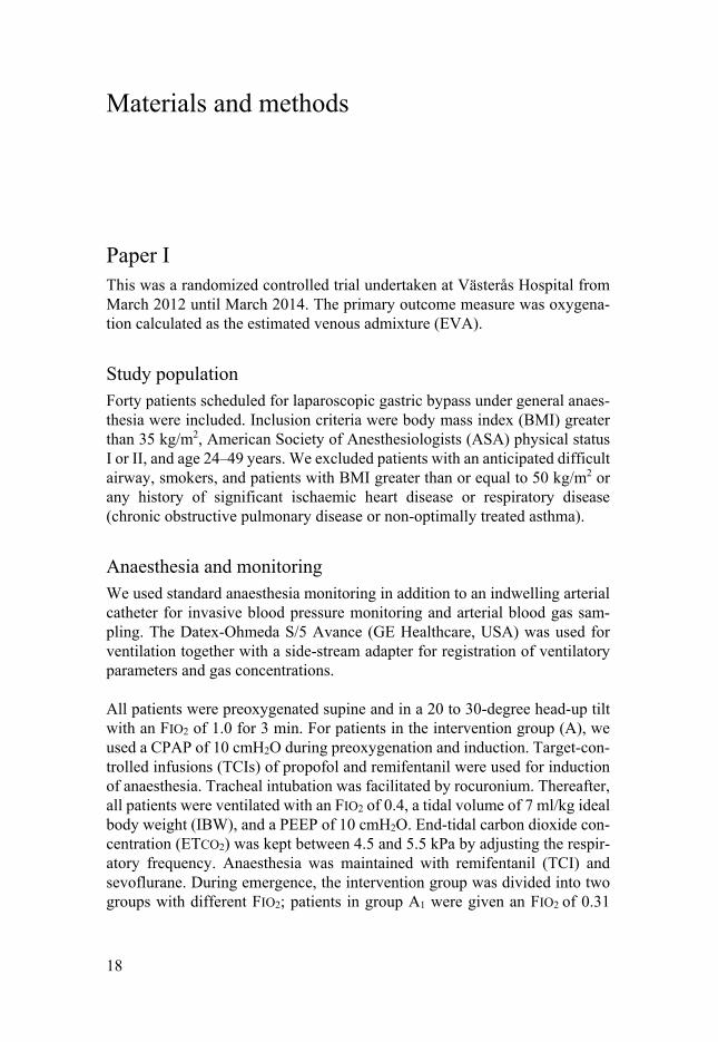
Computed tomography At the end of surgery, the endotracheal tube was clamped at end-expiration, and the ventilator was changed to a portable Vivo 50 (Breas Medical AB, Sweden), maintaining the same ventilator settings. A CT scan was obtained in the Radiology Department using a GE LightSpeed VCT XTe machine (GE Healthcare). With the patient in a supine position, we first obtained a frontal scout view at end-expiration. Thereafter, a single-slice transverse scan was performed, also at end-expiration (Fig. 1). A cursor was carefully positioned as basally as possible to achieve a single-slice scan approximately 5 mm above the right diaphragm dome.
Figure 1. Computed tomography images of one of the participating patients during anaesthesia. Frontal scout view (left) with cursor positioned approximately 5 mm above the right diaphragm dome. Basal axial scan with atelectasis visible in dorsal lung regions (right), in this case corresponding to 4.1% of total lung area.

21
The total lung area was measured by delineating the contours of both lungs. The atelectasis area was then carefully outlined posteriorly but with some mar-gin ventrally beyond the lung parenchyma haziness, using a separate region of interest (ROI) technique (Fig. 2).
Figure 2. Computed tomography measurements performed with the AW server soft-ware, in the same patient as in Figure 1. Region of interest (ROI) outlined for total lung area (left) and separate ROI outlined for the atelectasis area with some margin (right).
Workstation software (AW Server, GE Healthcare) was used to calculate the actual area of atelectasis, which was defined as -100 to +100 Hounsfield units (HU)13 (Fig. 3). The assessments were all performed by the same radiologist, who was blinded to the group assignment.
Figure 3. Histogram of the atelectasis region of interest performed using the AW server software, for the same patient as shown in Figures 1 and 2. The area of atelec-tasis was defined as -100 to +100 Hounsfield units (x-axis).
22
Arterial blood gases and estimation of venous admixture The blood-gas samples were obtained with the patients in a 15 to 20-degree head-up tilt. The first sample was drawn when the patients were breathing room air, before anaesthesia. The second sample was drawn during anaesthe-sia after the completion of surgery and with the patient still being ventilated with an FIO2 of 0.30–0.35.
We estimated oxygenation by calculating EVA, with the assumption that the arterio-venous oxygen content difference was 40 ml/l.
Paper III This randomized controlled trial investigated the effect of continuous PEEP on atelectasis formation during anaesthesia. The study was undertaken at Kö-ping Hospital between November 2015 and October 2016. The primary out-come measure was atelectasis area at the end of surgery.
Study population Twenty-four patients scheduled for non-abdominal day surgery were included in the study. Inclusion criteria were ASA physical status I and II, age 40–75 years and BMI less than 30 kg/m2. Patients with any significant respiratory or ischaemic heart disease were excluded, as were those with an anticipated dif-ficult airway. Active smokers and ex-smokers with a history of more than six pack-years were also excluded.
Anaesthesia and monitoring In addition to standard anaesthesia monitoring, we used an indwelling catheter in the radial artery to measure blood pressure. All patients received standard preoxygenation with FIO2 1.0 for 3 min. Induction and maintenance of anaes-thesia were performed with TCI of propofol and remifentanil. A single dose of rocuronium was used to facilitate tracheal intubation. The study subjects were ventilated using the portable Vivo 50 (Breas Medical AB) in a volume-controlled mode with an FIO2 of 0.30–0.35. Tidal volumes were set to 7 ml/kg IBW and the respiratory rate to 10 breaths/min and further adjusted to main-tain ETCO2 at approximately 5 kPa. The intervention group received PEEP 7 cmH2O or 9 cmH2O for patients with a BMI greater than or equal to 25 kg/m2, whereas the control group received zero PEEP (ZEEP). RMs were not used in either group.
23
Computed tomography After completion of surgery and while still anaesthetized, the patients were transported to the Radiology Department. This could be done without discon-necting or changing the ventilator settings, since a portable ventilator was used for mechanical ventilation during surgery. CT scanning, analysis, and calcu-lation of atelectasis area were performed in the same way as in study II, by a radiologist who was blinded to the group assignment and patient outcome.
Arterial blood gases and oxygenation The blood gas samples were drawn from the arterial line with the patients in a 15 to 20-degree head-up tilt. The first sample was obtained before anaesthesia when the patients were breathing room air. The second and third samples were collected midway through surgery and at the end of surgery with an FIO2 of 0.30–0.35. Oxygenation was estimated by calculating the ratio of the arterial oxygen partial pressure to the inspired oxygen fraction (PaO2/FIO2 ratio).
Paper IV This was a randomized controlled trial investigating the effects of PEEP ver-sus ZEEP during awakening from anaesthesia. The study was undertaken be-tween December 2017 and August 2018 at Köping Hospital. The primary out-come measure was the change in atelectasis from before awakening to after extubation.
Study population In total, 30 patients undergoing elective hernia repair or orthopaedic day sur-gery participated. The included study subjects were 40–75 years old and were considered ASA class I or II. Exclusion criteria were BMI greater than or equal to 30 kg/m2, any significant respiratory or ischaemic heart disease, and patients being active smokers or ex-smokers with a history of more than six pack-years.
Anaesthesia and monitoring Standard anaesthesia monitoring was supplemented with invasive blood pres-sure measurements via an arterial catheter placed in the radial artery. Routine preoxygenation with a slight head-up tilt and FIO2 1.0 for 3 min was followed by induction of anaesthesia with TCI of propofol and remifentanil and a single dose of rocuronium. Mechanical ventilation was performed with the portable Hamilton-T1 ventilator (Hamilton Medical, Switzerland). During surgery, the
24
ventilator settings were the same for both groups: FIO2 0.35, tidal volume 7 ml/kg IBW, and PEEP 7 or 9 cmH2O, with the higher setting in patients whose BMI was greater than or equal to 25 kg/m2. The respiratory frequency was set to 10 breaths/min and adjusted to maintain ETCO2 at approximately 5 kPa. RMs were not used.
Computed tomography and emergence from anaesthesia After completed surgery, the patients, still anaesthetized, were transported to the Radiology Department. A baseline CT scan was obtained using either the GE LightSpeed VCT XTe (GE Healthcare) or the Toshiba Aquilion PRIME (Canon Medical Systems, USA). In the control group, PEEP was maintained during emergence preoxygenation with FIO2 1.0 and subsequent awakening. In the intervention group, PEEP was withdrawn and set to zero (ZEEP) while FIO2 was still 0.35. The lungs were assumed to have achieved a new steady state EELV at ZEEP after 2 min,85 and thereafter emergence preoxygenation was started with FIO2 1.0 as for the control group. Extubation was undertaken in the post-anaesthesia care unit (PACU). Approximately 30 min after extu-bation, the patients were transported back to the Radiology Department for a second CT scan. This scanning was also done at end-expiration but with all study subjects fully awake and breathing room air. Calculation of atelectasis area was performed as described for studies II and III by a radiologist blinded to the study group assignment.
Arterial blood gases and oxygenation A baseline blood gas sample was drawn from the arterial line in the awake state when the patients were still breathing room air. The second blood gas was obtained at the end of surgery during mechanical ventilation with PEEP and FIO2 0.35. The third sample was drawn approximately 40 min after extu-bation, with all subjects again breathing room air. We estimated oxygenation by calculating the ratio of the arterial oxygen partial pressure to the inspired oxygen fraction (PaO2/FIO2 ratio).
25
Statistical analysis
All four studies were preceded by power calculations to assess the number of study subjects needed to detect a clinically significant reduction in the primary outcome measure. For study I, this meant a difference in arterial oxygen satu-ration (SaO2) of 2 percentage units between groups. For the remaining studies, it implied either a reduction in atelectasis area of 50% (studies II and III) or a 50% reduction in the increase in atelectasis area (study IV) compared with the respective control group.
In general, we did not assume that the data were normally distributed. There-fore, we used the Mann-Whitney U-test for comparisons between groups. Re-lated samples within each group were analysed with the Wilcoxon signed-rank test. For all tests, a two-sided P value of less than 0.05 was considered signif-icant, unless Bonferroni corrections were made for multiple comparisons. The 95% confidence intervals for the difference in medians were calculated with the Hodges-Lehmann estimator (study I) or derived by the percentile bootstrap method (studies III–IV). A post hoc sensitivity analysis to the primary out-come analysis was performed in study IV using a linear regression model after log transformation of data, with inclusion of the baseline atelectasis area as a covariate in the model. Statistical analyses were performed with IBM SPSS Statistics versions 20, 22, and 24 (IBM Corporation, USA) and R versions 3.4.2 and 3.5.1 (www.r-project.org).
26
Ethics
The four studies were approved by the Regional Ethics Committee in Uppsala and pre-registered at ClinicalTrials.gov. The study protocols were also ap-proved by the local Radiation Safety Committee before commencement.
CT scanning was included in the protocols for studies II–IV. The trade-off between possible study benefits and radiation exposure of the study partici-pants was carefully considered for each study. Full spiral CT scans of the thorax were not performed. Instead, each CT scan was limited to an initial scout and then a basal single-slice transverse scan. The total radiation dose received by each study participant was measured to be 0.3 mSv in studies II and III, and 0.6 mSv in study IV, which included two CT scans (CTDosimetry, Impactscan.org). A full thoracic CT scan is typically 5 mSv. For comparison, the natural background radiation in Sweden is measured to be 1.5 mSv/year. The annual average radiation dose received by the Swedish population, in-cluding medical examinations, is estimated to be 2.4 mSv/person (Swedish Radiation Safety Authority 2007). Moreover, most of the previously published studies using CT have used two or more slices, resulting in a higher radiation dose than in the studies included here.
The CT studies involved transportation of anaesthetized patients from the op-erating room to the Radiology Department, and after scanning was completed, to the PACU. The studies were performed at Köping Hospital, where the Ra-diology Department is located in the same building as the operating theatres but three floors below, with an elevator directly connecting the two units. Dur-ing transportation and CT, the patients were monitored and closely supervised by two consultant anaesthetists and one certified registered nurse anaesthetist. No complications related to transportation occurred during the studies.
Participation in studies II–IV prolonged the patient’s anaesthesia by approxi-mately 15 min. This fact, together with information regarding CT scanning and transportation, was explicitly communicated to the Regional Ethics Com-mittee and included in the written and oral information given to all partici-pants.

27
Results and discussion
Paper I All 40 included patients completed the study and all data were analysed. No complications were recorded. Baseline characteristics and physiological data were similar between groups, except for a lower median BMI and a greater proportion of men in the control group. There were no intergroup differences regarding ventilation or haemodynamic data during the anaesthesia.
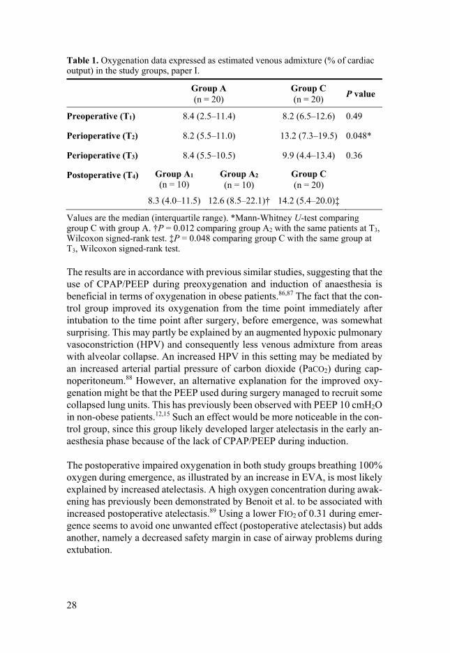
At 5 min after intubation (T2), EVA was lower in the intervention group (A) compared with the control group (C) (Table 1). The difference in medians was -5.0% (95% CI, -9.4 to -1.1%), P = 0.048.
At the end of surgery and before extubation (T3), EVA was unaltered in the intervention group. Oxygenation had improved in the control group as illus-trated by a lower EVA, and at this time point there was no longer any differ-ence between the groups.
One hour after emergence and extubation (T4), EVA had increased in the 30 patients given an FIO2 of 1.0. The group given an FIO2 of 0.31 during emer-gence (A1) maintained their oxygenation as illustrated by an unaltered EVA compared with before emergence.
28
Table 1. Oxygenation data expressed as estimated venous admixture (% of cardiac output) in the study groups, paper I.
Group A (n = 20)
Group C (n = 20) P value
Preoperative (T1) 8.4 (2.5–11.4) 8.2 (6.5–12.6) 0.49
Perioperative (T2) 8.2 (5.5–11.0) 13.2 (7.3–19.5) 0.048*
Perioperative (T3) 8.4 (5.5–10.5) 9.9 (4.4–13.4) 0.36
Postoperative (T4)
Group A1 (n = 10)
Group A2
(n = 10) Group C (n = 20)
8.3 (4.0–11.5) 12.6 (8.5–22.1)† 14.2 (5.4–20.0)‡
Values are the median (interquartile range). *Mann-Whitney U-test comparing group C with group A. †P = 0.012 comparing group A2 with the same patients at T3, Wilcoxon signed-rank test. ‡P = 0.048 comparing group C with the same group at T3, Wilcoxon signed-rank test.
The results are in accordance with previous similar studies, suggesting that the use of CPAP/PEEP during preoxygenation and induction of anaesthesia is beneficial in terms of oxygenation in obese patients.86,87 The fact that the con-trol group improved its oxygenation from the time point immediately after intubation to the time point after surgery, before emergence, was somewhat surprising. This may partly be explained by an augmented hypoxic pulmonary vasoconstriction (HPV) and consequently less venous admixture from areas with alveolar collapse. An increased HPV in this setting may be mediated by an increased arterial partial pressure of carbon dioxide (PaCO2) during cap-noperitoneum.88 However, an alternative explanation for the improved oxy-genation might be that the PEEP used during surgery managed to recruit some collapsed lung units. This has previously been observed with PEEP 10 cmH2O in non-obese patients.12,15 Such an effect would be more noticeable in the con-trol group, since this group likely developed larger atelectasis in the early an-aesthesia phase because of the lack of CPAP/PEEP during induction.
The postoperative impaired oxygenation in both study groups breathing 100% oxygen during emergence, as illustrated by an increase in EVA, is most likely explained by increased atelectasis. A high oxygen concentration during awak-ening has previously been demonstrated by Benoit et al. to be associated with increased postoperative atelectasis.89 Using a lower FIO2 of 0.31 during emer-gence seems to avoid one unwanted effect (postoperative atelectasis) but adds another, namely a decreased safety margin in case of airway problems during extubation.
29
Paper II All included patients received the allocated treatment and their data were an-alysed. Baseline characteristics and physiological data did not differ between the groups. Two patients had minor complications. One patient in the oxygen washout group had an episode of profound hypotension after induction, and one patient in the control group experienced a bronchospasm that required suctioning of the airways. These two patients had larger atelectasis than any other study subject.
The time required to complete the oxygen washout, i.e., for ETO2 to reach 25% or below, was a mean (SD) of 1.6 min (0.4 min). Peak airway pressures were below 30 cmH2O in all patients, and no episodes of desaturation were rec-orded. The group receiving standard treatment (control) required a mean (SD) of 20 min (3.3 min) to reach the targeted FIO2 of 0.30–0.35.
The area of atelectasis, expressed as percentage of the total lung area, was a median (IQR) 2.0 (1.5–2.7) in the oxygen washout group, and 1.8 (1.4–3.3) in the control group, P = 0.98.
There was no difference between the two groups for the absolute values of estimated venous admixture at the end of surgery. Comparisons within each group showed that oxygenation improved in the oxygen-washout group from the first (before anaesthesia) to the second (end of surgery) blood gas (Fig. 4). EVA decreased in this group from a mean (SD) of 7.6% (6.6%) to 3.9% (2.9%), P = 0.019. For the control group, there was no significant difference between EVA before anaesthesia and that at the end of surgery.

30
Figure 4. Changes in mean values of EVA for the two study groups in paper II, P = 0.028. The first blood gas was taken prior to the start of anaesthesia with all patients breathing room air. The second blood gas was taken at the end of surgery during me-chanical ventilation with an FIO2 of 0.30–0.35. EVA = estimated venous admixture (% of cardiac output).
The mean baseline value of EVA was higher in the oxygen-washout group (7.6%), however this was not significantly different to that in the control group (5.0%) (Fig. 4). A comparison of the groups regarding the change in oxygen-ation levels between the awake state before anaesthesia and the end of surgery showed that there was a significant difference between the groups. The change in EVA in the oxygen-washout group was (mean) -3.7 percentage units and the corresponding change in the control group was (mean) +0.6 percentage units, P = 0.028. This post hoc analysis of the study data was performed after the manuscript was published.
This was the first study to investigate the extent of atelectasis at the end of surgery in patients being ventilated with PEEP. Both groups exhibited notably small areas of atelectasis. A possible explanation is that the beneficial and combined effect of an early applied PEEP and preserved haemodynamics dur-ing induction had been previously underestimated. Although the control group was exposed to higher oxygen concentrations over a longer time, substantial airway closure following induction of anaesthesia was possibly avoided by the application of PEEP directly after tracheal intubation. Furthermore, the im-provement in oxygenation observed in the oxygen-washout group is likely ex-plained by a gradual recruitment effect of PEEP. Any such effect might be

31
greater in patients already exhibiting an increased EVA in the awake state, assuming an increased airway closure.
Although not being the primary intention of the study, the results of study II indicated that a moderate PEEP alone is sufficient to minimize atelectasis for-mation and to maintain oxygenation. This observation generated the hypothe-sis that was subsequently tested in study III.
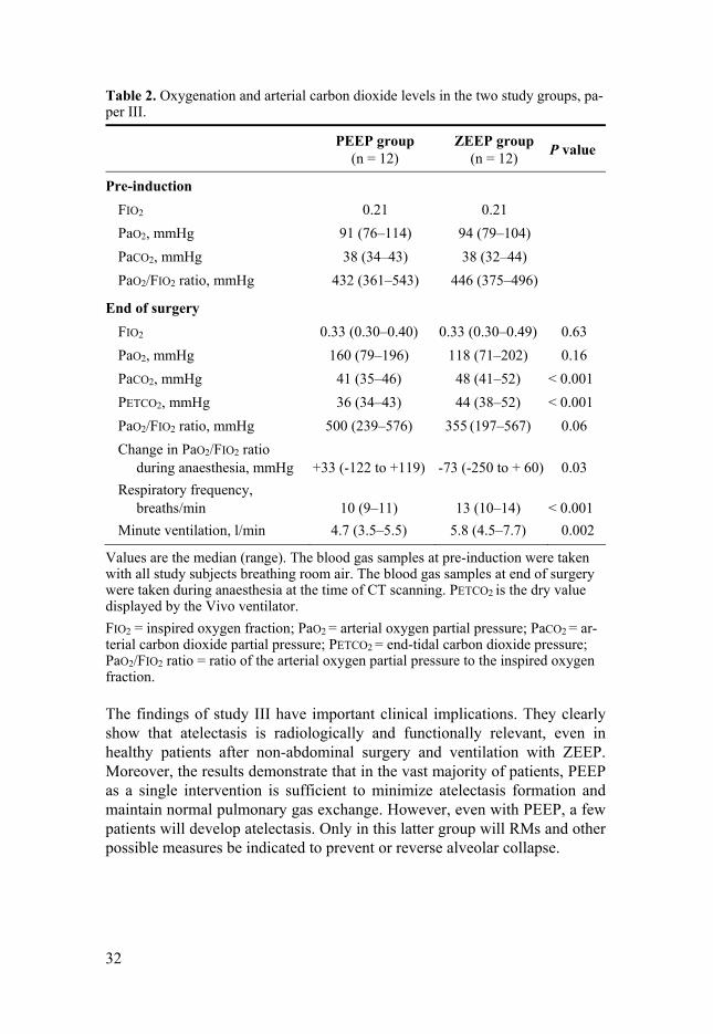
Paper III Baseline characteristics and physiological data were similar in the two study groups. All the 24 included patients received the allocated treatment and com-pleted the study. There was no difference between the study groups regarding the need for vasoactive drugs to maintain blood pressure during anaesthesia. No patient complications were recorded.
The atelectasis area, expressed as the percentage of total lung area, was a me-dian (range) 1.8 (0.3–9.9) in the PEEP group and 4.6 (1.0–10.2) in the ZEEP group. The difference in medians was 2.8% (95% CI, 1.7–5.7%), P = 0.002.
The PEEP group maintained their oxygenation and carbon dioxide elimina-tion, whereas the ZEEP group exhibited a decrease in PaO2/FIO2 ratio and higher carbon dioxide partial pressures (Table 2). At the end of surgery, the dynamic compliance for the PEEP group was a median (range) of 51 (29–71) ml/cmH2O and for the ZEEP group 34 (26–50) ml/cmH2O, P = 0.001.
32
Table 2. Oxygenation and arterial carbon dioxide levels in the two study groups, pa-per III.
PEEP group (n = 12)
ZEEP group (n = 12) P value
Pre-induction FIO2
PaO2, mmHg PaCO2, mmHg PaO2/FIO2 ratio, mmHg
0.21
91 (76–114) 38 (34–43)
432 (361–543)
0.21
94 (79–104) 38 (32–44)
446 (375–496)
End of surgery FIO2
PaO2, mmHg PaCO2, mmHg PETCO2, mmHg PaO2/FIO2 ratio, mmHg Change in PaO2/FIO2 ratio during anaesthesia, mmHg Respiratory frequency, breaths/min Minute ventilation, l/min
0.33 (0.30–0.40)
160 (79–196) 41 (35–46) 36 (34–43)
500 (239–576)
+33 (-122 to +119)
10 (9–11) 4.7 (3.5–5.5)
0.33 (0.30–0.49)
118 (71–202) 48 (41–52) 44 (38–52)
355 (197–567)
-73 (-250 to + 60)
13 (10–14) 5.8 (4.5–7.7)
0.63 0.16
< 0.001 < 0.001
0.06 0.03
< 0.001 0.002
Values are the median (range). The blood gas samples at pre-induction were taken with all study subjects breathing room air. The blood gas samples at end of surgery were taken during anaesthesia at the time of CT scanning. PETCO2 is the dry value displayed by the Vivo ventilator. FIO2 = inspired oxygen fraction; PaO2 = arterial oxygen partial pressure; PaCO2 = ar-terial carbon dioxide partial pressure; PETCO2 = end-tidal carbon dioxide pressure; PaO2/FIO2 ratio = ratio of the arterial oxygen partial pressure to the inspired oxygen fraction.
The findings of study III have important clinical implications. They clearly show that atelectasis is radiologically and functionally relevant, even in healthy patients after non-abdominal surgery and ventilation with ZEEP. Moreover, the results demonstrate that in the vast majority of patients, PEEP as a single intervention is sufficient to minimize atelectasis formation and maintain normal pulmonary gas exchange. However, even with PEEP, a few patients will develop atelectasis. Only in this latter group will RMs and other possible measures be indicated to prevent or reverse alveolar collapse.
33
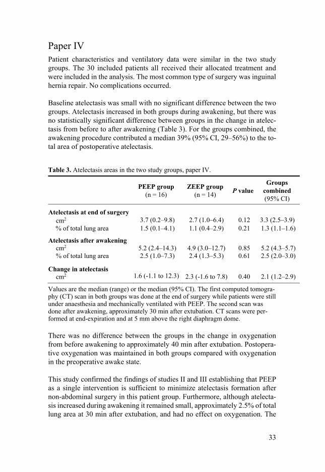
Paper IV Patient characteristics and ventilatory data were similar in the two study groups. The 30 included patients all received their allocated treatment and were included in the analysis. The most common type of surgery was inguinal hernia repair. No complications occurred.
Baseline atelectasis was small with no significant difference between the two groups. Atelectasis increased in both groups during awakening, but there was no statistically significant difference between groups in the change in atelec-tasis from before to after awakening (Table 3). For the groups combined, the awakening procedure contributed a median 39% (95% CI, 29–56%) to the to-tal area of postoperative atelectasis.
Table 3. Atelectasis areas in the two study groups, paper IV.
PEEP group (n = 16)
ZEEP group (n = 14) P value
Groups combined (95% CI)
Atelectasis at end of surgery cm2
% of total lung area
3.7 (0.2–9.8) 1.5 (0.1–4.1)
2.7 (1.0–6.4) 1.1 (0.4–2.9)
0.12 0.21
3.3 (2.5–3.9) 1.3 (1.1–1.6)
Atelectasis after awakening cm2 % of total lung area
5.2 (2.4–14.3) 2.5 (1.0–7.3)
4.9 (3.0–12.7) 2.4 (1.3–5.3)
0.85 0.61
5.2 (4.3–5.7) 2.5 (2.0–3.0)
Change in atelectasis cm2
1.6 (-1.1 to 12.3)
2.3 (-1.6 to 7.8)
0.40
2.1 (1.2–2.9)
Values are the median (range) or the median (95% CI). The first computed tomogra-phy (CT) scan in both groups was done at the end of surgery while patients were still under anaesthesia and mechanically ventilated with PEEP. The second scan was done after awakening, approximately 30 min after extubation. CT scans were per-formed at end-expiration and at 5 mm above the right diaphragm dome.
There was no difference between the groups in the change in oxygenation from before awakening to approximately 40 min after extubation. Postopera-tive oxygenation was maintained in both groups compared with oxygenation in the preoperative awake state.
This study confirmed the findings of studies II and III establishing that PEEP as a single intervention is sufficient to minimize atelectasis formation after non-abdominal surgery in this patient group. Furthermore, although atelecta-sis increased during awakening it remained small, approximately 2.5% of total lung area at 30 min after extubation, and had no effect on oxygenation. The
34
extent of postoperative atelectasis was the same in both groups, irrespective of whether PEEP was maintained or withdrawn during emergence preoxygen-ation. A reasonable interpretation of this finding is that preoxygenation with FIO2 1.0 before emergence is well tolerated in healthy patients undergoing non-abdominal day surgery. This in turn is made possible by minimal baseline atelectasis at the end of surgery combined with limited airway closure in the early postoperative period. In other patient categories, for example high-risk patients undergoing major surgery, the roles of the awakening procedure and PEEP during emergence preoxygenation still need clarification.
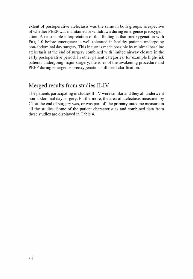
Merged results from studies II–IV The patients participating in studies II–IV were similar and they all underwent non-abdominal day surgery. Furthermore, the area of atelectasis measured by CT at the end of surgery was, or was part of, the primary outcome measure in all the studies. Some of the patient characteristics and combined data from these studies are displayed in Table 4.
35
Table 4. Merged data from CT studies II–IV.
PEEP during anaesthesia
(n = 66)
ZEEP during anaesthesia
(n = 12) P value
Male/female, n 44/22 9/3 –
Age, years 56 (50–63) 57 (50–68) –
Weight, kg 79 (70–87) 75 (67–82) –
BMI, kg/cm2 25 (23–27) 24 (22–27) –
IBW, kg 67 (60–75) 72 (61–74) –
ASA physical status I/II 42/24 8/4 –
Type of surgery, n Orthopaedic, extremity Inguinal hernia Other non-abdominal
31 31 4
6 6 0
– – –
PEEP level, cmH2O 0 6 7 8 9
0
15 19 9
23
12 0 0 0 0
– – – – –
Duration of surgery, min 53 (35–64) 46 (29–55) 0.15
Time at CT, min 105 (83–122) 96 (88–104) 0.20 Atelectasis, cm2 3.5 (2.5–5.7) 9.6 (6.8–13.3) < 0.001
Atelectasis, % of total lung area 1.6 (1.0–2.4) 4.6 (4.1–7.1) < 0.001
PaO2/FIO2 ratio awake, mmHg 425 (389–460) 446 (398–471) 0.36
EVA awake, % of cardiac output 5.5 (3.2–10.2) 5.0 (1.8–7.5) 0.34
PaO2/FIO2 ratio at CT, mmHg 504 (420–561) 355 (315–396) 0.001
EVA at CT, % of cardiac output 3.2 (1.2–6.7) 9.2 6.9–12.2) 0.007
Values are the median (interquartile range). t0 = start of preoxygenation. Computed tomography scans were done during anaesthesia at end-expiration and at 5–10 mm above the right diaphragm dome. PaO2/FIO2 ratio awake and EVA awake were calculated from arterial blood gases obtained with patients breathing room air before anaesthesia induction. PaO2/FIO2 ratio at CT and EVA at CT were calculated from blood gases obtained during general anaesthesia and FIO2 0.30–0.35, at the time of CT scanning. ASA = American Society of Anesthesiologists physical status; BMI = body mass in-dex; CT = computed tomography; EVA = estimated venous admixture; IBW = ideal body weight; PaO2/FIO2 ratio = ratio of the arterial oxygen partial pressure to the in-spired oxygen fraction.
36
General discussion
The studies included in this thesis explored factors associated with preventing or treating atelectasis formation during all three phases of anaesthesia, i.e., induction, maintenance, and awakening. The clinically most important finding is that PEEP, as a single intervention, reduces atelectasis formation to a neg-ligible level (median 1.6% of total lung area) in healthy patients undergoing non-abdominal surgery. The results also add support to the proposal that high FIO2 is well tolerated as long as airway closure is avoided.80 To put the above atelectasis in perspective, the area of atelectasis that has been observed in nor-mal-weight patients after induction of anaesthesia with zero PEEP, ranges be-tween 4 and 7% of the total lung area in a CT slice obtained close to the dia-phragm.13,80,81,90–94
Study I dealt in part with induction of anaesthesia. Preoxygenation before in-duction remains an integral safety measure to increase oxygen storage, be-cause difficulties with ventilation and intubation can occur unexpectedly.1,3 Preoxygenation efficiency has been shown to increase with the use of CPAP followed by mechanical ventilation with PEEP,86,95,96 or CPAP in combination with pressure-support ventilation.97–100 CPAP will increase EELV, and in combination with a high FIO2, apnoea time is prolonged and PaO2 is better maintained. In clinical anaesthesia, the one obvious effect of CPAP during preoxygenation is that the time required to achieve an ETO2 above 90% is shortened.98,99 In this context however, it is important to distinguish between efforts to improve or hasten preoxygenation and attempts to prevent atelecta-sis formation. Following preoxygenation with CPAP and subsequent tracheal intubation, early application of an adequate PEEP level is crucial to maintain-ing EELV. Otherwise, absorption atelectasis will follow, the extent of which might be more pronounced because of the newly oxygen-filled larger lung volume achieved by CPAP. Further, some as yet unpublished data from a re-cent study (Cajander et al., personal communication) indicate a possible in-creased risk of gastric insufflation when a PEEP is used before the airway is secured. This could potentially increase the risk of gastric regurgitation and aspiration. Until future studies have demonstrated its safe use, it is important to be aware of potential pitfalls using this technique and to perform a risk-benefit analysis for each patient.
37
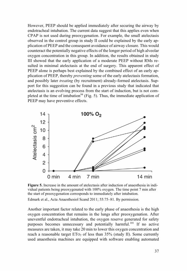
However, PEEP should be applied immediately after securing the airway by endotracheal intubation. The current data suggest that this applies even when CPAP is not used during preoxygenation. For example, the small atelectasis observed in the control group in study II could be explained by the early ap-plication of PEEP and the consequent avoidance of airway closure. This would counteract the potentially negative effects of the longer period of high alveolar oxygen concentration in this group. In addition, the results obtained in study III showed that the early application of a moderate PEEP without RMs re-sulted in minimal atelectasis at the end of surgery. This apparent effect of PEEP alone is perhaps best explained by the combined effect of an early ap-plication of PEEP, thereby preventing some of the early atelectasis formation, and possibly later treating (by recruitment) already-formed atelectasis. Sup-port for this suggestion can be found in a previous study that indicated that atelectasis is an evolving process from the start of induction, but is not com-pleted at the time of intubation94 (Fig. 5). Thus, the immediate application of PEEP may have preventive effects.
Figure 5. Increase in the amount of atelectasis after induction of anaesthesia in indi-vidual patients being preoxygenated with 100% oxygen. The time point 7 min after the start of preoxygenation corresponds to immediately after intubation. Edmark et al., Acta Anaesthesiol Scand 2011; 55:75–81. By permission.
Another important factor related to the early phase of anaesthesia is the high oxygen concentration that remains in the lungs after preoxygenation. After uneventful endotracheal intubation, the oxygen reserve generated for safety purposes becomes unnecessary and potentially harmful.101 If no active measures are taken, it may take 20 min to lower this oxygen concentration and reach a reasonable target ETO2 of less than 35% (study II). Some currently used anaesthesia machines are equipped with software enabling automated
38
end-tidal control, in which case the above will be less of a problem. Without this software, and despite the negative results of study II, it might still be rec-ommended that the pulmonary oxygen levels be lowered as soon as possible after successful intubation. This makes sense from a physiological point of view, given our knowledge about the mechanisms behind early atelectasis for-mation. In addition, any positive effect of the oxygen washout in study II might have been obscured by the effects of an early application of a moderate PEEP, which may have prevented early airway closure in this study group. The need to avoid residual high oxygen concentrations is probably more ur-gent in a situation where airway closure is more likely to occur, such as in obese patients undergoing laparoscopic surgery.
The early phases of anaesthesia have been the predominant focus for research in this field since it became known that atelectasis formation is closely related to induction of anaesthesia.41 This remained true after CT became the estab-lished method for quantifying atelectasis.12 Although Cai et al. used CT at the end of surgery to study the effect of different tidal volumes on atelectasis for-mation, their patients were ventilated with ZEEP and 100% O2,92 which is not consistent with current clinical practice. Studies II–IV in this thesis are the first published studies using CT to examine atelectasis at the end of surgery after ventilating patients with PEEP. The findings of these separate studies stand alone. However, the study subjects and the type of surgery they under-went were similar in the three studies, which offers an opportunity to combine the results to provide a clearer picture of the effects of PEEP during non-ab-dominal surgery (Table 4). For all 66 patients who received a moderate PEEP during mechanical ventilation, the atelectasis area at the end of surgery was a median (IQR) of 1.6% (1.0–2.4%) of total lung area (95% CI, 1.3–2.0%). None of these patients received any RMs, with the possible exception of the 12 patients in study II who underwent an oxygen washout manoeuvre that in-cluded larger tidal volumes (15 ml/kg IBW) and higher PEEP (10 cmH2O) for a mean of 1.6 min. For the 12 patients ventilated without PEEP, the area of atelectasis was a median (IQR) of 4.6% (4.1–7.1%), (95% CI, 4.1–7.1%). These combined results constitute a strong indication that healthy patients un-dergoing non-abdominal surgery should receive a moderate PEEP to minimize atelectasis and maintain oxygenation. Further, for this patient group, routine RMs seem unnecessary and should only be used when clearly indicated. The combined data also add support to previous findings that mechanical ventila-tion with minimal PEEP or ZEEP should be avoided, especially in combina-tion with low tidal volumes.102
The finding that PEEP is both necessary and sufficient to minimize atelectasis in most healthy patients undergoing non-abdominal surgery, also has im-portant implications for the awakening procedure. In study IV, the postopera-tive atelectasis area expressed as a percentage of total lung area was 2.5% for
39
both groups, regardless of whether PEEP was maintained and despite using FIO2 1.0 during emergence. This result is similar or better than those obtained in several previous studies testing the ability of CPAP, vital capacity manoeu-vres, and/or lower FIO2 during and after emergence to limit postoperative ate-lectasis formation.84,89,103 Our results indicate that minimal atelectasis at the end of surgery provides a beneficial starting-point for subsequent awakening, during which a high FIO2 seems well tolerated, possibly partly because of lim-ited postoperative airway closure after this type of surgery in healthy patients. Overall, an important clinical implication of the studies is that a high oxygen concentration during either early or late phases of anaesthesia is well tolerated provided that airway closure is avoided. In most healthy patients undergoing non-abdominal surgery, intra-operative airway closure can be counteracted by an early application of a moderate/adequate PEEP. Postoperative airway clo-sure appears to be clinically insignificant in pain-free patients after minor day surgery and after receiving short-acting anaesthetics. These patients probably have no or very little interference with normal tidal breathing and/or deep breathing.
The studies presented in this thesis are small but have the strengths of inves-tigating PEEP in healthy lungs under controlled conditions. In study II, the patients received 6 or 8 cmH2O depending on BMI. In studies III and IV, we used 7 or 9 cmH2O for the respective BMI categories. The slightly higher lev-els used in the latter two studies were in part chosen to see whether they would result in even smaller atelectasis without compromising haemodynamic sta-bility. Although these studies were not designed to investigate outcome dif-ferences between small increments in PEEP, we did not observe any additional beneficial effect with the higher levels. Thus, based on these results, a PEEP of 6 cmH2O is recommended for normal-weight healthy patients undergoing non-abdominal surgery. The higher level of 8 cmH2O is recommended for overweight patients with a BMI of 25–30 kg/m2. Higher and perhaps individ-ually titrated PEEP levels are recommended for obese patients with BMI greater than or equal to 30 kg/m2.104
Other factors affecting atelectasis formation The amount of atelectasis formed during general anaesthesia is associated with BMI,105, and possibly ASA physical status and smoking.84 There is also a pos-sible correlation with age, but this is a more complicated association. There is an age-dependent increase in closing capacity which exceeds functional resid-ual capacity (FRC) in older patients and results in airway closure already in the awake state before anaesthesia induction.106 This may paradoxically be protective against atelectasis formation because the high oxygen concentra-tion used during preoxygenation may not reach these already closed lung

40
units, and the alveoli may therefore be partially protected from collapse by their rich nitrogen content. On the other hand, chest wall compliance decreases with increasing age because of anatomical factors and ossification of rib car-tilage.107 If or when the lungs are affected by atelectasis, re-expansion may be more difficult in older patients.
In the three studies included in this thesis that quantified atelectasis, the par-ticipants were aged 40–75 years. Any observable negative effects related to increased closing capacity may have been blurred by the use of PEEP in most study subjects. The merged data for these studies, showed no detectable sig-nificant correlation between age and atelectasis formation. However, the stud-ies were not designed to investigate any such correlation. With respect to BMI, the study protocols included differentiated PEEP levels to diminish BMI as a confounder. Despite this, the merged data showed a slight positive linear cor-relation between BMI and atelectasis (r = 0.281, n = 78, P = 0.013; Pearson product-moment correlation). This could indicate that the chosen PEEP levels were too low for some overweight patients.
There is no published evidence that sex is correlated with atelectasis for-mation. Based on our current understanding of how atelectasis forms, such an association would be surprising. In the combined data obtained for this thesis, no correlation was found between sex and atelectasis.
As stated, one of the aims of this thesis was to investigate the physiological effects of PEEP (studies II–IV). To avoid interference from factors other than general anaesthesia, a fairly homogeneous study population, i.e., healthy pa-tients undergoing non-abdominal day surgery, was chosen. Despite this, out-liers with very small or very large atelectasis were identified in all three CT studies. In some cases, the large atelectasis could probably be explained by circumstances or characteristics that were close to fulfilling the exclusion cri-teria, for example, borderline BMI and/or smoking or recent lower respiratory tract infection. Excessive bronchial secretions, bronchospasm, and persistent or pronounced hypotension were identified as possible explanatory factors in some cases.
Anaesthesia-related hypotension is a potential but unrecognized factor affect-ing atelectasis formation. Decreased cardiac output and hypotension are com-mon during induction of anaesthesia, especially in older patients and/or pa-tients taking antihypertensive medication.108 Systemic hypotension causes a shift in pulmonary blood flow to the dorsal lung regions, and in an early study, was shown to be associated with both increased venous admixture and de-creased pulmonary dynamic compliance.109 These findings could in part be explained by the induction of atelectasis, which was not well known at that
41
time. An increased proportion of cardiac output, possibly with lower than nor-mal oxygen content, passing through dorsal lung regions represents ideal con-ditions for increased gas absorption from newly preoxygenated lung units,110 with subsequent atelectasis formation. Notably, few previous publications about atelectasis have provided information about whether, or how, haemody-namic stability was maintained during and after induction. Invasive blood pressure monitoring was an integral part of the three CT studies presented here, and study protocols mandated that mean arterial blood pressure was maintained at or above 60 mmHg. This might partly explain why the atelecta-sis areas observed in the present studies were generally smaller than those re-ported from earlier comparable studies. Interestingly, despite administration of vasopressors, substantial hypotension was observed in a small number of patients and was associated with larger atelectasis. Further, the late deteriora-tion in oxygenation observed in the control group in study III, possibly caused by late development of atelectasis, could also be explained by effective maintenance of blood pressure in the early phase of anaesthesia. Even if gas is trapped behind closed airways, a normal distribution of pulmonary blood flow avoids excessive gas absorption from susceptible lung units, and thus delays the collapse of alveoli. This hypothetical mechanism needs to be demonstrated, but would challenge the established concept that atelectasis al-ways forms early after induction of anaesthesia. If the hypothesis is shown to be correct, avoiding an increased proportion of pulmonary blood flow to areas with low alveolar ventilation (V̇A) to lung perfusion (Q̇) ratios (V̇A/Q̇) by maintaining blood pressure will be added to PEEP as a tool to prevent or delay atelectasis formation.
A small number of patients presented with larger than expected atelectasis, without any obvious complicating or explanatory factors. Individual patient characteristics, for example variations in thoracic or pulmonary elasticity, and specific events during anaesthesia may explain why some patients are more prone to develop atelectasis. In one of our patients, it is tempting to believe that a large heart caused increased pressure on the small airways, contributing to airway closure and atelectasis (Fig. 6). Any patient identified with signifi-cant oxygenation difficulties during anaesthesia may need a ventilatory strat-egy comprising both RMs and individualized PEEP to minimize atelectasis formation and the risk of postoperative pulmonary complications.18,104,111,112

42
Figure 6. Computed tomography scan of one of the participants in study III. Note the large heart and the substantial area of atelectasis evident in the pulmonary tissue below. The computed tomography scan was performed during anaesthesia at end-ex-piration approximately 5 mm above the right diaphragm dome.
Methodological aspects and study limitations General anaesthesia impairs oxygenation for two main reasons. First, atelec-tasis forms and leads to intrapulmonary shunting of venous blood.15,113 Sec-ond, increased perfusion to areas with already low V̇A/Q̇ leads to additional venous admixture of pulmonary end-capillary blood.114 Exact measurements of venous admixture requires sampling of mixed venous blood through a pul-monary artery catheter, but because of its invasiveness this is seldom done. Instead, the oxygenating capacity of the lungs is normally estimated, which can be done in several ways. The choice of oxygenation index is complex and remains controversial. In studies I and II, we chose to calculate and use EVA, because it has been suggested to be the best oxygenation index if mixed ve-nous blood is not collected.115 Nevertheless, use of the PaO2/ FIO2 ratio is more widespread, and this oxygenation index was used in studies III and IV. Im-paired oxygenation is a fundamental consequence of atelectasis and may serve as a surrogate indicator of the amount of atelectasis. However, an estimated or measured impairment in oxygenation can still not differentiate between the contribution from shunt (atelectasis) and V̇A/Q̇ mismatch. Therefore, CT re-mains the gold standard for quantifying atelectasis formation, and therefore

43
this method was used to directly measure atelectasis area in the study subjects participating in studies II–IV.
CT provides detailed images of pulmonary tissue and has been used since the 1980s to measure atelectasis.12,116 The CT workstation software enables quan-tification of lung zones with different levels of aeration based on varying at-tenuation values. Atelectasis, for example, has been defined as having attenu-ation values of -100 to +100 HU.13 Although the major part of the analysis is computerized, defining the border between the pleura and the dorsal thoracic wall must be done manually. Because atelectasis normally forms dorsally, and since it exhibits attenuation values close to those of the adjacent soft tissue on the inside of the thoracic cavity, the utmost care must be taken to accurately delineate the posterior border of the atelectasis and thus avoid calculation er-rors (Fig. 7). This procedure becomes even more critical with smaller areas of atelectasis. To rule out any inter-observer errors, the same radiologist per-formed the CT scan analysis in each of studies II–IV.
Figure 7. Example of a computed tomography scan of one of the participating pa-tients in study II. Note that the exact posterior border of the atelectasis area is diffi-cult to visualize. The computed tomography scan was done during anaesthesia at end-expiration and at 5–10 mm above the right diaphragm dome.

44
Instead of examining the entire thoracic cavity, most CT studies have used limited measurements to minimize radiation exposure to the study partici-pants. The basal lung regions are generally most affected by atelectasis;91 therefore, a basal CT slice just above the diaphragm has been considered rep-resentative to illustrate the extent of atelectasis formation. However, it is im-portant to acknowledge that this will underestimate the total amount of col-lapsed lung for two reasons. First, atelectasis distributed along the dorsal lung regions towards the apex is not included in the scan.117 Second, atelectasis comprises compressed lung tissue that has a higher tissue density than aerated lung tissue. Therefore, the actual area of collapsed lung is larger, and has been estimated to be four times greater than that estimated from the CT image.13
To conclude, the care of patients undergoing surgery is complex and must be versatile. This thesis focuses on a narrow aspect of respiratory care of patients during anaesthesia. After surgery and emergence from anaesthesia, continu-ous and professional postoperative care may be equally important to minimize the risk of PPCs, especially in high-risk patients. Several large trials of phys-iotherapy show promising results in preventing PPCs, especially after major surgery.118,119 This highlights the importance of taking a holistic approach to the perioperative care of patients undergoing anaesthesia and surgery. The work presented here highlights the crucial role of PEEP in counteracting air-way closure and atelectasis during anaesthesia and mechanical ventilation.
45
Conclusions
• In obese patients undergoing general anaesthesia for laparoscopic surgery, oxygenation can be preserved by using a CPAP of 10 cmH2O during pre-oxygenation and induction, followed by a PEEP of 10 cmH2O. Postoper-ative oxygenation might be impaired if a high FIO2 is used during emer-gence.
• An early oxygen washout after preoxygenation and intubation quickly re-stores nitrogen levels in the alveoli. In healthy patients undergoing non-abdominal surgery and being ventilated with a moderate PEEP, this ma-noeuvre has no further effect on the size of atelectasis at the end of sur-gery.
• For healthy patients undergoing non-abdominal surgery, a moderate PEEP is both necessary and sufficient to minimize atelectasis formation and maintain oxygenation. In this group of patients, RMs should only be uti-lized when clearly indicated. Mechanical ventilation with ZEEP is asso-ciated with several unfavourable physiological consequences and should be avoided.
• In most healthy day-surgery patients, postoperative atelectasis formation is limited without affecting oxygenation. Withdrawing PEEP before emer-gence preoxygenation does not reduce atelectasis formation after non-ab-dominal surgery. Conditional on a baseline open lung achieved by in-traoperative PEEP, emergence preoxygenation with 100% oxygen is well tolerated and thus recommended for maximum safety margins.
46
Strategy for minimizing atelectasis formation in perioperative care
This recommendation applies to healthy normal-weight patients undergoing non-abdominal surgery. In accordance with established guidelines,1 it encom-passes a maximum oxygen reserve during both induction and emergence.
1. Preoxygenate for 3 min with 100% O2 with the patient in a slight head-
up tilt position.
2. Use PEEP immediately when commencing mechanical ventilation after endotracheal intubation or insertion of a laryngeal mask airway.
3. Use PEEP 6 cmH2O if BMI < 25 kg/m2 and PEEP 8 cmH2O if BMI ≥ 25 kg/m2.
4. Set FIO2 to 0.35. Possibly use a higher fresh gas flow initially to achieve a more rapid reduction in ETO2.
5. Use RMs followed by individualized PEEP only if indicated by signs of atelectasis, i.e., decreased oxygenation with no other explanation. A sug-gested cut-off for when to consider an RM is SpO2 < 94% if ventilating with FIO2 0.35. Individual patient risks as well as type and duration of surgery must be considered.
6. Use 100% O2 during emergence preoxygenation and maintain PEEP.
7. In general, after total intravenous anaesthesia, supplementary oxygen en route to PACU is not necessary if spontaneous breathing is adequate as indicated by SpO2 ≥ 94%, and transport time is less than 5 min.
8. Give supplementary oxygen in PACU only when SpO2 < 94%.120
47
Future perspectives
Recent large international multicentre trials have reported conflicting results regarding protective ventilation during anaesthesia.59,60,62 Protective strategies comprising several components have been tested against “standard practice”, making it difficult to draw conclusions about which of the interventions was beneficial. At present, there seems to be a reasonable consensus that low tidal volumes should be routine, not only in critical care but also during mechanical ventilation in the operating theatre.112 On the other hand, the use of PEEP, or the level of PEEP, and the use of RMs is still a matter of debate, especially during major surgery. To make a significant contribution to the current knowledge, any new large trial should ideally be refined in terms of interven-tion, its study population, and its outcome measures. Moreover, if PPCs are chosen as an outcome measure, any intraoperative interventions regarding ventilation strategy should be accompanied by a detailed study plan and de-scription of the awakening procedure. This is because a chosen FIO2 and/or PEEP level during emergence preoxygenation and awakening may well have an impact on postoperative atelectasis formation and hence the incidence of PPCs, especially after major surgery or in high-risk patients.
Meanwhile, further explanatory studies are needed to clarify some of the ob-scure pathophysiological mechanisms involved in atelectasis formation. A worthwhile objective would be to explore the possible role of intraoperative hypotension as a contributor to atelectasis. Furthermore, a follow-up trial to our study IV could perhaps determine whether withdrawing or maintaining PEEP matters during emergence preoxygenation and awakening in patients undergoing major abdominal surgery. Such patients would probably exhibit more airway closure during the early postoperative phase, and therefore be more susceptible to high oxygen concentrations and more prone to develop postoperative atelectasis, compared with the patients investigated in study IV.
48
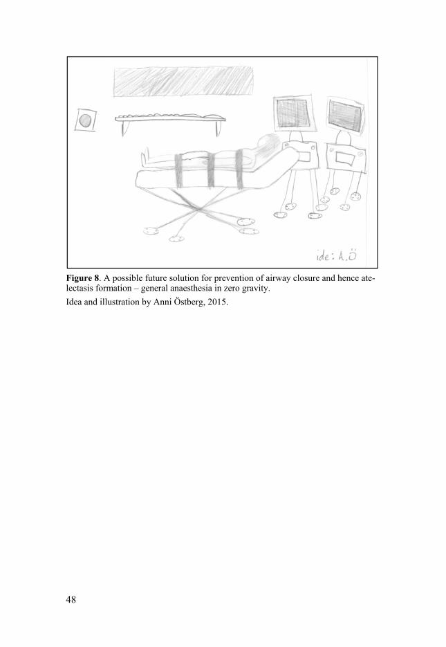
Figure 8. A possible future solution for prevention of airway closure and hence ate-lectasis formation – general anaesthesia in zero gravity. Idea and illustration by Anni Östberg, 2015.
49
Sammanfattning på svenska (Summary in Swedish)
Varje år genomförs i världen över 230 miljoner större operationer, de flesta i generell anestesi, d.v.s. patienterna sövs.28 Hos nästan alla vuxna patienter som sövs utvecklas atelektaser vilket innebär att delar av lungan faller sam-man. Det sker framför allt i lungavsnitt nära ryggen och diafragma (mellangär-det). Atelektaser leder till försämrad syresättning under operationen och ökar risken för postoperativa lungkomplikationer t.ex. lunginflammation.17–19 Dessa komplikationer är vanligare än hjärtkomplikationer och orsakar både förlängd vårdtid och ökade sjukvårdskostnader, liksom ökad dödlighet.21–27 Risken är störst hos äldre, överviktiga och multisjuka som genomgår stora op-erationer. Hos friska, normalviktiga personer som genomgår sövning och ki-rurgi återfår troligen lungan sin ursprungliga volym ganska snart efter väck-ningen, och för dem är riskerna små. Men eftersom ett så stort antal människor sövs och opereras årligen så har även små minskningar av risken stor bety-delse.
Bildandet av atelektaser förklaras dels av den höga syrgashalt som rutinmäss-igt och av säkerhetsskäl används vid nedsövningen, dels av en sänkt vilolung-volym när andningsmuskulaturen slappnar av när kroppen sövs.121 Den mins-kade vilolungvolymen orsakar avstängda partier i lungorna där alveolerna (lungblåsorna) nyligen fyllts med inandad ren syrgas. Atelektaser uppstår när den passerande lungcirkulationen suger åt sig syrgasen, varpå de avstängda lungblåsorna faller samman. Det är möjligt att blåsa upp de sammanfallna lungdelarna med hjälp av höga luftvägstryck, en så kallad rekryteringsmanö-ver, men det är oklart vilka patienter som skulle gynnas av detta. Det finns också potentiella biverkningar med lungrekrytering, till exempel minskar det venösa återflödet till hjärtat vilket påverkar blodcirkulationen negativt. Forsk-ningen kring atelektaser har huvudsakligen inriktat sig på att hitta sätt att för-hindra att de uppstår, genom t.ex. minskad initial syrgashalt, men detta leder samtidigt till minskad säkerhetsmarginal i händelse av svårigheter att ventilera patienten. Ett annat sätt är att motverka minskningen av vilolungvolymen. Inom intensivvården har man länge använt sig av ett utandningsmotstånd (PEEP) i samband med respiratorvård. PEEP har till viss del använts även vid mekanisk ventilation av sövda patienter som opereras, men tydliga riktlinjer
50
saknas då forskningen inte kunnat visa hur stort utandningsmotståndet bör vara, eller vilka patienter som har behov av det.
Detta avhandlingsarbete har fokuserat dels på betydelsen av syrgashalten un-der olika delar av anestesin, dels syftat till ökad kunskap kring effekterna av PEEP, både under anestesin och inför väckningen. Ett övergripande mål har varit att komma närmare ett optimalt sätt att ventilera sövda patienter, så att lungan hålls öppen och fri från atelektaser både under operationen och efter väckningen.
De arbeten som ingår i avhandlingen var samtliga kliniska och prospektiva randomiserade studier. I det första arbetet användes syresättning som ett sur-rogatmått för atelektaser hos överviktiga patienter som genomgick laparosko-pisk överviktskirurgi. I de följande studierna användes datortomografi (CT) för kvantifiering av atelektasyta hos friska patienter som genomgick dagkirur-gisk icke bukkirurgi.
Studie I: Två former av utandningsmotstånd, CPAP och PEEP, studerades un-der nedsövningen, liksom minskad syrgashalt vid väckningen. Gruppen med CPAP/PEEP under och efter nedsövningen hade bibehållen syresättning, till skillnad från jämförelsegruppen. Den grupp som fick en hög syrgashalt vid väckningen hade försämrad syresättning en timme postoperativt.
Studie II: En tidig syrgasutvädring i syfte att återställa en hög kvävgashalt i lungan och därmed stabilisera alveolerna visade sig inte ha effekt på mängden atelektaser i slutet av operationen. Både grupperna uppvisade endast små mängder atelektaser efter att ha ventilerats med ett måttligt PEEP, 6–8 cmH2O, under anestesin.
Studie III: Här jämfördes atelektasutveckling och syresättning i slutet av an-estesin hos två grupper som ventilerades antingen med eller utan ett måttligt PEEP. Gruppen med PEEP uppvisade statistiskt signifikant mindre atelektaser och bättre syresättning än gruppen utan PEEP.
Studie IV: I detta arbete undersöktes mängden atelektaser med CT före och efter väckning. Jämförelse gjordes mellan en grupp med bibehållet PEEP un-der rutinmässig väckning med 100% syrgas, och en grupp där PEEP avlägs-nades precis innan syrgasen höjdes till 100% inför väckningen. Vid en tid-punkt ungefär 30 min efter väckning hade grupperna likartad mängd små ate-lektaser samt bibehållen syresättning jämfört med innan sövning.
Avhandlingen visar att bibehållen vilolungvolym utgör nyckeln till undvi-kande av atelektasutveckling under anestesi, särskilt vid användande av de höga syrgashalter som generellt rekommenderas i samband med nedsövning

51
och väckning. Att lungan är öppen och fri från atelektaser när väckningen på-börjas verkar vara fördelaktigt. Det viktigaste fyndet av klinisk relevans är att ett måttligt PEEP som enda intervention resulterar i minimala mängder ate-lektaser hos friska patienter som genomgår icke bukkirurgi. Ett måttligt PEEP kan därför rekommenderas till alla friska vuxna som genomgår anestesi.
52
Acknowledgements
Lennart Edmark, my friend and co-supervisor. Lennart’s contribution to the current state of knowledge in the field of atelectasis is far greater than has been acknowledged. He has walked alongside me for every step of this work. His contribution to my joy in doing research and the subsequent genesis of this thesis is beyond description.
Mats Enlund, my main supervisor, colleague and former head of the Centre for Clinical Research, for his long friendship and many wise inputs during this work.
Arnar Thorisson, in times of struggle, and otherwise, it is a privilege to work with a down-to-earth Icelander whose standard answer is “No problem”.
Henrik Zetterström, venous admixture expert and co-author deluxe. You send him what you believe is a perfect manuscript and it comes back with brilliant suggestions for improvement regarding language, style, and content.
Göran Hedenstierna, who was already involved in studies on pulmonary physiology at the time I was born in 1971. It has been an honour to be a part of the research that he founded.
Emma-Karin Englund, anaesthetic nurse and supervisor at the Day Surgery Unit in Köping. Emma is like an enzyme, she facilitates processes. Things just run very smoothly when she is around.
The staff at the Day Surgery Unit in Köping, especially Annika Arvidsson and Lotta Pettersson, who have assisted in identifying eligible patients.
The staff at the Radiology Department in Köping, especially Kauser Ab-dulkadir and Fleury Ndongozi. The cooperation with these helpful people has given us truly unique opportunities to carry out our research projects.
Johan Bring, a statistician sent from heaven. The moment I realized he was married to the sister of one of my best friends was hilarious.
53
Jerzy Leppert, founder of the Centre for Clinical Research. It has been a priv-ilege to take part in the opportunities provided by this institution.
The three Ms at the Centre for Clinical Research, Maria Dell’ Uva Karlsson, Maria Pettersson, and Mariana Ehn, for always being helpful and support-ive.
Egil Henrikssen, who always stopped and asked how things were going, and always gave a few encouraging words. He is deeply missed.
All my wonderful colleagues at the Department of Anaesthesia and Intensive Care in Västerås. And the many helpful workmates in Västerås and Köping who have walked corridors and stairs to the laboratory with arterial blood gas syringes. An endless procession of blood gases, I know. Thank you.
Håkan Scheer and Tina Svedberg, present and former head of department, for their supportive attitude to this project.
All friends and colleagues across Sweden and elsewhere, especially my friends from medical school at Uppsala University and the lovely people at the Department of Anaesthesia and Intensive Care in Whangarei, New Zea-land.
Allmänna Idrottsklubben, AIK, for providing my brain with golden oppor-tunities to now and then focus on something completely different. We are eve-rywhere.
My two brothers, Birger and Gustav and their wonderful families.
My dear relatives in Finland and my fabulous mother-in-law, Kati Saranen. Among them I always feel welcome.
My sister-in-law Outi Riihimäki, who was slightly ahead of me as a PhD student, for stimulating discussions and sharing of the laughs and horrors that seem to go hand in hand with doing research.
My lovely daughters Anni and Elsa, seeing you grow up to the sensible, strong, and independent women you have become is overwhelming. I am so proud of you both.
Katri, my dear wife. From the very first moment when I told you that I was considering doing this, I have felt your strong support and encouragement. For that, and a lot of other things, I love you.

54
References
1. Frerk C, Mitchell VS, McNarry AF, Mendonca C, Bhagrath R, Patel A, O’Sullivan EP, Woodall NM, Ahmad I: Difficult Airway Society 2015 guidelines for management of unanticipated difficult intubation in adults. Br J Anaesth 2015; 115:827–48
2. Bell MD: Routine pre-oxygenation - a new “minimum standard” of care? Anaesthesia 2004; 59:943–5
3. Nimmagadda U, Salem MR, Crystal GJ: Preoxygenation: Physiologic basis, benefits, and potential risks. Anesth Analg 2017; 124:507–17
4. Wahba RW: Perioperative functional residual capacity. Can J Anaesth 1991; 38:384–400
5. Hedenstierna G, McCarthy G, Bergström M: Airway closure during mechanical ventilation. Anesthesiology 1976; 44:114–23
6. Gilmour I, Burnham M, Craig DB: Closing capacity measurement during general anesthesia. Anesthesiology 1976; 45:477–82
7. Juno J, Marsh HM, Knopp TJ, Rehder K: Closing capacity in awake and anesthetized-paralyzed man. J Appl Physiol 1978; 44:238–44
8. Bergman NA, Tien YK: Contribution of the closure of pulmonary units to impaired oxygenation during anesthesia. Anesthesiology 1983; 59:395–401
9. Joyce CJ, Baker AB, Kennedy RR: Gas uptake from an unventilated area of lung: computer model of absorption atelectasis. J Appl Physiol 1993; 74:1107–16
10. Rothen H., Sporre B, Engberg G, Wegenius G, Reber A, Hedenstierna G: Prevention of atelectasis during general anaesthesia. Lancet 1995; 345:1387–91
11. Dantzker DR, Wagner PD, West JB: Instability of lung units with low Va/Q ratios during O2 breathing. J Appl Physiol 1975; 38:886–95
12. Brismar B, Hedenstierna G, Lundquist H, Strandberg A, Svensson L, Tokics L: Pulmonary densities during anesthesia with muscular relaxation - a proposal of atelectasis. Anesthesiology 1985; 62:422–8
13. Lundquist H, Hedenstierna G, Strandberg A, Tokics L, Brismar B: CT-assessment of dependent lung densities in man during general anaesthesia. Acta Radiol 1995; 36:626–32
14. Duggan M, Kavanagh BP: Pulmonary atelectasis: a pathogenic perioperative entity. Anesthesiology 2005; 102:838–54
55
15. Tokics L, Hedenstierna G, Strandberg A, Brismar B, Lundquist H: Lung collapse and gas exchange during general anesthesia: effects of spontaneous breathing, muscle paralysis, and positive end-expiratory pressure. Anesthesiology 1987; 66:157–67
16. Hedenstierna G, Rothen HU: Respiratory function during anesthesia: effects on gas exchange. Compr Physiol 2012; 2:69–96
17. van Kaam AH, Lachmann RA, Herting E, Jaegere A De, Iwaarden F van, Noorduyn LA, Kok JH, Haitsma JJ, Lachmann B: Reducing atelectasis attenuates bacterial growth and translocation in experimental pneumonia. Am J Respir Crit Care Med 2004; 169:1046–53
18. Miskovic A, Lumb AB: Postoperative pulmonary complications. Br J Anaesth 2017; 118:317–34
19. Tusman G, Böhm SH, Warner DO, Sprung J: Atelectasis and perioperative pulmonary complications in high-risk patients. Curr Opin Anaesthesiol 2012; 25:1–10
20. Restrepo RD, Braverman J: Current challenges in the recognition, prevention and treatment of perioperative pulmonary atelectasis. Expert Rev Respir Med 2015; 9:97–107
21. Lawrence VA, Hilsenbeck SG, Mulrow CD, Dhanda R, Sapp J, Page CP: Incidence and hospital stay for cardiac and pulmonary complications after abdominal surgery. J Gen Intern Med 1995; 10:671–8
22. Smith PR, Baig MA, Brito V, Bader F, Bergman MI, Alfonso A: Postoperative pulmonary complications after laparotomy. Respiration 2010; 80:269–74
23. McAlister FA, Bertsch K, Man J, Bradley J, Jacka M: Incidence of and risk factors for pulmonary complications after nonthoracic surgery. Am J Respir Crit Care Med 2005; 171:514–7
24. Khuri SF, Henderson WG, DePalma RG, Mosca C, Healey NA, Kumbhani DJ, Participants in the VA National Surgical Quality Improvement Program: Determinants of long-term survival after major surgery and the adverse effect of postoperative complications. Ann Surg 2005; 242:326–43
25. LAS VEGAS investigators: Epidemiology, practice of ventilation and outcome for patients at increased risk of postoperative pulmonary complications: LAS VEGAS – an observational study in 29 countries. Eur J Anaesthesiol 2017; 34:492–507
26. Serpa Neto A, Hemmes SN, Barbas CS, Beiderlinden M, Fernandez-Bustamante A, Futier E, Hollmann MW, Jaber S, Kozian A, Licker M, Lin W-Q, Moine P, Scavonetto F, Schilling T, Selmo G, Severgnini P, Sprung J, Treschan T, Unzueta C, Weingarten TN, Wolthuis EK, Wrigge H, Gama de Abreu M, Pelosi P, Schultz MJ, PROVE Network investigators: Incidence of mortality and morbidity related to postoperative lung injury in patients who have undergone abdominal or thoracic surgery: a systematic review and meta-analysis. Lancet Respir Med 2014; 2:1007–15
56
27. Fernandez-Bustamante A, Frendl G, Sprung J, Kor DJ, Subramaniam B, Martinez Ruiz R, Lee J-W, Henderson WG, Moss A, Mehdiratta N, Colwell MM, Bartels K, Kolodzie K, Giquel J, Vidal Melo MF: Postoperative pulmonary complications, early mortality, and hospital stay following noncardiothoracic surgery: A multicenter study by the Perioperative Research Network Investigators. JAMA Surg 2017; 152:157–66
28. Weiser TG, Regenbogen SE, Thompson KD, Haynes AB, Lipsitz SR, Berry WR, Gawande AA: An estimation of the global volume of surgery: a modelling strategy based on available data. Lancet 2008; 372:139–44
29. Slutsky AS: Lung injury caused by mechanical ventilation. Chest 1999; 116:9S–15S
30. Mauri T, Cambiaghi B, Spinelli E, Langer T, Grasselli G: Spontaneous breathing: a double-edged sword to handle with care. Ann Transl Med 2017; 5:292–292
31. Slutsky AS: History of mechanical ventilation. From Vesalius to ventilator-induced lung injury. Am J Respir Crit Care Med 2015; 191:1106–15
32. Avignon PD, Hedenstrom G, Hedman C: Pulmonary complications in respirator patients. Acta Med Scand Suppl 1956; 316:86–90
33. Dreyfuss D, Saumon G: Ventilator-induced lung injury: lessons from experimental studies. Am J Respir Crit Care Med 1998; 157:294–323
34. Slutsky AS, Ranieri VM: Ventilator-induced lung injury. N Engl J Med 2013; 369:2126–36
35. Muscedere JG, Mullen JB, Gan K, Slutsky AS: Tidal ventilation at low airway pressures can augment lung injury. Am J Respir Crit Care Med 1994; 149:1327–34
36. Hussein O, Walters B, Stroetz R, Valencia P, McCall D, Hubmayr RD: Biophysical determinants of alveolar epithelial plasma membrane wounding associated with mechanical ventilation. AJP Lung Cell Mol Physiol 2013; 305:L478–84
37. Davidovich N, DiPaolo BC, Lawrence GG, Chhour P, Yehya N, Margulies SS: Cyclic stretch-induced oxidative stress increases pulmonary alveolar epithelial permeability. Am J Respir Cell Mol Biol 2013; 49:156–64
38. Suki B, Hubmayr R: Epithelial and endothelial damage induced by mechanical ventilation modes. Curr Opin Crit Care 2014; 20:17–24
39. Chu EK, Whitehead T, Slutsky AS: Effects of cyclic opening and closing at low- and high-volume ventilation on bronchoalveolar lavage cytokines. Crit Care Med 2004; 32:168–74
40. Güldner A, Braune A, Ball L, Silva PL, Samary C, Insorsi A, Huhle R, Rentzsch I, Becker C, Oehme L, Andreeff M, Vidal Melo MF, Winkler T, Pelosi P, Rocco PR, Kotzerke J, Gama de Abreu M: Comparative effects of volutrauma and atelectrauma on lung inflammation in experimental acute respiratory distress syndrome. Crit Care Med 2016; 44:e854-65

57
41. Bendixen HH, Whyte H, Laver MB: Impaired oxygenation in surgical patients during general anesthesia with controlled ventilation. N Engl J Med 1963; 269:991–6
42. Frumin MJ, Bergman NA, Holaday DA, Rackow H, Salanitre E: Alveolar-arterial O2 differences during artificial respiration in man. J Appl Physiol 1959; 14:694–700
43. Finley TN, Lenfant C, Haab P, Piiper J, Rahn H: Venous admixture in the pulmonary circulation of anesthetized dogs. J Appl Physiol 1960; 15:418–24
44. Amato MB, Barbas CS, Medeiros DM, Magaldi RB, Schettino GP, Lorenzi-Filho G, Kairalla RA, Deheinzelin D, Munoz C, Oliveira R, Takagaki TY, Carvalho CR: Effect of a protective-ventilation strategy on mortality in the acute respiratory distress syndrome. N Engl J Med 1998; 338:347–54
45. The Acute Respiratory Distress Syndrome Network: Ventilation with lower tidal volumes as compared with traditional tidal volumes for acute lung injury and the acute respiratory distress syndrome. N Engl J Med 2000; 342:1301–8
46. Esteban A, Anzueto A, Frutos F, Alía I, Brochard L, Stewart TE, Benito S, Epstein SK, Apezteguía C, Nightingale P, Arroliga AC, Tobin MJ, Mechanical Ventilation International Study Group: Characteristics and outcomes in adult patients receiving mechanical ventilation: a 28-day international study. JAMA 2002; 287:345–55
47. Gajic O, Dara SI, Mendez JL, Adesanya AO, Festic E, Caples SM, Rana R, St Sauver JL, Lymp JF, Afessa B, Hubmayr RD: Ventilator-associated lung injury in patients without acute lung injury at the onset of mechanical ventilation. Crit Care Med 2004; 32:1817–24
48. Gajic O, Frutos-Vivar F, Esteban A, Hubmayr RD, Anzueto A: Ventilator settings as a risk factor for acute respiratory distress syndrome in mechanically ventilated patients. Intensive Care Med 2005; 31:922–6
49. Determann RM, Royakkers A, Wolthuis EK, Vlaar AP, Choi G, Paulus F, Hofstra J, de Graaff MJ, Korevaar JC, Schultz MJ: Ventilation with lower tidal volumes as compared with conventional tidal volumes for patients without acute lung injury: a preventive randomized controlled trial. Crit Care 2010; 14:R1
50. Serpa Neto A, Cardoso SO, Manetta JA, Pereira VG, Espósito DC, Pasqualucci MD, Damasceno MC, Schultz MJ: Association between use of lung-protective ventilation with lower tidal volumes and clinical outcomes among patients without acute respiratory distress syndrome: a meta-analysis. JAMA 2012; 308:1651–9
51. Blum JM, Maile M, Park PK, Morris M, Jewell E, Dechert R, Rosenberg AL: A description of intraoperative ventilator management in patients with acute lung injury and the use of lung protective ventilation strategies. Anesthesiology 2011; 115:75–82
52. Kilpatrick B, Slinger P: Lung protective strategies in anaesthesia. Br J Anaesth 2010; 105:i108–16
58
53. Fernández-Pérez ER, Keegan MT, Brown DR, Hubmayr RD, Gajic O: Intraoperative tidal volume as a risk factor for respiratory failure after pneumonectomy. Anesthesiology 2006; 105:14–8
54. Michelet P, D’Journo X-B, Roch A, Doddoli C, Marin V, Papazian L, Decamps I, Bregeon F, Thomas P, Auffray J-P: Protective ventilation influences systemic inflammation after esophagectomy: a randomized controlled study. Anesthesiology 2006; 105:911–9
55. Schultz MJ, Haitsma JJ, Slutsky AS, Gajic O: What tidal volumes should be used in patients without acute lung injury? Anesthesiology 2007; 106:1226–31
56. Serpa Neto A, Hemmes SN, Barbas CS, Beiderlinden M, Biehl M, Binnekade JM, Canet J, Fernandez-Bustamante A, Futier E, Gajic O, Hedenstierna G, Hollmann MW, Jaber S, Kozian A, Licker M, Lin W, Maslow AD, Memtsoudis SG, Reis Miranda D, Moine P, Ng T, Paparella D, Putensen C, Ranieri M, Scavonetto F, Schilling T, Schmid W, Selmo G, Severgnini P, Sprung J, et al.: Protective versus conventional ventilation for surgery: A systematic review and individual patient data meta-analysis. Anesthesiology 2015; 123:66–78
57. Ladha K, Vidal Melo MF, McLean DJ, Wanderer JP, Grabitz SD, Kurth T, Eikermann M: Intraoperative protective mechanical ventilation and risk of postoperative respiratory complications: hospital based registry study. BMJ 2015; 351:h3646
58. Wolthuis EK, Choi G, Dessing MC, Bresser P, Lutter R, Dzoljic M, Poll T van der, Vroom MB, Hollmann M, Schultz MJ: Mechanical ventilation with lower tidal volumes and positive end-expiratory pressure prevents pulmonary inflammation in patients without preexisting lung injury. Anesthesiology 2008; 108:46–54
59. Severgnini P, Selmo G, Lanza C, Chiesa A, Frigerio A, Bacuzzi A, Dionigi G, Novario R, Gregoretti C, de Abreu MG, Schultz MJ, Jaber S, Futier E, Chiaranda M, Pelosi P: Protective mechanical ventilation during general anesthesia for open abdominal surgery improves postoperative pulmonary function. Anesthesiology 2013; 118:1307–21
60. Futier E, Constantin J-M, Paugam-Burtz C, Pascal J, Eurin M, Neuschwander A, Marret E, Beaussier M, Gutton C, Lefrant J-Y, Allaouchiche B, Verzilli D, Leone M, Jong A De, Bazin J-E, Pereira B, Jaber S, IMPROVE Study Group: A trial of intraoperative low-tidal-volume ventilation in abdominal surgery. N Engl J Med 2013; 369:428–37
61. Hemmes SN, Neto AS, Schultz MJ: Intraoperative ventilatory strategies to prevent postoperative pulmonary complications. Curr Opin Anaesthesiol 2013; 26:126–33
62. PROVE Network Investigators for the Clinical Trial Network of the European Society of Anaesthesiology, Hemmes SN, Gama de Abreu M, Pelosi P, Schultz MJ: High versus low positive end-expiratory pressure during general anaesthesia for open abdominal surgery (PROVHILO trial): a multicentre randomised controlled trial. Lancet 2014; 384:495–503
59
63. Goldenberg NM, Steinberg BE, Lee WL, Wijeysundera DN, Kavanagh BP: Lung-protective ventilation in the operating room: time to implement? Anesthesiology 2014:184–8
64. Hedenstierna G, Edmark L, Perchiazzi G: Postoperative lung complications: have multicentre studies been of any help? Br J Anaesth 2014; 114:541–3
65. Tusman G, Böhm SH, Vazquez de Anda GF, Campo JL do, Lachmann B: “Alveolar recruitment strategy” improves arterial oxygenation during general anaesthesia. Br J Anaesth 1999; 82:8–13
66. Tusman G, Böhm SH, Suarez-Sipmann F, Turchetto E: Alveolar recruitment improves ventilatory efficiency of the lungs during anesthesia. Can J Anesth 2004; 51:723–7
67. Rothen HU, Sporre B, Engberg G, Wegenius G, Hedenstierna G: Re-expansion of atelectasis during general anesthesia: a computed tomography study. Br J Anaesth 1993; 71:788–95
68. Grasso S, Mascia L, Del Turco M, Malacarne P, Giunta F, Brochard L, Slutsky AS, Marco Ranieri V: Effects of recruiting maneuvers in patients with acute respiratory distress syndrome ventilated with protective ventilatory strategy. Anesthesiology 2002; 96:795–802
69. Odenstedt H, Lindgren S, Olegård C, Erlandsson K, Lethvall S, Åneman A, Stenqvist O, Lundin S: Slow moderate pressure recruitment maneuver minimizes negative circulatory and lung mechanic side effects: evaluation of recruitment maneuvers using electric impedance tomography. Intensive Care Med 2005; 31:1706–14
70. Boussarsar M, Thierry G, Jaber S, Roudot-Thoraval F, Lemaire F, Brochard L: Relationship between ventilatory settings and barotrauma in the acute respiratory distress syndrome. Intensive Care Med 2002; 28:406–13
71. Futier E, Marret E, Jaber S: Perioperative positive pressure ventilation: an integrated approach to improve pulmonary care. Anesthesiology 2014; 121:400–8
72. Coppola S, Froio S, Chiumello D: Protective lung ventilation during general anesthesia: is there any evidence? Crit Care 2014; 18:210
73. Canet J, Gallart L, Gomar C, Paluzie G, Vallès J, Castillo J, Sabaté S, Mazo V, Briones Z, Sanchis J: Prediction of postoperative pulmonary complications in a population-based surgical cohort. Anesthesiology 2010; 113:1338–50
74. Sabaté S, Mazo V, Canet J: Predicting postoperative pulmonary complications. Curr Opin Anaesthesiol 2014; 27:201–9
75. Barbosa FT, Castro AA, de Sousa-Rodrigues CF: Positive end-expiratory pressure (PEEP) during anaesthesia for prevention of mortality and postoperative pulmonary complications. Cochrane Database Syst Rev 2014; 6
60
76. Güldner A, Kiss T, Serpa Neto A, Hemmes SN, Canet J, Spieth PM, Rocco PR, Schultz MJ, Pelosi P, Gama de Abreu M: Intraoperative protective mechanical ventilation for prevention of postoperative pulmonary complications: a comprehensive review of the role of tidal volume, positive end-expiratory pressure, and lung recruitment maneuvers. Anesthesiology 2015; 123:692–713
77. Pelosi P, Hedenstierna G, Ball L, Edmark L, Bignami E: The real role of the PEEP in operating room: pros & cons. Minerva Anestesiol 2018; 84:1–14
78. Hill JD, Main FB, Osborn JJ, Gerbode F: Correct use of respirator on cardiac patient after operation. Arch Surg 1965; 91:775–8
79. Bindslev L, Hedenstierna G, Santesson J, Norlander O, Gram I: Airway closure during anaesthesia, and its prevention by positive end expiratory pressure. Acta Anaesthesiol Scand 1980; 24:199–205
80. Neumann P, Rothen HU, Berglund JE, Valtysson J, Magnusson A, Hedenstierna G: Positive end-expiratory pressure prevents atelectasis during general anaesthesia even in the presence of a high inspired oxygen concentrations. Acta Anaesthesiol Scand 1999; 43:295–301
81. Rusca M, Proietti S, Schnyder P, Frascarolo P, Hedenstierna G, Spahn DR, Magnusson L: Prevention of atelectasis formation during induction of general anesthesia. Anesth Analg 2003; 97:1835–9
82. Walkey AJ, Sorbo L Del, Hodgson CL, Adhikari NK, Wunsch H, Meade MO, Uleryk E, Hess D, Talmor DS, Thompson BT, Brower RG, Fan E: Higher PEEP versus lower PEEP strategies for patients with acute respiratory distress syndrome: A systematic review and meta-analysis. Ann Am Thorac Soc 2017; 14:S297–303
83. Futier E: Positive end-expiratory pressure in surgery: good or bad? Lancet 2014; 384:472–4
84. Edmark L, Auner U, Lindbäck J, Enlund M, Hedenstierna G: Post-operative atelectasis – a randomised trial investigating a ventilatory strategy and low oxygen fraction during recovery. Acta Anaesthesiol Scand 2014; 58:681–8
85. Grivans C, Lundin S, Stenqvist O, Lindgren S: Positive end-expiratory pressure-induced changes in end-expiratory lung volume measured by spirometry and electric impedance tomography. Acta Anaesthesiol Scand 2011; 55:1068–77
86. Coussa M, Proietti S, Schnyder P, Frascarolo P, Suter M, Spahn DR, Magnusson L: Prevention of atelectasis formation during the induction of general anesthesia in morbidly obese patients. Anesth Analg 2004; 98:1491–5
87. Harbut P, Gozdzik W, Stjernfält E, Marsk R, Hesselvik JF: Continuous positive airway pressure/pressure support pre-oxygenation of morbidly obese patients. Acta Anaesthesiol Scand 2014; 58:675–80
61
88. Strang CM, Ebmeyer U, Maripuu E, Hachenberg T, Hedenstierna G: Improved ventilation-perfusion matching by abdominal insufflation (pneumoperitoneum) with CO2 but not with air. Minerva Anestesiol 2013; 79:617–25
89. Benoît Z, Wicky S, Fischer J-F, Frascarolo P, Chapuis C, Spahn DR, Magnusson L: The effect of increased FIO(2) before tracheal extubation on postoperative atelectasis. Anesth Analg 2002; 95:1777–81
90. Rothen HU, Sporre B, Engberg G, Wegenius G, Reber A, Hedenstierna G: Atelectasis and pulmonary shunting during induction of general anaesthesia – can they be avoided? Acta Anaesthesiol Scand 1996; 40:524–9
91. Reber A, Engberg G, Wegenius G, Hedenstierna G: Lung aeration. The effect of pre-oxygenation and hyperoxygenation during total intravenous anaesthesia. Anaesthesia 1996; 51:733–7
92. Cai H, Gong H, Zhang L, Wang Y, Tian Y: Effect of low tidal volume ventilation on atelectasis in patients during general anesthesia: a computed tomographic scan. J Clin Anesth 2007; 19:125–9
93. Edmark L, Kostova-Aherdan K, Enlund M, Hedenstierna G: Optimal oxygen concentration during induction of general anesthesia. Anesthesiology 2003; 98:28–33
94. Edmark L, Auner U, Enlund M, Östberg E, Hedenstierna G: Oxygen concentration and characteristics of progressive atelectasis formation during anaesthesia. Acta Anaesthesiol Scand 2011; 55:75–81
95. Herriger A, Frascarolo P, Spahn DR, Magnusson L: The effect of positive airway pressure during pre-oxygenation and induction of anaesthesia upon duration of non-hypoxic apnoea. Anaesthesia 2004; 59:243–7
96. Gander S, Frascarolo P, Suter M, Spahn DR, Magnusson L: Positive end-expiratory pressure during induction of general anesthesia increases duration of nonhypoxic apnea in morbidly obese patients. Anesth Analg 2005; 100:580–4
97. Baillard C, Fosse JP, Sebbane M, Chanques G, Vincent F, Courouble P, Cohen Y, Eledjam JJ, Adnet F, Jaber S: Noninvasive ventilation improves preoxygenation before intubation of hypoxic patients. Am J Respir Crit Care Med 2006; 174:171–7
98. Delay JM, Sebbane M, Jung B, Nocca D, Verzilli D, Pouzeratte Y, Kamel M El, Fabre JM, Eledjam JJ, Jaber S: The effectiveness of noninvasive positive pressure ventilation to enhance preoxygenation in morbidly obese patients: A randomized controlled study. Anesth Analg 2008; 107:1707–13
99. Tanoubi I, Drolet P, Fortier LP, Donati F: Inspiratory support versus spontaneous breathing during preoxygenation in healthy subjects. A randomized, double blind, cross-over trial. Ann Fr Anesth Reanim 2010; 29:198–203
62
100. Futier E, Constantin J-MM, Pelosi P, Chanques G, Massone A, Petit A, Kwiatkowski F, Bazin J-EE, Jaber S: Noninvasive ventilation and alveolar recruitment maneuver improve respiratory function during and after intubation of morbidly obese patients: A randomized controlled study. Anesthesiology 2011; 114:1354–63
101. Martin DS, Grocott MP: Oxygen therapy and anaesthesia: too much of a good thing? Anaesthesia 2015; 70:522–7
102. Levin M a., McCormick PJ, Lin HM, Hosseinian L, Fischer GW: Low intraoperative tidal volume ventilation with minimal PEEP is associated with increased mortality. Br J Anaesth 2014; 113:97–108
103. Edmark L, Auner U, Hallén J, Lassinantti-Olowsson L, Hedenstierna G, Enlund M: A ventilation strategy during general anaesthesia to reduce postoperative atelectasis. Ups J Med Sci 2014; 119:242–50
104. Pereira SM, Tucci MR, Morais CC, Simões CM, Tonelotto BF, Pompeo MS, Kay FU, Pelosi P, Vieira JE, Amato MB: Individual positive end-expiratory pressure settings optimize intraoperative mechanical ventilation and reduce postoperative atelectasis. Anesthesiology 2018; 129:1070–81
105. Eichenberger A-S, Proietti S, Wicky S, Frascarolo P, Suter M, Spahn DR, Magnusson L: Morbid obesity and postoperative pulmonary atelectasis: an underestimated problem. Anesth Analg 2002; 95:1788–92
106. Leblanc P, Ruff F, Milic-Emili J: Effects of age and body position on “airway closure” in man. J Appl Physiol 1970; 28:448–51
107. Janssens JP, Pache JC, Nicod LP: Physiological changes in respiratory function associated with ageing. Eur Respir J 1999; 13:197–205
108. Comfere T, Sprung J, Kumar MM, Draper M, Wilson DP, Williams BA, Danielson DR, Liedl L, Warner DO: Angiotensin system inhibitors in a general surgical population. Anesth Analg 2005; 100:636–44
109. Skene DS, Sullivan SF, Patterson RW: Pulmonary shunting and lung volume during hypotension induced with trimetaphan. Br J Anaesth 1978; 50:339–44
110. Dale WA, Rahn H: Rate of gas absorption during atelectasis. Am J Physiol Content 1952; 170:606–15
111. Qaseem A, Snow V, Fitterman N, Hornbake ER, Lawrence VA, Smetana GW, Weiss K, Owens DK, Aronson M, Barry P, Casey DE, Cross JT, Fitterman N, Sherif KD, Weiss KB, Clinical Efficacy Assessment Subcommittee of the American College of Physicians: Risk assessment for and strategies to reduce perioperative pulmonary complications for patients undergoing noncardiothoracic surgery: a guideline from the American College of Physicians. Ann Intern Med 2006; 144:575–80
112. Mills GH: Respiratory complications of anaesthesia. Anaesthesia 2018; 73:25–33
113. Hedenstierna G, Tokics L, Strandberg A, Lundquist H, Brismar B: Correlation of gas exchange impairment to development of atelectasis during anaesthesia and muscle paralysis. Acta Anaesthesiol Scand 1986; 30:183–91
63
114. Tokics L, Hedenstierna G, Svensson L, Brismar B, Cederlund T, Lundquist H, Strandberg A: V/Q distribution and correlation to atelectasis in anesthetized paralyzed humans. J Appl Physiol 1996; 81:1822–33
115. Zetterström H: Assessment of the efficiency of pulmonary oxygenation. The choice of oxygenation index. Acta Anaesthesiol Scand 1988; 32:579–84
116. Damgaard-Pedersen K, Qvist T: Pediatric pulmonary CT-scanning. Anaesthesia-induced changes. Pediatr Radiol 1980; 9:145–8
117. Reber A, Engberg G, Sporre B, Kviele L, Rothen HU, Wegenius G, Nylund U, Hedenstierna G: Volumetric analysis of aeration in the lungs during general anaesthesia. Br J Anaesth 1996; 76:760–6
118. Pasquina P, Tramér MR, Granier J-M, Walder B: Respiratory physiotherapy to prevent pulmonary complications after abdominal surgery. Chest 2006; 130:1887–99
119. Boden I, Skinner EH, Browning L, Reeve J, Anderson L, Hill C, Robertson IK, Story D, Denehy L: Preoperative physiotherapy for the prevention of respiratory complications after upper abdominal surgery: pragmatic, double blinded, multicentre randomised controlled trial. BMJ 2018; 360
120. O’Driscoll BR, Howard LS, Davison AG: BTS guideline for emergency oxygen use in adult patients. Thorax 2008; 63:vi1-vi68
121. Hedenstierna G, Edmark L: Effects of anesthesia on the respiratory system. Best Pract Res Clin Anaesthesiol 2015; 29:273–84
Acta Universitatis UpsaliensisDigital Comprehensive Summaries of Uppsala Dissertationsfrom the Faculty of Medicine 1566
Editor: The Dean of the Faculty of Medicine
A doctoral dissertation from the Faculty of Medicine, UppsalaUniversity, is usually a summary of a number of papers. A fewcopies of the complete dissertation are kept at major Swedishresearch libraries, while the summary alone is distributedinternationally through the series Digital ComprehensiveSummaries of Uppsala Dissertations from the Faculty ofMedicine. (Prior to January, 2005, the series was publishedunder the title “Comprehensive Summaries of UppsalaDissertations from the Faculty of Medicine”.)
Distribution: publications.uu.seurn:nbn:se:uu:diva-380317
ACTAUNIVERSITATIS
UPSALIENSISUPPSALA
2019