pulpal and periradicular response to caries and periradicular response to caries.pdf · of tertiary...
TRANSCRIPT
Pulpal and Periradicular Responseto CariesCurrent Management and Regenerative Options
Sami M.A. Chogle, BDS, DMD, MSDa,b,c,*, Harold E. Goodis, DDSb,d,e,Bassam Michael Kinaia, DDS, MSf,g,h
KEYWORDS
� Pulp capping � Vital pulp therapy � Tertiary dentin � Pulp exposure� Periapical pathology � Periradicular lesion � Periodontal regeneration
KEY POINTS
� The pulp-dentin complex is a strategic and dynamic barrier to various insults that plaguethe dentition. Researchers have yet to understand the complete potential of this constantlyshifting junction and its components, the predentin, dentinal tubules, odontoblast layer;their processes; and the vascular and neural elements.
� The most common cause of injury to the pulp-dentin complex is the carious breakdown ofthe enamel and dentin, leading to pathologic changes in the pulp and periradicular area.
� In recent years, there has been a change in the restorative management of caries. Classi-cally, complete removal of caries was a basic principle strictly taught for many years indental schools. The current emphasis, however, is on strategies to preserve dentin andprotect the pulp, sometimes with incomplete removal of caries.
INTRODUCTION
The dentin and pulp function physiologically as a single unit, the pulp-dentin complex.This complex is a dynamic tissue that responds to mechanical, bacterial, or chemical
Conflict of Interest: The authors declare that there is no conflict of interest related to thisarticle.a Postgraduate Program in Endodontics, Endodontics Department, The Boston University Institutefor Dental Research and Education, PO Box 505097, Dubai Healthcare City, Dubai, United ArabEmirates; b Department of Endodontics, The Goldman School of Dental Medicine, Boston, MA,USA; c Department of Endodontics, Case School of Dental Medicine, Cleveland, OH, USA; d TheBoston University Institute for Dental Research and Education, PO Box 505097, Dubai HealthcareCity, Dubai, United Arab Emirates; e Department of Endodontics, The University of CaliforniaSchool of Dentistry, San Francisco, CA, USA; f Postgraduate Periodontology Program, PeriodontologyDepartment, The Boston University Institute for Dental Research and Education, PO Box 505097,Dubai Healthcare City, Dubai, United Arab Emirates; g Department of Periodontology and OralBiology, The Goldman School of Dental Medicine, Boston, MA, USA; h University of Detroit Mercy –School of Dentistry, Detroit, MI, USA* Corresponding author. 3149 Sebor Road, Shaker Heights, OH 44120.E-mail address: [email protected]
Dent Clin N Am 56 (2012) 521–536doi:10.1016/j.cden.2012.05.003 dental.theclinics.com0011-8532/12/$ – see front matter � 2012 Elsevier Inc. All rights reserved.
Chogle et al522
irritation in several ways to decrease that irritation.1 The vitality and dentin repair potentialof the pulp are dependent on the survival of the odontoblasts beneath the site of injury.2
Apart from dentinogenesis, odontoblasts also play important roles as defense cells3 andas thermal andmechanical sensory receptors.4,5 Thus, the net effect of caries or a restor-ative procedure on the pulp is the result of a complex interaction of many factors. Thesefactors include the thickness and permeability of the intervening dentin, the health of theunderlying pulp, mechanical injury to odontoblast processes during cavity preparation,the possible toxicity of the restorative material, and microbial leakage.6
Although caries are the principal reason for placement of initial restorations, it isimportant to discriminate between pulp management of a carious insult and the eventsthat can affect the pulp in the absence of caries. The latter include injurious events thatoccur after deep cavity preparation7 or bacterial leakage from restorations.8 This articlesummarizes current understanding of the management of carious insults to the pulpand the subsequent effects on the periradicular tissues as they relate to current viewson tissue regeneration.
PULP RESPONSE TO CARIES
Contrary to most connective tissues, the dental pulp does not tolerate injury easily andis more vulnerable for 3 reasons: (1) it is a large volume of tissue with a small volume ofblood supply; (2) it is a terminal circulation with few, if any, collateral vessels; and (3) itis confined in calcified tissue walls.9,10 As a result, early caries lesions produce cyto-plasmic changes in the odontoblasts that are evident at the ultrastructural level.11
Before an active lesion reaches the dentinoenamel junction, a significant reductionin the cytoplasm-to-nucleus ratio of odontoblasts and a concomitant reduction in pre-dentin thickness have been observed.12 The dynamics of caries progression cannotbe explained solely on a chemical basis and are influenced by interaction betweenthe metabolic activities of the bacterial biofilms and the response from odontoblasts.Generation of microbial metabolic products and matrix degradation products and therelease of growth factors from the dentin extracellular matrix influence diseaseprogression. At the same time, the tubular characteristics of dentin and the extentof tubular sclerosis derived from the reactionary response of the pulp-dentin complexaffect the permeability of dentin.13,14 Due to this variability in caries progression, thereis no single response to the disease. Rather, the pulp-dentin complex exhibits a broadspectrum of responses that represents a summation of injury, defense, and repairevents.15 The relative contributions and interactions of these interrelated responsesare critical in determining the fate of the pulp-dentin complex and its ability to survivethe caries assault. Although discussion of these interactions and their is beyond thescope of this article, a few examples provide a brief overview.A positive hydrostatic pressure from the pulpal circulation results in outward fluid
flow when tubules are exposed. Outward fluid flow is a transudate of plasma andmay contribute to pulpal protection because it contains proteins (immunoglobulins,albumin, and fibrinogen) and minerals (calcium and phosphates).14 Outward fluidflow also limits the rate of diffusion of noxious agents in a pulpal direction.16 To initiateinjury, bacterial acids, soluble plaque metabolic products, and cell wall componentshave to diffuse pulpward against an outward flow of dentinal fluid.17 Although outwardfluid flow reduces the rates of permeation of microbial and chemical solutes,18 endo-toxins derived from the lipopolysaccharides may be observed in vital pulps of humancarious teeth.19 Alternatively, the buffering action of dentin (0.6 mm or thicker) caneffectively buffer bacterial acids found in carious dentin, such as lactic and aceticacids, and avoid direct injury to the pulp.20 Taken together, these findings may explain
Pulpal Injury and Current Management Options 523
why injury responses to the odontoblasts and subodontoblastic cells are apparent inactive carious lesions that involve more than a quarter of the thickness of enamel.21
In response to the carious insult, the pulp-dentin complex initiates both innate22 andadaptive immune response.23 Innate immunity plays an important role in shallowcaries after the initial enamel caries reaches the dentinoenamel junction. During thisinitial stage, pulpal responses are likely low grade and chronic.24 The transition frominnate to adaptive immunity probably occurs in irreversibly inflamed pulps that areseparated by less than 2 mm of deep carious dentin.25 This transition may also beinfluenced by repair reactions involving dentinal sclerosis and tertiary dentinogenesis,which modify the permeability of the dentin matrix.When the pulp is subjected to a gradually progressive insult, amajor part of the pulp’s
response is the deposition ofmineralswithin the dentinal tubules, occluding the tubulesagainst further ingress of noxious stimuli. Caries lesions may progress slowly or rapidlyor become arrested. Consequently, sclerosis of the dentinal tubules is either absent orminimal in rapidly progressing active lesions. In the absence of complete occlusion orwith minimal tubular occlusion, rapid diffusion of the metabolic and degradation prod-ucts could overwhelm the pulp’s defensive responses and result in pulpal inflammation,absence of tertiary dentinogenesis, and severe pulpal injury.26 Conversely, a trans-parent zone of sclerotic dentin observed at the base of the caries-affected dentin canreduce dentin permeability and impede the diffusion of bacterial products or solubilizedmatrix components along the tubules, thereby delaying lesion progression. Becausethe thickness of this zone is higher in slow progressing and arrested lesions,27 thisformof defense and healing of pulp tissue aremore favorable in response to slowly pro-gressing or arrested lesions.28
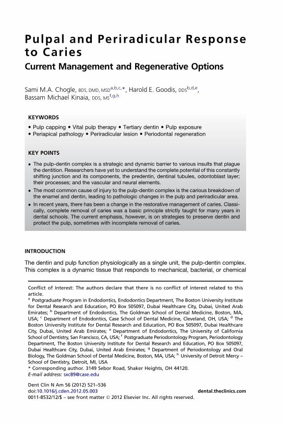
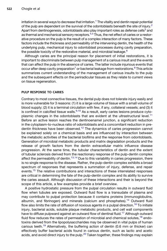
Additionally, the pulp-dentin complex may react to stimuli with tertiary dentin forma-tion. Unlike primary and secondary dentinogenesis, tertiary dentinogenesis isa focused reaction in the vicinity of the dentin that is directly affected by the cariesprocess. Recent reports have redefined tertiary dentinogenesis in relation to the natureof the injury. The term, reactionary dentinogenesis, has been adopted to describe thesecretion of a tertiary dentinmatrix by primary odontoblasts that have survived injury tothe tooth (Fig. 1). This is a wound healing reaction to produce circumpulpal dentin inresponse to slowly progressing dentinal caries.29 Conversely, reparative dentinogen-esis refers to the secretion of tertiary dentin after the death of the primary odontoblastsunderlying the injury, after the differentiation of odontoblast-like cells (Fig. 2).15,30
Reparative dentin formation occurs in response to deep dentinal caries and representsamore complex sequence of biologic events compared to reactionary dentinogenesis,including progenitor cell recruitment and differentiation.31 When the pulp is exposed inadvanced lesions, reparative dentinogenesis results in dentin bridge formation, whichrestores the functional integrity of the pulp-dentin complex. In actively progressingadvanced lesions involving pulpal exposure, however, the inflammatory reactionsmay become acute and uncontrolled as bacteria approach and penetrate the pulp.Although inflammation is regarded as a defense response, severe reactions can resultfrom continued ingress of bacteria, producing irreversible destruction of the pulp thateventually results in pulpal necrosis and development of periradicular lesions.32
VITAL PULP THERAPY
Vital pulp therapy is aimed at sealing the pulp after injury and stimulating the formationof tertiary dentin.33 This can be achieved through direct and indirect pulp capping,pulpotomy, and other therapies that protect the pulp from the chemical, bacterial,mechanical, and thermal insults due to attrition, erosion, caries, restoration
Fig. 1. Illustration of a tooth with caries involving the superficial layer of dentin. Bioactivemolecules are released by cariously involved dentin (a), which increase secretory activity ofodontoblasts and result in deposition of reactionary dentin (b).
Chogle et al524
procedures, and restoration placement.34 The dental pulp, when exposed, mayrespond favorably to application of a variety of materials used in pulp capping proce-dures.35 Many studies have confirmed the formation of hard tissue over the site of theexposure.36–40 This may demonstrate that the dental pulp has an intrinsic capacity toheal. The clinical outcomes differ, however, in their inferences as to the predictabilityof hard tissue formation. The factors affecting the outcome of pulpal capping proce-dures may be categorized broadly as those related to the pulp exposure and the mate-rial used to seal the exposure.
Fig. 2. Illustration of a tooth with caries involving the pulp-dentin complex leading to deathof underlying odontoblasts. Bioactive molecules are released by cariously involved dentin (a)into the subjacent pulp and cause proliferation/differentiation of precursor cells (c). Theseodontoblast-like cells migrate to site of pulpal injury and deposit reparative dentinmatrix (b).
Pulpal Injury and Current Management Options 525
Indirect Pulp Treatment
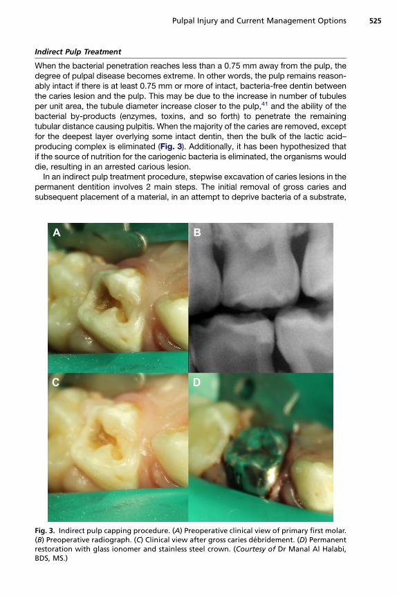
When the bacterial penetration reaches less than a 0.75 mm away from the pulp, thedegree of pulpal disease becomes extreme. In other words, the pulp remains reason-ably intact if there is at least 0.75 mm or more of intact, bacteria-free dentin betweenthe caries lesion and the pulp. This may be due to the increase in number of tubulesper unit area, the tubule diameter increase closer to the pulp,41 and the ability of thebacterial by-products (enzymes, toxins, and so forth) to penetrate the remainingtubular distance causing pulpitis. When the majority of the caries are removed, exceptfor the deepest layer overlying some intact dentin, then the bulk of the lactic acid–producing complex is eliminated (Fig. 3). Additionally, it has been hypothesized thatif the source of nutrition for the cariogenic bacteria is eliminated, the organisms woulddie, resulting in an arrested carious lesion.In an indirect pulp treatment procedure, stepwise excavation of caries lesions in the
permanent dentition involves 2 main steps. The initial removal of gross caries andsubsequent placement of a material, in an attempt to deprive bacteria of a substrate,
Fig. 3. Indirect pulp capping procedure. (A) Preoperative clinical view of primary first molar.(B) Preoperative radiograph. (C) Clinical view after gross caries debridement. (D) Permanentrestoration with glass ionomer and stainless steel crown. (Courtesy of Dr Manal Al Halabi,BDS, MS.)
Chogle et al526
prevent a direct carious pulpal exposure, and remineralize the remaining caries lesion,with a subsequent return to normal tissuepH.42 In vitro studies of severely carious teeth,however, found that clinical observationsof dentin color changesandmineral increasesin the remaining carious dentin do not always represent a change in the bacterialcontent.43 Although the microbiologic bioburden may be reduced, it is still present. Ina clinical setting, retaining a layer of carious dentin (indirect procedures) presents thedilemmamost clinicians face in deciding how to treat these lesions.Continued researchand clinical trials are needed to develop the appropriate case selection guidelines,treatment approaches, and materials needed to maximize clinical success.
Pulp Exposure
The classic studies of Kakehashi and colleagues44 demonstrate bacterial infection asa critical etiologic factor for pulpal necrosis. The extent of damage from microbialcontamination may vary based on the size and chronicity of the exposure, pulp status,and material used to seal the exposure.Several studies suggest that the size of the pulpal exposure may influence case
selection because large pulpal exposures may have greater risk of microleakageand be difficult to restore.45,46 ppartial pulpotomies after traumatic crown fractures,however, have demonstrated a 96% success rate with close to 3-year follow-up,including pulpal exposures ranging from 0.5 mm to 4.0 mm.47 Thus, the size of theexposure may not play a major role, at least within this range.The duration of pulpal contamination, although important, remains a controversial
factor in terms of successful pulp capping. Many clinicians believe that longer periodsof contamination by oral microorganisms and debris reduce the chance of success.This is supported by results from animal studies that indicate that the success ofCa(OH)2 pulp capping is reduced from 93% to 56% when microbial contaminationis extended from 1 hour to 7 days.48–50 Alternatively, clinical studies in youngerpatients with up to 3 months of pulp exposure demonstrate a 93% radiographicsuccess rate for partial pulpotomy at a mean follow-up of 4.5 years.47,51 As a result,the superficial pulp in younger patients seems more resistant to bacterial invasionthan the mature pulp in older patients. Furthermore, the size of the pulp chamberand root canal systems of younger patients mitigates toward a larger volume of pulpaltissue, hence greater success in younger patients.The inflammatory response of the pulp to bacteria and their by-products and the
trauma of caries removal may increase the amount of bleeding of exposed dentalpulp tissue. This can adversely affect the effective seal against bacterial invasionand lead to development of a chronic inflammatory infiltrate and inhibition of tertiarydentin formation. Therefore, the use of a hemostatic agent may be useful in vitalpulp procedures to clot the capillaries within the subjacent pulp tissue.46 Severalstudies have examined the use of hemostatic agents placed over the exposure tohalt hemorrhage with conflicting results, such as sodium hypochlorite, ferric sulfate,and chlorhexidine digluconate.52–55 Further work should better define the use of theseagents, especially when used in combination with other materials, such as mineraltrioxide aggregate (MTA), that is now suggested for these procedures.
Materials of Pulp Capping
For a successful outcome in vital pulp therapy, the healing response must demon-strate rapid hard tissue formation at the pulp-material interface with minimal inflamma-tory response. This desirable healing process should occur when any substance isapplied directly to the pulpal exposure site that is capable of stimulating dentinogen-esis.56 One study using a cell culture model system reported that calcium hydroxide
Pulpal Injury and Current Management Options 527
(Ca[OH]2)inhibited macrophage function and reduced inflammatory reactions whenused in direct pulp capping and pulpotomy procedures.57 In another study, an adhe-sive system applied to exposed human pulp tissue caused large areas of neutrophilinfiltration and death of odontoblasts, thereby inhibiting pulp repair.58 Together, thesestudies suggest that the sealing ability of the agent, the method of placement (eg,minimizing the impaction of pulp capping agents in dental pulp), and the chemicalnature of the pulp capping material are all critical factors in desirable pulpal healing.
Calcium hydroxideThe introduction of Ca(OH)2, from a historical perspective, played an important role inthe development of vital pulp therapy. Recently, it has been shown that calcium ionsreleased from Ca(OH)2 stimulate fibronectin synthesis by dental pulp cells, which inturn may induce the differentiation of pulp progenitor cells into mineralized tissue-producing phenotypes.59 Cross-sections of pulps treated for more than 6 weeksdemonstrated a superior amorphous layer of tissue debris and Ca(OH)2, a middle layerof a coarse meshwork of fibers identified as fibrodentin, and an inner layer showingtubular osteodentin.60 Apart from the ability to form a dentin bridge in the subjacentpulp tissue, Ca(OH)2 has demonstrated additional benefits, such as antimicrobialcharacteristics. In a primate study with a 1-year to 2-year follow-up, Ca(OH)2-induceddentin bridge formation occurred in 78 of 91 (85%) exposed and contaminated dentalpulps, whereas 10% of the pulps in the study sample became necrotic. Despite thesuccessful use of Ca(OH)2 as a pulp capping agent for 60 years,61 predictableoutcomes remain a problem. For example, a retrospective study that examinedCa(OH)2 pulp capping of carious exposures in 123 teeth, revealed that 45% failed inthe 5-year group and 80% failed in the 10-year group.62 Additionally a summary ofseveral primate studies involving direct pulp capping with Ca(OH)2 reported severalinflamed and infected pulps after a follow-up period of 1 years to 2 years.63 The inves-tigators questioned the long-term efficacy of commercially available Ca(OH)2 bases,particularly in light of the potential for microleakage.63 This has led to newer studiescomparing it with other materials. One such material, MTA, has generated greatinterest for direct pulp capping and vital pulp therapy.
Mineral trioxide aggregateMTA is the material of choice for correcting procedural errors as well as for root-endfilling material in apicoectomy procedures. In both instances, the material has beenshown e tissue compatible, encouraging the formation of new cementum-like hardtissue with restoration of the periodontal ligament, and is considered to have signifi-cant osteogenic potential.64–66 MTA is currently the alternative material for Ca(OH)2in direct pulp capping procedures (Fig. 4). MTA was compared with Ca(OH)2 in youngpermanent teeth undergoing apexogenesis (coronal pulpotomy, retention of rootsystem vital pulp tissue, and immature root formation).67 Two of 14 teeth in theCa(OH)2 group failed because of pain and swelling, whereas all in the MTA groupseemed successfully treated. When MTA and Ca(OH)2 were compared in directpulp capping procedures in dog teeth,68 MTA presented a higher success rate thanCa(OH)2, with a lower occurrence of infection and pulpal necrosis. A more recentrandomized clinical trial compared the pulpal responses with iatrogenic pulpotomyperformed in healthy human teeth using MTA or Dycal.69 Pulpal wounds treatedwithMTAweremostly free from inflammation after 1 week and coveredwith a compacthard tissue barrier within 3 months whereas teeth treated with Dycal revealeddistinctly less consistent formation of a hard tissue barrier and presence of pulpalinflammation at up to 3 months. Collectively, the results of all these studies indicate
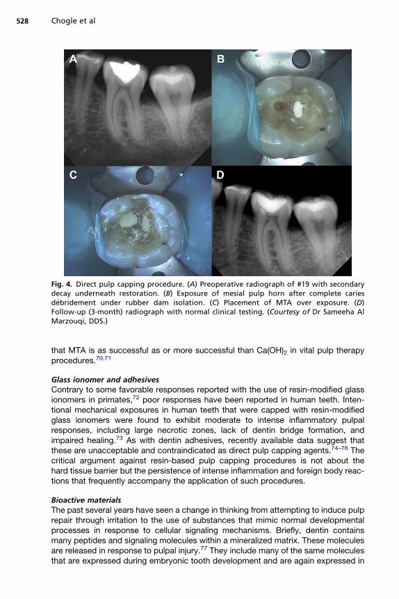
Fig. 4. Direct pulp capping procedure. (A) Preoperative radiograph of #19 with secondarydecay underneath restoration. (B) Exposure of mesial pulp horn after complete cariesdebridement under rubber dam isolation. (C) Placement of MTA over exposure. (D)Follow-up (3-month) radiograph with normal clinical testing. (Courtesy of Dr Sameeha AlMarzouqi, DDS.)
Chogle et al528
that MTA is as successful as or more successful than Ca(OH)2 in vital pulp therapyprocedures.70,71
Glass ionomer and adhesivesContrary to some favorable responses reported with the use of resin-modified glassionomers in primates,72 poor responses have been reported in human teeth. Inten-tional mechanical exposures in human teeth that were capped with resin-modifiedglass ionomers were found to exhibit moderate to intense inflammatory pulpalresponses, including large necrotic zones, lack of dentin bridge formation, andimpaired healing.73 As with dentin adhesives, recently available data suggest thatthese are unacceptable and contraindicated as direct pulp capping agents.74–76 Thecritical argument against resin-based pulp capping procedures is not about thehard tissue barrier but the persistence of intense inflammation and foreign body reac-tions that frequently accompany the application of such procedures.
Bioactive materialsThe past several years have seen a change in thinking from attempting to induce pulprepair through irritation to the use of substances that mimic normal developmentalprocesses in response to cellular signaling mechanisms. Briefly, dentin containsmany peptides and signaling molecules within a mineralized matrix. These moleculesare released in response to pulpal injury.77 They include many of the same moleculesthat are expressed during embryonic tooth development and are again expressed in
Pulpal Injury and Current Management Options 529
dental tissues in response to pathologic conditions.78 Because the pulp-dentincomplex demonstrates great regenerative potential, a suitable bioactive substancecould recruit a population of multipotent mesenchymal progenitor cells to producenew hard tissue. Investigations of these bioactive substances, although in theirinfancy, include the roles of stem cells and genetic recruitment. These strategiesinvolve selective activation of genes and other proteins necessary in dentinogenesisand can generate new, biologically based approaches to pulpal healing.31
PERIRADICULAR DISEASE
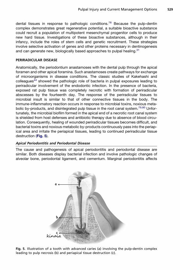
Anatomically, the periodontium anastamoses with the dental pulp through the apicalforamen and other apical foramina. Such anastamoses create pathways for exchangeof microorganisms in disease conditions. The classic studies of Kakehashi andcolleagues44 showed the pathologic role of bacteria in pulpal exposures leading toperiradicular involvement of the endodontic infection. In the presence of bacteria,exposed rat pulp tissue was completely necrotic with formation of periradicularabscesses by the fourteenth day. The response of the periradicular tissues tomicrobial insult is similar to that of other connective tissues in the body. Theimmune-inflammatory reaction occurs in response to microbial toxins, noxious meta-bolic by-products, and disintegrated pulp tissue in the root canal system.79,80 Unfor-tunately, the microbial biofilm formed in the apical end of a necrotic root canal systemis shielded from host defenses and antibiotic therapy due to absence of blood circu-lation. Consequently, healing of wounded periradicular tissues becomes difficult, andbacterial toxins and noxious metabolic by-products continuously pass into the periap-ical area and irritate the periapical tissues, leading to continued periradicular tissuedestruction (Fig. 5).
Apical Periodontitis and Periodontal Disease
The cause and pathogenesis of apical periodontitis and periodontal disease aresimilar. Both diseases display bacterial infection and involve pathologic changes ofalveolar bone, periodontal ligament, and cementum. Marginal periodontitis affects
Fig. 5. Illustration of a tooth with advanced caries (a) involving the pulp-dentin complexleading to pulp necrosis (b) and periapical tissue destruction (c).
Chogle et al530
the coronal periodontal tissues, whereas apical periodontitis affects apical periodontaltissues. Bone loss is one of characteristic feature with crestal bone loss in periodontaldisease and apical resorption in apical periodontitis. Apical periodontitis, however,does not have a direct communication with the oral cavity and the main source ofpathogenesis originates through the root canal system. Periodontal disease, alterna-tively, has a direct communication with the microflora of the oral cavity, making it moredifficult to isolate.81 The intimate relationship between the root canal system and theperiodontium results in multiple pathways connecting the pulpal and the periodontaltissues where microbial by-products may affect neighboring tissues. A primaryendodontic lesion may be complicated with secondary periodontal involvement ora concomitant periodontal disease on the same tooth.82,83 Even though pathologyinvolving pulpal and/or periodontal tissues may be separate disease entities, eachprimary disease may mimic characteristics as well as etiologically influence theprogression of the other, often making diagnosis and treatment planning in such casesa challenge for clinicians. An interdisciplinary approach is often used to establish theappropriate treatment plan.
Management and Treatment Options
Despite the location (apically, crestally, or a combination) of the diseased tissues,treatment options aim to repair and regenerate the lost structures. Various treatmentmethods have been proposed, ranging from the use of bone grafts,84 guided tissueregeneration (GTR),85,86 molecular biologic agents (enamel matrix derivatives),87 andgrowth and differentiation factors88,89 to the promising use of stem cells90,91 (seethe article elsewhere in this issue by Sedgley and colleagues). Bone grafts generallyresulted in repair that is healing of a wound by tissues that do not fully restore thearchitecture or the function of the lost part (ie, healing by long junctional epithelium).92
GTR, enamel matrix derivatives, and growth and differentiation factors heal by regen-eration that constitutes the reproduction of a lost or injured part. GTR is a procedurethat attempts to regenerate lost periradicular structures through differential tissueresponses by using a barrier (Fig. 6).93 Therefore, restoration of the periodontal appa-ratus occurs by regeneration of connective tissue, cementum, and bone rather thanepithelium. Healing by regeneration is favorable compared with repair, because thegoal of periodontal therapy is to restore the periodontal tissues to their original biologicstructure. Although many materials have been used to regenerate the lost structures,the concept of GTR remains the major principle in the treatment methodology usedtoday to manage periodontal-endodontic tissue/bony defects.
Guided Tissue Regeneration
GTR constitutes the reproduction of a lost or injured tissue. Melcher94 suggested that4 different cell types dictate the type of periodontal healing that occurs. These cellsoriginate from the gingival epithelial tissue, lamina propria of connective tissue, alve-olar bone, and periodontal ligament. Cells derived from periodontal ligament and alve-olar bone have the potential to heal by true regeneration compared with cells from thelamina propria of gingiva or gingival epithelial tissue. Understanding barrier-mediatedselective cell repopulation gave rise to the concept of epithelial exclusion to restorelost periodontal tissue and obtain new attachments. In periodontal disease, the crestalregion is primarily affected because it is exposed to the microflora of the oral cavity.The use of a barrier is important to isolate the periodontal defect from the soft tissuesand allow adequate space and time for periodontal ligament and alveolar bone cells toreconstitute the lost periodontal attachment apparatus. GTR therapy in intrabony andfurcation defects has been used with favorable results.85,86
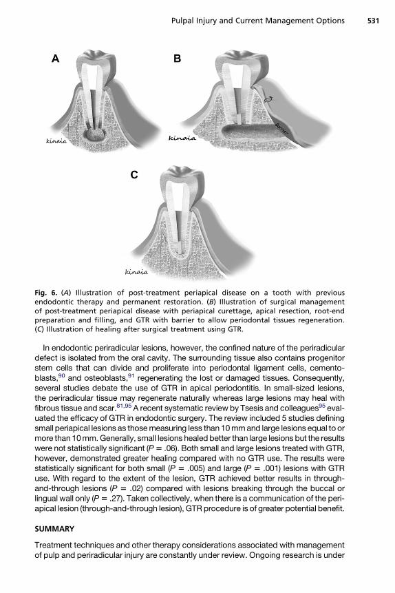
Fig. 6. (A) Illustration of post-treatment periapical disease on a tooth with previousendodontic therapy and permanent restoration. (B) Illustration of surgical managementof post-treatment periapical disease with periapical curettage, apical resection, root-endpreparation and filling, and GTR with barrier to allow periodontal tissues regeneration.(C) Illustration of healing after surgical treatment using GTR.
Pulpal Injury and Current Management Options 531
In endodontic periradicular lesions, however, the confined nature of the periradiculardefect is isolated from the oral cavity. The surrounding tissue also contains progenitorstem cells that can divide and proliferate into periodontal ligament cells, cemento-blasts,90 and osteoblasts,91 regenerating the lost or damaged tissues. Consequently,several studies debate the use of GTR in apical periodontitis. In small-sized lesions,the periradicular tissue may regenerate naturally whereas large lesions may heal withfibrous tissue and scar.81,95 A recent systematic review by Tsesis and colleagues95 eval-uated the efficacy of GTR in endodontic surgery. The review included 5 studies definingsmall periapical lesions as thosemeasuring less than 10mmand large lesions equal to ormore than10mm.Generally, small lesions healed better than large lesions but the resultswere not statistically significant (P5 .06). Both small and large lesions treated with GTR,however, demonstrated greater healing compared with no GTR use. The results werestatistically significant for both small (P 5 .005) and large (P 5 .001) lesions with GTRuse. With regard to the extent of the lesion, GTR achieved better results in through-and-through lesions (P 5 .02) compared with lesions breaking through the buccal orlingual wall only (P5 .27). Taken collectively, when there is a communication of the peri-apical lesion (through-and-through lesion), GTR procedure is of greater potential benefit.
SUMMARY
Treatment techniques and other therapy considerations associated with managementof pulp and periradicular injury are constantly under review. Ongoing research is under
Chogle et al532
way to better understand intricacies related to caries, pulp, and periradicularresponses. Significant advances in understanding of the molecular basis of the pulpaland periradicular injury and healing response should lead to significant new, biologi-cally based pulp therapies. In this issue, readers can gain insight into clinical practicesand techniques, current understanding, and future directions for regenerative medi-cine for the field of endododontics.
REFERENCES
1. Goldberg M, Farges JC, Lacerda-Pinheiro S, et al. Inflammatory and immunolog-ical aspects of dental pulp repair. Pharmacol Res 2008;58:137–47.
2. About I, Murray PE, Franquin JC, et al. The effect of cavity restoration variables onodontoblast cell number and dental repair. J Dent 2001;29:109–17.
3. Farges JC, Keller JF, Carrouel F, et al. Odontoblasts in the dental pulp immuneresponse. J Exp Zool B Mol Dev Evol 2009;312B:425–36.
4. Magloire H, Couble ML, Thillichon$Prince B, et al. Odontoblast: a mechano-sensory cell. J Exp Zool B Mol Dev Evol 2009;312B:416–24.
5. Son AR, Yang YM, Hong JH, et al. Odontoblast TRP channels and thermo/mechanical transmission. J Dent Res 2009;88:1014–9.
6. Mjor IA, Odont D. Pulp-dentin biology in restorative dentistry. Part 2: initial reac-tions to preparation of teeth for restorative procedures. Quintessence Int 2001;32:537–51.
7. Wisithphrom K, Murray Pf, About I, et al. Interactions between cavity preparationand restoration events and their effects on pulp vitality. Int J Periodontics Restor-ative Dent 2006;26:596–605.
8. Bergenholtz G. Evidence for bacterial causation of adverse pulpal responses inresin-based dental restorations. Crit Rev Oral Biol Med 2000;11:467–80.
9. Stanley HR. Pulpal response to dental techniques and materials. Dent Clin NorthAm 1971;15:115–26.
10. Kim S. Microcirculation of the dental pulp in health and disease. J Endod 1985;11:465–71.
11. Magloire H, Joffre A, Couble ML, et al. Ultrastructural alterations of human odon-toblasts and collagen fibers in the pulpal border zone beneath early carieslesions. Cell Mol Biol 1981;27:437–43.
12. Bjørndal L, Darvann T, Thylstrup A. A quantitative light microscopic study of theodontoblast and subodontoblastic reactions to active and arrested enamel carieswithout cavitation. Caries Res 1998;32:59–69.
13. Mjor IA. Dentin permeability: the basis for understanding pulp reactionsandadhesive technology. Braz Dent J 2009;20:3–16.
14. Pashley DH, Pashley H, Carvalho RM, et al. The effects of dentin permeability onrestorative dentistry. Dent Clin North Am 2002;46:211–45.
15. Smith AJ. Pulpal responses to caries and dental repair. Caries Res 2002;36:223–32.
16. Puapichartdumrong P, Ikeda H, Suda H. Outward fluid flow reduces inward diffu-sion of bacterial lipopolysaccharide across intact and demineralised dentine.Arch Oral Biol 2005;50:707–13.
17. Hahn CL, Liewehr FR. Relationships between caries bacteria, host responsesand clinical signs and symptoms of pulpitis. J Endod 2007;33:213–9.
18. Pashley DH, Matthews WG. The effects of outward forced convective flow oninward diffusion in human dentine in vitro. Arch Oral Biol 1993;38:577–82.
Pulpal Injury and Current Management Options 533
19. Khabbaz MG, Anastasiadis PL, Sykaras SN. Determinat ion of endotoxins in thevital pulp of human carious teeth: association with pulpal pain. Oral Surg OralMed Oral Pathol 2001;91:587–93.
20. Camps J, Pashley DH. Buffering action of human dentin in vitro. J Adhes Dent2000;2:39–50.
21. Brannstrom M, Lind PO. Pulpal response to early dental caries. J Dent Res 1965;44:1045–50.
22. Hahn CL, Liewehr FR. Innate immune responses of the dental pulp to caries.J Endod 2007;33:643–51.
23. Hahn CL, Liewehr FR. Update on the adaptive immune responses of the dentalpulp. J Endod 2007;33:773–81.
24. Trowbridge HO. Pathogenesis of pulpitis resulting from dental caries. J Endod1981;7:52–60.
25. Reeves R, Stanley HR. The relationship of bacterial penetration and pulpalpathosis in carious teeth. Oral Surg Oral Med Oral Pathol 1966;22:59–65.
26. Bjørndal L. The caries process and its effect on the pulp: the science is changingand so is our understanding. J Endod 2008;34(Suppl 7):52–5.
27. Zheng L, Hilton JF, Habelitz S, et al. Dentin caries activity status related to hard-ness and elasticity. Eur J Oral Sci 2003;111:243–52.
28. Bjørndal L, Mjor IA. Pulp-dentin biology in restorative dentistry, Part 4: dentalcaries-characteristics of lesions and pulpal reactions. Quintessence Int 2001;32:717–36.
29. Duque C, Hebling J, Smith AJ, et al. Reactionary dentinogenesis after app lyingrestorative materials and bioactive dentin matrix molecules as liners in deep cavi-ties prepared in nonhuman primate teeth. J Oral Rehabil 2006;33:452–61.
30. Tecles O, Laurent P, Zygouritsas S, et al. Activation of human dental pulpprogenitor/stem cells in response to odontoblast injury. Arch Oral Biol 2005;50:103–8.
31. Sloan AJ, Smith AJ. Stem cells and the dental pulp: potential roles in dentineregeneration and repair. Oral Dis 2007;13:151–7.
32. Bergenholtz G. Pathogenic mechanisms in pulpal disease. J Endod 1990;16:98–101.
33. Tziafas D, Smith AJ, Lesot H. Designing new treatment strategies in vital pulptherapy. J Dent 2000;28:77–92.
34. Burke FM, Samarawickrama DY. Progressive changes in the pulpo-dentinalcomplex and their clinical consequences. Gerodontology 1995;12:57–66.
35. Tjaderhane L. The mechanism of pulpal wound healing. Aust Endod J 2002;28:68–74.
36. CvekM, Cleaton-Jones PE, Austin JC, et al. Pulp reactions to exposure after exper-imental crown fractures or grinding in adult monkeys. J Endod 1982;8:391–7.
37. Zander HA, Glass RL. The healing of phenolized pulp exposure. Oral Surg OralMed Oral Pathol 1949;2:803–10.
38. Mastenon JB. Inherent healing potential of the dental pulp. Br Dent J 1966;120:430–6.
39. Torneck CD, Moe H, Howley TP. The effect of calcium hydroxide on porcine pulpfibroblasts in vitro. J Endod 1983;9:131–5.
40. Cox CF, Bergenholtz G, Fitzgerald M, et al. Capping of the dental pulp mechan-ically exposed to the oral microflora-A 5-week observation of wound healing in themonkey. J Oral Pathol 1982;11:327–39.
41. CvekM. A clinical report on partial pulpotomy and capping with calcium hydroxidein permanent incisors with complicated crown fracture. J Endod 1978;4:232–7.
Chogle et al534
42. Mejilre I, Cvek M. Partial pulpotomy in young permanent teeth with deep cariouslesions. Endod Dent Traumatol 1993;9:238–42.
43. Isermann GT, Kaminski EJ. Pulpal response to minimal exposure in presence ofbacteria and Dycal. J Endod 1979;5:322–7.
44. Kakehashi S, Stanley HR, Fitzgerald RJ. The effects of surgical exposures ofdental pulps in germ-free and conventional laboratory rats. Oral Surg Oral MedOral Pathol 1965;20:340–9.
45. Segura JJ, Llamas R, Rubio-Manzanares AJ, et al. Calcium hydroxide inhibitssubstrate adherence capacity of macrophages. J Endod 1997;23:444–7.
46. Hebling J, Giro EM, Costa CA. Biocompatibility of an adhesive system applied toexposed human dental pulp. J Endod 1999;25:676–82.
47. Hafez AA, Cox CF, Tarim B, et al. An in vivo evaluation of hemorrhage control usingsodium hypochlorite and direct capping with a one- or two- component adhesivesystem in exposed nonhuman primate pulps. Quintessence Int 2002;33:261–72.
48. Accorinte Mde L, Loguercio AD, Reis A, et al. Response of human pulp cappedwith a bonding agent after bleeding control with hemostatic agents. Oper Dent2005;30:147–55.
49. Accorinte Mde L, Loguercio AD, Reis A, et al. Effects of hemostatic agents on thehistomorphologic response of human dental pulp capped with calciumhydroxide. Quintessence Int 2007;38:843–52.
50. Silva AF, Tarquinio SB, Demarco FF, et al. The influence of haemostatic agents onhealing of healthy human dental pulp tissue capped with calcium hydroxide. IntEndod J 2006;39:309–16.
51. Pitt Ford TR. Pulpal response to Procal for capping exposures in dog’s teeth. J BrEndod Soc 1979;12:67–72.
52. Pitt Ford TR. Pulpal response to a calcium hydroxide material for capping expo-sures. Oral Surg Oral Med Oral Pathol 1985;59:194–7.
53. Brannstrom M, Nyborg H, Stromberg T. Experiments with pulp capping. OralSurg Oral Med Oral Pathol 1979;48:347–52.
54. Heys DR, Cox CF, Heys RJ, et al. Histological considerations of direct pulpcapping agents. J Dent Res 1981;60:1371–9.
55. Cox CF, Bergenholtz G, Heys DR, et al. Pulp capping of dental pulp mechanicallyexposed to oral microflora: a 1-2 year observation of wound healing in themonkey. J Oral Pathol 1985;14:156–68.
56. Stanley HR. Criteria for standardizing and increasing credibility of direct pulpcapping studies [special issue]. Am J Dent 1998;11:S17–34.
57. Franz FE, Holz J, Baume LJ. Ultrastructure (SEM) of dentine bridging in thehuman dental pulp. J Biol Buccale 1984;12:239–46.
58. Nair PN, Duncan HF, Pitt Ford TR, et al. Histological, ultrastructural and quantita-tive investigations on the response of healthy human pulps to experimentalcapping with mineral trioxide aggregate: a randomized controlled trial. Int EndodJ 2008;41:128–50.
59. Cox CF, Subay RK, Ostro E, et al. Tunnel defects in dentin bridges: their formationfollowing direct pulp capping. Oper Dent 1996;21:4–11.
60. Ziafras D, Belibasakis G, Veis A, et al. Dentin regeneration in vital pulp therapy:design principles. Adv Dent Res 2001;15:96–100.
61. Barthel CR, Rosenkranz B, Leuenberg A, et al. Pulp capping of carious expo-sures: treatment outcome after 5 and 10 years: a retrospective study. J Endod2000;26:525–8.
62. Murray PE, Garcia-Godoy F. The incidence of pulp healing defects with directcapping materials. Am J Dent 2006;19:171–7.
Pulpal Injury and Current Management Options 535
63. do Nascimento AB, Fontana UF, Teixeria HM, et al. Biocompatibility of a resin-modified glass-ionomer cement applied as pulp capping in human teeth. Am JDent 2000;13:28–34.
64. Accorinte ML, Loguercio AD, Reis A, et al. Response of human pulps cappedwith different self-etch adhesive systems. Clin Oral Investig 2008;12:119–27.
65. Cui C, Zhou X, Chen X, et al. The adverse effect of self-etching adhesivesystems on dental pulp after direct pulp capping. Quintessence Int 2009;40:e26–34.
66. Bas‚ ak F, Vural IM, Kaya E, et al. Vasorelaxant effect of a self-etch adhesivesystem through calcium antagonistic action. J Endod 2008;34:1202–6.
67. Salako N, Joseph B, Ritwik P, et al. Comparison of bioactive glass, mineraltrioxide aggregate, ferric sulfate, and formocresol as pulpotomy agents in ratmolar. Dent Traumatol 2003;19:314–20.
68. EI-Meligy OA, Avery DR. Comparison of mineral trioxide aggregate andcalcium hydroxide as pulpotomy agents in young permanent teeth (apexogene-sis). Pediatr Dent 2006;28:399–404.
69. Witherspoon DE. Vital pulp therapy with new materials: new directions and treat-ment perspectives-Permanent teeth. J Endod 2008;34(Suppl 7):S25–8.
70. Bogen G, Kim JS, Bakland LK. Direct pulp capping with mineral trioxide aggre-gate: an observational study. J Am Dent Assoc 2008;139:305–15.
71. Silva TA, Rosa AL, Lara VS. Dentin matrix proteins and soluble factors: intrinsicregulatory signals for healing and resorption of dental and periodontal tissues.Oral Dis 2004;10:63–74.
72. About I, Mitsiadis TA. Molecular aspects of tooth pathogenesis and repair: in vivoand in vitro models. Adv Dent Res 2001;15:59–62.
73. Marshall GW Jr, Marshall SJ, Kinney JH, et al. The dentin substrate: structure andproperties related to bonding. J Dent 1997;25:441–58.
74. Cox CF, Halez AA, Akimoto N, et al. Biocompatibility of primer, adhesive and resincomposite systems on non-exposed and exposed pulps on non-human primateteeth [special issue]. Am J Dent 1998;11:S55–63.
75. Bonecker M, Toi C, Cleaton-Jones P. Mutans Streptococci and lactobacilli incarious dentin before and after a traumatic restorative treatment. J Dent 2003;31:413–28.
76. Washington JT, Schneiderman E, Spears R, et al. Biocompatibility and osteo-genic potential of new generation endodontic materials established by usingprimary osteoblasts. J Endod 2011;37:1166–70.
77. Zairi A, Lambrianidis T, Pantelidou O, et al. Periradicular tissue responses to bio-logically active molecules or MTA when applied in furcal perforation of dogs’teeth. Int J Dent 2012;2012:257832.
78. Al-Hiyasat AS, Al-Sa’Eed OR, Darmani H. Quality of cellular attachment to variousroot-end filling materials. J Appl Oral Sci 2012;20:82–8.
79. Moller AJ, Fabricius L, Dahlen G, et al. Influence on periapical tissues of indige-nous oral bacteria and necrotic pulp tissue in monkeys. Scand J Dent Res 1981;89:475–84.
80. Fabricius L, Dahlen G, Ohman AE, et al. Predominant indigenous oral bacteriaisolated from infected root canals after varied times of closure. Scand J DentRes 1982;90:134–44.
81. Lin L, Chen M, Ricucci D, et al. Guided tissue regeneration in periapical surgery.J Endod 2010;36:618–25.
82. Simon JH, Glick DH, Frank AL. The relationship of endodontic-periodonticlesions. J Periodontol 1972;43:202–8.
Chogle et al536
83. Rotstein I, Simon JH. Diagnosis, prognosis and decision-making in the treat-ment of combined periodontal-endodontic lesions. Periodontol 2000 2004;34:165–203.
84. Schallhorn RG. Present status of osseous grafting procedures. J Periodontol1977;48(9):570–6.
85. Murphy KG, Gunsolley JC. Guided tissue regeneration for the treatment of peri-odontal intrabony and furcation defects. A systematic review. Ann Periodontol2003;8(1):266–302.
86. Kinaia BM, Steiger J, Neely AL, et al. Treatment of class II molar furcation involve-ment: meta-analyses of reentry results. J Periodontol 2011;82(3):413–28.
87. Esposito M, Grusovin M, Papanikolaou N, et al. Enamel matrix derivative (Emdo-gain) for periodontal tissue regeneration in intrabony defects. A Cochranesystematic review. Eur J Oral Implantol 2009;2:247–66.
88. Howell TH, Fiorellini JP, Paquette DW, et al. A phase I/II clinical trial to evaluatea combination of recombinant human platelet-derived growth factor-BB andrecombinant human insulin-like growth factor-I in patients with periodontaldisease. J Periodontol 1997;68(12):1186–93.
89. Sigurdsson TJ, Lee MB, Kubota K, et al. Periodontal repair in dogs: recombinanthuman bone morphogenetic protein-2 significantly enhances periodontal regen-eration. J Periodontol 1995;66(2):131–8.
90. Gronthos S, Zannettino AC, Hay SJ, et al. Molecular and cellular characterizationof highly purified stromal stem cells derived from human bone marrow. J Cell Sci2003;116:1827–35.
91. Seo BM, Miura M, Gronthos S, et al. Investigation of multipotent postnatal stemcells from human periodontal ligament. Lancet 2004;364(9429):149–55.
92. The American Academy of Periodontology. Glossary of periodontal terms. 4thedition. Chicago: The American Academy of Periodontology; 2001. p. 47.
93. The American Association of Endodontics. Glossary of endodontic terms. 7thedition. Chicago: The American Academy of Periodontology; 2003. p. 26.
94. Melcher AH. On the repair potential of periodontal tissues. J Periodontol 1976;47(5):256–60.
95. Tsesis I, Rosen E, Tamse A, et al. Effect of guided tissue regeneration on theoutcome of surgical endodontic treatment: a systematic review and meta-anal-ysis. J Endod 2011;37(8):1039–45.