putting structure into context: fitting of atomic models into electron microscopic and electron...
TRANSCRIPT
Putting structure into context: fitting of atomic models intoelectron microscopic and electron tomographic reconstructionsNiels Volkmann
Available online at www.sciencedirect.com
A complete understanding of complex dynamic cellular
processes such as cell migration or cell adhesion requires the
integration of atomic level structural information into the larger
cellular context. While direct atomic-level information at the
cellular level remains inaccessible, electron microscopy,
electron tomography and their associated computational
image processing approaches have now matured to a point
where sub-cellular structures can be imaged in three
dimensions at the nanometer scale. Atomic-resolution
information obtained by other means can be combined with
this data to obtain three-dimensional models of large
macromolecular assemblies in their cellular context. This article
summarizes some recent advances in this field.
Address
Sanford-Burnham Medical Research Institute, 10901 N Torrey Pines
Road, La Jolla, CA 92037, USA
Corresponding author: Volkmann, Niels ([email protected])
Current Opinion in Cell Biology 2012, 24:141–147
This review comes from a themed issue on
Cell structure and dynamics
Edited by Jason Swedlow and Gaudenz Danuser
Available online 5th December 2011
0955-0674/$ – see front matter
# 2011 Elsevier Ltd. All rights reserved.
DOI 10.1016/j.ceb.2011.11.002
IntroductionRecognition and cooperative interaction among molecules
in large assemblies are fundamental for dynamic processes
in living cells. Understanding of how these assemblies
work often requires structural information at the atomic
level. Nuclear magnetic resonance (NMR) spectroscopy
and X-ray crystallography are well-established approaches
for obtaining atomic structures of individual molecules and
domains. However, atomic structures of large macromol-
ecular assemblies remain more difficult to obtain with
these methods. These complexes can be too large to be
amenable to NMR and often exhibit a large degree of
flexibility that hampers crystallization attempts.
Electron microscopy has been a powerful tool for investi-
gating biological structures for several decades now, but
only recently steps towards achieving its full potential
have begun to come to fruition. High-resolution electron
microscopy studies of purified macromolecules are starting
www.sciencedirect.com
to rival X-ray crystallography in the resolution achievable
and it is now possible to image frozen hydrated cells and
tissue sections at close-to-native conditions at resolutions
that allow identification and analysis of molecular com-
ponents [1,2]. Technical advances in electron microscopy
equipment, in methods of specimen preparation, and in
computational image reconstruction methods have all been
essential in enabling the remarkable progress we have
witnessed in the last few years. It is now possible to achieve
resolutions of 0.5 nm or better not only from two-dimen-
sional crystals [3] or helically symmetrical objects [4], but
also from icosahedral virus particles [5] and even from
smaller, less symmetric particles [6��,7]. Electron tomogra-
phy, the most widely applicable method for obtaining
three-dimensional information by electron microscopy,
can now be combined efficiently with localization and
dynamics data from light microscopy [8,9�] and potentially
allows investigation of entire mammalian cells at molecular
resolution [10], paving the way for structure-based systems
biology [11].
Since the potential of the method has been realized, more
and more methods are emerging that target efficient incorp-
oration of atomic-level information into reconstructions
derived by electron microscopy. Here, we will describe
recent advances and open challenges in this field with an
emphasis on assembly reconstructions at intermediate
resolution (1–3 nm) and tomographic reconstructions of
eukaryotic cells as they relate to the actin cytoskeleton, a
major determinant of dynamic cellular processes such as
directed cell migration and focal adhesion dynamics.
Reconstructions of isolated cytoskeletalassembliesMany biological assemblies occur naturally in helical form,
particularly cytoskeleton filaments. These filamentous
structures are not usually amenable to crystallization due
to their natural tendency to polymerize. Image processing
of electron microscopy data can take advantage of the
helical symmetry and can, in principle, achieve near-atomic
resolution [4,12,13]. However, owing to the intrinsic flexi-
bility of filamentous actin [14], the structure determination
of actin-filament assemblies generally does not extended
into the subnanometer range. The notable exception to this
rule is the recent three-dimensional structure of rabbit
skeletal actin filaments, which was determined at a resol-
ution of better than 0.7 nm [15��]. Electron microscopy
reconstructions of actin filaments in complex with binding
partners most commonly fall into the 1.2–2.5 nm resolution
range. Examples of actin filaments with bound domains of
cytoskeletal proteins that were recently solved include
Current Opinion in Cell Biology 2012, 24:141–147
142 Cell structure and dynamics
actin in complex with tropomyosin [16], myosin binding
protein C [17,18], a-actinin [19], eps8 [20], drebrin [21],
coronin-1A [22], talin [23], and fimbrin [24]. Three-dimen-
sional structures of actin filaments crosslinked by villin [25]
and vinculin [26] as well as of arp2/3-mediated actin
branches [27] have been determined by electron tomogra-
phy at resolutions of about 2.5–3.5 nm.
Fitting of atomic models into intermediateresolution density mapsIn the 1–3 nm resolution range that is most generally
achievable for actin-based cytoskeletal assemblies, it is
possible to map individual subunits and thus to under-
stand the general architecture of the assemblies. This
intermediate resolution also gives a solid basis for fitting
high-resolution structures of smaller entities into the
reconstructions. The resulting models are often referred
to as ‘pseudo-atomic’ models to hint at the fact that the
accuracy of the atom positioning is of limited resolution.
This is an unfortunate and confusing term because the
models are built out of actual atoms and the term ‘pseudo
atom’ is often used to denote atom-like representations of
entire residues or other groups of atoms in coarse-grained
molecular modeling [28], direct phasing approaches
[29,30] or nuclear magnetic resonance calculations [31].
Until recently, correlation-based rigid-body fitting [32–34]
has beenthe mostcommonly usedtool toachieve the goalof
fitting high-resolution structures into reconstructions from
electron microscopy. Because of the increased availability
of subnanometer resolution reconstructions where second-
ary structural elements are often visible as rods (a-helices)
and sheets (b-sheets), significant efforts have been directed
towards developing ‘flexible’ fitting methods that allow the
high-resolution structures to be distorted in some way,
subject to different types of constraints, in order to improve
the fit with the density [35–49]. However, these flexible
fitting methods are not necessarily useful for resolutions
above the subnanometer range where secondary structural
elements are not discernible. Recent test calculations
indicate that the conceptually simpler modular rigid-body
fitting of domain structures often surpasses the achievable
accuracy of the models obtained by flexible fitting [50��].
The need for validation toolsAt intermediate resolution, depending on the shape of
the structure, the number of parameters that can be
determined can be severely limited. Care must be taken
that the number of the degrees of freedom used during
the fitting procedure does not exceed the number of
independent observations. Otherwise, overfitting will
inevitably ensue. Even the fitting of a rigid body using
only the six rotational and translational degrees of
freedom can lead to ambiguities in the resulting models
[51]. Recently developed methods for incorporating
data from other data sources such as Forster resonance
energy transfer [52�], proteomics [53�], or sparse distance
Current Opinion in Cell Biology 2012, 24:141–147
restraints [54�] into the fitting process are helping to
resolve some of these ambiguities.
However, it is clear that rigorous and objective statistics-
based evaluation criteria are needed to corroborate
conclusions drawn from these models. The inherent
uncertainties could be expressed by considering the
various possible conformational changes and orien-
tations that fit the observed data equally well. A prom-
ising step into this direction is the use of statistical tools
to obtain confidence intervals for the orientation
parameters in modular fitting of rigid body domains,
which allows determining ensembles of structures that
fit the data equally well [50��]. These ensembles can
then be used to estimate the uncertainties in the posi-
tioning of the atoms or in interaction surfaces.
Unfortunately, statistical procedures are sometimes
applied in a very casual manner to density fitting pro-
cedures so that the conclusions deduced are not always
reliable. A recent example is the comparison of different
scoring functions for the quality of fit [55]. The authors
calculate and compare confidence intervals by assuming
that all scoring functions follow Gaussian, normal distri-
butions. However, it is well known that, for example, the
correlation coefficient, one of the scoring functions ana-
lyzed, does not follow a Gaussian distribution at all and
needs to be subjected to a variance-stabilizing variable
transformation [56] before reliable confidence intervals
can be calculated. Since no normality tests were per-
formed for the other scoring functions and a visual inspec-
tion of the distributions does not convey a particularly
Gaussian shape for any of those, the confidence intervals
calculated under the normality assumption cannot be
considered to be supported by the data.
Cellular electron tomographyElectron tomography is the most widely applicable
method for obtaining three-dimensional information of
large assemblies. In fact, it is the only method suitable for
investigating unique structures such as organelles, cells,
and tissues at a relatively high resolution of 4–8 nm. The
reasons for the limited resolution as compared to other
electron microscopy techniques are manifold. Primary
bottlenecks include extremely low signal-to-noise ratios
and low contrast in the tomograms. Technical develop-
ments are under way to improve upon both of these
issues. Phase plates that are mounted inside the micro-
scope can modify the microscope characteristics so that
the contrast in tomograms is significantly improved
[57,58]. Direct electron detection devices promise to
improve the signal-to-noise ratio of the cameras as well
as optimize the efficiency of signal detection [59].
Structure determination by electron microscopy and ima-
ge reconstruction requires the sample to be thin enough to
allow transmission of the image forming electrons. This
www.sciencedirect.com
Fitting into electron microscopy reconstructions Volkmann 143
limits the sample thickness to about 500 nm, which is
sufficient for small bacteria [60] and viruses [61] but can
be problematic for eukaryotic cells. Cryo-sectioning is one
method that can produce thin sections [62–65] but tends
to produce cutting artifacts. Focused ion beam milling
can also produce thin enough sections [9�,66,67] but the
technique is still in its infancy. Alternatively, electron
tomography can be restricted to thin regions of the cell.
Electron cryo-tomography has first been used to investi-
gate the actin cytoskeleton at the cell edges of dictyoste-
lium cells [68,69]. More recently, cell edges of eukaryotic
cells have also been investigated [70–72]. The compu-
tational tools for interpreting these dense, crowded tomo-
grams are still in their infancy and, for the most part,
interpretation is carried out manually. The inherent
subjectivity of the process has led to a major controversy
in the field [73,74], corroborating the need for objective
computational tools in this area.
Extracting structural information fromelectron tomogramsThe primary tool for extracting molecular level information
from electron tomograms is currently based on template
matching [75–78]. It has, so far, mostly been applied to the
detection of isolated macromolecular assemblies such as
ribosomes [79,80], but has also been used for detecting
filaments [81] and membranes [82,83]. The major chal-
lenge in this framework is to distinguish true positive from
false positive detections. The detection performance
depends on tomogram-specific parameters such as sample
thickness, data acquisition settings, and the degree of
molecular crowding. It also depends on target-specific
parameters, such as abundance in the cell, molecular
weights, and cellular abundance of assemblies with similar
structural signature competing for detections.
In a recent study, proteomics experiments for detecting the
identity and concentrations of cellular proteins of the
pathogen Leptospira interrogans were performed and com-
bined with electron-tomography-based template matching
to detect spatial localizations [78,84]. This experiment
allows estimating the detection performance of the tem-
plate matching approach in light of the proteomics data.
The study showed that ribosomes can be discovered at an
estimated true positive rate of better than 90% but dis-
covery rates higher than 50% are difficult to achieve for
targets of smaller molecular weights, indicating that there
is room for improvements. The detection of low abundance
target assemblies did not work out at all. A recently
introduced alternative approach for detecting macromol-
ecules in cellular tomograms is based on an initial template-
free classification using rotation-invariant features of the
tomogram, which is then refined using a Gaussian Hidden
Markov Random Field [85]. The advantage of this
approach is that it does not depend on templates. However,
the current performance on simulated data is relatively
poor and indicates that further development is necessary
www.sciencedirect.com
before this approach will be a viable alternative to tem-
plate-based methods.
Correlating high-resolution information withelectron tomogramsThe quality and resolution of the raw densities of macro-
molecular assemblies extracted as subvolumes from elec-
tron tomograms are generally not good enough for direct
structural interpretation or meaningful fitting of atomic
models. In order to boost the signal to make this feasible,
the subvolumes must be aligned, classified, and averaged.
Several approaches have been developed recently to
address these issues [86–92]. A recent study uses kernel
density estimator self-organizing maps for classification of
the extracted subvolumes [93��], which shows very
encouraging results not only for the classification step itself
but also for cross-validation of template-matching algor-
ithms applied to electron tomograms. Once classification,
alignment and averaging is achieved, the quality of the
density maps is greatly improved and fitting of high-resol-
ution atomic models can be pursued in an analogous
fashion to that used for other electron microscopy recon-
structions. Because the resolution tends to be lower than
that of single-particle reconstructions (current limit about
2.5 nm), it is of major importance to minimize the degrees
of freedom exploited in the fitting process and to employ
validation procedures to detect ambiguities.
Concluding remarksElectron microscopy and electron tomography, in conjunc-
tion with computational tools for integrating atomic-resol-
ution information, are already making it possible to provide
a bridge between cell biological function and molecular
mechanism. With further improvements in experimental
methods and hardware, in conjunction with emerging tech-
nologies such as correlative light and electron microscopy
[8,9] and iPALM [94,95], these approaches will not only
allow high-resolution mapping and interpretation of macro-
molecular assemblies and cytoskeleton elements in eukar-
yotic cells but will also allow direct correlation with
dynamics information from life-cell imaging. Current bot-
tlenecks include the relatively low signal-to-noise ratio and
high noise level of electron tomograms as well as the lack of
validation tools for incorporating atomic level information.
However, in both areas promising progress has been made
in the last few years. In summary, electron microscopy is
likely to provide major contributions for defining the
detailed spatiotemporal framework that is necessary for
pushing the understanding of cell structure and dynamics
to the next level. Further technical progress combined with
systematic integration of atomic-resolution and dynamics
information should allow electron microscopy to be a major
player in the future of structural cell biology.
AcknowledgementsI would like to thank Dr. Dorit Hanein for critically reading the manuscriptand for providing valuable input. I thank Dr. Roman Koning for kindly
Current Opinion in Cell Biology 2012, 24:141–147
144 Cell structure and dynamics
Figure 1
(a)
modularize(subdomains)
segmentmonomer
iterativemodular
fitting
applysymmetry
fitting
extractfilament
segmentdetect
filaments
(b)
(c)(d)
(e)
(f) (g)
(h)
(i)
Current Opinion in Cell Biology
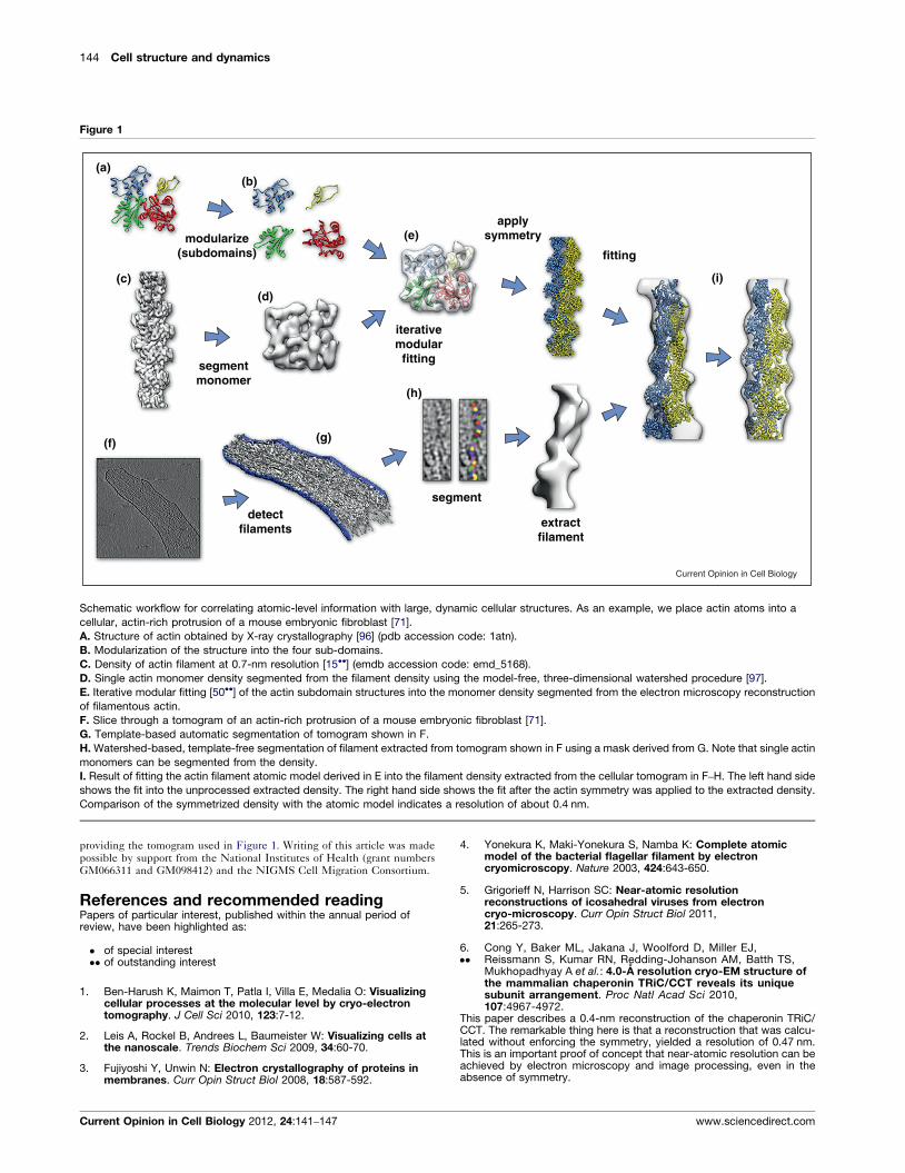
Schematic workflow for correlating atomic-level information with large, dynamic cellular structures. As an example, we place actin atoms into a
cellular, actin-rich protrusion of a mouse embryonic fibroblast [71].
A. Structure of actin obtained by X-ray crystallography [96] (pdb accession code: 1atn).
B. Modularization of the structure into the four sub-domains.
C. Density of actin filament at 0.7-nm resolution [15��] (emdb accession code: emd_5168).
D. Single actin monomer density segmented from the filament density using the model-free, three-dimensional watershed procedure [97].
E. Iterative modular fitting [50��] of the actin subdomain structures into the monomer density segmented from the electron microscopy reconstruction
of filamentous actin.
F. Slice through a tomogram of an actin-rich protrusion of a mouse embryonic fibroblast [71].
G. Template-based automatic segmentation of tomogram shown in F.
H. Watershed-based, template-free segmentation of filament extracted from tomogram shown in F using a mask derived from G. Note that single actin
monomers can be segmented from the density.
I. Result of fitting the actin filament atomic model derived in E into the filament density extracted from the cellular tomogram in F–H. The left hand side
shows the fit into the unprocessed extracted density. The right hand side shows the fit after the actin symmetry was applied to the extracted density.
Comparison of the symmetrized density with the atomic model indicates a resolution of about 0.4 nm.
providing the tomogram used in Figure 1. Writing of this article was madepossible by support from the National Institutes of Health (grant numbersGM066311 and GM098412) and the NIGMS Cell Migration Consortium.
References and recommended readingPapers of particular interest, published within the annual period ofreview, have been highlighted as:
� of special interest�� of outstanding interest
1. Ben-Harush K, Maimon T, Patla I, Villa E, Medalia O: Visualizingcellular processes at the molecular level by cryo-electrontomography. J Cell Sci 2010, 123:7-12.
2. Leis A, Rockel B, Andrees L, Baumeister W: Visualizing cells atthe nanoscale. Trends Biochem Sci 2009, 34:60-70.
3. Fujiyoshi Y, Unwin N: Electron crystallography of proteins inmembranes. Curr Opin Struct Biol 2008, 18:587-592.
Current Opinion in Cell Biology 2012, 24:141–147
4. Yonekura K, Maki-Yonekura S, Namba K: Complete atomicmodel of the bacterial flagellar filament by electroncryomicroscopy. Nature 2003, 424:643-650.
5. Grigorieff N, Harrison SC: Near-atomic resolutionreconstructions of icosahedral viruses from electroncryo-microscopy. Curr Opin Struct Biol 2011,21:265-273.
6.��
Cong Y, Baker ML, Jakana J, Woolford D, Miller EJ,Reissmann S, Kumar RN, Redding-Johanson AM, Batth TS,Mukhopadhyay A et al.: 4.0-A resolution cryo-EM structure ofthe mammalian chaperonin TRiC/CCT reveals its uniquesubunit arrangement. Proc Natl Acad Sci 2010,107:4967-4972.
This paper describes a 0.4-nm reconstruction of the chaperonin TRiC/CCT. The remarkable thing here is that a reconstruction that was calcu-lated without enforcing the symmetry, yielded a resolution of 0.47 nm.This is an important proof of concept that near-atomic resolution can beachieved by electron microscopy and image processing, even in theabsence of symmetry.
www.sciencedirect.com
Fitting into electron microscopy reconstructions Volkmann 145
7. Ludtke SJ, Baker ML, Chen DH, Song JL, Chuang DT, Chiu W: Denovo backbone trace of GroEL from single particle electroncryomicroscopy. Structure 2008, 16:441-448.
8. Hanein D, Volkmann N: Correlative light-electron microscopy.Adv Protein Chem Struct Biol 2011, 82:91-99.
9.�
Rigort A, Bauerlein FJ, Leis A, Gruska M, Hoffmann C, Laugks T,Bohm U, Eibauer M, Gnaegi H, Baumeister W, Plitzko JM:Micromachining tools and correlative approaches for cellularcryo-electron tomography. J Struct Biol 2010, 172:169-179.
This paper describes a complete pipeline for correlative electron and lightcryo-microscopy, including milling of the sample within a cryo FocusedIon Beam instrument.
10. Noske AB, Costin AJ, Morgan GP, Marsh BJ: Expeditedapproaches to whole cell electron tomography and organellemark-up in situ in high-pressure frozen pancreatic islets.J Struct Biol 2008, 161:298-313.
11. Volkmann N, Hanein D: Electron microscopy in the context ofsystems biology. In Structural Bioinformatics. Edited by Gu J,Bourne PE. Wiley-Blackwell; 2009:143-170.
12. Miyazawa A, Fujiyoshi Y, Unwin N: Structure and gatingmechanism of the acetylcholine receptor pore. Nature 2003,423:949-955.
13. Sachse C, Chen JZ, Coureux PD, Stroupe ME, Fandrich M,Grigorieff N: High-resolution electron microscopy of helicalspecimens: a fresh look at tobacco mosaic virus. J Mol Biol2007, 371:812-835.
14. Galkin VE, Orlova A, Schroder GF, Egelman EH: Structuralpolymorphism in F-actin. Nat Struct Mol Biol 2010,17:1318-1323.
15.��
Fujii T, Iwane AH, Yanagida T, Namba K: Direct visualization ofsecondary structures of F-actin by electron cryomicroscopy.Nature 2010, 467:724-728.
This paper describes the structure of the actin filament at 0.68 nmresolution. This is the highest resolution structure of filamentous actinobtained so far, providing a much better structural basis for interpretingactin-based assemblies.
16. Li X, Tobacman LS, Mun JY, Craig R, Fischer S, Lehman W:Tropomyosin position on F-actin revealed by EMreconstruction and computational chemistry. Biophys J 2011,100:1005-1013.
17. Orlova A, Galkin VE, Jeffries CM, Egelman EH, Trewhella J: TheN-terminal domains of myosin binding protein C can bindpolymorphically to F-actin. J Mol Biol 2011, 412:379-386.
18. Mun JY, Gulick J, Robbins J, Woodhead J, Lehman W, Craig R:Electron microscopy and 3D reconstruction of F-actindecorated with cardiac myosin-binding protein C (cMyBP-C).J Mol Biol 2011, 410:214-225.
19. Galkin VE, Orlova A, Salmazo A, Djinovic-Carugo K, Egelman EH:Opening of tandem calponin homology domains regulatestheir affinity for F-actin. Nat Struct Mol Biol 2010, 17:614-616.
20. Hertzog M, Milanesi F, Hazelwood L, Disanza A, Liu H, Perlade E,Malabarba MG, Pasqualato S, Maiolica A, Confalonieri S et al.:Molecular basis for the dual function of Eps8 on actindynamics: bundling and capping. PLoS Biol 2010, 8:e1000387.
21. Grintsevich EE, Galkin VE, Orlova A, Ytterberg AJ, Mikati MM,Kudryashov DS, Loo JA, Egelman EH, Reisler E: Mapping ofdrebrin binding site on F-actin. J Mol Biol 2010, 398:542-554.
22. Galkin VE, Orlova A, Brieher W, Kueh HY, Mitchison TJ,Egelman EH: Coronin-1A stabilizes F-actin by bridgingadjacent actin protomers and stapling opposite strands of theactin filament. J Mol Biol 2008, 376:607-613.
23. Gingras AR, Bate N, Goult BT, Hazelwood L, Canestrelli I,Grossmann JG, Liu H, Putz NS, Roberts GC, Volkmann N et al.:The structure of the C-terminal actin-binding domain of talin.EMBO J 2007, 27:458-469.
24. Galkin VE, Orlova A, Cherepanova O, Lebart MC, Egelman EH:High-resolution cryo-EM structure of the F-actin-fimbrin/plastin ABD2 complex. Proc Natl Acad Sci U S A 2008,105:1494-1498.
www.sciencedirect.com
25. Hampton CM, Liu J, Taylor DW, DeRosier DJ, Taylor KA: The 3Dstructure of villin as an unusual F-Actin crosslinker. Structure2008, 16:1882-1891.
26. Janssen ME, Kim E, Liu H, Fujimoto LM, Bobkov A, Volkmann N,Hanein D: Three-dimensional structure of vinculin bound toactin filaments. Mol Cell 2006, 21:271-281.
27. Rouiller I, Xu XP, Amann KJ, Egile C, Nickell S, Nicastro D, Li R,Pollard TD, Volkmann N, Hanein D: The structural basis of actinfilament branching by Arp2/3 complex. J Cell Biol 2008,180:887-895.
28. Xia Z, Gardner DP, Gutell RR, Ren P: Coarse-grained model forsimulation of RNA three-dimensional structures. J Phys ChemB 2010, 114:13497-13506.
29. Dorset DL: Direct phase determination in protein electroncrystallography: the pseudo-atom approximation. Proc NatlAcad Sci U S A 1997, 94:1791-1794.
30. Lunin VY, Lunina NL, Petrova TE, Vernoslova EA, Urzhumtsev AG,Podjarny AD: On the ab initio solution of the phase problem formacromolecules at very low resolution: the few atoms modelmethod. Acta Crystallogr D Biol Crystallogr 1995,51:896-903.
31. Wuthrich K, Billeter M, Braun W: Pseudo-structures for the 20common amino acids for use in studies of proteinconformations by measurements of intramolecular proton-proton distance constraints with nuclear magnetic resonance.J Mol Biol 1983, 169:949-961.
32. Volkmann N, Hanein D: Quantitative fitting of atomic modelsinto observed densities derived by electron microscopy.J Struct Biol 1999, 125:176-184.
33. Roseman AM: Docking structures of domains into maps fromcryo-electron microscopy using local correlation. ActaCrystallogr D Biol Crystallogr 2000, 56:1332-1340.
34. Chacon P, Wriggers W: Multi-resolution contour-based fittingof macromolecular structures. J Mol Biol 2002, 317:375-384.
35. Tama F, Miyashita O, Brooks CL3: Flexible multi-scale fitting ofatomic structures into low-resolution electron density mapswith elastic network normal mode analysis. J Mol Biol 2004,337:985-999.
36. Hinsen K, Reuter N, Navaza J, Stokes DL, Lacapere J-J: Normalmode-based fitting of atomic structure into electron densitymaps: application to sarcoplasmic reticulum Ca-ATPase.Biophys J 2005, 88:818-827.
37. Gao H, Frank J: Molding atomic structures into intermediate-resolution cryo-EM density maps of ribosomal complexesusing real-space refinement. Structure 2005, 13:401-406.
38. Suhre K, Navaza J, Sanejouand YH: NORMA: a tool for flexiblefitting of high-resolution protein structures into low-resolutionelectron-microscopy-derived density maps. Acta Crystallogr DBiol Crystallogr 2006, 62:1098-1100.
39. Velazquez-Muriel JA, Valle M, Santamaria-Pang A, Kakadiaris IA,Carazo JM: Flexible fitting in 3D-EM guided by the structuralvariability of protein superfamilies. Structure 2006,14:1115-1126.
40. Schroder GF, Brunger AT, Levitt M: Combining efficientconformational sampling with a deformable elastic networkmodel facilitates structure refinement at low resolution.Structure 2007, 15:1630-1641.
41. Kovacs JA, Yeager M, Abagyan R: Damped-dynamics flexiblefitting. Biophys J 2008, 95:3192-3207.
42. Topf M, Lasker K, Webb B, Wolfson H, Chiu W, Sali A: Proteinstructure fitting and refinement guided by cryo-EM density.Structure 2008, 16:295-307.
43. Jolley CC, Wells SA, Fromme P, Thorpe MF: Fitting low-resolution cryo-EM maps of proteins using constrainedgeometric simulations. Biophys J 2008, 94:1613-1621.
44. Tan RKZ, Devkota B, Harvey SC: YUP.SCX: coaxing atomicmodels into medium resolution electron density maps. J StructBiol 2008, 163:163-174.
Current Opinion in Cell Biology 2012, 24:141–147

146 Cell structure and dynamics
45. Rusu M, Birmanns S, Wriggers W: Biomolecular pleiomorphismprobed by spatial interpolation of coarse models.Bioinformatics 2008, 24:2460-2466.
46. Trabuco LG, Villa E, Mitra K, Frank J, Schulten K: Flexible fitting ofatomic structures into electron microscopy maps usingmolecular dynamics. Structure 2008, 16:673-683.
47. DiMaio F, Tyka MD, Baker ML, Chiu W, Baker D: Refinement ofprotein structures into low-resolution density maps usingrosetta. J Mol Biol 2009, 392:181-190.
48. Zhu J, Cheng L, Fang Q, Zhou ZH, Honig B: Building and refiningprotein models within cryo-electron microscopy density mapsbased on homology modeling and multiscale structurerefinement. J Mol Biol 2010, 397:835-851.
49. Zheng W: Accurate flexible fitting of high-resolution proteinstructures into cryo-electron microscopy maps using coarse-grained pseudo-energy minimization. Biophys J 2011,100:478-488.
50.��
Volkmann N: Confidence intervals for fitting of atomic modelsinto low-resolution densities. Acta Crystallogr D Biol Crystallogr2009, 65:679-689.
This paper describes a statistical procedure that allows obtaining con-fidence intervals for the orientation parameters of structures fitted intoreconstructions derived by electron microscopy. The paper also showsthat an iterative modular fitting approach outperforms flexible fittingapproaches in most cases.
51. Volkmann N, Hanein D: Docking of atomic models intoreconstructions from electron microscopy. Methods Enzymol2003, 374:204-225.
52.�
Xu XP, Rouiller I, Slaughter BD, Egile C, Kim E, Unruh JR, Fan X,Pollard TD, Li R, Hanein D, Volkmann N: Three-dimensionalreconstructions of Arp2/3 complex with bound nucleationpromoting factors. Embo J, 2011 Sep 20. [Epub ahead of print].
This paper describes the structure of Arp2/3 complex with bound nuclea-tion promoting factors. The iterative modular fitting of the complexcomponents into the density was augmented by a novel probabilitymapping procedure for FRET distance data.
53.�
Alber F, Dokudovskaya S, Veenhoff LM, Zhang W, Kipper J,Devos D, Suprapto A, Karni-Schmidt O, Williams R, Chait BT et al.:Determining the architectures of macromolecular assemblies.Nature 2007, 450:683-694.
This paper describes a procedure for integrating data from electronmicroscopy, proteomics and biophysical methods into a structure con-sistent with all data sources.
54.�
Campos M, Francetic O, Nilges M: Modeling pilus structuresfrom sparse data. J Struct Biol 2011, 173:436-444.
This paper describes the integration of generalized NMR restraints andparameters from an electron microscopy reconstruction in a commonframework to obtain models of helically symmetric bacterial pili.
55. Vasishtan D, Topf M: Scoring functions for cryoEM densityfitting. J Struct Biol 2011, 174:333-343.
56. Fisher RA: On the ‘probable error’ of a coefficient of correlationdeduced from a small sample. Metron 1921, 1:1-32.
57. Murata K, Liu X, Danev R, Jakana J, Schmid MF, King J,Nagayama K, Chiu W: Zernike phase contrast cryo-electronmicroscopy and tomography for structure determination atnanometer and subnanometer resolutions. Structure 2010,18:903-912.
58. Danev R, Kanamaru S, Marko M, Nagayama K: Zernike phasecontrast cryo-electron tomography. J Struct Biol 2010,171:174-181.
59. Milazzo AC, Moldovan G, Lanman J, Jin L, Bouwer JC, Klienfelder S,Peltier ST, Ellisman MH, Kirkland AI, Xuong NH: Characterization ofa direct detection device imaging camera for transmissionelectron microscopy. Ultramicroscopy 2010, 110:744-747.
60. Jensen GJ, Briegel A: How electron cryotomography is openinga new window onto prokaryotic ultrastructure. Curr Opin StructBiol 2007, 17:260-267.
61. Fu CY, Johnson JE: Viral life cycles captured in three-dimensions with electron microscopy tomography. Curr OpinVirol 2011, 1:125-133.
Current Opinion in Cell Biology 2012, 24:141–147
62. Bos E, SantAnna C, Gnaegi H, Pinto RF, Ravelli RB, Koster AJ, deSouza W, Peters PJ: A new approach to improve the quality ofultrathin cryo-sections; its use for immunogold EM andcorrelative electron cryo-tomography. J Struct Biol 2011,175:62-72.
63. Al-Amoudi A, Castano-Diez D, Devos DP, Russell RB, Johnson GT,Frangakis AS: The three-dimensional molecular structure ofthe desmosomal plaque. Proc Natl Acad Sci U S A 2011,108:6480-6485.
64. Bouchet-Marquis C, Hoenger A: Cryo-electron tomography onvitrified sections: a critical analysis of benefits and limitationsfor structural cell biology. Micron 2011, 42:152-162.
65. Gruska M, Medalia O, Baumeister W, Leis A: Electrontomography of vitreous sections from cultured mammaliancells. J Struct Biol 2008, 161:384-392.
66. Hayles MF, de Winter DA, Schneijdenberg CT, Meeldijk JD,Luecken U, Persoon H, de Water J, de Jong F, Humbel BM,Verkleij AJ: The making of frozen-hydrated, vitreous lamellasfrom cells for cryo-electron microscopy. J Struct Biol 2010,172:180-190.
67. Marko M, Hsieh C, Schalek R, Frank J, Mannella C: Focused-ion-beam thinning of frozen-hydrated biological specimensfor cryo-electron microscopy. Nat Methods 2007,4:215-217.
68. Medalia O, Weber I, Frangakis AS, Nicastro D, Gerisch G,Baumeister W: Macromolecular architecture in eukaryotic cellsvisualized by cryoelectron tomography. Science 2002,298:1209-1213.
69. Medalia O, Beck M, Ecke M, Weber I, Neujahr R, Baumeister W,Gerisch G: Organization of actin networks in intact filopodia.Curr Biol 2007, 17:79-84.
70. Patla I, Volberg T, Elad N, Hirschfeld-Warneken V, Grashoff C,Fassler R, Spatz JP, Geiger B, Medalia O: Dissecting themolecular architecture of integrin adhesion sites by cryo-electron tomography. Nat Cell Biol 2010, 12:909-915.
71. Koning RI, Zovko S, Barcena M, Oostergetel GT, Koerten HK,Galjart N, Koster AJ, Mieke Mommaas A: Cryo electrontomography of vitrified fibroblasts: microtubule plus ends insitu. J Struct Biol 2008, 161:459-468.
72. Urban E, Jacob S, Nemethova M, Resch GP, Small JV: Electrontomography reveals unbranched networks of actin filamentsin lamellipodia. Nat Cell Biol 2010, 12:429-435.
73. Yang C, Svitkina T: Visualizing branched actin filaments inlamellipodia by electron tomography. Nat Cell Biol 2011,13:1012-1013.
74. Small JV, Winkler C, Vinzenz M, Schmeiser C: Reply: Visualizingbranched actin filaments in lamellipodia by electrontomography. Nat Cell Biol 2011, 13:1013-1014.
75. Bohm J, Frangakis AS, Hegerl R, Nickell S, Typke D,Baumeister W: From the cover: toward detecting andidentifying macromolecules in a cellular context: templatematching applied to electron tomograms. Proc Natl Acad Sci US A 2000, 97:14245-14250.
76. Frangakis AS, Bohm J, Forster F, Nickell S, Nicastro D, Typke D,Hegerl R, Baumeister W: Identification of macromolecularcomplexes in cryoelectron tomograms of phantom cells. ProcNatl Acad Sci U S A 2002, 99:14153-14158.
77. Forster F, Medalia O, Zauberman N, Baumeister W, Fass D:Retrovirus envelope protein complex structure in situ studiedby cryo-electron tomography. Proc Natl Acad Sci U S A 2005,102:4729-4734.
78. Beck M, Malmstrom JA, Lange V, Schmidt A, Deutsch EW,Aebersold R: Visual proteomics of the human pathogenLeptospira interrogans. Nat Methods 2009, 6:817-823.
79. Brandt F, Etchells SA, Ortiz JO, Elcock AH, Hartl FU,Baumeister W: The native 3D organization of bacterialpolysomes. Cell 2009, 136:261-271.
80. Ortiz JO, Brandt F, Matias VR, Sennels L, Rappsilber J,Scheres SH, Eibauer M, Hartl FU, Baumeister W: Structure of
www.sciencedirect.com
Fitting into electron microscopy reconstructions Volkmann 147
hibernating ribosomes studied by cryoelectron tomography invitro and in situ. J Cell Biol 2010, 190:613-621.
81. Rigort A, Gunther D, Hegerl R, Baum D, Weber B, Prohaska S,Medalia O, Baumeister W, Hege H-C: Automated segmentationof electron tomograms for a quantitative description of actinfilament networks [Internet]. J Struct Biol, 2011 Sep 1, [Epubahead of print].
82. Lebbink MN, van Donselaar E, Humbel BM, Hertzberger LO,Post JA, Verkleij AJ: Induced membrane domains as visualizedby electron tomography and template matching. J Struct Biol2009, 166:156-161.
83. Lebbink MN, Jimenez N, Vocking K, Hekking LH, Verkleij AJ,Post JA: Spiral coating of the endothelial caveolar membranesas revealed by electron tomography and template matching.Traffic 2010, 11:138-150.
84. Malmstrom J, Beck M, Schmidt A, Lange V, Deutsch EW,Aebersold R: Proteome-wide cellular protein concentrations ofthe human pathogen Leptospira interrogans. Nature 2009,460:762-765.
85. Xu M, Beck M, Alber F: Template-free detection ofmacromolecular complexes in cryo electron tomograms.Bioinformatics 2011, 27:69-76.
86. Bartesaghi A, Sprechmann P, Liu J, Randall G, Sapiro G,Subramaniam S: Classification and 3D averaging with missingwedge correction in biological electron tomography. J StructBiol 2008, 162:436-450.
87. Forster F, Hegerl R: Structure determination in situ byaveraging of tomograms. Methods Cell Biol 2007, 79:741-767.
88. Forster F, Pruggnaller S, Seybert A, Frangakis AS: Classificationof cryo-electron sub-tomograms using constrainedcorrelation. J Struct Biol 2008, 161:276-286.
www.sciencedirect.com
89. Schmid MF, Booth CR: Methods for aligning and for averaging3D volumes with missing data. J Struct Biol 2008, 161:243-248.
90. Winkler H, Zhu P, Liu J, Ye F, Roux KH, Taylor KA: Tomographicsubvolume alignment and subvolume classification applied tomyosin V and SIV envelope spikes. J Struct Biol 2009, 165:64-77.
91. Scheres SH, Melero R, Valle M, Carazo JM: Averaging of electronsubtomograms and random conical tilt reconstructionsthrough likelihood optimization. Structure 2009, 17:1563-1572.
92. Stolken M, Beck F, Haller T, Hegerl R, Gutsche I, Carazo J-M,Baumeister W, Scheres SHW, Nickell S: Maximum likelihoodbased classification of electron tomographic data. J Struct Biol2011, 173:77-85.
93.��
Yu Z, Frangakis AS: Classification of electron sub-tomogramswith neural networks and its application to template-matching. J Struc Biol 2011, 174:494-504.
This paper describes how kernel density estimator self-organizing mapscan be used to classify three-dimensional subtomograms and to narrowdown the number of false positives after template matching.
94. Shtengel G, Galbraith JA, Galbraith CG, Lippincott-Schwartz J,Gillette JM, Manley S, Sougrat R, Waterman CM,Kanchanawong P, Davidson MW et al.: Interferometricfluorescent super-resolution microscopy resolves 3D cellularultrastructure. Proc Natl Acad Sci U S A 2009, 106:3125-3130.
95. Kanchanawong P, Shtengel G, Pasapera AM, Ramko EB,Davidson MW, Hess HF, Waterman CM: Nanoscale architectureof integrin-based cell adhesions. Nature 2010, 468:580-584.
96. Kabsch W, Mannherz HG, Suck D, Pai EF, Holmes KC: Atomicstructure of the actin:DNase I complex. Nature 1990, 347:37-44.
97. Volkmann N: A novel three-dimensional variant of thewatershed transform for segmentation of electron densitymaps. J Struct Biol 2002, 138:123.
Current Opinion in Cell Biology 2012, 24:141–147