pxl01 in sodium hyaluronate results in increased prg4 · pdf fileoriginal article pxl01 in...
TRANSCRIPT

ORIGINAL ARTICLE
PXL01 in sodium hyaluronate results in increased PRG4 expression:a potential mechanism for anti-adhesion
Sara Edsfeldta,b, Bj€orn Holma,b, Margit Mahlapuuc, Carol Renod, David A. Hartd and Monica Wiiga,b
aDepartment of Surgical Sciences, Hand Surgery, Uppsala University, Uppsala, Sweden; bDepartment of Hand Surgery, Uppsala UniversityHospital, Uppsala, Sweden; cPergamum AB, Stockholm, Sweden; dDepartment of Surgery, McCaig Institute for Bone and Joint Health,University of Calgary, Calgary, Canada
ABSTRACTPurpose: To investigate the anti-adhesive mechanisms of PXL01 in sodium hyaluronate (HA) by usingthe rabbit lactoferrin peptide, rabPXL01 in HA, in a rabbit model of healing tendons and tendonsheaths. The mechanism of action for PXL01 in HA is interesting since a recent clinical study of thehuman lactoferrin peptide PXL01 in HA administered around repaired tendons in the hand showedimproved digit mobility.Materials and methods: On days 1, 3, and 6 after tendon injury and surgical repair, reverse transcript-ase-quantitative polymerase chain reaction (RT-qPCR) was used to assess mRNA expression levels forgenes encoding the mucinous glycoprotein PRG4 (also called lubricin) and a subset of matrix proteins,cytokines, and growth factors involved in flexor tendon repair. RabPXL01 in HA was administeredlocally around the repaired tendons, and mRNA expression was compared with untreated repaired ten-dons and tendon sheaths.Results: We observed, at all time points, increased expression of PRG4 mRNA in tendons treated withrabPXL01 in HA, but not in tendon sheaths. In addition, treatment with rabPXL01 in HA led to repres-sion of the mRNA levels for the pro-inflammatory mediators interleukin (IL)-1b, IL-6, and IL-8 in tendonsheaths.Conclusions: RabPXL01 in HA increased lubricin mRNA production while diminishing mRNA levels ofinflammatory mediators, which in turn reduced the gliding resistance and inhibited the adhesion for-mation after flexor tendon repair.
ARTICLE HISTORYReceived 8 May 2016Revised 15 August 2016Accepted 23 August 2016
KEYWORDSAdhesions; flexor tendons;lubricin; proteoglycan 4;PXL01
Introduction
PXL01 is a synthetic peptide derived from the human milkprotein, lactoferrin. In vitro studies in human cell lines haveshown that PXL01 exhibits an inhibitory effect on importanthallmarks of adhesion formation by reducing secretion ofinflammatory cytokines, promoting fibrinolysis, and reducinginfections (1). Further, PXL01 in sodium hyaluronate (HA)reduced post-surgical adhesions in experimental models ofabdominal surgery in rats (1) and flexor tendon repair sur-gery in rabbits (2,3). A recent clinical trial investigating theefficacy and safety of adjuvant treatment with PXL01 in HAafter flexor tendon injury and repair in the hand showedincreased digit mobility in PXL01 in HA-treated fingers com-pared to placebo (4). However, the molecular mechanismsunderlying the effect of PXL01 in HA on adhesion formationand tendon healing have not been clarified. In this study, weused the corresponding peptide derived from rabbit lactofer-rin (rabPXL01) to study the effects in a rabbit model of ten-don surgery. We hypothesized that treatment with rabPXL01in HA would lead to reduced inflammation and increasedsynthesis of proteoglycan 4 (PRG4), also called lubricin, after
tendon injury and repair in rabbits. Increased lubricin levelsand reduced inflammation could subsequently lead to inhib-ition of adhesion formation.
PRG4 is an interesting gene, and individuals carryingmutant variants of the gene can suffer a number of connect-ive tissue complications (5). The protein it encodes is amucinous glycoprotein secreted into synovial fluid by syn-ovial fibroblasts (6) and the superficial zone cells of articularcartilage (7) and menisci (8). It has lubricating properties onarticular cartilage (9), reduces synovial cell overgrowth, andprotects the surface of the cartilage (10). Lubricin has alsobeen found on the flexor tendon surface (11), and recentstudies have shown that lubricin reduces tendon-glidingresistance (12,13). In the eye, the ocular surface epitheliaexpress PRG4, which reduces friction between the corneaand conjunctiva (14). There is evidence that PRG4 may play arole in the biomechanics of rabbit ligaments during aging(15). PRG4 may also have anti-inflammatory effects itself (16).The expression of PRG4 is inhibited by tumor necrosis factora (TNF-a) and interleukin (IL)-1, and stimulated by transform-ing growth factor b (TGF-b) (11). Thus, interfering with these
CONTACT Monica Wiig, MD, PhD [email protected], [email protected] Department of Surgical Sciences, Hand Surgery, UppsalaUniversity, PO Box 256, SE-751 05 Uppsala, Sweden� 2016 The Author(s). Published by Informa UK Limited, trading as Taylor & Francis Group.This is an Open Access article distributed under the terms of the Creative Commons Attribution License (http://creativecommons.org/licenses/by/4.0/), which permits unrestricted use,distribution, and reproduction in any medium, provided the original work is properly cited.
UPSALA JOURNAL OF MEDICAL SCIENCES, 2017VOL. 122, NO. 1, 28–34http://dx.doi.org/10.1080/03009734.2016.1230157
inflammatory molecules may potentially increase lubricin lev-els after a tendon injury.
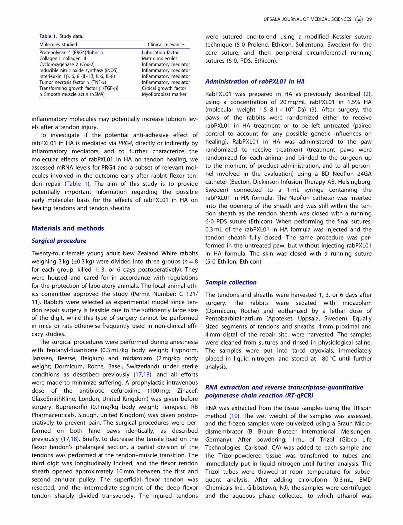
To investigate if the potential anti-adhesive effect ofrabPXL01 in HA is mediated via PRG4, directly or indirectly byinflammatory mediators, and to further characterize themolecular effects of rabPXL01 in HA on tendon healing, weassessed mRNA levels for PRG4 and a subset of relevant mol-ecules involved in the outcome early after rabbit flexor ten-don repair (Table 1). The aim of this study is to providepotentially important information regarding the possibleearly molecular basis for the effects of rabPXL01 in HA onhealing tendons and tendon sheaths.
Materials and methods
Surgical procedure
Twenty-four female young adult New Zealand White rabbitsweighing 3 kg (±0.3 kg) were divided into three groups (n¼ 8for each group; killed 1, 3, or 6 days postoperatively). Theywere housed and cared for in accordance with regulationsfor the protection of laboratory animals. The local animal eth-ics committee approved the study (Permit Number: C 121/11). Rabbits were selected as experimental model since ten-don repair surgery is feasible due to the sufficiently large sizeof the digit, while this type of surgery cannot be performedin mice or rats otherwise frequently used in non-clinical effi-cacy studies.
The surgical procedures were performed during anesthesiawith fentanyl-fluanisone (0.3mL/kg body weight; Hypnorm,Janssen, Beerse, Belgium) and midazolam (2mg/kg bodyweight; Dormicum, Roche, Basel, Switzerland) under sterileconditions as described previously (17,18), and all effortswere made to minimize suffering. A prophylactic intravenousdose of the antibiotic cefuroxime (100mg; Zinacef,GlaxoSmithKline, London, United Kingdom) was given beforesurgery. Buprenorfin (0.1mg/kg body weight; Temgesic, RBPharmaceuticals, Slough, United Kingdom) was given postop-eratively to prevent pain. The surgical procedures were per-formed on both hind paws identically, as describedpreviously (17,18). Briefly, to decrease the tensile load on theflexor tendon’s phalangeal section, a partial division of thetendons was performed at the tendon–muscle transition. Thethird digit was longitudinally incised, and the flexor tendonsheath opened approximately 10mm between the first andsecond annular pulley. The superficial flexor tendon wasresected, and the intermediate segment of the deep flexortendon sharply divided transversely. The injured tendons
were sutured end-to-end using a modified Kessler suturetechnique (5-0 Prolene, Ethicon, Sollentuna, Sweden) for thecore suture, and then peripheral circumferential runningsutures (6-0, PDS, Ethicon).
Administration of rabPXL01 in HA
RabPXL01 was prepared in HA as previously described (2),using a concentration of 20mg/mL rabPXL01 in 1.5% HA(molecular weight 1.5–8.1� 106 Da) (3). After surgery, thepaws of the rabbits were randomized either to receiverabPXL01 in HA treatment or to be left untreated (pairedcontrol to account for any possible genetic influences onhealing). RabPXL01 in HA was administered to the pawrandomized to receive treatment (treatment paws wererandomized for each animal and blinded to the surgeon upto the moment of product administration, and to all person-nel involved in the evaluation) using a BD Neoflon 24GAcatheter (Becton, Dickinson Infusion Therapy AB, Helsingborg,Sweden) connected to a 1mL syringe containing therabPXL01 in HA formula. The Neoflon catheter was insertedinto the opening of the sheath and was still within the ten-don sheath as the tendon sheath was closed with a running6-0 PDS suture (Ethicon). When performing the final sutures,0.3mL of the rabPXL01 in HA formula was injected and thetendon sheath fully closed. The same procedure was per-formed in the untreated paw, but without injecting rabPXL01in HA formula. The skin was closed with a running suture(5-0 Ethilon, Ethicon).
Sample collection
The tendons and sheaths were harvested 1, 3, or 6 days aftersurgery. The rabbits were sedated with midazolam(Dormicum, Roche) and euthanized by a lethal dose ofPentobarbitalnatrium (Apoteket, Uppsala, Sweden). Equallysized segments of tendons and sheaths, 4mm proximal and4mm distal of the repair site, were harvested. The sampleswere cleaned from sutures and rinsed in physiological saline.The samples were put into tared cryovials, immediatelyplaced in liquid nitrogen, and stored at –80 �C until furtheranalysis.
RNA extraction and reverse transcriptase-quantitativepolymerase chain reaction (RT-qPCR)
RNA was extracted from the tissue samples using the TRIspinmethod (19). The wet weight of the samples was assessed,and the frozen samples were pulverized using a Braun Micro-dismembrator (B. Braun Biotech International, Melsungen,Germany). After powdering, 1mL of Trizol (Gibco LifeTechnologies, Carlsbad, CA) was added to each sample andthe Trizol-powdered tissue was transferred to tubes andimmediately put in liquid nitrogen until further analysis. TheTrizol tubes were thawed at room temperature for subse-quent analysis. After adding chloroform (0.3mL; EMDChemicals Inc., Gibbstown, NJ), the samples were centrifugedand the aqueous phase collected, to which ethanol was
Table 1. Study data.
Molecules studied Clinical relevance
Proteoglycan 4 (PRG4)/lubricin Lubrication factorCollagen I, collagen III Matrix moleculesCyclo-oxygenase 2 (Cox-2) Inflammatory mediatorInducible nitric oxide synthase (iNOS) Inflammatory mediatorInterleukin 1b, 6, 8 (IL-1b, IL-6, IL-8) Inflammatory mediatorTumor necrosis factor a (TNF-a) Inflammatory mediatorTransforming growth factor b (TGF-b) Critical growth factora Smooth muscle actin (aSMA) Myofibroblast marker
UPSALA JOURNAL OF MEDICAL SCIENCES 29
added to precipitate the RNA (70%, 0.6mL). Using an RNeasyTotal RNA Kit (Qiagen, Chatsworth, CA), RNA was eluted andquantified fluorometrically with Sybergreen (Mandel, Guelph,Ontario, Canada). RNA was further processed for reverse tran-scription to cDNA using a Qiagen Omniscript kit (OmniscriptRT Kit, Qiagen, Mississauga, Ontario, Canada). cDNA was pre-pared for qPCR using IQ SYBR Green Supermix (Bio-RadLaboratories Mississauga, Ontario, Canada). Primers previouslyutilized for rabbit tissues were added (Table 2) and the sam-ples amplified using real-time PCR (iCycler iQ, Real Time PCRDetection System, Bio-Rad Laboratories). 18S was used as thehouse-keeping gene for normalization, and non-reverse
transcribed RNA was used as a negative control to detectpossible DNA contamination (none of the samples containeddetectable DNA; data not shown).
Statistical analysis
Levels of mRNA for a subset of mediators involved in thehealing of flexor tendons and tendon sheaths were assessed1, 3, and 6 days postoperatively. Flexor tendons and tendonsheaths treated with rabPXL01 in HA were compared with itsuntreated paired control using repeated measures ANOVA.Table 3 is a supplement reporting the remaining data. All
Table 2. Primer sequences.
Gene Primer sequence Base pairs Source
18S Forward sequence TGG TCG CTC GCT CCT CTC C 360 NR_003286Reverse sequence CGC CTG CTG CCT TCC TTG G
aSMA Forward sequence GTG TGA GGA AGA GGA CAG CA 446 X60732Reverse sequence TAC GTC CAG AGG CAT AGA GG
Collagen I Forward sequence GAT GCG TTC CAG TTC GAG TA 312 Personal communicationReverse sequence GGT CTT CCG GTG GTC TTG TA
Collagen III Forward sequence TTA TAA ACC AAC CTC TTC CT 255 Personal communicationReverse sequence TAT TAT AGC ACC ATT GAG AC
COX-2 Forward sequence CAA ACT GCT CCT GAA ACC CAC TC 82 NM_001082388Reverse sequence GCT ATT GAC GAT GTT CCA GAC TCC
IL-1b Forward sequence GCC GAT GGT CCC AAT TAC AT 121 M26295Reverse sequence ACA AGA CCT GCC GGA AGC T
IL-6 Forward sequence CCT GCC TGC TGA GAA TCA CTT 51 AF469048Reverse sequence CGA GAT ACA TCC GGA ACT CCA T
IL-8 Forward sequence CAA CCT TCC TGC TGT CTC TG 145 NM_001082293Reverse sequence GGT CCA CTC TCA ATC ACT CT
iNOS Forward sequence CTG TGA CGT CCA GCG CTA CA 119 AF469048Reverse sequence GCA CGG CGA TGT TGA TCT CTC GCC CT
PRG4 Forward sequence GAA CGT GCT ATA GGA CCT TC 287 NM_00127709Reverse sequence CAG ACT TTG GAT AAG GTC TGC C
TGF-b Forward sequence CGG CAG CTG TAC ATT GAC TT 271 AF000133Reverse sequence AGC GCA CGA TCA TGT TGG AC
TNF-a Forward sequence TCT AGT CAA CCC TGT GGC CC 51 NM_00108Reverse sequence GCC CGA GAA GCT GAT CTG AG
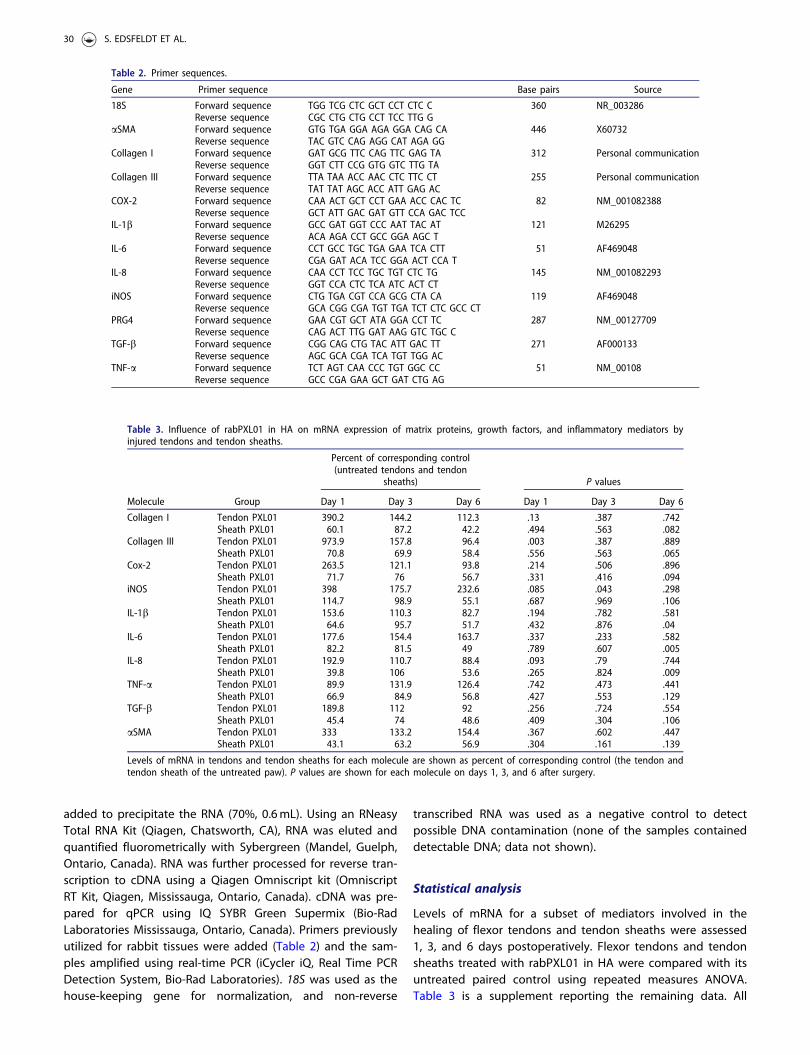
Table 3. Influence of rabPXL01 in HA on mRNA expression of matrix proteins, growth factors, and inflammatory mediators byinjured tendons and tendon sheaths.
Percent of corresponding control(untreated tendons and tendon
sheaths) P values
Molecule Group Day 1 Day 3 Day 6 Day 1 Day 3 Day 6
Collagen I Tendon PXL01 390.2 144.2 112.3 .13 .387 .742Sheath PXL01 60.1 87.2 42.2 .494 .563 .082
Collagen III Tendon PXL01 973.9 157.8 96.4 .003 .387 .889Sheath PXL01 70.8 69.9 58.4 .556 .563 .065
Cox-2 Tendon PXL01 263.5 121.1 93.8 .214 .506 .896Sheath PXL01 71.7 76 56.7 .331 .416 .094
iNOS Tendon PXL01 398 175.7 232.6 .085 .043 .298Sheath PXL01 114.7 98.9 55.1 .687 .969 .106
IL-1b Tendon PXL01 153.6 110.3 82.7 .194 .782 .581Sheath PXL01 64.6 95.7 51.7 .432 .876 .04
IL-6 Tendon PXL01 177.6 154.4 163.7 .337 .233 .582Sheath PXL01 82.2 81.5 49 .789 .607 .005
IL-8 Tendon PXL01 192.9 110.7 88.4 .093 .79 .744Sheath PXL01 39.8 106 53.6 .265 .824 .009
TNF-a Tendon PXL01 89.9 131.9 126.4 .742 .473 .441Sheath PXL01 66.9 84.9 56.8 .427 .553 .129
TGF-b Tendon PXL01 189.8 112 92 .256 .724 .554Sheath PXL01 45.4 74 48.6 .409 .304 .106
aSMA Tendon PXL01 333 133.2 154.4 .367 .602 .447Sheath PXL01 43.1 63.2 56.9 .304 .161 .139
Levels of mRNA in tendons and tendon sheaths for each molecule are shown as percent of corresponding control (the tendon andtendon sheath of the untreated paw). P values are shown for each molecule on days 1, 3, and 6 after surgery.
30 S. EDSFELDT ET AL.

tests were two-sided, and P< .05 was considered statisticallysignificant.
Results
Rupture rate
Three tendon ruptures occurred after repair surgery (two rup-tures in the same rabbit): two of the untreated tendons andone of the rabPXL01 in HA-treated tendons, giving a rupturerate of 8% for the untreated tendons and 4% for therabPXL01 in HA-treated tendons.
The effect of rabPXL01 in HA on the expression of PRG4mRNA in tendon and tendon sheaths
At all time points levels of PRG4 mRNA were higher in ten-dons treated with rabPXL01 in HA compared to untreated
tendons. On the other hand, levels of PRG4 mRNA were simi-lar in treated and untreated sheaths (Figure 1).
Influence of rabPXL01 in HA on mRNA levels forinflammatory mediators, transforming growth factor,the myofibroblast marker aSMA, and collagens I and III
In tendon sheaths treated with rabPXL01 in HA, mRNA levelsof IL-1b, IL-6, and IL-8 were lower than in untreated sheathson day 6 (Figure 2, Table 3). Levels of mRNA for iNOS tendedto be higher in rabPXL01 in HA-treated tendons than inuntreated tendons at all time points, but with significant dif-ferences detected only on day 3 (P¼ .043, Table 3).The mRNA level of collagen III in rabPXL01 in HA-treated ten-dons was higher than that of untreated tendons on day 1(P¼ .003). RabPXL01 in HA treatment did notsignificantly influence mRNA levels for any of the other mole-cules assessed, neither in tendons nor in tendon sheaths(Table 3).
Figure 1. Expression of PRG4 mRNA in flexor tendons and tendon sheaths treated with rabPXL01 in HA compared to untreated tendons and tendon sheaths 1, 3,and 6 days postoperatively. The following significance symbols are used: ���P< .001; ��P< .01; �P< .05.
UPSALA JOURNAL OF MEDICAL SCIENCES 31

Discussion
Previous studies on adjuvant treatment with PXL01 in HAhave shown an increase in digit range of motion in bothhumans (4) and rabbits (2,3) without elevating the rate ofrupture. The principal finding of the present study was anincreased expression of PRG4 mRNA on days 1, 3, and 6 aftersurgery in tendons treated with rabPXL01 in HA. These find-ings suggest that rabPXL01 in HA treatment leads to anincrease in lubricin production, and subsequently reduces thegliding resistance and inhibits adhesion formations afterflexor tendon repair. The present findings are consistent withthose reported by Hayashi et al. (12) who compared tendonsin PRG4 knockout, heterozygous, and wild-type mice andshowed that the gliding resistance of intrasynovial tendonsfrom PRG4 knockout mice was higher than in the othergroups. Adjuvant treatment with lubricin has also been eval-uated in canine models of flexor tendon repair (13,20–22).
Carbodiimide-derivatized HA and lubricin-treated tendonsexhibited lower gliding resistance than tendons treated withHA alone (13). However, two of these studies that showedpositive effects on digit function also reported adverseeffects on tendon healing and decreased repair strength(21,22). These investigations suggested that carbodiimide-derivatized HA and lubricin may prevent cellular adhesionsto tendon surfaces and enable lubrication of the tendon dur-ing flexion and extension, but this treatment also interferesnegatively with tendon healing. In the present study weobserved higher levels of PRG4 mRNA in rabPXL01 in HA-treated tendons, but no macroscopically obvious damage tothe tendon tissue or altered rate of post-surgical tendon rup-ture. Previous studies using PXL01 in HA have shown noincrease in the rate of tendon rupture and no tendon dam-ages (2,3).
There were no detectable differences in mRNA levels inthe tendons for the mediators IL-1, TNF-a, and TGF-b
Figure 2. Expression of mRNA for IL-1b (top), IL-6 (middle), and IL-8 (bottom) in flexor tendons and tendon sheaths treated with rabPXL01 in HA compared tountreated tendons and tendon sheaths 6 days after surgery. The following significance symbols are used: ���P< .001; ��P< .01; �P< .05.
32 S. EDSFELDT ET AL.

between treated and untreated groups. However, we foundincreased levels of PRG4 mRNA in rabPXL01 in HA-treatedtendons but not in tendon sheaths. Receptors for PXL01 arenot presently known, but likely the differences between PRG4mRNA expression in tendons and tendon sheaths depend onPXL01 affinity for a tendon cell-specific receptor. Whetherthese cells are endogenous tendon cells or cells recruited tothe injury site requires further investigation. These findingsregarding PRG4 point to an interesting new target on whichto focus future investigations, as well as to confirm that themRNA changes observed are followed by increased proteinproduction and secretion.
We have previously shown that PXL01 has anti-inflamma-tory effects on human macrophages by reducing the releaseof pro-inflammatory cytokines IL-1b, IL-6, and IL-8 (1).Consistent with these observations, the present studyrevealed a significant and co-ordinated suppression of mRNAlevels for IL-1b, L-6, and IL-8 in tendon sheaths 6 days aftersurgery. Prolonged inflammation in the wound-healing cas-cade can lead to excessive adhesion formation, whereasdown-regulation of the inflammatory response is believed torestrict proliferation and remodeling, leading to preventionof scarring (23). Suppression of inflammation may thus beone additional complimentary mechanism behind theimprovement in digit mobility that has been shown afteradjuvant treatment with PXL01 in HA (2–4).
The healing mechanisms in the tendon differ from themechanisms operative in the tendon sheath. There is, forexample, a change in collagen expression with increasingtype III collagen in both the injured tendon and tendonsheath directly after an injury, whereas the level of collagen Iremains unaltered in the tendon and increases in the tendonsheath at a later stage (17). This shift in collagen expressionis believed to decrease tendon strength (24). In the presentstudy, we observed higher levels of mRNA for both collagenI and collagen III in tendons treated with rabPXL01 in HA, atthe earliest time point studied, day 1. An increase of bothcollagen I and collagen III production early in the healingprocess might create a stronger tendon tissue that can betterwithstand forces created during early active rehabilitation,with limited risk of repair rupture (24). Therefore, increasedproduction of collagens I and III early after rabPXL01 in HAtreatment might help to reduce the risk of repair rupture,but further studies are needed to support this notion.
HA as carrier allows for a slow release of rabPXL01 com-bined with initial lubricating properties around the tendon.HA has also shown some anti-inflammatory effects itself byreducing the concentration of inflammatory mediators (25).The current study design does not enable us to differentiatebetween the molecular effects of rabPXL01 versus HA, whichis a limitation of the experiment. However, there were severalreasons for the chosen design. Firstly, this study designmimics the design of the recently published randomized clin-ical trial where the treatment with a combination of PXL01 inHA improved hand function (4). Secondly, in the same rabbitmodel of tendon injury, adjuvant treatment with the combin-ation of PXL01 in HA resulted in improved digit mobilitycompared to HA alone (3). Finally, using PXL01 alone is nottechnically feasible in this in vivo model, as the peptide does
not remain at the wound site when applied in saline, butrather diffuses away rapidly.
In this study, tissue samples were harvested 1, 3, and6 days after surgery. These time points were selected toaddress potential changes in the expression profile of differ-ent wound-healing mediators. It is known that molecularevents such as those associated with an inflammatoryresponse, which subsequently ‘set the stage’ for scar forma-tion, take place shortly after the injury (26). Relevant to thispoint, Berglund et al. (18) detected temporal alterations inthe pattern of mRNA expression for IL-1b and hyaluronansynthases, which reached peak levels 3 and 6 days after ten-don repair, respectively. A limitation of the present study isthat mRNA expression may not completely mirror the proteinlevels, since there may be post-transcriptional regulation ofprotein synthesis.
In summary, our results suggest that the anti-adhesiveeffect of PXL01 in HA involves an increased production ofPRG4 in tendons and a decreased expression of inflammatorymediators in tendon sheaths.
Acknowledgements
The authors thank Britt-Marie Andersson for skillful laboratory assistance.
Disclosure statement
S.E. and M.W. have accepted consulting fees from Pergamum AB. C.R.,D.A.H., and B.H. declare no conflicts of interest. M.M. is an employee ofPergamum AB. Pergamum AB was involved in designing the study andprovided the rabPXL01 in HA formulation. However, Pergamum AB hasnot performed the surgery or expression analysis.
Funding
This work was supported by Alberta Innovates Health SolutionsOsteoarthritis Team Grant [200700596] Uppsala University Hospital,10.13039/501100005423 Swedish Society for Strategic Research.
References
1. Nilsson E, Bjorn C, Sjostrand V, Lindgren K, Munnich M, Mattsby-Baltzer I, et al. A novel polypeptide derived from human lactofer-rin in sodium hyaluronate prevents postsurgical adhesion forma-tion in the rat. Ann Surg. 2009;250:1021–8.
2. Wiig M, Olmarker K, Hakansson J, Ekstrom L, Nilsson E, Mahlapuu M.A lactoferrin-derived peptide (PXL01) for the reduction of adhesionformation in flexor tendon surgery: an experimental study in rabbits.J Hand Surg Eur Vol. 2011;36:656–62.
3. Hakansson J, Mahlapuu M, Ekstrom L, Olmarker K, Wiig M. Effectof lactoferrin peptide (PXL01) on rabbit digit mobility after flexortendon repair. J Hand Surg Am. 2012;37:2519–25.
4. Wiig ME, Dahlin LB, Friden J, Hagberg L, Larsen SE, Wiklund K,et al. PXL01 in Sodium hyaluronate for improvement of handrecovery after flexor tendon repair surgery: randomized controlledtrial. PLoS One. 2014;9:e110735. doi: 10.1371/journal.pone.0110735.
5. Alazami AM, Al-Mayouf SM, Wyngaard CA, Meyer B. Novel PRG4mutations underlie CACP in Saudi families. Hum Mutat.2006;27:213. doi: 10.1002/humu.9399.
6. Jay GD, Britt DE, Cha CJ. Lubricin is a product of megakaryocytestimulating factor gene expression by human synovial fibroblasts.J Rheumatol. 2000;27:594–600.
UPSALA JOURNAL OF MEDICAL SCIENCES 33

7. Schumacher BL, Block JA, Schmid TM, Aydelotte MB, Kuettner KE.A novel proteoglycan synthesized and secreted by chondrocytesof the superficial zone of articular cartilage. Arch BiochemBiophys. 1994;311:144–52.
8. Schumacher BL, Schmidt TA, Voegtline MS, Chen AC, Sah RL.Proteoglycan 4 (PRG4) synthesis and immunolocalization in bovinemeniscus. J Orthop Res. 2005;23:562–8.
9. Swann DA, Slayter HS, Silver FH. The molecular structure of lubri-cating glycoprotein-I, the boundary lubricant for articular cartilage.J Biol Chem. 1981;256:5921–5.
10. Rhee DK, Marcelino J, Baker M, Gong Y, Smits P, Lefebvre V, et al.The secreted glycoprotein lubricin protects cartilage surfaces andinhibits synovial cell overgrowth. J Clin Invest. 2005;115:622–31.
11. Rees SG, Davies JR, Tudor D, Flannery CR, Hughes CE, Dent CM,et al. Immunolocalisation and expression of proteoglycan 4 (cartil-age superficial zone proteoglycan) in tendon. Matrix Biol. 2002;21:593–602.
12. Hayashi M, Zhao C, Thoreson AR, Chikenji T, Jay GD, An KN, et al.The effect of lubricin on the gliding resistance of mouse intrasyno-vial tendon. PLoS One. 2013;8:e83836. doi: 10.1371/journal.pone.0083836.
13. Taguchi M, Sun YL, Zhao C, Zobitz ME, Cha CJ, Jay GD, et al.Lubricin surface modification improves tendon gliding after ten-don repair in a canine model in vitro. J Orthop Res. 2009;27:257–63.
14. Schmidt TA, Sullivan DA, Knop E, Richards SM, Knop N, Liu S, et al.Transcription, translation, and function of lubricin, a boundarylubricant, at the ocular surface. JAMA Ophthalmol. 2013;131:766–76.
15. Thornton GM, Lemmex DB, Ono Y, Beach CJ, Reno CR, Hart DA,et al. Aging affects mechanical properties and lubricin/PRG4 geneexpression in normal ligaments. J Biomech. 2015;48:3306–11.
16. Alquraini A, Garguilo S, D'Souza G, Zhang LX, Schmidt TA, Jay GD,et al. The interaction of lubricin/proteoglycan 4 (PRG4) with toll-like receptors 2 and 4: an anti-inflammatory role of PRG4 in
synovial fluid. Arthritis Res Ther. 2015;17:353. doi: 10.1186/s13075-015-0877-x.
17. Berglund M, Reno C, Hart DA, Wiig M. Patterns of mRNA expres-sion for matrix molecules and growth factors in flexor tendoninjury: differences in the regulation between tendon and tendonsheath. J Hand Surg Am. 2006;31:1279–87.
18. Berglund M, Hart DA, Wiig M. The inflammatory response and hya-luronan synthases in the rabbit flexor tendon and tendon sheathfollowing injury. J Hand Surg Eur Vol. 2007;32:581–7.
19. Reno C, Marchuk L, Sciore P, Frank CB, Hart DA. Rapid isolation oftotal RNA from small samples of hypocellular, dense connectivetissues. Biotechniques. 1997;22:1082–6.
20. Zhao C, Sun YL, Jay GD, Moran SL, An KN, Amadio PC. Surfacemodification counteracts adverse effects associated with immobil-ization after flexor tendon repair. J Orthop Res. 2012;30:1940–4.
21. Zhao C, Hashimoto T, Kirk RL, Thoreson AR, Jay GD, Moran SL,et al. Resurfacing with chemically modified hyaluronic acid andlubricin for flexor tendon reconstruction. J Orthop Res. 2013;31:969–75.
22. Zhao C, Sun YL, Kirk RL, Thoreson AR, Jay GD, Moran SL,et al. Effects of a lubricin-containing compound on the resultsof flexor tendon repair in a canine model in vivo. J BoneJoint Surg Am. 2010;92:1453–61.
23. Szpaderska AM, DiPietro LA. Inflammation in surgical wound heal-ing: friend or foe? Surgery. 2005;137:571–3.
24. Wang XT, Liu PY, Tang JB. Tendon healing in vitro: genetic modifi-cation of tenocytes with exogenous PDGF gene and promotion ofcollagen gene expression. J Hand Surg Am. 2004;29:884–90.
25. Mitsui Y, Gotoh M, Nakama K, Yamada T, Higuchi F, Nagata K.Hyaluronic acid inhibits mRNA expression of proinflammatorycytokines and cyclooxygenase-2/prostaglandin E(2) production viaCD44 in interleukin-1-stimulated subacromial synovial fibroblastsfrom patients with rotator cuff disease. J Orthop Res. 2008;26:1032–7.
26. Diegelmann RF, Evans MC. Wound healing: an overview of acute,fibrotic and delayed healing. Front Biosci. 2004;9:283–9.
34 S. EDSFELDT ET AL.