pyrophosphorylases in solanum tuberosum' - plant physiology
TRANSCRIPT

Plant Physiol. (1993) 101: 1073-1080
Pyrophosphorylases in Solanum tuberosum’
IV. Purification, Tissue Localization, and Physicochemical Properties of UDP-Glucose Pyrophosphorylase
joseph R. Sowokinos**, james P. Spychalla3, and Sharon 1. Desborough4
Depar tment of Horticultural Science, University of Minnesota, St. Paul, Minnesota 551 08
~
l h e enzyme UDP-glucose pyrophosphorylase (UCPase) from potato (Solanum tuberosum L. cv Norchip) tubers was purified 177-fold to near homogeneity and to a specific activity of 1099 international unitslmg of protein. l h e molecular mass of the puri- fied enzyme was 53 kD as determined by SDS-PACE and gel filtration. lmmunological and activity assays detected UGPase at similar levels in potato stems, stolons, and tubers. Leaves and roots contained lower levels of UCPase activity and protein. Lineweaver- Burk plots for substrates inorganic pyrophosphate and UDP-glu- cose were linear in the pyrophosphorolytic diredion, yielding K,,, values of 0.13 and 0.14 mM, respectively. However, Lineweaver- Burk plots for the substrates glucose-1-P and UTP were biphasic in nature when UCPase was assayed in the direction of UDP-glucose synthesis. At physiological substrate concentrations (Le. from 0.05- 0.20 mM), K,,, values of 0.08 mM (glucose-1-P) and 0.12 mM (UTP) were obtained. When substrate concentrations increased above 0.20 mM, K,,, values increased to 0.68 mM (glucose-1-P) and 0.53 mM (UTP). lhese kinetic patterns of potato UCPase suggest a “negative cooperative effect” (A. Conway, D.E. Koshland, Ir. [1968] Biochemistry 7: 401 1-4022) with respect to the substrates glucose- 1-P and UTP. l h e biphasic substrate saturation curves were similar to the kinetics of the dimeric form of UCPase purified from Salmonella typhimurium (T. Nakae [1971] J Biol Chem 246 4404- 4411). l h e in vivo significance of the enzyme’s “negative co- operativity” in the direction of UDP-glucose synthesis and potato sweetening i s discussed.
~ ~~
An actively growing potato (Solanum tuberosum L.) tuber utilizes Suc imported from leaves to supply the carbon and energy for cellular growth and starch synthesis. Imported
’ Agricultural Experiment Station, University of Minnesota Scien- tific Joumal Series No. 19967.
* Present address: Red River Valley Potato Research Laboratory, 311 Fifth Avenue NE, East Grand Forks, MN 56721. Red River Valley Potato Research Laboratory is cooperatively operated by the Midwest Area, Agricultural Research Service, U.S. Department of Agriculture, the Minnesota Agricultural Experiment Station, the North Dakota Agricultural Experiment Station, and the Red River Valley Potato Grower‘s Association.
Present address: Molecular Genetics Department, Cambridge Laboratory, Centre for Plant Science Research, Colney Lane, Nor- hich, NR4 7UJ, UK.
* Present address: Department of Plant Biology, Life Sciences Building 2, Room 417, Southem Illinois University, Carbondale, IL 62901.
* Corresponding author; fax 1-218-773-2207.
Suc is most likely degraded by cytosolic Suc synthase to form Fru and UDP-Glc (refs. in Mares et al., 1985; ap Rees and Morrell, 1990). If the two hexose moieties of Suc are to be routed to starch formation, they must first enter the hexose- phosphate pool. Fru can be readily phosphorylated via fruc- tokinase or hexokinase. However, if UDP-Glc is to enter the hexose-phosphate pool, one of the a-/3 phosphodiester bonds of UDP-Glc must be cleaved. One enzyme capable of this activity is UGPase (UTP:a-o-glucose-1-phosphate uridylyl transferase, EC 2.7.7.9), which catalyzes the reversible reaction:
UDP-Glc + PPi Glc-1-P + UTP
The Glc-1-P formed may then cross the amyloplast mem- brane directly en route to support starch synthesis (refs. in Sowokinos, 1990). Glc-I-P formed can also enter respiratory pathways following conversion by PGM and PHI (Mares et al., 1985).
Conversely, a sprouting tuber is in a sucroneogenic mode, degrading starch and exporting SUC. The pathway of sucro- neogenesis from starch to Suc also invokes UGPase to pro- duce the cytosolic intermediate UDP-Glc, which is a substrate for the rate-limiting SPS reaction (Sung et al., 1988; Sowok- inos, 1990). UDP-Glc is also the glucosyl donor for the synthesis of oligosaccharides and extraplastidal polysaccha- rides. UDP-Glc can also be derivatized to other nucleoside diphospho-sugars that are used in the synthesis of pectic substances, hemicelluloses, glycolipids, and other assorted glycosylated molecules (Goodwin and Mercer, 1983).
The metabolic importance of UGPase in the direction of UDP-Glc formation is particularly evident in mutant strains of Dictyostelium, which have reduced activity levels of this enzyme. The morphogenesis of the mutant strains is arrested in stages of growth that require stored glycogen to be mobi- lized for use in cellulose formation (Dimond et al., 1976). Similarly, UGPase activity may determine, in part, a potato clone’s ability to convert stored starch into sugars. The activity of this enzyme has been shown to be correlated with the
Abbreviations: n, Hill coefficient; PGM, phosphoglucomutase; PHI, phosphohexoseisomerase; SA, specific activity; SPS, sucrose phosphate synthase; TPBS, Tween-20 phosphate buffered saline; UGPase, UDP-glucose pyrophosphorylase; V,,,, maximum reaction velocit y .
1073 www.plantphysiol.orgon November 18, 2018 - Published by Downloaded from
Copyright © 1993 American Society of Plant Biologists. All rights reserved.

1074 Sowokinos et al. Plant Physiol. Vol. 101, 1993
amount of Glc that tubers of diverse potato genotypes accu- mulate in storage (Sowokinos, 1990).
We undertook the purification and characterization of UGPase as a prelude to cloning and genetic manipulation of its activity leve1 in potato tubers. UGPase has been purified from bacteria, yeast, and slime mold as well as from a number of other mammalian and plant tissues including potato (refs. in Tumquist and Hansen, 1973; Hondo et al., 1983; Nakano et al., 1989; Vella and Copeland, 1990). We report an alter- native chromatographic schedule that yields a purified UGPase with kinetic and structural properties differing from those previously reported for plant UGPases. Immunological and activity localization of this enzyme in various potato tissues are also presented.
MATERIALS AND METHODS
Chemicals
Phosphoglucomutase, Glc-6-P dehydrogenase, UDP-Glc dehydrogenase, EDTA, Tris, glycylglycine, NAD, NADP, ADP-Glc, UDP-Glc, 3-P-glycerate, hexose-phosphate sub- strates, Glc-1,6-diphosphate, and other enzyme assay com- ponents were obtained from Sigma. The chromatography materials phenyl-Sepharose CL-4B, DEAE-Sephacel, and CM-Sepharose were purchased from Pharmacia. Bio-Gel A- 0.5M and bovine gamma-globulin were purchased from Bio- Rad Lab~ratories.~
Preparation of Acetone Powders
The potato variety Norchip (Solanum tuberosum L.) was used as the source of enzyme during the course of this study. Acetone powders were prepared from immature tubers (200 g average size) as previously described (Sowokinos, 1976). Two hundred grams of fresh tissue, selected at random from five healthy tubers, were thinly sliced and immediately fro- zen. Each sample was blended at slow speed for three I-min intervals in a 4-L explosion-proof Waring blender containing 1.5 L of cold acetone (-20OC). The residue was washed five times with 150 mL of cold acetone and then dried ovemight in a vacuum desiccator. The dried samples were stored in airtight glass jars at -2OOC over desiccant.
Extraction of UGPase from Various Potato Tissues
Tissues (i.e. stolons, tubers, stems, leaves, and roots) were removed from immature field-grown plants (cv Norchip). Each sample was briefly washed, diced, and frozen in liquid Nz. A11 extraction steps were conducted at 4OC unless other- wise indicated. One-half gram of each tissue was homoge- nized in a mortar and pestle with 1 mL of buffer consisting of 100 m~ Hepes, pH 7.5, 5 mM MgC12, 1 m~ EDTA, 2 mM GSH, and 0.1% Na2HS03 (w/v). One percent PVP (w/v) was added to the leaf extraction buffer. After a 10-min grinding period, the suspensions were centrifuged at 27,OOOg for 15 min. Precipitates were washed with 0.7 mL of buffer and recentrifuged, and the two supematants from each tissue
Mention of company or trade name does not imply endorsement by the U.S. Department of Agriculture over others not named.
type were combined. Each extract was dialyzed ovemight against the buffer (40 volumes) without sodium bisulfite or PVP, with one change. The dialysate was centrifuged to remove inactive protein. Extracts were immediately measured for enzyme activity.
Measurement of Pyrophosphorylase Activity
Pyrophosphorolysis of UDP-Glc was assayed using the one-step method previously described (Sowokinos, 1976). Reaction mixtures contained in 1 mL, pH 8.5, 80 pmol of glycylglycine, 1 pmol of UDP-Glc, 5 pmol of MgC12, 1 unit each of PGM and Glc-6-P dehydrogenase, 20 pmol of Cys, 0.02 pmol of Glc-1,6-diP, 0.6 pmol of NADP, and 0.01 to 0.05 unit of diluted enzyme. The reactions were initiated by the addition of 2.5 pmol of PPi. The formation of NADPH (340 nm) was recorded continuously at 37OC with a Beckman model DU-40 spectrophotometer, until a loss of initial linear reaction rate occurred. One unit of activity in the pyrophos- phorolysis direction is defined as that amount of enzyme that catalyzes the formation of 1 pmol of Glc-1-P/min. SA is defined as IU/mg of protein.
Synthesis of UDP-Glc was determined using a two-step assay system. In the first part, reaction mixtures, pH 8.5, contained 80 pmol of glycylglycine, 8 pmol of UTP, 10 pmol of MgC12,300 pg of BSA, and 0.01 to 0.02 unit of the purified enzyme. After a 2-min incubation period at 37OC, the reac- tions were initiated by the addition of 10 pmol of Glc-I-P to give a total volume of 1 mL. Following a 20-min reaction period, the formation of UDP-Glc was terminated by placing a11 tubes into a boiling water bath for 2 min. The mixtures were centrifuged for 10 min at 2500g, and the supematants were collected.
In the second part, the amount of UDP-Glc formed was quantified using a modification of the methods described by Franke and Sussman (1971). Each reaction mixture, pH 8.5, contained 130 pmol of glycylglycine, 6 pmol of NAD, 0.05 unit of UDP-Glc dehydrogenase, and 200 to 500 pL of supematant from the first part to give a total volume of 1.0 mL. The formation of NADH was recorded at 37OC until a maximum change in optical density occurred at 340 nm. The absorbance difference before and after the addition of the supematant from the first part was used to calculate the amount of UDP-Glc formed. One unit of activity in the synthesis direction is defined as that amount of enzyme necessary to catalyze the formation of 1 pmol of UDP-Glc/ min.
Substrates and other assay components were saturating so that the rate of each reaction measured was linear with respect to enzyme concentration and time under the experi- mental conditions used.
Purification of UGPase
Extraction and (NH,),S04 Precipitation
A11 steps were conducted at 4OC unless indicated otherwise. Forty grams of acetone powder were mixed with 400 mL of 1 X extraction buffer (pH 7.5) without Na2HS03. The slurry was stirred slowly for 40 min and centrifuged at 27,OOOg for 20 min. The pellet was washed once with 40 mL of buffer,
www.plantphysiol.orgon November 18, 2018 - Published by Downloaded from Copyright © 1993 American Society of Plant Biologists. All rights reserved.

Potato Tuber UDP-Glucose Pyrophosphorylase 1075
centrifuged, and the two supernatants were combined. After solid (NH4),S04 addition (36.1 %, w/v), the precipitate was discarded and (NH4)+3O4 (20.1%, w/v) was added to the supematant. The resulting precipitate (60-90% ammonium sulfate fraction) was collected after centrifugation and redis- solved in 30 mL of buffer.
Column Chromatography
The protein solution was loaded (0.1 mL/min) onto a hydrophobic interaction column (phenyl-Sepharose CL-4B, 2.6 X 30 cm) previously equilibrated with 1.3 M ammonium sulfate. The protein was eluted (0.8 mL/min) with a reverse linear gradient ranging from 1.3 to O M ammonium sulfate in 10 mM Hepes (pH 7.0). Fractions containing the activity peak were pooled (70 mL) and concentrated to 2.4 mL using an Amicon ultrafiltration system with a YM 30 membrane. The concentrate was loaded (0.1 mL/min) onto a gel-filtration column (Bio-Gel A-0.5 M, 1.6 X 66 cm) previously equili- brated with the l x extraction buffer without NaZS03 but containing 100 mM NaC1. Fractions containing the peak ac- tivity (3 tubes/l2 mL) were collected and dialyzed for 20 h against 40 volumes (480 mL) of 50 mM Tris-HC1 buffer, pH 7.5, with one change at 10 h. The dialysate was loaded (0.2 mL/min) onto an anion-exchange column (DEAE-Sephacel, 1 X 16 cm) equilibrated with 50 mM Tris-HC1, pH 7.5. The protein was eluted in the same buffer with a linear gradient from O to 0.2 M NaC1. The activity peak was collected in a 12-mL volume and dialyzed for 6 h against 40 volumes (480 mL) of 10 mM sodium phosphate buffer, pH 6.0, with one change at 3 h. The dialysate was loaded (0.25 mL/min) onto a cation-exchange column (CM-Sepharose, 1 X 10 cm) equil- ibrated in 10 mM sodium phosphate buffer, pH 6.0. UGPase protein did not bind to the column and was collected in the first few fractions using the same buffer. The final 20-mL fraction was concentrated to 6.5 mL using Amicon ultrafiltration.
Protein Determination
Protein content of chromatographic isolates was quanti- tated according to the method of Bradford (1976). Protein content of fresh tissue extracts prepared for immunoblotting was determined according to the method of Rubin and War- ren (1977). Bovine gamma-globulin was used as the protein standard.
Gel-Filtration Chromatography
A Bio-Gel column (1.6 X 66 cm) was used to determine the molecular mass of the purified UGPase from potato tuber. The column was equilibrated with the lx extraction buffer without Na2HS03 but containing 100 mM NaCl. Molecular mass standards run simultaneously with the potato protein were P-amylase (200 kD), alcohol dehydrogenase (150 kD), BSA (66 kD), ovalbumin (45 kD), and carbonic anhydrase (29 kD).
SDS-PACE
SDS-PAGE was run with 0.75-mm thick slab gels contain- ing 10% acrylamide according to the procedure of Laemmli
(1970) as described previously (Sowokinos and Preiss, 1982). Molecular mass standards used were myosin (205 kD), P- galactosidase (116 kD), phosphorylase b (97.4 kD), BSA (66 kD), ovalbumin (45 kD), and carbonic anhydrase (29 kD). Proteins were identified using silver stain (Integrated Sepa- ration Systems, Hyde Park, MA).
Nondenaturing PACE
A native polyacrylamide gel was run with 1.0-mm thick slab gels containing 7.5% acrylamide according to the pro- cedure of Laemmli (1970). Proteins were identified using silver stain.
Antibody Production
UGPase isolated during the final CM-Sepharose purifica- tion step was further purified with preparative SDS-PAGE. Protein was electroeluted from the gel into dialysis tubing (mo1 wt cutoff 12,000-14,000), concentrated using a Centri- con-30 microconcentrator, and mixed with complete Freund's adjuvant for the initial immunization (300 pg) of a New Zealand White rabbit. Two subsequent immunizations (75 p g , at 2-week intervals) were given using incomplete Freund's adjuvant. Final bleeding occurred after two additional weeks.
h"oblott ing
Protein was extracted from various tissues of glasshouse- grown potato plants (i.e. leaf, stem, stolon, tuber, and root) and from cultured potato cells (i.e. callus and suspension). Tissues (0.3-0.5 g) were homogenized in four volumes of SDS-PAGE buffer (v/w), sonicated, heated in boiling water for 3 min, centrifuged at 12,OOOg, and filtered through an Amicon disk membrane (0.45 pm). After SDS-PAGE, proteins were transferred to polyvinylidene difluoride membranes according to the method of LeGendre and Matsudaira (1988). The membranes were blocked in 5% (v/v) chicken egg white in TPBS (0.05% [v/v] Tween-20, 10 m~ Na2HP04/NaH2P04 [pH 7.51, and 0.9% [w/v] NaCl), washed in TPBS, blocked with 0.01% (w/v) biotin in TPBS, washed in TPBS, incubated in whole rabbit antisera (1 : lOOO) in TPBS, incubated in horse- radish peroxidase-labeled avidin-complexed biotinylated goat anti-rabbit immunoglobulin G (Vector Laboratories, Bur- lingame, CA), washed in PBS, and incubated in a substrate solution (0.05% [w/v] 4-chloro-1-napthol, 17% [v/v] metha- nol, 0.025% [v/v] H202 in PBS) and rinsed with water.
RESULTS AND DISCUSSION
Purification of UCPase
Table I summarizes the purification of UGPase from potato tubers. The yield of activity was low (7%), but the purified enzyme demonstrated a high SA of 1099 units/mg of protein.
Figure 1 shows the results of the SDS-PAGE analysis of each of the purification steps. It is noted that the final purified enzyme preparation was near homogeneity (lane 6 contains 1 pg of the final CM-Sepharose isolate). Two lightly stained, faster-migrating bands appear on the SDS-PAGE gel. Based on the sensitivity of the silver stain used (0.3-0.5 ng), enzyme
www.plantphysiol.orgon November 18, 2018 - Published by Downloaded from Copyright © 1993 American Society of Plant Biologists. All rights reserved.

1076 Sowokinos et al. Plant Physiol. Vol. 101, 1993
Table 1. Purification of UGPase from S. tuberosum L (cv Norchip)
Fraction
Acetone powderAmmonium sulfate
(60-90%)Phenyl-Sepharose CL-4BBio-Gel A-0.5 MDEAE-SephacelCM-Sepharose
TotalActivityunits'
17,20811,150
10,0287,3741,3821,220
TotalProtein
mg
2,782437
6129
1.581.11
SpecificActivity
units mg"1 protein6.2
25.5
164254875
1,099
Yield
%
10065
5843
87
Purification
fold
4.1
26.541.0
141177
' Unit, The amount of enzyme necessary to catalyze the formation of 1 n<mo\ of Clc-1-P per min.
purity appears to be greater than 99.8%. It is also possiblethat the faint, faster-migrating bands are artifacts from partialbreakdown of the basic subunit that can occur during elec-trophoresis. This is suggested by the fact that the sum of themass of the two lower bands equals the molecular mass ofthe upper UGPase subunit.
Properties of UGPase
Subunit Molecular Mass
The molecular mass of the potato UGPase subunit wasdetermined to be 53 kD by denaturing SDS-PAGE (Fig. 1)and gel-filtration chromatography (data not shown). Poly-clonal antisera produced against the purified enzyme re-vealed the presence of only a single band on the immunoblotagainst total tuber protein separated by SDS-PAGE (Fig. 2,lane 4). This band aligned with the 53-kD subunit of thepurified enzyme (lane 3). Preimmune serum showed noreaction with either purified or total protein samples (lanes 1and 2, respectively). The antisera inhibited 98% of the activityof purified preparations of UGPase (data not shown). Collec-tively, these results indicate that potato UGPase has a subunitmolecular mass of about 53 kD. Preliminary evidence withnondenaturing PAGE of the purified enzyme suggested that
-205-116-97.4-66
-45
-29
1 2 3 4 5 6 7
Figure 1. SDS-PACE of purified potato tuber UCPase stained withsilver. Lanes contain the following fractions: 1, acetone powder (25^g); 2, ammonium sulfate precipitate (15 Mg)/' 3, phenyl-Sepharose(10 Mg); 4, Bio-Gel (10 Mg); 5, DEAE-Sephacel (1 Mg); 6, CM-Sepha-rose (1 Mg); and 7, molecular mass standards (8 Mg each).
potato UGPase can have a dimeric structure in vitro (datanot shown).
The potato tuber monomeric subunit mass agrees with thereported values of 53 and 55 kD for the enzyme from Typhalatifolia pollen (Hondo et al., 1983) and slime mold (Frankeand Sussman, 1971), respectively. Previously, Nakano et al.(1989) indicated that potato UGPase probably exists as amonomeric polypeptide with an approximate molecular massof 50 kD. Based on the coding sequence of a cDNA forUGPase isolated from a cDNA library of immature potatotuber, Katsube et al. (1990) predicted a subunit mass of51,783. Although a monomeric nature for potato UGPase iscertainly possible, a multimeric nature would be consistentwith the multimeric forms reported for UGPase from slimemold (Franke and Sussman, 1971) and animal tissues (Turn-quist and Hansen, 1973). The enzyme from slime mold,which shows extensive amino acid homology to UGPase
1 2 3 4 5Figure 2. Specificity of anti-UGPase serum as demonstrated byreaction with a western blot developed from SDS-PAGE of the finalCM-Sepharose and total tuber protein fractions. Lanes 1 and 3contain 0.2 \i% of purified UGPase. Lanes 2 and 4 contain 100 Mg oftotal tuber protein. Lanes 1 and 2 were probed with preimmuneserum (1:1000) and lanes 3 and 4 were probed with whole rabbitanti-UGPase (1:1000). Lane 5 contains 100 Mg of total tuber proteinstained with Coomassie blue R-250. www.plantphysiol.orgon November 18, 2018 - Published by Downloaded from
Copyright © 1993 American Society of Plant Biologists. All rights reserved.
Potato Tuber UDP-Clucose Pyrophosphorylase 1077
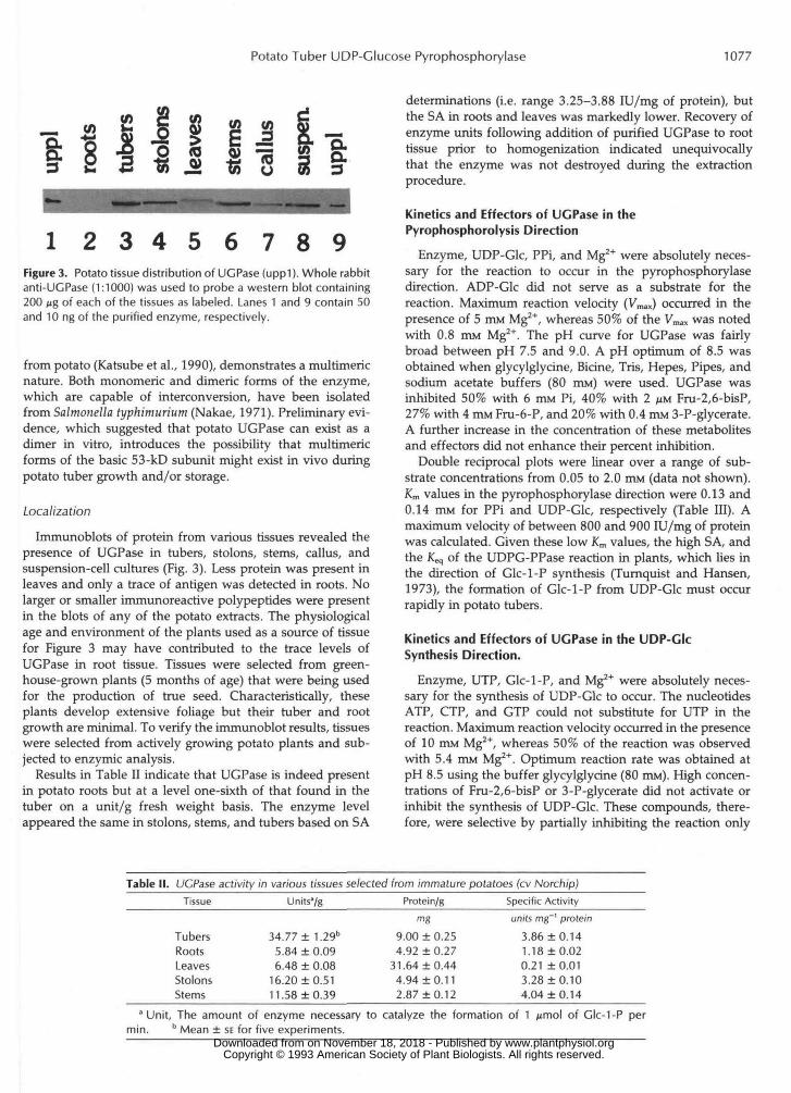
1 2 3 4 5 6 7 8 9Figure 3. Potato tissue distribution of UCPase (uppl). Whole rabbitanti-UCPase (1:1000) was used to probe a western blot containing200 Mg of each of the tissues as labeled. Lanes 1 and 9 contain 50and 10 ng of the purified enzyme, respectively.
from potato (Katsube et al., 1990), demonstrates a multimericnature. Both monomeric and dimeric forms of the enzyme,which are capable of interconversion, have been isolatedfrom Salmonella typhimurium (Nakae, 1971). Preliminary evi-dence, which suggested that potato UGPase can exist as adimer in vitro, introduces the possibility that multimericforms of the basic 53-kD subunit might exist in vivo duringpotato tuber growth and/or storage.
Localization
Immunoblots of protein from various tissues revealed thepresence of UGPase in tubers, stolons, stems, callus, andsuspension-cell cultures (Fig. 3). Less protein was present inleaves and only a trace of antigen was detected in roots. Nolarger or smaller immunoreacrive polypeptides were presentin the blots of any of the potato extracts. The physiologicalage and environment of the plants used as a source of tissuefor Figure 3 may have contributed to the trace levels ofUGPase in root tissue. Tissues were selected from green-house-grown plants (5 months of age) that were being usedfor the production of true seed. Characteristically, theseplants develop extensive foliage but their tuber and rootgrowth are minimal. To verify the immunoblot results, tissueswere selected from actively growing potato plants and sub-jected to enzymic analysis.
Results in Table II indicate that UGPase is indeed presentin potato roots but at a level one-sixth of that found in thetuber on a unit/g fresh weight basis. The enzyme levelappeared the same in stolons, stems, and tubers based on SA
determinations (i.e. range 3.25-3.88 lU/mg of protein), butthe SA in roots and leaves was markedly lower. Recovery ofenzyme units following addition of purified UGPase to roottissue prior to homogenization indicated unequivocallythat the enzyme was not destroyed during the extractionprocedure.
Kinetics and Effectors of UGPase in thePyrophosphorolysis Direction
Enzyme, UDP-Glc, PPi, and Mg2+ were absolutely neces-sary for the reaction to occur in the pyrophosphorylasedirection. ADP-Glc did not serve as a substrate for thereaction. Maximum reaction velocity (Vmax) occurred in thepresence of 5 mM Mg2+, whereas 50% of the Vmax was notedwith 0.8 HIM Mg2+. The pH curve for UGPase was fairlybroad between pH 7.5 and 9.0. A pH optimum of 8.5 wasobtained when glycylglycine, Bicine, Tris, Hepes, Pipes, andsodium acetate buffers (80 mM) were used. UGPase wasinhibited 50% with 6 mM Pi, 40% with 2 ^M Fru-2,6-bisP,27% with 4 mM Fru-6-P, and 20% with 0.4 HIM 3-P-glycerate.A further increase in the concentration of these metabolitesand effectors did not enhance their percent inhibition.
Double reciprocal plots were linear over a range of sub-strate concentrations from 0.05 to 2.0 mM (data not shown).Km values in the pyrophosphorylase direction were 0.13 and0.14 mM for PPi and UDP-Glc, respectively (Table III). Amaximum velocity of between 800 and 900 lU/mg of proteinwas calculated. Given these low Km values, the high SA, andthe Keq of the UDPG-PPase reaction in plants, which lies inthe direction of Glc-l-P synthesis (Turnquist and Hansen,1973), the formation of Glc-l-P from UDP-Glc must occurrapidly in potato tubers.
Kinetics and Effectors of UGPase in the UDP-GlcSynthesis Direction.
Enzyme, UTP, Glc-l-P, and Mg2+ were absolutely neces-sary for the synthesis of UDP-Glc to occur. The nucleotidesATP, CTP, and GTP could not substitute for UTP in thereaction. Maximum reaction velocity occurred in the presenceof 10 mM Mg2+, whereas 50% of the reaction was observedwith 5.4 mM Mg2+. Optimum reaction rate was obtained atpH 8.5 using the buffer glycylglycine (80 DIM). High concen-trations of Fru-2,6-bisP or 3-P-glycerate did not activate orinhibit the synthesis of UDP-Glc. These compounds, there-fore, were selective by partially inhibiting the reaction only
Table II. UCPase activity in various tissues selected from immature potatoes (cv Norchip)Tissue
TubersRootsLeavesStolonsStems
Units'/g
34.77 ± 1.29b
5.84 ± 0.096.48 ± 0.08
16.20 ±0.5111. 58 ±0.39
Protein/gmg
9.00 + 0.254.92 ± 0.27
31.64 + 0.444.94 + 0.112.87 ±0.12
Specific Activityunits mg"' protein
3.86 + 0.141.18 ±0.020.21 +0.013.28 ±0.104.04 ±0.1 4
'Unit, The amount of enzyme necessary to catalyze the formation of 1min. b Mean + SE for five experiments.
of Clc-1-P per
www.plantphysiol.orgon November 18, 2018 - Published by Downloaded from Copyright © 1993 American Society of Plant Biologists. All rights reserved.

1078 Sowokinos et al. Plant Physiol. Vol. 101, 1993
Table 111. Kinetic constants of purified UCPase from S. tuberosum L. (cv Norchip)
Reaction Substrate Concentration Range K"
mM mM mM unitsb mg-' protein
Pyrophosphorolysis PPi 0.05-2.0 0.13 0.11 827 UDP-Glc 0.05-2.0 0.14 0.12 890
Synthesis UTP 0.05-5.0 0.18 0.17 94 0.05-0.2 0.12 72 >0.25 0.53 131
Glc-1-P 0.05-5.0 0.17 0.18 69 0.05-0.2 0:08 44 >0.25 0.68 138
a Nakano et al., 1989. Unit, The amount of enzyrne necessary to catalyze the formation of 1 pmol of Glc-1-P (pyrophosphorolysis) or 1 pmol of UDP-Glc (synthesis) per min.
in the pyrophosphorolysis direction. Inorganic phosphate maximally inhibited the synthesis reaction only 30% at a high concentration of 12 mM.
Although the K,,, values for the pyrophosphorolytic sub- strates (i.e. PPi and UDP-Glc) were similar to the values reported for the potato UDPG-PPase by Nakano et al. (1989) (see Table 111), kinetics in the synthesis direction were dis- tinctly different. It must be initially emphasized that the cultivars used to purify the enzyme were different in both cases. Figure 4 shows the Lineweaver-Burk plot obtained when the concentration of Glc-1-P was varied from 0.05 to 5.0 mM. The equation for the line, y = 0.2521 + 0.0437~ (Y = 0.932, Fl,3z = 208) appeared to adequately explain a11 the data points represented (P < 0.0001). The K,,, for Glc-1-P (0.17 mM) was similar to the value reported by Nakano et al. (1989) (Table 111).
To test the validity of the regression equation, standardized residuals were analyzed according to the procedure of Sokal and Rohlf (1981). A residual plot of the errors of the observed velocities from those predicted by the regression equation
on the concentration of Glc-1-P (data not shown). The curved residual distribution violated the assumption of random er- rors in a regression analysis. This could result if the relation- ship between the rate of UGPase activity and Glc-1-P con- centration was actually nonlinear or biphasic in nature. Regression analysis indicated a change in kinetic behavior at a Glc-1-P concentration near 0.20 mM. To investigate this further, two separate regressions were calculated based on either low (0.05-0.20 mM) or high (>0.20 m) concentrations of Glc-1-P (Fig. 4). As Glc-1-P concentration increased above 0.25 m ~ , the K , of UGPase for Glc-1-P increased 8.5-fold from 0.08 to 0.68 mM (Table 111). Corresponding n values increased from 0.40 to 0.92 as the concentration of Glc-1-P increased (Fig. 5). There appeared to be an increased inter- action between Glc- 1 -P binding sites as the concentration of the substrate increased.
A similar pattern of kinetics and distribution of residuals was observed when UTP was used as a substrate (data not shown). Calculations from two separate regression equations revealed that the K, for UTP increased from 0.12 to 0.53 mM
suggested that errors from the mean velocie were depkdent as the concentration of UTP increased above 0.25 m ~ .
Figure 4. A Lineweaver-Burk plot of purified UGPase reaction in the direction of UDP-Glc synthesis. The reciprocal of each reaction ve- locity was plotted against the reciprocal of Glc- I -P (concentration range 0.05-5.0 mM).
www.plantphysiol.orgon November 18, 2018 - Published by Downloaded from Copyright © 1993 American Society of Plant Biologists. All rights reserved.

Potato Tuber UDP-Glucose Pyrophosphorylase 1079
Deviation of the synthesis reaction from Michaelis-Menten kinetics did not appear to be an artifact of the assay system used. Each experiment was conducted severa1 times with basically the same result. Initially, a one-step continuous spectrophotometric assay was used to determine the rate of UDP-Glc synthesis. Because another enzyme was present in this coupled system (i.e. UDP-Glc dehydrogenase), it was suspected that some other aspect of the reaction mixture might be responsible for the nonlinear kinetics. To eliminate this possibility, the assay was adapted to a two-step process. Step one involved simply the formation of UDP-Glc with purified potato UGPase. A11 assay components were saturat- ing so that the velocity of the reaction was limited solely by substrate concentration rather than by incubation time or enzyme concentration. Step two involved the quantitation of the UDP-Glu formed via the UDP-Glc dehydrogenase sys- tem. This latter step was verified with UDP-Glc standards and was found to be linear from 0.01 to 2.0 pmol of UDP- Glc per tube. Aliquots of the step-one extracts were adjusted so that the level of UDP-Glc introduced into step two fel1 within this linear concentration range.
Data obtained at this time, however, do not explain the molecular basis for these kinetics. One possibility is that increased substrate concentration causes a change in the three-dimensional-structure of the basic 53-kD polypeptide subunit, which in tum alters its catalytic properties. Fitzger- ald et al. (1969) showed a change in the catalytic properties of a single polypeptide species of UGPase from rat mammary glands. This conversion was a function of pH, temperature, and enzyme concentration. They indicated that although the K,,, for UTP and the molecular mass stayed the same, its V,, increased markedly. The V,,, of the potato enzyme also increased 2- to 3-fold as the concentration of either UTP or Glc-1 -P increased, respectively (Table 111). Additionally, a higher multimeric form of the 53-kD polypeptide could be formed, which exhibits different catalytic properties.
In S. typhimurium, Nakae and Nikaido (1971) observed multiple forms of UGPase that demonstrated real differences in their catalytic properties. They demonstrated an enzyme form IIIb (a dimer, molecular mass 80 kD) that gave nonlinear Lineweaver-Burk plots of l/v versus 1/[S] when the concen- trations of UTP and Glc-I-P were varied in the assay mixture. They attributed this deviation from Michaelis-Menten kinet- ics to the conversion of one of the monomers (IIIa) to a dimer. They explained the increased K, values observed as a 'neg- ative cooperative effect" as described by Conway and Kosh- land (1968), where the binding of one substrate molecule makes the binding of another molecule of the same substrate at a second binding site more difficult. This dimer also yielded strictly linear kinetics when the reaction was measured in the pyrophosphorolysis direction. These kinetic characteristics of the bacterial dimer were similar to the kinetic pattem de- scribed with UGPase purified from cv Norchip potatoes. The conversion of the bacterial monomer to a dimer was shown to be temperature sensitive, stimulated by the presence of NaCl, and irreversible in nature (Nakae, 1971). Hill coeffi- cients less than 1.0 (Fig. 5) supported a negative cooperative effect with the potato enzyme with respect to the binding of Glc-I-P. Whether multimeric forms of UGPase occur in vivo in the intact potato tubers remains to be determined.
. ." -5.0 - 4.0 - 3.0 - 2.0
log GIc- I-P
Figure 5. A Hill equation plot of the data in Figure 4.
Role of UGPase in Potato Sweetening
Due to the differences in the substrate saturation kinetics between the synthetic and pyrophosphorolytic reactions, it is tempting to suggest a regulatory function for UGPase during tuber sweetening. Studies of potato carbohydrate metabolism have suggested that cold sensitivity of glycolytic enzymes diverts products of starch breakdown (i.e. Glc-I-P) toward Suc formation (ap Rees et al., 1981). Altered temperature coefficients of key glycolytic enzymes in the cold (3-5OC), however, do not explain why certain potato clones sweeten at warmer temperatures (10-15OC) and others do not. Potato sweetening is a complex metabolic process and it is obvious that key regulatory steps also exist directly between the hexose-P's and free sugars. It was previously found that levels of UGPase activity were correlated ( r = 0.92) with the amount of Glc (i.e. product of Suc hydrolysis) that was accumulated by genetically diverse potato clones in the cold (Sowokinos, 1990). SPS requires high concentrations of UDP- Glc ( K , = 2.5 m ~ ) to achieve 50% of its maximum catalytic activity in potatoes (Murata, 1972). This level is approxi- mately 10-fold higher than the concentration of UDP-Glc in mature tubers (Morre11 and ap Rees, 1986). During periods when hexose-Ps are elevated in potato tubers, a coarse met- abolic control via UGPase could supply varying levels of UDP-Glc and directly affect the velocity of the rate-limiting SPS reaction. Sicher (1986) also indicated that availability of UDP-Glc may be limiting the SPS reaction in leaf tissues.
There may also be a fine metabolic control where change in substrate concentration alters the catalytic properties of UGPase (Table 111 and Fig. 4). In stored tubers, cytosolic Glc- 1-P may be directed toward respiration via PGM or toward sucroneogenesis via UGPase (Fig. 6). When the concentration of Glc-I-P is low (0.20 m~ or less), PGM with a K, for Glc- 1-P of 0.06 m~ (Takamiya and Fukui, 1978) preferentially cycles carbon toward respiratory pathways and energy pro-
www.plantphysiol.orgon November 18, 2018 - Published by Downloaded from Copyright © 1993 American Society of Plant Biologists. All rights reserved.

1080 Sowokinos et al. Plant Physiol. Vol. 101, 1993
(G I ycol y sis 1 Cytosol \
Sucrose-6-P
S6P-Ptase I Sucrose /
UGPase
Amylop ,a~ \ v r Q Z m M . Km.0.68mM p n v e r t a s e Glucose + Fructose
(Gluconeogenesis)
Figure 6. Competing enzyme systems for cytosolic Clc-1-P (GlP) directing carbons toward glycolytic or gluconeogenic pathways. Enzymes represented include UGPase, PGM, PHI, SPS, SUC-6-P phosphatase (S6P-Ptase), and invertase.
duction. Under situations when Glc-1-P is increased, as it is during low temperature stress (Pollock and ap Rees, 1975; Isherwood, 1976), the negative cooperativity would increase the K, of potato UGPase for Glc-1-P 9-fold from 0.08 to 0.68 mM. Through this fine metabolic control, PGM would then demonstrate a 10-fold greater affinity for Glc-1-P than UGPase. Because glycolysis is also restricted, this could help explain the increase in Glc-I-P, Glc-6-P, and Fru-6-P ob- served in the cold-stored potatoes (Pollock and ap Rees, 1975). Because the efficiency of glycolysis is decreased at cold temperatures, decreasing the relative activity of potato UGPase may counter this effect by limiting the accumulation of free sugars.
ACKNOWLEDCMENTS
The technical assistance and statistical analysis provided by Irene Shea and Jeff Alberts, respectively, are greatly appreaated.
Received July 13, 1992; accepted December 4, 1992. Copyright Clearance Center: 0032-0889/93/101/1073/08.
LITERATURE ClTED
ap Rees T, Dixon WL, Pollock CJ, Franks F (1981) Low temperature sweetening of higher plants. In J Freind, MJC Rhodes, eds, Recent Advances in the Biochemistry of Fruits and Vegetables. Academic Press, New York, pp 41-61
ap Rees T, Morrell S (1990) Carbohydrate metabolism in developing potatoes. Am Potato J 67: 835-847
Bradford MM (1976) A rapid and sensitive method for the quanti- tation of miaogram quantities of protein utilizing the principle of protein-dye binding. Anal Biochem 72: 248-254
Conway A, Koshland DE Jr (1968) Negative cooperativity in enzyme action: the binding of diphosphopyridine nucleotide to glyceraldehyde 3-phosphate dehydrogenase. Biochemistry 7:
Dimond RL, Farnsworth PA, Loomis WF (1976) Isolation and characterization of mutations affecting UDPG pyrophosphorylase activity in Dictyostelium discoideum. Dev Biol 5 0 169-181
Fitzgerald DK, Chen S, Ebner KE (1969) Time-dependent increase
4011-4022
in uridine diphosphate glucose pyrophosphorylase activity in vitro. Biochim Biophys Acta 178 491-498
Franke J, Sussman M (1971) Synthesis of uridine diphosphate glucose pyrophosphorylase during the development of Dictyoste- lium discoideum. J Biol Chem 246 6381-6388
Goodwin TW, Mercer E1 (1983) Introduction to Plant Biochemistry. Pergamon Press, Oxford, New York, pp 227-272
Hondo T, Hara A, Funaguma T (1983) The purification and some properties of the UDP-glucose pyrophosphorylase from the pollen of Typhya latifolia Linne. Plant Cell Physiol24 61-69
Isherwood FA (1976) Mechanism of starch-sugar interconversion in Solanum tuberosum. Phytochemistry 1 5 33-41
Katsube T, Kazuta Y, Mori H, Nakano K, Tanizawa K, Fukui T (1990) UDP-glucose pyrophosphorylase from potato tuber: cDNA cloning and sequencing. J Biochem 108 321-326
Laemmli UK (1970) Cleavage of structural proteins during the assembly of the head of bacteriophage T4. Nature 227: 680-685
LeGendre N, Matsudaira P (1988) Direct microsequencing from Immobilon-P transfer membrane. BioTechniques 6 154-159
Mares DJ, Sowokinos JR, Hawker JS (1985) Carbohydrate metab- olism in developing potato tubers. In PH Li, ed, Potato Physiology. Academic Press, Orlando, FL, pp 279-327
Morrell S, ap Rees T (1986) Sugar metabolism in developing tubers of Solanum tuberosum. Phytochemistry 2 5 1579-1589
Murata T (1972) Sucrose phosphate synthase from various plant origins. Agric Biol Chem 3 6 1877-1884
Nakae T (1971) Multiple molecular forms of uridine diphosphate glucose pyrophosphorylase from Salmonella typhimurium. 111. In- terconversion of the various forms. J Biol chem 246 4404-4411
Nakae T, Nikaido H (1971) Multiple molecular forms of uridine diphosphate glucose pyrophosphorylase from Salmonella typhi- murium. I. Catalytic properties of various forms. J Biol Chem 246
Nakano K, Omura Y, Tagaya M, Fukui T (1989) UDP-glucose pyrophosphorylase from potato tuber: purification and character- ization. J Biochem 106 528-532
Pollock CJ, ap Rees T (1975) Effect of cold on glucose metabolism by callus and tubers of Solanum tuberosum. Phytochemistry 1 4
Rubin RW, Warren RW (1977) Quantitation of microgram amounts of protein in SDS-mercaptoethanol-Tris electrophoresis sample buffer. Anal Biochem 83: 773-777
Sicher RC (1986) Sucrose biosynthesis in photosynthetic tissue: rate- controlling factors and metabolic pathway. Physiol Plant 67:
Sokal RR, Rohlf FJ (1991) Biometry: The Principles and Practice of Statistics in Biological Research, Ed 2. WH Freeman and Company, New York, pp 539-540
Sowokinos JR (1976) Pyrophosphorylases in Solanum tuberosum. I. Changes in ADP-glucose and UDP-glucose pyrophosphorylase activities associated with starch biosynthesis during tuberization, maturation, and storage of potatoes. Plant Physiol57: 63-68
Sowokinos JR (1990) Stress-induced alterations in carbohydrate metabolism. In M Vayda, W Park, eds, Molecular and Cellular Biology of the Potato. C.A.B. Intemational, Wallingford, UK, pp
Sowokinos JR, Preiss J (1982) Pyrophosphorylases in Solanum tub- erosum. 111. Purification, physical, and catalytic properties of ADP- glucose pyrophosphorylase in potatoes. Plant Physiol 69
Sung SJS, Xu DP, Galloway CM, Black CC Jr (1988) A reassessment of glycolysis and gluconeogenesis in higher plants. Physiol Plant
Takamiya S, Fukui T (1978) Purification and multiple forms of phosphoglucomutase from potato tubers. Plant Cell Physiol 1 9
Turnquist RL, Hansen RG (1973) Uridine diphosphoryl glucose pyrophosphorylase. In PD Boyer, ed, The Enzymes, Ed 3, Vol VIII. Academic Press, New York, pp 51-71
Vella J, Copeland L (1990) UDP-glucose pyrophosphorylase from the plant fraction of nitrogen-fixing soybean nodules. Physiol Plant 7 8 140-146
4386-4396
1903-1906
118-121
137-158
1459-1466
7 2 650-654
759-768
www.plantphysiol.orgon November 18, 2018 - Published by Downloaded from Copyright © 1993 American Society of Plant Biologists. All rights reserved.